ASTROCYTIC Gq-GPCR-LINKED IP3R-DEPENDENT Ca2+ SIGNALING DOES NOT MEDIATE NEUROVASCULAR COUPLING IN MOUSE VISUAL CORTEX IN VIVO
Daniel E. Bonder
A thesis submitted to the faculty at the University of North Carolina at Chapel Hill in partial fulfillment of the requirements for the degree of Doctor of Philosophy in the Graduate School
(Curriculum of Neurobiology).
Chapel Hill 2014
Approved by: Ken D. McCarthy Thomas Kash
Mohanish Deshmukh Ben Philpot
iii ABSTRACT
Daniel E. Bonder: Astrocytic Gq-GPCR-linked IP3R-dependent Ca2+ signaling does not mediate neurovascular coupling in mouse visual cortex in vivo
(Under the direction of Ken D. McCarthy)
Local blood flow is modulated in response to changing patterns of neuronal activity (Roy and Sherrington, 1890), a process termed neurovascular coupling. It has been proposed that astrocytic Gq-GPCR-linked IP3R-dependent Ca2+ signaling drives this
process, though in vivo tests of this hypothesis are largely lacking. We examined the impact of astrocytic Gq-GPCR and IP3R-dependent Ca2+ signaling on cortical blood flow in awake, responsive mice using multiphoton laser-scanning microscopy and novel genetic tools that enable the selective manipulation of astrocytic signaling pathways in vivo. Selective
iv
ACKNOWLEDGEMENTS
My family: my father, Ed, for providing guidance on science, life and everything else; my mother, Carol, for being an immovable and unshakeable anchor; my older brother, Matt, for sharing the pain, joy and wisdom of experience; my younger brother, Kev, for always listening and never judging; my future parents-in-law, Tom and Diane, for unwavering support, hospitality and kindness.
My friends: Ryan, DK, Katie, Elise, Glenn, Becca, Becca, Kristine, Steve, Rob, Jason, Kyle, Todd, Jen, Dembowski, Kristin, Dave, Jeremy, Varun, Ben, Katy, Vicki, for good times passed and those to come, and for accepting me for me.
v
My home: my cutie, Corey, and my kitty cotecs, Cas and Mimi, for sticking by me, encouraging me to grow, and filling my life with joy and meaning.
Others of note: my thesis adviser, Ken, for providing the space and freedom I needed to develop this research project and find myself; the organizers of Carolina Kickstart Entrepreneurship Teams, Andy Kant and Lisa Heimbach, for helping me explore
opportunities beyond research; my therapist, Margaret Rhee, for providing much-needed guidance in a time of pain and confusion; my trainer, Marty Heben, for teaching me and pushing me to achieve; the director of TIBBS and my career coach, Erin Hopper, for being perhaps the only person to listen to me expound on complicated thoughts, dreams and chains of logic and reply that it makes perfect sense to you, and for giving me the positive feedback and courage to make a difficult decision.
vi PREFACE
My beliefs about science and its ideal, my personal journey through graduate school, and why I decided on the career and life path I aim to follow are summarized, or more appropriately can be “deduced”, from the following quotes from a literary character who became one of my muses.
“From long habit the train of thoughts ran so swiftly through my mind that I arrived at the conclusion without being conscious of the intermediate steps.”
“It was easier to know it than to explain why I know it. If you were asked to prove that two and two made four, you might find some difficulty, and yet you are quite sure of the fact.”
“One’s ideas must be as broad as Nature if they are to interpret Nature.”
“From a drop of water a logician could infer the possibility of an Atlantic or a Niagara without having seen or heard of one or the other.”
Sherlock Holmes, A Study in Scarlet
vii
“It is a capital mistake to theorize before one has data. Insensibly one begins to twist facts to suit theories, instead of theories to suit facts.”
Sherlock Holmes, A Scandal in Bohemia
“The world is full of obvious things which nobody by any chance ever observes.”
Sherlock Holmes, The Hound of the Baskervilles
“There is nothing more deceptive than an obvious fact.”
Sherlock Holmes, The Bascombe Valley Mystery
“No, it is not selfishness or conceit...If I claim full justice for my art, it is because it is an impersonal thing – a thing beyond myself. Crime is common. Logic is rare. Therefore it is upon the logic rather than the crime that you should dwell. You have degraded what should have been a course of lectures into a series of tales.”
Sherlock Holmes, The Adventure of the Copper Beeches
“I confess that I have been as blind as a mole, but it is better to learn wisdom late than never to learn it at all.”
viii
TABLE OF CONTENTS
LIST OF FIGURES ... xi
LIST OF ABBREVIATIONS AND SYMBOLS ...xiii
CHAPTER 1: INTRODUCTION ... 1
THE WHAT AND THE WHY ... 1
What is neurovascular coupling? ... 1
Why do we suspect astrocytes are involved? ... 3
ASTROCYTE BIOLOGY: A HISTORICAL PERSPECTIVE ... 5
A brief overview of the beginnings of astrocyte biology ... 5
Astrocyte biology in the era of electrophysiology and biochemistry ... 8
Astrocytic Ca2+ signaling: The Great Glial Revolution arrives ... 16
THE MODERN ERA: PUTATIVE FUNCTIONS OF ASTROCYTES ...19
Gliotransmission ... 19
Astrocyte-to-Neuron Lactate Shuttle (ANLS) ... 24
Gap junction coupled astrocyte networks ... 28
Neurovascular Coupling ... 32
CHAPTER 2: THESIS RESEARCH ... 37
OVERVIEW ...37
ix
MATERIALS AND METHODS ...41
Mice ... 41
Adeno-associated viral (AAV) injection for expression of GCaMP or hM3Dq ... 41
Chronic optical imaging through Polished, Reinforced Thinned Skull (PoRTS) windows ... 42
Fluorescence immunochemistry ... 43
In vivo loading of Oregon Green BAPTA-1 and Sulforhodamine-101 dye ... 43
Multiphoton imaging in lightly sedated, responsive mice ... 44
Visual Stimulation ... 45
Ca2+ imaging and analysis... 46
Blood flow imaging and analysis ... 47
RESULTS ...49
Selective stimulation of astrocytic hM3Dq does not alter basal visual cortical blood flow ... 49
Visual cortical astrocytes do not display observable somatic Ca2+ elevations following visual stimulation ... 55
Perivascular astrocyte endfeet do not display observable Ca2+ elevations following visual stimuli ... 57
x
Genetic deletion of astrocytic IP3R-dependent Ca2+ signaling does not alter neurovascular coupling in lightly
sedated, responsive mice ... 65
DISCUSSION ...68
CHAPTER 3: FUTURE DIRECTIONS IN NEUROVASCULAR COUPLING ... 73
THE CHALLENGE: AN ALTERNATIVE NEEDED ...73
Glial K+ siphoning hypothesis of neurovascular coupling ... 74
Conducted vasodilation via vascular endothelium ... 75
Intersection with astrocytic Ca2+ signaling ... 77
Drawing inferences from available data ... 79
Arguments against glial K+ siphoning mediating neurovascular coupling ... 83
PROPOSED EXPERIMENTAL DESIGN TO TEST THE K+ SIPHONING HYPOTHESIS ...85
Overview ... 85
Suggested Genetic Models and Pharmacology ... 87
Required Controls and Potential Difficulties ... 88
CHAPTER 4: CONCLUSIONS ... 92
IS ASTROCYTIC Ca2+ SIGNALING CRUCIAL FOR BRAIN FUNCTION? ...92
The IP3R2 KO mouse line: a case study in cognitive dissonance ... 92
“Funny-looking neurons” ... 95
CONCLUSION ... 100
xi
LIST OF FIGURES
CHAPTER 1: INTRODUCTION
Figure 1.Measuring human brain activity using fMRI ... 2
Figure 2. Astrocytes in the CNS ... 7
Figure 3.Passive electrical properties of astrocytes ... 10
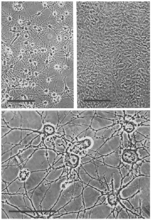
Figure 4.Primary glial cultures ... 14
Figure 5.Intercellular Ca2+ waves in cultured astrocytes ... 17
Figure 6.The tripartite synapse ... 21
Figure 7.Current thinking on astrocytic Ca2+ and synaptic modulation ... 24
Figure 8.Basis of astrocyte-neuron lactate shuttle ... 26
Figure 9.Gap junction-coupled astrocyte networks ... 30
Figure 10.Current model of neurovascular coupling ... 34
CHAPTER 2: THESIS RESEARCH Figure 1.Expression of transgenes using AAV vectors does not lead to lasting astrocytic reactivity or microglial activation ... 51
Figure 2. Basal cortical blood flow is unaffected by stimulation of astrocytic hM3Dq ... 54
Figure 3. Mouse visual cortical astrocytes do not display observable somatic Ca2+ elevations following visual stimulation ... 56
Figure 4. Cortical arterioles dilate in the absence of observable Ca2+ elevations in perivascular astrocyte endfeet ... 59
Figure 5. Neuropil regions display Ca2+ elevations that correlate very well with stimulus-evoked arteriole dilations in terms of amplitude and kinetics ... 61
Figure 6. Air puff startle elicits widespread astrocytic Ca2+ elevation and cortical blood flow changes ... 64
xii
CHAPTER 3: FUTURE DIRECTIONS IN NEUROVASCULAR COUPLING
xiii
LIST OF ABBREVIATIONS AND SYMBOLS
AAV Adeno-associated virus
Ca2+ Calcium ion
Cl- Chloride ion
cKO Conditional knockout
CNO Clozapine-N Oxide
Gq-GPCR Gq G protein-coupled receptor
IP3 Inositol 1,4,5-trisphosphate
IP3R Inositol 1,4,5-trisphosphate receptor
K+ Potassium ion
KO Knockout
Na+ Sodium ion
CHAPTER 1: INTRODUCTION
THE WHAT AND THE WHY
What is neurovascular coupling?
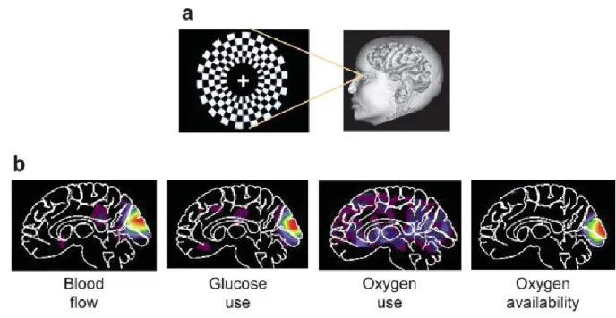
Neurovascular coupling is a well-documented phenomenon in which local blood flow to active regions of the brain is increased, similar to how blood flow to skeletal muscles increases during exercise. First described by researchers Roy and Sherrington in the year 1890, neurovascular coupling has garnered interest in the basic research and medical communities for its contribution to the Blood Oxygen Level-Dependent (BOLD) signal detected by functional magnetic resonance imaging (fMRI). Functional MRI is an incredibly powerful technique that permits noninvasive imaging of human brain activity in real time with millimeter spatial resolution.
2
Figure 1 (Raichle and Mintun, 2006). Measuring human brain activity using fMRI. Presentation of a visual stimulus to an individual (a) results in increased blood flow to active regions of the brain, wherein neurons consume glucose and oxygen (b). Blood flow changes and metabolic rates underlie the BOLD signal measured in fMRI.
3
and hyperoxia in vivo does not alter evoked blood flow increases (Lindauer et al., 2010; Mishra et al., 2011). The magnitude of blood flow increase and oxygen delivery far exceeds oxygen usage (Lin et al., 2010). These data suggest that metabolic demand, more
accurately neuronal metabolic demand, in itself is not driving neurovascular coupling in vivo. Instead, it is possible that neurotransmitter-related signaling events are responsible for hemodynamic changes in brain (Attwell and Iadecola, 2002). This idea does not
necessarily conflict with the hypothesis that astrocytic glycolysis drives neurovascular coupling (Paulson et al., 2010), as it is plausible that these cellular processes could be interdependent.
Why do we suspect astrocytes are involved?
The linear coupling of blood flow increases and rates of glucose metabolism provides one indication that astrocytes might be central to mediating functional hemodynamic
4
for gaseous signaling compounds like nitric oxide (NO), must be transported/transmitted/translated through an astrocytic compartment.
Within the last decade, a wealth of direct evidence has accumulated indicating that astrocytes are capable of modulating vascular diameter by means of intracellular Ca2+ elevations stimulated by the activation of G protein-coupled receptors linked to Gq signaling cascades (Gq-GPCRs), resulting in Ca2+-dependent release of various vasoactive
molecules from endfeet onto cerebral arterioles, most notably cyclooxygenase derivatives (Attwell et al., 2010; Petzold and Murthy, 2011; Newman, 2013; Howarth, 2014). This hypothesis (which serves as the basis for the present thesis project, described in detail in Chapter 2) is attractive because it provides a plausible and relatively straightforward, direct mechanism linking neuronal activity to local blood flow modulation. A further implication is that BOLD signals are a decent proxy for neuronal activity (assuming that astrocytic Ca2+ elevations translate neuronal activity “faithfully”, as most data appeared to indicate) – excellent news for fMRI researchers and diagnosticians. Unfortunately, the results of more recent in vivo work, including the data and conclusions described in detail in Chapter 2, strongly indicate that this hypothesis is oversimplified or perhaps flatly incorrect.
5
ASTROCYTE BIOLOGY: A HISTORICAL PERSPECTIVE
A brief overview of the beginnings of astrocyte biology
Astrocytes belong to a class of non-neuronal support cells in the central nervous system (CNS) termed “glia”, “glial cells”, or “neuroglia”. The name originates from the work of Rudolf Carl Virchow, a 19th century doctor, scientist, and politician in Germany, in which Virchow argued for the existence of sheets of connective tissue lying underneath the
ventricular ependymal layer of the brain, penetrating into and filling all areas of the brain and separating neural tissue from vascular elements (Somjen, 1988). He named this connective tissue “Nervenkitt”, roughly translating to “nerve-glue” from which the term “neuroglia” (or simply “glia”) is derived (Somjen, 1988). While Virchow was not the first to actually identify non-neuronal cell types of the brain, his work did appear to bring about awareness of the potential importance of the non-neuronal components of the brain in possibly regulating brain function or structure (Somjen, 1988).
Early studies on glia focused primarily on distinguishing the various subtypes within this family of cells – protoplasmic and fibrous astrocytes, oligodendrocytes, and microglia – on the basis of histological techniques and light microscopy. For this purpose, the silver staining method of Camillo Golgi proved to be a highly valuable development. This
6
demonstrated the ability of microglial cells to phagocytose dying cells (del Rio-Hortega, 1933).
Sufficient circumstantial evidence was provided to additionally begin formulating plausible inferences about other functions of glia aside from structural support. Santiago Ramon y Cajal postulated that glia “insulate” nerve fibers (Ramon y Cajal, 1920),
preempting our eventual knowledge that myelin is produced by oligodendrocytes or that many synapses are fully or partially ensheathed by astrocytic compartments. Golgi argued that glial cells “feed” neurons (Golgi, 1885-1886), a fore-runner to our current understanding of astrocyte-to-neuron metabolic relationships. Even by the year 1910, the notion that glial cells could secrete substances towards the purposes of modulating brain function, a highly-studied phenomenon we now term “gliotransmission”, was entering the mainstream
7
Figure 2 (Ramon y Cajal, 1913). Astrocytes in the CNS. Drawing by Ramon y Cajal showing protoplasmic astrocytes (A) with pericellular processes (a, b) and perivascular processes (c), and neurons (B).
8
(Berlucchi, 2002), structures that would only later be characterized in more detail and be named “synapses”. Tanzi’s belief predicted modern-day studies on spine growth and dynamics during learning tasks.
In terms of imaging experimental technique, the field has advanced significantly, though this a relatively recent occurrence. In place of the Golgi stain we have powerful microscopic and fluorescent imaging tools, and have added the ability to manipulate
astrocytic function through pharmacology and emerging genetic capabilities. It is fascinating though that conceptually, astrocyte biologists are only recently investigating (or perhaps more accurately, able to investigate) phenomena inferred by others over a century ago. What seems perfectly clear, though, is that despite a paucity of solid evidence to justify their inferences the majority of early researchers in the field have always believed or assumed that glia are more than simply brain glue. Objectively, given what was known in the late 1800s and early 1900s, the alternative “brain glue” hypothesis was equally supportable. The assumption that astrocytes are more than simply passive or purely supportive brain elements has served as a guiding foundational principle for the field of glial biology ever since, despite its being rooted in unsubstantiated inference rather than hard facts. This idea will be investigated more thoroughly later in this chapter.
Astrocyte biology in the era of electrophysiology and biochemistry
Work primarily with insect nervous systems during the 1950s emphasized a metabolic relationship between glial cells and neurons. Histological analysis of central ganglia of Rhodnius prolixus revealed that, while there is no nutrient circulation into the depths of the ganglia, different types of glial cells “have extensive cytoplasm which
9
ganglion cells” (Wigglesworth, 1959). “Classical” glial cells and specialized perineurium cells within the insect nervous system were found to express high levels of metabolism-related enzymes such as esterases (Wigglesworth, 1958) and succinic dehydrogenase
(Wigglesworth, 1956). Glycogen and triglycerides were also found in abundance in
perineurium and glial cells, respectively (Wigglesworth, 1960). Interestingly, satellite cells of mammalian peripheral nervous system ganglia displayed a similar metabolic and enzymatic activity profile (Schmitt, 1958). This led researchers to believe that nutrients were stored in glial compartments and supplied to neurons via glial cells.
The advent of electrophysiological techniques in the middle part of the 20th century provided a novel method for studying the properties and functions of glial cells in the nervous system. It became appreciated that an important component of inter-neuronal communication is the action potential, a specialized cellular process driven by electrical and chemical gradients across neuronal membranes (Hodgkin and Huxley, 1952).
Electrophysiology made it possible to study similar phenomena in glial cells. Do astrocytes communicate by electrical impulses similar to neurons? Might astrocytes influence neuronal activity through electrical activity?
Unlike neurons, astrocytes do not display rapid, impulse-like electrical signals. Instead, depolarizing stimuli result in graded, slow alterations in astrocytic membrane potential (Hild et al., 1958; Hild and Tasaki, 1962). These types of “glial responses” were evoked experimentally with electrical stimulus strengths exceeding those that would be produced by action potentials or other neuronal membrane potential changes (Hild and Tasaki, 1962). Direct neuronal stimulation, as opposed to field stimulation, proved
10
successful neuronal conduction. These studies suggested a limited, supportive role for astrocytes in brain function.
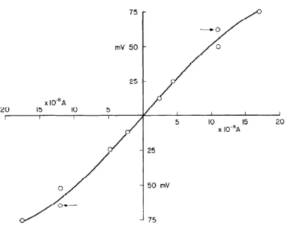
Figure 3 (Kuffler and Potter, 1964). Passive electrical properties of astrocytes. Connective glial cells in the leech CNS display a passive response to changes in membrane potential, indicated by a near-linear input resistance over a range of electrical potentials.
11
function and slow electrical processes such as potentials measured from the surface of the brain (what is measured in an electroencephalogram or EEG) or spreading depression (Hild et al., 1958), an abnormal condition.
The potential importance of other electrophysiological aspects of astrocytes was not immediately recognized. Compared to neurons, astrocytes have a notably low membrane resistance such that electrical currents produced by neurons would flow through glial
cytoplasm rather than the extracellular space in between neurons and glia (Hild and Tasaki, 1962). Additionally, astrocytes possess special low-resistance connections (what would later be identified as gap junctions) that allow for direct electrical communication between cells (Kuffler and Potter, 1964). Similar intercellular connections were not found between neurons and glia (Kuffler and Potter, 1964). Several years following these results, a potential utility for these specialized electrical connections was inferred based on the realization that glial depolarization is the result of extracellular K+ accumulation during neuronal firing (Orkland et al., 1966). The authors of this work argued that connected astrocyte networks could act as K+ “spatial buffers”, transporting extracellular K+ away from areas of high concentration to areas of low K+ concentration (Orkland et al., 1966). At the time it was unclear how this phenomenon, if true, could contribute to greater brain function. Even its functional importance as a homeostatic mechanism was understood to be limited: “Available
experiments do not support the idea that currents in glial cells influence neurons…” (Orkland et al., 1966, p. 805).
12
mechanism (Newman, 1984). This cellular phenomenon was demonstrated to be a more effective method for buffering retinal K+ levels than simple ionic diffusion through the extracellular space (Newman et al., 1984). Analogously, contacts between blood vessels and astrocytic processes within the cerebrum are nearly ubiquitous, a fact recognized by previously mentioned 19th century pioneers. This raised the possibility that similar
mechanisms could operate within the brain as well. This suspicion was supported with the finding that endfeet of cerebral astrocytes possess roughly tenfold higher K+ conductance than other cellular regions (Newman, 1986).
It was with this in mind that researchers took a bold conceptual step forward, trying to link individual cellular processes to greater brain functions and activities. As early as the year 1890, functional alterations in the blood supply to the brain had been documented (Roy and Sherrington, 1890). Roy and Sherrington (1890) proposed that “the brain possesses an intrinsic mechanism by which its vascular supply can be varied locally in correspondence with local variations of functional activity” (p. 105). Could K+ extruded by astrocytes onto blood vessels during bouts of neuronal firing be the mechanism coupling neuronal activity to functional blood flow dynamics? Early mathematical modeling suggested it could (Paulson and Newman, 1987).
Glial biology was progressing on other fronts as well. Advancements in chemical and cell culture techniques led to an incredibly diverse volume of work in the 1970s and 1980s cataloguing diverse previously unrecognized functions of astrocytes. In large part, these studies were made possible by the ability to obtain pure primary non-neuronal (McCarthy and Partlow, 1976) and pure astroglial cell cultures (McCarthy and de Vellis, 1980), which offered significant advantages over other cell separation and purification methods.
13
electrophysiology, performing ligand binding studies, or observing receptor-activated generation of intracellular second messengers such as cAMP or IP3 (Murphy and Pearce, 1987). By these methods, many groups began describing astrocytic responsiveness to noradrenaline through alpha and beta adrenoceptors, acetylcholine through muscarinic receptors, histamine, 5-hydroxytryptamine (5HT, serotonin), dopamine, amino acids
(glutamate, GABA), adenosine, peptides, prostanoids, and drugs such as benzodiazepines (reviewed in Murphy and Pearce, 1987).
Data from culture experiments indicated that astrocytes express nearly the same profile of receptors as neurons. In addition to metabotropic receptors coupled to the breakdown of membrane lipids (IP3 generation, known to stimulate Ca2+ mobilization) or activation of adenylate cyclase and cAMP generation (reviewed in Murphy and Pearce, 1987), ionotropic receptors coupled to ion current flux were also found in cultured
14
15
It was also recognized that astrocytes interacted with neurotransmitters in other ways besides receptor-mediated events. It was known that the brain has high affinity transport systems for the uptake of the primary neurotransmitters, glutamate and GABA, that were localized to synaptosomes (Logan and Snyder, 1972). Later work identified similar
neurotransmitter uptake systems in glial cells (Henn, 1976; Schousboe, 1981). Over time, uptake mechanisms for a diverse range of neurotransmitters were detected within cultured astrocytes, including for serotonin (Katz and Kimelberg, 1985) and the catecholamines, noradrenaline and dopamine (Semenoff and Kimelberg, 1985). Glutamate-induced depolarization of cultured astrocytes (Kettenmann et al., 1984) were attributed to uptake mechanisms (Kettenmann and Schachner, 1985). More precise details on the electrogenic nature of glial neurotransmitter uptake mechanisms were characterized shortly thereafter (Brew and Attwell, 1987).
Despite the advantages of purified cultures and the experimental possibilities they opened up, it was also argued that isolated, cultured astrocytes should not necessarily be considered identical to their in vivo counterparts (Juurlink and Hertz, 1985). In particular, it was argued that cultured astrocytes express such a wide variety of receptors because they acquire de-differentiated properties or undergo arrested development due to the culture procedures or conditions, or isolation from other cells types (Murphy and Pearce, 1987). This concern appears to have been taken seriously. As other biochemical and imaging technologies became available in ensuing years, the use of primary astrocyte cultures gradually diminished. In the modern era most labs no longer make prominent use of the technique.
16
of some previous suspicions, such as the idea that astrocytes store glycogen and maintain an important metabolic relationship with neurons that is governed in part by neuronal activity (Pentreath, 1982; Seal and Pentreath, 1985). The significance of these observations seems to have been unclear to many, and understandably so given the technical limitations of primary astrocyte cultures and novelty of the findings. Based simply on the available data, it would be accurate to frame astrocytic function solely in terms of homeostatic regulation. On the other hand, it would not necessarily be unreasonable to infer beyond the data and see a more dynamic brain element entirely – astrocytes as intrinsic and essential components for information processing or storage.
Time would tell.
Astrocytic Ca2+ signaling: The Great Glial Revolution arrives
The landscape of glial biology was massively transformed in the early 1990s because of the development of reliable cell-permeable chemical Ca2+ indicator dyes, in particular fluo-3 (Minta et al., 1989). This new generation of indicators provided significant advantages over previous dyes, such as fura-2, being excited by visible light as opposed to UV and offering greater convenience for fluorescence microscopy. It was now more
tractable to examine, in real time, some of the signaling events downstream of astrocytic metabotropic receptor activation.
17
year, wherein researchers observed oscillatory elevations in cytosolic Ca2+ in primary cultured astrocytes (using fluo-3) which could propagate as waves throughout the entire astrocyte network (Cornell-Bell et al., 1990). According to the authors, “These propagating waves of calcium suggest that networks of astrocytes may constitute a long-range signaling system within the brain” (Cornell-Bell et al., 1990, p. 470).
Building on this work, it was demonstrated that in organotypic brain slices, in which local tissue structure and network architecture is preserved, astrocytes display similar single-cell Ca2+ elevations and multi-cell Ca2+ waves (Dani et al., 1992). These results clearly indicated, to the authors, “that astrocytes may have a much more dynamic and active role in brain function than has been generally recognized” (Dani et al., 1992). Additional work by other groups suggested that these astrocytic Ca2+ elevations often coincide with electrical bursts from neurons in co-cultures, but also that neuronal activity does not always elicit the relatively slowly propagating astrocytic Ca2+ signals (Murphy et al., 1993).
18
After this point in time the focus of the greater glial biology community would shift almost exclusively to astrocytic Ca2+ signaling. At the deeper conceptual level astrocyte biology became synonymous with astrocytic Ca2+ signaling, much in the same way that neuronal function is understood through the action potential and synaptic release. This is ultimately the spirit of what I term the “Great Glial Revolution” – the paradigm shift from “astrocytes as nerve-glue” to “astrocytes as active partners in brain function”. In most cases, astrocytic “activity” is understood in terms of Ca2+ dynamics. This framework will be explored more fully in Chapter 4.
Glial biology within the last two decades has seen a period of rapid conceptual and technical innovation. Cultures were replaced by acute slices, and gradually labs are
developing in vivo experimental capability. Genetically-encoded Ca2+ indicator proteins like GCaMP have surpassed indicator dyes in sensitivity, signal-to-noise, and response
amplitude, further facilitating sophisticated long-term in vivo studies. All of these developments helped to shape our current understanding of the diverse functions of astrocytes, specifically astrocytic Ca2+ signaling, in the brain.
Simultaneously though, the field has become known for the raw contention and starkly opposing beliefs among different investigators and groups on topics like
gliotransmission (Fiacco et al., 2007; Agulhon et al., 2010) or the function of astrocytes in neurovascular coupling (Nizar et al., 2013; Takata et al., 2013), topics which will be
discussed in more detail later in this chapter. This is a strange situation we find ourselves in. As the number of observations grows and as more “facts” accumulate, seemingly the less confident we are in what we “know” about astrocytes. It is as if there is an inverse
19
THE MODERN ERA: PUTATIVE FUNCTIONS OF ASTROCYTES
Gliotransmission
Gliotransmission is the idea that astrocytes actively release substances in a Ca2+ -dependent manner for the purpose of modulating neuronal activity. Intrinsic to this notion is the concept of the “tripartite synapse” composed of the pre- and post-synaptic neuronal elements and the perisynaptic glial compartment.
In the first demonstration of signaling from astrocytes to neurons, researchers applied a focal electrical stimulation to single astrocytes, which prompted a spreading Ca2+ wave among multiple astrocytes and led to increases in intracellular Ca2+ in neurons resting atop the astrocyte layer (Nedergaard, 1994). A similar effect was noted by a separate group that same year (Parpura et al., 1994). Application of bradykinin to cultured astrocytes resulted in the release of glutamate into the culture medium, which was sufficient to induce elevations of neuronal Ca2+ through NMDA receptors (Parpura et al., 1994).
It was possible that these results were artifacts of astrocytes in culture conditions, perhaps due to functional dedifferentiation (Juurlink and Hertz, 1985). In an effort to address such concerns, several labs began testing for astrocytic Ca2+ elevations in response to neurotransmitter receptor stimulation in acute slice preparations in which local brain network architecture is preserved. Agonists of adrenergic receptors were reported to evoke Ca2+ elevations in hippocampal astrocytes in situ (Duffy and MacVicar, 1995). Electrical
stimulation of the hippocampal Schaffer collateral neuronal fiber pathway similarly induced elevations of astrocytic Ca2+ within the stratum radiatum of the CA1 region that were
20
and McCarthy, 2000; Araque et al., 2002), and histamine (Shelton and McCarthy, 2000). This proved true when washing on receptor agonists or stimulating fiber pathways to evoke synaptic release of neurotransmitters.
Astrocytic Ca2+-induced release of glutamate could produce a diverse range of effects on neuronal activity. Most notably, astrocytic Ca2+ elevations led to NMDAR-dependent slow inward currents (SICs) in adjacent neurons and reduce the magnitude of excitatory and inhibitory post-synaptic currents (Araque et al., 1998b). Astrocytic Ca2+ elevation could also alter the frequency of miniature post-synaptic potentials (Araque et al., 1998a). These data opened the possibility for modulation of neuronal network dynamics via astrocytic Ca2+ signaling in response to neuronal activity. For instance, interaction of
astrocytes and inhibitory neurons could lead to astrocyte-mediated potentiation of miniature inhibitory post-synaptic currents in pyramidal neurons (Kang et al., 1998). Results such as these gave rise to the notion of the “tripartite synapse” in which astrocytes constitute an equal partner with pre- and post-synaptic neuronal elements (Araque et al., 1999).
Initially it was unclear the mechanism by which astrocytic Ca2+ elevation led to changes in neuronal activity. One early study concluded that gap junction-mediated
21
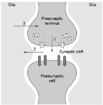
Figure 6 (Araque et al., 1999). The tripartite synapse. Neurons signal to one another via neurotransmitter release at the synaptic cleft (1). Neurotransmitter spillover during neuronal firing is detected by astrocytes surrounding in the synapse (2). Astrocytes signal back to neurons (3), thereby modulating neuronal activity as active partners in synaptic transmission.
22
While Ca2+-dependent exocytosis is traditionally the primary mechanism believed to underlie gliotransmission-like effects, other non-vesicular release pathways have been proposed as well. These include reversal of glutamate transporters (Szatkowski et al., 1990), release from volume-sensitive membrane channels (Kimelberg et al., 1990; Takano et al., 2005), release through functional hemichannels composed of gap junction subunits (Ye et al., 2003), lysosomal exocytosis (Zhang et al., 2007), and release through other membrane channels such as the two-pore K+ channel TREK-1 or the Ca2+-activated anion channel BEST1 (Woo et al., 2012).
In many ways, studies examining gliotransmission have been the driving force behind progress in glial biology for decades now. In particular, this area of research pushed the further characterization of astrocytic Ca2+ dynamics. Questions about whether astrocytes are involved in modulating neuronal activity date back to the very beginnings of glial biology.
However, fewer ideas within the glial field are more hotly contested and
controversial than gliotransmission. On the surface, this is due in large part because of discrepant results between different studies and investigative groups. For instance, uncaging IP3 within astrocytes to evoke Ca2+ elevations is sufficient to increase the
frequency of AMPA spontaneous excitatory post-synaptic currents (Fiacco and McCarthy, 2004; Fiacco et al., 2007). But evoking astrocytic Ca2+ by expressing and stimulating an exogenous Gq-GPCR does not alter neuronal activity (Fiacco et al., 2007; Wang et al., 2013a). Clamping astrocytic Ca2+ with BAPTA blocks long-term potentiation (LTP) in
neighboring neurons (Henneberger et al., 2010). Yet a genetic mouse line lacking astrocytic IP3R-dependent Ca2+ elevation and displays no observable Ca2+ signaling, the IP3R2 KO, has normal basal neuronal activity (Petravicz et al., 2008) and plasticity (Agulhon et al., 2010). In contrast, a mouse line inducibly expressing a dominant negative synaptobrevin II in astrocytes (dn-SNARE) to prevent Ca2+-dependent exocytosis produced defects in
23
(Pascual et al., 2005). However, a separate genetic model designed to block astrocytic exocytosis using tetanus neurotoxin (TeNT) found no effect on basal synaptic activity (Lee et al., 2014). Furthermore, not all studies are in agreement on whether astrocytes express the necessary Ca2+-dependent exocytosis machinery (Li et al., 2013).
These discrepancies are not minor. Rather many of these observations are flatly inconsistent with the idea that astrocytes actively regulate neuronal activity in a Ca2+ -dependent manner, especially if Ca2+ elevations are a function of Gq-GPCR and
downstream IP3R activity. The prominent belief among many investigators is that discrepant results are due to technical limitations (Agulhon et al., 2008; Volterra et al., 2014) or
24
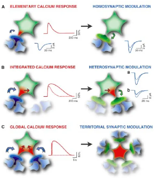
Figure 7 (Volterra et al., 2014). Current thinking on astrocytic Ca2+ and synaptic modulation. Different types of Ca2+ signals can be evoked in astrocytes depending on the nature and strength of local synaptic activity. In turn, these varying astrocytic Ca2+ elevations could function on diverse spatial or temporal scales to modulate activity of individual neurons or neuronal networks.
Astrocyte-to-Neuron Lactate Shuttle (ANLS)
25
vessels. Based on this fact, early researchers inferred that astrocytes might play the critical role of “feeding” neurons. Some of the initial studies examining this possibility in insect model systems were consistent with the idea (Wigglesworth, 1956, 1958, 1959, 1960).
The idea gained substantial traction with the finding that astrocytic glutamate uptake stimulates the utilization of glucose and production of lactate through aerobic glycolysis in cultured astrocytes (Pellerin and Magistretti, 1994). This was significant when considered alongside the previous finding that lactate can sustain neuronal activity in brain slices in the absence of glucose (Schurr et al., 1988). It was also becoming appreciated that glycogen, a stored form of glucose within cells, is primarily localized to astrocytes in brain (Ignacio et al., 1990). Subsequent histochemical studies characterized the expression profiles of lactate dehydrogenase enzymes (Bittar et al., 1996; Pellerin et al., 1998) and monocarboxylate transporters (Pellerin et al., 1998) in neurons and astrocytes. These data were consistent with the idea that astrocytes might generate lactate for neuronal consumption.
In vivo, the activity-dependent release of lactate in striatum was shown to be
26
Figure 8 (Pellerin and Magistretti, 2012). Basis of astrocyte-neuron lactate shuttle. Glutamate uptake by astrocytes near synapses stimulates astrocytic glucose uptake and utilization by glycolysis. Lactate (or pyruvate) produced through glycolysis can be released to fuel neuronal metabolism.
However, the astrocyte-neuron lactate shuttle (ANLS) hypothesis was called into question by other groups. Interestingly, the initial observation of glutamate-induced
utilization of glucose by astrocytes in culture was found to be heavily dependent on culture conditions, with many conditions yielding negative results (Hertz et al., 1998). Analyses of enzyme kinetics and substrate availability within the brain appeared inconsistent with ANLS (Chih et al., 2001).
27
dendrites displaying immediate oxidative metabolism during periods of activity followed by delayed astrocytic glycolysis (Kasischke et al., 2004). Activity-stimulated glucose utilization observed in real time in vivo using blood-born fluorescent glucose analogues like 6-deoxy-N-(7-nitrobenz-2-oxa-1,3-diazol-4-yl)-aminoglucose (6-NBDG) indicated that astrocytic glucose uptake was markedly increased during brain activity whereas neuronal glucose uptake remained constant (Chuquet et al., 2010). The loss of neuronal activity caused by systemic insulin-induced hypoglycemia was prevented by infusion of lactate into the brain (Wyss et al., 2011). Brain activity led to an increase in lactate oxidation in vivo, and lactate was preferred as an energy substrate when both lactate and glucose were available (Wyss et al., 2011).
The importance of lactate shuttling to neurons was explored in two recent in vivo studies. Glycogenolysis and lactate release by astrocytes were found to be essential for long-term memory formation and LTP in the hippocampus (Suzuki et al., 2011). Impeding astrocyte-neuron lactate transport by disrupting monocarboxylate transporter expression produced amnesia, but could be reversed with application of exogenous lactate provided that neuronal lactate uptake was intact (Suzuki et al., 2011). Similar effects were noted when glycogenolysis was impaired pharmacologically (Newman et al., 2011), suggesting that astrocytic glycogen breakdown and lactate release are necessary for memory
formation. Importantly, application of lactate could rescue memory deficits but application of glucose could not, suggesting that neurons preferentially use lactate as an energy source during physiological activity (Newman et al., 2011; Suzuki et al., 2011). Lactate might have other functions in the brain other than as a metabolic substrate or byproduct. In the locus coeruleus, astrocyte-derived lactate can excite neurons to release noradrenaline
28
Not all observations agree with the astrocyte-neuron lactate shuttle hypothesis, however. Lactate release from astrocytes depends on glucose oxidation through glycolysis, but there is evidence that oxidative metabolism and glutamate oxidation are both used to fuel astrocyte metabolism (Hertz et al., 2007; Dienel, 2013). Many perisynaptic astrocytic processes contain mitochondria, permitting glutamate oxidation for ATP generation (Dienel, 2013). In a number of different experimental preparations neurons are capable of
upregulating glucose transport and glycolytic processes to meet energy needs (Dienel, 2012). There is a net release of lactate from the brain during periods of activity, suggesting lactate is not locally oxidated (Dienel, 2012, 2013). Mathematical modeling indicates that astrocytic glycogen breakdown serves the purpose of preserving extracellular glucose levels for neuronal glycolysis and oxidative metabolism, rather than generation of lactate for
shuttling to neurons (DiNuzzo et al., 2010, 2012). An alternative hypothesis to ANLS which considers these data states that lactate might be an “opportunistic”, glucose-sparing substrate under the proper conditions or in certain circumstances, but that glucose is probably the major fuel for neurons (Dienel, 2012).
It should be noted that the debate on the validity of ANLS is primarily over whether or not lactate is the preferred fuel source for neurons under normal conditions. Astrocytes are understood to play a critical role in maintaining neuronal energy demand, though whether this is through lactate shuttling, glucose shunting, or other mechanisms is unclear.
Gap junction coupled astrocyte networks
29
confirmed in brain slices (Gutnick et al., 1981). Astrocytes are extensively connected via gap junctions, which allow the passage of ions and electrical currents, and small molecules less than 1000 Da in size (Giaume and McCarthy, 1996). Astrocyte coupling was initially
proposed as a syncitium-like organization, although focus within the field has shifted away from this idea towards functional and plastic networks that operate in concert with neuronal networks (Giaume and Liu, 2012).
One important aspect of astrocytic networks is that gap junctional communication between astrocytes is modified by neuronal activity (Rouach et al., 2004). This phenomenon was first reported in an astrocyte-neuron co-culture system, in which dye- and electrical-coupling between astrocytes was enhanced by the presence of cerebellar neurons (Fischer and Kettenmann, 1985). Dye-coupling was increased in astrocytes after stimulation of the frog optic nerve (Marrero and Orkland, 1996). Conditions that are known to induce plasticity or significant neuronal activity changes, such as an enriched environment, increased expression of genes related to astrocytic coupling, like Cx30, in addition to genes related to neuronal structure and activity (Rampon et al., 2000). A direct relationship between neuronal activity and astrocytic gap junctional communication was demonstrated in striatal astrocyte-neuron co-cultures. The presence of astrocyte-neurons in the culture augmented the degree of functional coupling between astrocytes, as assessed by intercellular Ca2+ wave propagation (Rouach et al., 2000). Modulation of gap junctional communication could also be modulated in the reverse direction. The removal of cultured neurons by application of neurotoxin or suppression of spontaneous neuronal activity with pharmacological agents both decreased glial coupling (Rouach et al., 2000).
30
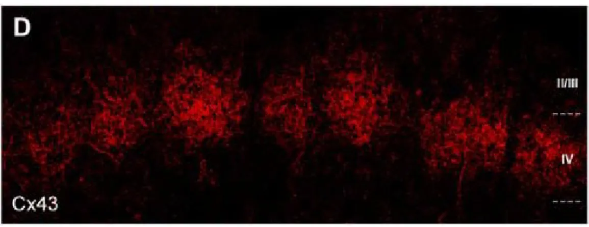
immunochemical data indicate that interconnected astrocyte networks are also arranged into barrels. In particular, gap junctional communication is much stronger within barrels than between barrels, and genetic models that lack neuronal barrel organization also lack astrocytic barrel organization (Houades et al., 2008). Similar structural segregation was observed in the olfactory bulb (Roux et al., 2011), where single glomeruli process input derived from single odorants. Suppression of neuronal activity in vivo by application of tetrodotoxin (TTX) or sensory deprivation reduces intraglomerular gap junctional coupling by modulation of Cx30, but not Cx43, through a mechanism dependent on extracellular K+ and K+ influx into astrocytes (Roux et al., 2011).
Figure 9 (Houades et al., 2008). Gap junction-coupled astrocyte networks. Astrocytes are extensively coupled by gap junctions composed of Cx43 and Cx30 that allow intercellular passage of ions and small molecules (<1000 Da). The structural organization of astrocytic coupling often mirrors local neuronal architecture, as in rodent barrel cortex above.
31
absence of extracellular glucose, coupled astrocyte networks are able to maintain neuronal activity by shuttling glucose or lactate across multiple cells to areas of energetic demand (Rouach et al., 2008).In contrast, neuronal activity cannot be maintained in a genetic mouse model lacking astrocytic Cx43 and Cx30 (Rouach et al., 2008). Intercellular glucose
transport could possibly be a function of Na2+ currents and intercellular Na2+ waves
generated by astrocytic glutamate uptake (Bernardinelli et al., 2004). The nature of evoked astrocytic Na2+ signals in acute slices depends on the strength of stimulation, with Na2+ transients propagating across multiple astrocytes with stronger stimulus intensities (Langer and Rose, 2009). Further, spread of Na2+ throughout astrocyte networks is impaired by interference with gap junctional communication and absent altogether in the Cx43, Cx30 double KO mouse line (Langer et al., 2012), indicating that astrocytic Na2+ signals travel intercellularly through gap junctions.
Interfering with astrocytic gap junctional coupling also leads to defects in synaptic transmission including increased basal transmission and release probability, impaired LTP, and enhanced LTD (Pannasch et al., 2011). These defects are attributed to increases in extracellular glutamate and K+ levels, which are in turn due to slower clearance rates by Cx43, Cx30-deficient astrocytes (Pannasch et al., 2011), which is consistent with
impairments in Na2+ uptake or intercellular dissipation noted in other studies (Langer et al., 2012). Synaptic transmission in mice with deficient astrocytic gap junctional coupling could also altered by changes in the ability of astrocyte networks to buffer extracellular K+ levels. For example, while radial K+ redistribution in hippocampal slices does not require intact astrocytic coupling, the presence of gap junctions facilitates K+ clearance and increases the threshold for epileptiform events (Wallraff et al., 2006).
32
Cx43, Cx30 double KO mouse brain (Lutz et al., 2009). These findings suggest that intercellular communication among coupled astrocyte networks or between astrocytes and oligodendrocytes is critical for myelin integrity and therefore for neuronal function beyond the synapse.
The majority of studies in the field of glial biology have considered astrocytes as single units, insofar as experimental design is concerned – for example, uncaging ions or molecules in single astrocytes. Functional coupling between astrocytes as part of larger functional networks is one of the most intriguing and unique characteristics of these cells. Though it is still a relatively unexplored area, available data already indicates that gap junctional communication is likely a central component of astrocytic function in the brain.
Neurovascular Coupling
As the brain processes information, blood flow to active regions is increased. This vascular phenomenon is termed functional hyperemia and also commonly referred to as neurovascular coupling. It is believed to occur to keep pace with increased neuronal metabolic demand, replenishing glucose and oxygen that is consumed at the onset of activity. While the first observation of functional hyperemia was made by Roy and
33
systems (Gq, Gs, Gi) that could be stimulated by neuronal activity in primary cultures or in situ (Murphy and Pearce, 1987; Porter and McCarthy, 1997).
Further studies revealed that large astrocytic Ca2+ signals can cause arteriole dilations or constrictions in acute slices or retinal preparations. Specifically, stimulating neuronal activity (Zonta et al., 2003; Metea and Newman, 2006; Straub et al., 2006) or activation of astrocytic Gq-GPCRs using agonists (Zonta et al., 2003; Mulligan and MacVicar, 2004) or directly uncaging Ca2+ or IP
3 in single astrocytes (Mulligan and
MacVicar, 2004; Straub et al., 2006) leads to slow vascular changes correlated to evoked Ca2+ elevations in astrocytes. Interfering with astrocytic signals using mGluR antagonists (Zonta et al., 2003), inhibitors of ER Ca2+ release (Straub et al., 2006) or injecting individual astrocytes with BAPTA (Mulligan and MacVicar, 2004) decreases the magnitude of the evoked vascular changes. Similar results were obtained when uncaging Ca2+ in perivascular astrocyte endfoot structures in vivo (Takano et al., 2006). Astrocytic Ca2+ elevations activate arachidonic acid metabolism and release of various vasoactive products (Zonta et al., 2003; Mulligan and MacVicar, 2004; Metea and Newman, 2006; Takano et al., 2006; Petzold et al., 2008).
34
Figure 10 (Petzold and Murthy, 2011). Current model of neurovascular coupling. Neuronal activity stimulates astrocytic Gq-GPCRs leading to Ca2+ elevation and Ca2+-dependent release of vasoactive arachidonic acid metabolites. This model is based largely on in situ studies.
However, not all experimental results are in agreement with the current
35
astrocytes can cause arteriole dilations (Zonta et al., 2003; Mulligan and MacVicar, 2004; Filosa et al., 2006; Metea and Newman, 2006; Takano et al., 2006; Girouard et al., 2010) or shown separately that physiological stimulus evokes blood flow increases (Kleinfeld et al., 1998; Devor et al., 2007; Tian et al., 2010) or astrocytic Ca2+ elevations (Wang et al., 2006; Petzold et al., 2008; Schummers et al., 2008; Nimmerjahn et al., 2009). A primary prediction of the model is that perivascular astrocytic Ca2+ elevation should precede the vascular response. Much of the available correlative in vivo data do not support this.
Recent studies have tested the causal relationship between astrocytic Ca2+ signals and vascular responses in vivo. Electrical forepaw stimulation in anesthetized mice led to reliable dilations of cortical arterioles, but adjacent astrocyte endfeet displayed inconsistent Ca2+ elevations (Nizar et al., 2013). When Ca2+ elevations were observed, they were delayed relative to the onset of blood flow increase (Nizar et al., 2013). Further, vascular responses to stimuli in vivo are preserved in the IP3R2 KO mouse model, in which astrocytic IP3R-dependent Ca2+ signaling is eliminated (Nizar et al., 2013; Takata et al., 2013). In contrast, a separate study described rapid astrocytic Ca2+ signals, the amplitude of which correlate well with the amplitude of evoked blood flow increases (Lind et al., 2013). Traditional microscopic frame imaging (1-2 Hz) is too slow to detect these rapid signals. Furthermore, in vivo experiments with anesthetized animals are confounded by the
36
37
CHAPTER 2: THESIS RESEARCH
Experimental evidence demonstrating that astrocytic Gq-GPCR-linked IP3R-dependent Ca2+ signaling does not mediate neurovascular coupling in mouse visual cortex in vivo
OVERVIEW
Local blood flow is modulated in response to changing patterns of neuronal activity (Roy and Sherrington, 1890), a process termed neurovascular coupling. It has been
38
39 INTRODUCTION
Understanding how cerebral vascular dynamics are coupled to neuronal activity is of intense scientific and clinical interest. Blood flow changes serve as the basis for functional MRI (Kim and Ogawa, 2012), which is the best method for noninvasively imaging human brain activity in real time. The underlying cellular and molecular mechanisms driving
neurovascular coupling have remained elusive and controversial (Mishra et al., 2011; Nizar et al., 2013; Takata et al., 2013).
Substantial evidence utilizing pharmacological approaches to manipulate astrocytic signaling in situ supports the hypothesis that functional hemodynamics are mediated by Gq-GPCR-linked Ca2+-dependent processes (Zonta et al., 2003; Mulligan and MacVicar, 2004; Metea and Newman, 2006; Straub et al., 2006; Gordon et al., 2008; Girouard et al., 2010; He et al., 2012; Stobart et al., 2013). It remains unknown to what degree functional
hemodynamics in the acute slice mirror processes of neurovascular coupling in vivo. It was recently reported that cortical arteriole dilations in anesthetized mice occur in the absence of observable Ca2+ elevations in adjacent astrocyte compartments (Nizar et al., 2013).
40
requires the development and application of models that enable experimentation under near-physiological conditions.
In this study, we utilized cutting-edge genetic tools and physiological stimuli to test the hypothesis that astrocytic Gq-GPCR-linked IP3R-dependent Ca2+ signaling mediates neurovascular coupling in awake, lightly sedated, responsive mice. We used the hM3Dq DREADD designer receptor system (Armbruster et al., 2007; Agulhon et al., 2013) to selectively stimulation astrocytic Gq-GPCR signaling cascades in vivo. To selectively eliminate IP3R-dependent Ca2+ release downstream of Gq-GPCR activity, we employed the IP3R2 KO mouse model (Petravicz et al., 2008). These genetic technologies overcome the major limitations of pharmacological tools such as non-selective drugs, caged molecules or chelators, and enable precise control over astrocytic Gq-GPCR-linked IP3R-dependent Ca2+ signaling in vivo. In addition to cell-permeable Ca2+ dyes, we utilized genetically-encoded Ca2+ sensors, cyto-GCaMP3 and Lck-GCaMP6s, for enhanced detection of astrocytic Ca2+ dynamics (Chen et al., 2013; Shigetomi et al., 2013). Overall, our experimental approach represents a significant advance over previous methods.
Our results establish that astrocytic Gq-GPCR-linked IP3R-dependent Ca2+ signaling is not a central mediator of neurovascular coupling in visual cortex of lightly sedated,
41 MATERIALS AND METHODS
Mice
All mice were housed in the animal facilities at the University of North Carolina, Chapel Hill in accordance with Institutional Animal Care and Use Committee guidelines. IP3R2 KO mice were generated as described previously (Li et al., 2005). IP3R2 +/- mice were interbred to generate homozygous full mutant mice (IP3R2–/–) and littermate controls (IP3R2 +/+). GFAP-cre mice were provided by Dr. Michael Sofroniew at the University of California Los Angeles. The Gfap-cre transgene was designed containing Cre recombinase and the entire mouse glial fibrillary acidic protein (/Gfap/) gene, driven by the Gfap promoter sequence, as described (Gregorian et al., 2009). All mice were maintained on a C57Bl/6 background and C57Bl/6 littermates were used as wild type controls. Both males and females were used for all studies.
Adeno-associated viral (AAV) injection for expression of GCaMP or hM3Dq
AAV8-GFAP-DIO-42
GqDREADD-mCherry, 1 x 10⁹ genome copies/uL. AAV was generated by UNC Vector Core Services. Following successful injection, the needle was left in the cortex for at least 5 minutes to allow for diffusion of the AAV away from the needle track. The incision was sealed with Vetbond and a surgical staple, covered with antibiotic ointment, and mice were given a single injection of antibiotic subcutaneously (ciprofloxacin, 5 mg/kg body weight dose). 4 weeks were given to allow for full transduction of virally-delivered genes and
sufficient re-growth of thinned section of skull prior to installation of chronic optical windows.
Chronic optical imaging through Polished, Reinforced Thinned Skull (PoRTS)
windows
Naïve adult mice or mice injected with AAV at least 4 weeks previously were anesthetized with isoflurane (3-4% for induction, 1-2% for surgery, 100% oxygen). Body temperature was maintained at 37.4° Celsius (Fine Science Tools TR-200). PoRTS optical windows were prepared as previously described (Drew et al., 2010), with a minor
43 Fluorescence immunochemistry
Mice were perfusion fixed with 4% paraformaldehyde (PFA) in phosphate-buffered saline (PBS) three months after AAV injection and two months after PoRTS installation. Brains were removed and fixed in PFA for an additional 24 hours, rinsed in PBS, placed in 30% sucrose in PBS solution for 24 hours, frozen in OCT, and cut in 40 micron sections on a Reichert-Jung Cryo-cut 1800 cryostat. Block solution (20% normal goat serum, 0.1% triton X-100) was placed on the slides for 2 hours at room temperature prior to antibody
treatments. For astrocytic GFAP staining, a Cy3-conjugated mouse anti-GFAP monoclonal antibody (Sigma) was used at a 1:500 dilution in PBS. For microglial staining, a rabbit anti-Iba1 monoclonal antibody (Wako) was used at a 1:500 dilution in PBS. For enhancement of GCaMP signal, mouse (Sigma) or rabbit (Invitrogen) anti-GFP antibodies were used at 1:500 dilution. Primary antibodies remained on the slides for 24 hours at 4°C, followed by appropriate secondary antibodies (Alexa Fluor) for two hours at room temperature at a 1:1000 dilution in PBS. Sections were rinsed in PBS and mounted with Vectashield fluorescence mounting medium with DAPI (Vector). Widefield images were acquired on a Zeiss Axioskop.
In vivo loading of Oregon Green BAPTA-1 and Sulforhodamine-101 dye
44
in Cortex Buffer containing 30 μM sulforhodamine 101 to label astrocytes (Nimmerjahn et al., 2004). 40 nL of dye solution was injected at a depth of 250 um using similar methods as for AAV injection (see above). 1.2% agarose gel and a 3 mm cover slip were placed over the craniotomy, which was then sealed with dental cement (Parkell). For hydration and energy substrate a subcutaneous injection of Lactated Ringer’s solution containing 5% dextrose was administered following surgery and prior to imaging.
Multiphoton imaging in lightly sedated, responsive mice
Initially, mice were anesthetized with isoflurane (3-4% for induction, 1-2% for
maintenance, 100% oxygen). Chlorprothixene sedative was administered subcutaneously at doses between 0.4-3 mg/kg body weight. While anesthetized, mice were placed on a water-heated temperature controlled pad (Adroit Medical HTP-1500) and head-restrained
underneath the microscope objective. In preparation for imaging, isoflurane was decreased and maintained well below anesthetizing levels (0-0.2%, 100% oxygen). The animal was allowed to stabilize under these conditions for at least 20 minutes prior to imaging. Under these conditions, mice are awake and responsive – they typically will not move during experiments, but if otherwise startled (by turning on the lights or touching the animal) they will struggle.
A custom two-photon microscope, converted from an Olympus Fluoview 300 system, with a 60x, 0.9 numerical aperture water immersion objective, and Hamamatsu
45
directly measure blood flow within individual cortical capillaries (see below). For experiments requiring CNO injection, a 30-gauge catheter was inserted intraperitoneally to permit
injection of vehicle or CNO solution during imaging without needing to touch or disturb the animal.
Visual Stimulation
VisionWorks software was used to generate drifting grating visual stimuli presented on a 7-inch VGA monitor (Lilliput 669GL) placed 10 cm from the mouse’s eye at a 45 degree angle from normal. Stimuli consisted of 100% black/white contrast drifting square wave gratings (spatial frequency of 0.05 cycles/degree, drift speed of 2 cycles/second) presented at various angled orientations. A black, cardboard shroud was constructed and attached to the visual stimulus screen. This shroud fits just around the eye contralateral to optical window and prevents light from the stimulus screen from contaminating the imaging.
46 Ca2+ imaging and analysis
ImageJ and Matlab software was used for image analysis. Image sequences were first motion-corrected using the Multistack Reg plugin for ImageJ. Ca2+ dynamics were obtained using custom Matlab scripts generously provided by Dr. Spencer Smith (University of North Carolina at Chapel Hill). These scripts allowed for semi-automated or freehand selection of regions of interest (ROIs). For OGB-1/SR-101 imaging data, ROIs were drawn around neuronal (OGB-1 positive, SR-101 negative) or astrocytic (OGB-1 positive, SR-101 positive) cell bodies. In the case of astrocytic ROIs, these were generated from the SR-101 channel to limit contamination from surrounding neuropil. The routine calculated the average pixel intensity ΔF/F0 with background noise subtracted for each ROI, and computed Ca2+ response averages based off precise trigger times tracked through Spike 2. Raw
fluorescence data was mildly filtered using a 5-point period exponential moving average function (α=1/3) prior to averaging multiple responses.
For GCaMP imaging data, ROIs were drawn around astrocyte endfeet adjacent to cortical arterioles. Arterioles could be identified based on two criteria: 1) arterioles display spontaneous vasomotion whereas venules do not, and 2) blood in post-arteriole capillaries flows away from the vessel, whereas for venules blood flows towards the vessel. Endfoot ROIs were drawn slightly larger than the physical cellular structure because arteriole dilations often caused endfeet to move in the XY plane, producing false fluorescence changes. This necessitated drawing enlarged ROIs to ensure that the entire endfoot
remained within the ROI during arteriole dilations. Contamination from surrounding neuropil regions was not a concern as GCaMP was selectively expressed in astrocytes.
47
x 8.35 microns) box ROIs was tiled across the imaging field, with fluorescence changes extracted as described above.
Blood flow imaging and analysis
For imaging blood flow, a high-molecular weight dextran-conjugated Rhodamine dye (Sigma #R9379) was dissolved into a 5% solution with saline and injected into the tail vein. Blood flow was measured in two ways. First, as in Figure 1C-E and 3E, F, relative volumetric blood flow changes can be estimated based on the degree of arteriole diameter change in response to stimuli. The estimation involves use of the Hagen-Poiseuille equation of fluid dynamics, which states that volumetric flux varies as a function of the fourth power of the vessel radius. To ascertain estimates of changes in vessel radius, images of vascular rhodamine were filtered using a 2-pixel median filter and binarized. Large ROIs were drawn around the vessel cross-section. By tracking fluorescence changes of the binarized images an area measurement in pixels was obtained. From this we derived changes in radius. Our estimated volumetric blood flow increases of 40% (Figure 3D, E, Figure 4B) to 50% (Figure 3F) correspond to arteriole dilations of approximately 8-12% in diameter, similar to what has been described in other studies (Takano et al., 2006). Tracking arteriole dilations permits simultaneous monitoring of cellular Ca2+ dynamics and blood flow changes in response to stimuli in vivo.
Volumetric blood flow changes were also directly measured by tracking erythrocyte velocities within cortical capillaries (Schaffer et al., 2006), as in Figure 3A and 4D, E.
48
(1.5 ms/line). Matlab scripts, generously provided by Dr. Chris Schaffer (Cornell University), were used to calculate erythrocyte velocities from the resulting XT images. Compared to estimating changes in arteriole radius, this method of measuring blood flow is direct and has much greater temporal resolution.
49 RESULTS
Selective stimulation of astrocytic hM3Dq does not alter basal visual cortical blood
flow
Firmly elucidating the physiological role of astrocytic Gq-GPCR and Ca2+ signaling in neurovascular coupling has proven a difficult task due to reliance on pharmacological tools and acute slice preparations. To begin clarifying the role of astrocytic Gq-GPCR cascades and Ca2+ signaling in modulating cortical blood flow in vivo, we expressed the hM3Dq designer receptor and Lck-GCaMP6s in astrocytes using an AAV delivery system (see methods). The hM3Dq viral construct included a double-inverted open reading frame (DIO) element, which prohibits expression of hM3Dq in the absence of Cre-dependent
recombination (Cardin et al., 2010). Astrocyte-selective expression of hM3Dq was therefore achieved by injecting the AAV into GFAP-Cre mice. Four weeks following AAV injection, mice were outfitted with a Polished, Reinforced Thinned Skull (PoRTS) cranial window (Drew et al., 2010), permitting chronic optical access to the visual cortex. Rhodamine dye was injected into the tail vein for monitoring changes in blood flow to stimuli based on direct measures within capillaries or arteriole diameter fluctuations (see Methods).
50
ipsilateral to the AAV injection and PoRTS window were not detectably altered compared to the contralateral cortex (Figure 1A-C, n = 3 mice). GFAP labeling remained sparse in AAV-injected cortex (Figure 1A, B right panels), similar to the non-AAV-injected contralateral cortex (Figure 1A, B left panels). This was true even near the center of AAV injection close to where the injection needle penetrated (Figure 1C).
Altered microglial morphology is also indicative of cortical trauma (Xu et al., 2007). Iba1+ microglia from AAV-injected cortex (Figure 1D, E right panels) were indistinguishable from microglia in non-injected contralateral cortex (Figure 1D, E left panels) on the basis of gross morphology, indicating an absence of microglial reactivity near the injection and window site. These data suggest that expression of transgenes such as Lck-GCaMP6s in cortical astrocytes does not result in lasting glial reactivity.
Basal astrocytic Ca2+ activity in lightly sedated, responsive mice was readily detected by Lck-GCaMP6s (Figure 2A-C, see Methods). Diverse types of astrocytic Ca2+ elevations were detected throughout the imaging field such as “single peaks”, “multi-peaks” or