NSDHL MUTATIONS ASSOCIATED WITH CHILD SYNDROME IDENTIFIED IN ORAL VERRUCIFORM XANTHOMA
George I. Getz
A thesis submitted to the faculty of the University of North Carolina at Chapel Hill in partial fulfillment of the requirements for the degree of Master of Science in the School of Dentistry
(Periodontology).
Chapel Hill 2018
Approved by:
Jonathan Reside
Antonio Amelio
Ricardo Padilla
ABSTRACT
GEORGE I. GETZ: NSDHL Mutations Associated with CHILD Syndrome Identified in Oral Verruciform Xanthoma
(Under the direction of Jonathan Reside)
While the etiology of Verruciform xanthoma (VX) lesions remains unclear, recent
evidence suggests the possible role of a mutation in the NAD(P)-dependent steroid
dehydrogenase-like (NSDHL) gene in cutaneous lesions. The aim of this study is to evaluate
the presence of mutations of the NSDHL gene in cases of oral VX.
A total of 112 oral VX lesions were diagnosed at the UNC Pathology Laboratory and
Biopsy Service between 2005-2017. DNA was extracted from the archived formalin-fixed
and paraffin tissue blocks in a subset of 20 patients. Polymerase chain reaction was then
used to screen for the presence of four known germline mutations in the NSDHL gene
associated with congenital hemidysplasia with ichthyosiform nevus and limb defects
(CHILD) Syndrome and one somatic mutation that was identified in VX lesions in a previous
study with no known CHILD syndrome association.
A total of eight of the tissue samples had known missense mutations associated with
CHILD syndrome. Furthermore, two of these aforementioned eight tissue samples also had
additional missense mutations previously identified in VX lesions. Thus, oral VX lesions
ACKNOWLEDGEMENTS
This dissertation would not have been possible without the guidance and
assistance of many individuals who contributed to the preparation and completion of this
study.
First and foremost, my utmost gratitude to my mentor Dr. Jonathan Reside and
committee members Drs. Antonio Amelio, Ricardo Padilla, and Ingeborg De Kok whose
encouragement and dedication has been my inspiration in the completion of this project.
Specifically, Dr. Amelio and his team: Mr. Kshitij Sharma, Ms. Adele Musicant,
Dr. Miranda Carper, Ms. Shaily Aghera, Ms. Chloe Twomey, Ms. Saumya Goel, and Ms.
Sophia Raterman for their guidance, assistance, and camaraderie during the bench
research portion of this study.
Dr. Adam Lietzan for his assistance with the protein structure analysis.
Ms. Deanna DeCoursey at Eton Biosciences, Inc. for bearing with my numerous
requests with the Sanger sequencing.
Co-residents Drs. Brenda Lopez, Bruno Herrera, and Megumi Williamson who
helped me counter the tribulations of our residency life with a bit of humor.
My parents George and Carolinda for always supporting me in whatever my
endeavor happened to be without question or judgment.
TABLE OF CONTENTS
LIST OF TABLES ... vii
LIST OF FIGURES ... viii
LIST OF ABBREVIATIONS ... ix
CHAPTER 1: INTRODUCTION ... 1
VERRUCIFORM XANTHOMA ... 1
NAD[P]H STEROID DEHYDROGENASE-LIKE (NSDHL) GENE ... 7
CONGENITAL HEMIDYSPLASIA WITH ICHTHYOSIFORM NEVUS AND LIMB DEFECTS (CHILD) SYNDROME ... 8
CK SYNDROME ...10
IMPLICATIONS OF IDENTIFYING THE ROLE OF THE NSDHL GENE IN ORAL VERRUCIFORM XANTHOMA ...12
REFERENCES ...15
CHAPTER 2: NSDHL MUTATIONS ASSOCIATED WITH CHILD SYNDROME IDENTIFIED IN ORAL VERRUCIFORM XANTHOMA ...20
INTRODUCTION ...20
MATERIALS AND METHODS ...21
Archived Samples Collection ...21
DNA Extraction from Samples...21
Plasmid Construction ...21
Polymerase Chain Reaction Amplification of Exons 4 and 6 of the NSDHL gene ...22
PCR Purification and Sanger Sequencing of PCR Amplicons ...24
Analysis of Sanger Sequencing ...24
RESULTS ...25
CONCLUSIONS ...33
LIST OF TABLES
Table 1. Clinicopathologic Characteristics of Test Samples ...35
Table 2. Clinicopathologic Characteristics of Control Samples ...36
Table 3. Summary of NSDHL Mutational Analysis in Oral VX Test Samples ...37
LIST OF FIGURES
Figure 1. Clinical Photographs Submitted with Tissue Biopsies to UNC Oral
Pathology Laboratory and Biopsy Service ...13
Figure 2. Histologic Images from Oral Verruciform Xanthoma and Oral Mucocele Lesions (H&E Staining) ...13
Figure 3. NSDHL Gene ...14
Figure 4. NSDHL Mutations of Interest ...39
Figure 5. Plasmid Engineered with Human HSDHL Mutant Template ...39
Figure 6. Study Primer Pairs with HEK293T DNA to Confirm PCR Amplification ...40
Figure 7. Student Exon 4 Sense/Exon 6 Antisense Primers with Constructed Plasmid ...40
Figure 8. Sanger Sequencing, Exon 4 Amplicons (Controls) ...41
Figure 9. Sanger Sequencing, Exon 6 Amplicons (Controls) ...41
Figure 10. Consensus Multiple Sequence Alignment of Exon 4 Sanger Sequencing...42
Figure 11. Sanger Sequencing, Exon 4 Amplicons (Test Sample Mutations) ...43
Figure 12. Consensus Multiple Sequence Alignment of Exon 6 Sanger Sequencing...44
Figure 13. Sanger Sequencing, Exon 6 Amplicons (Test Sample Mutations) ...44
LIST OF ABBREVIATIONS
CCR2 C-C chemokine receptor type 2
CHILD Congenital Hemidysplasia with Ichthyosiform Erythroderma and Limb Defects
CI Confidence Interval
CNS Central nervous system
DNA Deoxyribonucleic acid
H&E Hematoxylin-eosin
HPV Human Papillomavirus
mRNA Messenger ribonucleic acid
MCP-1 Monocyte chemoattractant protein-1
NAD[P]H Nicotinamide adenine dinucleotide phosphate
NSDHL NAD[P]H steroid dehydrogenase-like protein
PCR Polymerase chain reaction
PG Poryphyromonas gingivalis
RNA Ribonucleic acid
UNC University of North Carolina
CHAPTER 1:
INTRODUCTION
VERRUCIFORM XANTHOMA
VX primarily affects the oral mucosa. First described by Shafer in 1971, it most
commonly presents with a well-circumscribed verrucous or papillomatous appearance,
however, in some instances VX may appear flat, polypoid or sessile (Shafer, 1971) (Figure
1). It usually occurs as a small (2mm–2cm), solitary, asymptomatic, slow growing, white,
pink, grey, or yellowish lesion (Neville, 1986). Oral VX lesions are most commonly found
on the gingiva (40.9-70.6%) (Philipsen et al., 2003), as well as the mandibular ridge, palate,
floor of the mouth, lip, and mucobuccal fold (Shafer, 1971). Lesions have also been
identified on the faucial pillars and the upper respiratory tract (Sathish et al., 2013; Travis et
al., 1989). The differential diagnosis for oral VX includes squamous papilloma, verruca
vulgaris, condyloma acuminatum, verrucous leukoplakia, squamous cell carcinoma, and
gingival epulis (Qi et al., 2014). Histological evaluation following excisional biopsy is the
primary means of diagnosis. Treatment via excision is usually curative for oral VX lesions as
recurrence is rare (Shetty et al., 2013); only three recurrent cases have ever been described,
and all were localized to the hard palate (Nowparast et al., 1981). In cutaneous VX,
however, persistent recurrence is reported, although the condition eventually resolves
(Connolly et al., 2003). No malignant transformation of VX has ever been reported, although
carcinoma, likely due to the degenerative epithelial changes in these oral lesions (Neville et
al., 1986).
While the majority of oral VX lesions are isolated and solitary, multifocal lesions
have been described in association with several diseases and conditions, including chronic
graft-versus-host disease. However, multifocal lesions may also occur in otherwise healthy
patients; four solitary gingival VX lesions were once identified in a patient with no systemic
disease (Qi et al., 2014).
Extraoral VX lesions were first identified and described by Santa Cruz and Martin in
1979. Extraoral lesions are most commonly found involving the anogenital mucosa and
epidermis, although lesions have also been identified on the penis and scrotum (Santa Cruz et
al., 1979, Philipsen et al., 2003). In 2013, a 8mm diameter solitary VX lesion was found on
the forearm of an 82-year-old male with an unremarkable medical history and no history of
trauma to the area (Blankenship et al., 2013). Multiple extraoral VX lesions are usually
associated with conditions such as epidermal nevi, lymphedema, and CHILD syndrome
(Neville, 1986).
No data is available concerning the prevalence or incidence of oral VX. During the
course of a 12-year period, 6 VX lesions were diagnosed from a total of 24,245 specimens in
a university oral pathology laboratory setting, for a frequency of 0.025% (Buchner et al.,
1981). In a 2002 literature review of 282 cases, oral VX occurred in females (mean age, 54.9
years) and males (mean age, 44.2 years) in a 1:1.1 female:male ratio. 109 out of the 282
cases were from Japanese patients, but a comparison with non-Japanese patients showed few
Histologically, in oral VX, the epithelium has a pebbly or verruciform surface,
although it occasionally may appear flat (Nowparast et al., 1981). There is invagination of
hyperkeratosis into crypts in the stratified squamous epithelium (Huang et al., 1986).
Hematoxylin and eosin (H&E) staining of this parakeratin will produce a characteristic
orange color, clearly separating the epithelium associated with the lesion from the
surrounding epithelium. Often the parakeratinized surface will appear rough and will display
infiltrations of neutrophils. Uniform elongation of rete pegs is characteristically present.
However, the hallmark of VX is the presence of foamy (lipid-laden) cells, or xanthoma cells,
within the connective tissue papillae between the rete ridges (Figure 2). These xanthoma
cells do not usually extend deeper into the connective tissue than the tips of the rete pegs
(Neville, 1980). Isolated foamy cells have also been noted within the epithelium in some
cases (Cobb et al., 1976). Histologically, differentiation from other lesions that have foamy
cells is not difficult as VX is the only lesion known to have these type cells confined to the
connective tissue papillae (Shetty et al., 2013). Although there was some disagreement about
the origin of the foam cells in the past, immunohistochemistry suggests that foam cells
descend from macrophages, as they are positive for CD-68 and cathepsin B antibodies
(Mostafa et al.,1993; Oliveira et al., 2001). Scattered lymphocytes and plasma cells may also
be present in the connective tissue. Additional staining with Scharlach R stain has
demonstrated the presence of lipid granules within the xanthoma cells and the overlying
epithelium (Zegarelli et al., 1975). Periodic acid-Schiff positive, diastase-resistant granules
have been found in the cytoplasm of the xanthoma cells (Shafer et al., 1971; Zegarelli et al.,
be identified (Mostafa et al., 1993), but it is thought that presence of these microbes most
likely represents a secondary infection (Neville, 1980).
Electron microscopy of gingival oral VX lesions revealed the presence of lipid-laden
macrophages (Zegarelli et al., 1975). A typical macrophage had an irregular nuclear profile
with prominent nucleolus and the cytoplasm containing numerous large lipid vacuoles.
Epithelial cell degeneration was evident with VX. Lipid containing vacuoles were identified
in epithelial cells as well in the intercellular spaces between epithelial cells (Zegarelli et al.,
1975). Light microscopy of cutaneous VX from the penis displayed numerous lipid vacuoles
in melanocytes as well. Some of these were seen to be within the disrupted basal layer of the
epithelium where they could reach the foam cells in the dermis (Balus et al., 1991).
There are a several theories on the etiology and pathogenesis of VX. Early on, noting
that VX commonly occurs at areas of masticatory mucosa, it was proposed that epithelial
degradation initiates formation of the characteristic foam cells (Zegarelli et al., 1975). The
epithelium becomes entrapped with the crypts within the stratified squamous epithelium and
is not lost from the body. The entrapped epithelium then degenerates, eventually forming
lipids. The products of this breakdown elicit an inflammatory response, explaining the
neutrophil infiltrate that is often seen within the epithelium and a mostly chronic cell
infiltrate in the submucosa (Zegarelli et al., 1975). It is unclear, however, how the initial
crypt that would necessitate the entrapment of epithelial cells form, although it is proposed it
may be caused by a local irritant (Mehra et al., 2005). Later studies, however, observed foam
cell deposition in areas of little epithelial degradation and without entrapment of epithelial
cells (Travis et al., 1989). In a study of a multifocal VX in the upper aerodigestive tract of a
identified within the same individual. This suggested that the squamous epithelium
progresses through flat and papillary stages, before becoming verrucous. Citing that
macrophages are known to produce a variety of growth factors (Nathan, 1987), it was
suggested that the foam cells might play a role in inducing epithelial hyperplasia. Therefore,
it is believed that the accumulation of foam cells is the primary abnormality in the early
lesion and that increasing epithelial hyperplasia and inflammation are secondary
manifestations (Travis et al., 1989).
In order to discover the local factors that may regulate the recruitment and
accumulation of the xanthoma cells in the connective tissue papilla, the cellular and
molecular pathways of chronic inflammation have been studied using immunohistochemical
techniques. It was suggested that keratinocyte hyperplasia may be initiated and maintained
by T cell-mediated inflammation as a response to altered keratinocytes. Furthermore, they
discovered MCP-1 localized in the basal layer of VX and CCR2 expressed in its foam cells.
MCP-1 is a potent macrophage attractor and CCR2 up-regulates macrophage and T-cell
trafficking. They suggested that this MCP-1/CCR2 axis may serve to induce macrophages in
the sub-basal layer (Ide et al., 2008). This may explain why oral VX is most commonly
identified on gingiva tissues, as gingivitis and chronic periodontitis are examples of T cell
activation and keratinocyte expression of MCP-1. In fact, P. gingivalis can up-regulate the
secretion of MCP-1 (Kuramitsu et al.,2002).
Lending further credence that VX may be the result of a chronic inflammatory
process, Rawal et al., 2007, in the first study of its kind, characterized the phenotypes of the
foam cells in VX at different oral locations. They found that the resident and chronic
cells, consistent with a chronic reactive process. This was found consistently, regardless of
the location of the VX lesions.
The source of the lipid contained in foam cells has been the subject of investigation,
in the hope that this may lead to the identifying the pathophysiology of VX. It has been
suggested by some (Huguet et al., 1995; Kishimoto et al., 1998; Wu et al.,2003) that it may
derive from serum lipoprotein. However, squamous epithelium are active sites of lipid
biosynthesis (Ide et al., 1998), these lipids increase in conditions of chronic inflammatory
dermatoses (Uchiyama et al., 2000), and the ultrastructure of VX reveals membrane-bound
lipid-laden vacuoles and foamy macrophages within the epithelium (Zegarelli et al., 1975).
Therefore, it appears that the theory that the degradation of the epithelium, and the resultant
uptake of the keratocyte’s lipids by macrophages in the dermis, may, in fact, lead to the
formation of the pathognomonic foam cells of VX (Balus et al., 1991).
As the clinical manifestation is often verruciform or papillary, and common locations
for VX are the oral cavity or genitalia, a viral etiology, and specifically Human Papilloma
Virus (HPV), has been suggested (Mehra et al., 2005). However, multiple studies have failed
to prove such a connection. While HPV DNA has been found in condyloma-appearing VX
(Hu et al., 2005) and one case report found an association between HPV type 6a particles
found in the nucleus of keratinocytes of a cutaneous VX (scrotum) lesion after PCR and
sequence analysis (Khaskhely et al., 2000), no signs of HPV infection have ever been
detected.
There are several other diseases and syndromes that have shown an association with
VX. There is one case study where an association was found with both a rare type of
capillary syndrome, a condition where vascular permeability leads to hemodynamic disorder.
The locations of the multifocal papillary lesions in both cases were in the lower extremities.
It was reasoned that accumulation of lipids in subcutaneous tissues gave rise to the
characteristic foamy cells in their patient’s VX, the result of atypical peripheral lymphatics
and permeable capillaries, allowing exudation into subcutaneous tissue. Indeed, Hunter et al.
proposed this mechanism for VX in 1970 (Wu et al., 2003).
More recently, VX has been associated with CHILD syndrome, a rare x-linked
autosomal dominant disorder caused by a mutation of theNAD[P]H steroid
dehydrogenase-like (NSHDL) gene involved in cholesterol biosynthesis (Mehra et al., 2005). The lesions in
CHILD syndrome and VX have a similar morphological and histological appearance,
including the appearance of foam cells confined to the connective tissue papillae. The
authors surmised that this inactivation of the NSDHL gene also plays a role in sporadic VX.
NAD[P]H STEROID DEHYDROGENASE-LIKE (NSHDL) GENE
Cholesterol is a major precursor of steroid hormones, influencing embryonic and
postnatal development. The NSHDL gene encodes for a 3b-hydroxysteroid dehydrogenase
located within the membranes of endoplasmic reticulum and on the surface of intracellular
lipid storage droplets that is involved in one the later steps of the biosynthesis pathway of
cholesterol, where C-4 methyl groups are removed (Caldas et al., 2003; Preiksaitene et al.,
2015). Its chromosomal location is on the long arm of the X chromosome at position 28
(Xq28). NSHDL has 8 exons (Figure 3) encoding an mRNA of 1581 base pairs, with the first
methionine codon located at exon 2. The encoded NSDHL protein is composed of 373
cholesterol synthesis. A disruption of the expression of this gene can manifest itself in the
lack of formation and maintenance of an intact cutaneous barrier, explaining, among other
sequelae, the ichthyosiform erythroderma that are characteristic for CHILD syndrome (Mi et
al., 2015). Loss of function mutations in NSHDL also cause an even more rare congenital
disease, CK syndrome (Morimoto et al., 2011; Kanungo et al., 2013).
CONGENITAL HEMIDYSPLASIA WITH ICHTHYOSIFORM NEVUS AND LIMB DEFECTS (CHILD) SYNDROME
CHILD syndrome is a rare x-linked dominant syndrome, with only 60 cases reported
worldwide (Souich et al., 2015). It is a multisystem birth defect that is usually male-lethal,
with only one documented male case likely due to postzygotic mutation (Happle et al., 1996).
First described in 1903 by Otto Sachs, symptoms usually present at birth or within the first
few weeks of development (Bittar and Happle, 2004). It is usually characterized by an
inflammatory epidermal nevus with an often wax-like scaling showing a unique lateralization
pattern (usually on the right side) (Hummel et al., 2003) and a midline demarcation, although
a single case has been found with symmetrical skin lesions (König et al., 2002). In addition,
structural defects may be found ipsilateral to major cutaneous involvement, including hearing
loss, the shortening or absence of limbs and hypoplasia of the brain, lung, heart, and the
kidney (Happle et al., 1980; König et al., 2002). It has been hypothesized that the unilateral
manifestations in CHILD Syndrome could result from interference by mutant cells with a
small population of organizer cells determining the laterality decision at the midline
(Hummel et al., 2003).
Because of the typical clinical and histopathological aspects of the CHILD syndrome
involvement. CHILD syndrome has been diagnosed in patients with only the ipsilateral
lesions, with no ipsilateral skeletal defects (Ko et al., 2008). Female carriers may
occasionally be found clinically healthy or only having minimal involvement due to
X-inactivation (lyonization) or skewed X-X-inactivation (McLarren et al., 2010, Yu et al., 2018).
As a result, numerous sporadic occurrences of CHILD syndrome may be found to be familial
cases after genetic testing (Bittar et al., 2006).
Histology of the CHILD syndrome lesions show similar structures as found in VX. A
typical epidermal lesion will display acanthosis and marked parakeratosis with lymphocyte
infiltration involving the upper part of the dermis and a considerable number of neutrophils
present in the epidermis. Finally, the dermal papillae contain considerable numbers of foamy
cells (Happle et al., 1996). These similarities were even seen in a mild case of CHILD
syndrome. A biopsy from a persistent VX appearing lesion from the left side of the vulva (a
common location for extraoral VX lesions) of an otherwise asymptomatic 27-year old female
revealed a broad parakeratotic corneal layer, neutrophil aggregation, and foam cells in the
dermal papillae. Molecular analysis confirmed a mutation of the NSDHL consistent with
those seen in CHILD syndrome (Gantner et al., 2014).
Mutations in the NSDHL at the Xq28 chromosome locus were identified as the likely
cause for CHILD syndrome (König et al., 2000). Soon after, 4 different mutations of the
NSDHL gene were reported in subjects with CHILD syndrome (Bornholdt et al., 2005), and,
to date, 20 unique mutations at different exons in the NSDHL gene have been found to cause
CHILD syndrome (Mi et al., 2015). While the majority of these mutations have been
missense or nonsense mutations, cases with frameshift mutations, splice-site mutations, and
al., 2005; Yu et al., 2018, Yang et al., 2015). The mutations have also been reported at all 8
exons in various studies (Mi et al., 2015). Although there is great variability in the sequelae
of CHILD syndrome, phenotypic variability does not appear to be associated with a specific
type or location of the mutations. The end result of any type of mutation appears to be loss
of function for the enzyme. It is believed that a non-functional NSDHL may cause the
characteristics of CHILD syndrome through the resulting lack of cholesterol, other sterols
downstream of the block in biosynthesis, or by accumulations of intermediate products
upstream of the resultant of NSDHL expression (Bornholdt et al., 2005). Abnormalities in
cholesterol synthesis would be significant as cholesterol plays a role in the establishment and
upkeep of a complete cutaneous barrier. Accumulation of the toxic pathway metabolites
could also contribute to the characteristic ichthyosis. In one study, a compounded lovastatin
and cholesterol topical medication wad designed to treat CHILD syndrome lesions by
bypassing hepatic first-pass metabolism and allowing access to cutaneous and extracutaneous
tissues). This was compared to a purely cholesterol-based control treatment (Paller et al.,
2011). The treatment with cholesterol alone did not resolve the lesions. However, the
addition of the statin appeared to address the toxic metabolites while the supplemental
cholesterol countered the cholesterol deficiency. Following 3 months of treatment, the
previously affected epidermis was of normal thickness, with a regular orthokeratinization and
normal stratum granulosum (Mi et al., 2015.
CK SYNDROME
CK Syndrome, named for the initials of an affected patient, is an intellectual
dysmorphia (Souich et al., 2009). Only 24 cases, from 3 unrelated families, have been
identified, and its prevalence is unknown (Souich et al., 2015). Like CHILD syndrome, it is
an x-linked disorder where the primary etiology is dysfunction of the NSDHL gene at the
Xq28 chromosome. Unlike the x-linked dominant disease CHILD syndrome, it is inherited
in a recessive manner, affecting males, and its primary sequelae are mild to severe
intellectual disability, behavior problems (aggression, attention deficit hyperactivity disorder,
and irritability), and dysmorphia including amygdaliform eyes, a high nasal bridge, high
arched palate, long and slender fingers and toes, scoliosis, micrognathia, and a crowded
dentition (Souich et al., 2009; Morimoto et al., 2012; Kanungo et al., 2013). The different
clinical phenotypes may be explained because of the different alleles of the NSDHL gene.
CHILD syndrome patients have null alleles that result in the complete loss of function of
NSDHL. On the other hand, in CK Syndrome, patients possess hypomorphic
temperature-sensitive alleles that result in the partial loss of the functional NSHDL protein (McLarren et
al., 2010). The exact process by which this occurs is unclear. The use of quantitative reverse
transcriptase PCR, immunoblotting, and immunohistochemistry to determine NSDHL protein
expression in peripheral tissues identified NSDHL expression in all non-CNS tissues with
malformations in CK syndrome (and CHILD syndrome) (Morimoto et al., 2012). Indeed, all
of these tissues have been shown to synthesize cholesterol. Furthermore, this established that
NSHDL gene expression was restricted to specific cells within the tissues. Autonomous and
non-autonomous cellular mechanisms could occur because of impairment of the normal
biological processes by cholesterol deficiency, toxicity of cholesterol biosynthesis
intermediates or their metabolites. This may contribute to the particular features of CK
IMPLICATIONS OF IDENTIFYING THE ROLE OF THE NSDHL GENE IN ORAL VERRUCIFORM XANTHOMA
A previous study was conducted to identify direct associations between CHILD
syndrome and Verruciform xanthoma (Mehra et al., 2005). The lesions were perioral or
extraoral, including the labium, groin, scrotum, thigh, calf, abdomen, and upper lip. While
missense mutations that had been reported for CHILD syndrome were not identified, a
different missense mutation in a common exon (exon 6) of the NSDHL gene was identified
in 2 out the 7 patients included in the study. To date, no association between the mutations
that cause CHILD Syndrome and oral VX lesions has been researched.
VX cutaneous lesions are associated with CHILD syndrome. As the syndrome is
caused by mutations of the NSDHL gene, there is a possibility that oral VX lesions may also
be associated with a mutation in the NSDHL. Any mutations noted may help to establish an
etiologic theory for VX and support further efforts to evaluate risk of NSDHL mutations in
Figure 1. Clinical Photographs Submitted with Tissue Biopsies to UNC Oral Pathology Laboratory and Biopsy Service. (A) Test Sample 4. (B) Test Sample 9.
Figure 2. Histologic Images from Oral Verruciform Xanthoma and Oral Mucocele Lesions (H&E Staining). (A) Test Sample 16 at increasing magnification (xanthoma cell indicated by arrow). (B) Control Sample 1 (oral mucocele).
Figure 1. Clinical photographs submitted with tissue biopsies to UNC Oral Pathology Laboratory and Biopsy Service A. Test Sample 4. B. Test Sample 9
A. B.
Figure 2. A. Test Sample #16 (H&E staining) at increasing magnification (xanthoma cell indicated by arrow). B. Control Sample 1 (oral mucocele).
A.
REFERENCES
Balus, Breathnach AS, O’Grady AJ. Ultrastructural observations on “foam cells” and the source of their lipid in verruciform xanthoma. J Am Acad Dermatol. 1991;24(5):760-764.
Bittar M, Happle R. CHILD syndrome avant la lettre. J Am Acad Deratol. 2004;50(2 Suppl):S34-37.
Bittar M, Happle R, Grzeschik K-HH, Leveleki L, Hertl M, Bornholdt D, et al. CHILD syndrome in 3 generations: the importance of mild or minimal skin lesions. Arch Dermatol. 2006;142(3):348-351.
Blankenship W, Zech L, Mirzabeigi M, Venna S. Verruciform xanthoma of the upper-‐
extremity in the absence of chronic skin disease or syndrome: a case report and review of the literature. J Cutan Pathol. 2013;40(8):745-752.
Bornholdt D, König A, Happle R, Leveleki L, Bittar M, Danarti R, et al. Mutational spectrum of NSDHL in CHILD syndrome. J Med Genet. 2005;42(2):e17.
Buchner A, Hansen LS, Merrell PW. Verruciform xanthoma of the oral mucosa. Report of five cases and review of the literature. Arch Dermatol. 1981;117:563-565.
Caldas H, Herman GE. NSDHL, an enzyme involved in cholesterol biosynthesis, traffics through the Golgi and accumulates on ER membranes and on the surface of lipid droplets. Hum Mol Genet 2003;12:2981-2991.
Cobb CM, Holt R, Denys FR. Ultrastructural features of the verruciform xanthoma. J Oral Pathol. 1976; 5:42-51.
Connolly SB, Lewis EJ, Lindholm JS, Zelickson BD, Zachary CB, Tope WD. Management of cutaneous verruciform xanthoma. J Am Acad Dermatol. 2000;42(2 Pt 2):343–347.
Gantner S, Rütten A, Requena L, Gassenmaier G, Landthaler M, Hafner C. CHILD syndrome with mild skin lesions: histopathologic clues for the diagnosis. J Cutan Pathol. 2014;41(10):787–790.
Happle R, Koch H, Lenz W. The CHILD syndrome: congenital hemidysplasia with ichthyosiform erythroderma and limb defects. Eu K Pediatr. 1980; 134:27-33.
Happle R, Effendy I, Megahed M, Orlow S, Küster W. CHILD syndrome in a boy. Am J Med Genet. 1996;62(2):192–194.
Huang JS, Tseng CC, Jin YT, Huang CC, Wong TY, Chen HA, et al. Verruciform xanthoma. Case report and literature review. J Periodontol. 1996;67(2):162-165.
Huguet P, Toran N, Tarragona J. Cutaneous verruciform xanthoma arising on a congenital lymphoedematous leg. Histopathology. 1995;26(3):277-279.
Hummel M, Cunningham D, Mullett C, Kelley R, Herman G. Left-‐sided CHILD syndrome caused by a nonsense mutation in the NSDHL gene. Am J Med Genet A. 2003;122A(3):246-251.
Hunter JAA, Morely WN, Peterkin GAG. Xanthomatosis secondary to lymphedema. Trans St John Hosp Dermatol Soc. 1970;56:143-148.
Ide F, Obara K, Yamada H, Mishima K, Saito I, Kusama K. Cellular basis of verruciform xanthoma: immunohistochemical and ultrastructural characterization. Oral Dis.
2008;14(2):150-157.
Khaskhely NM, Uezato H, Kamiyama T, Maruno M, Kariya KI, Oshiro M, Nonake S. Association of human papillomavirus Type 6 with a verruciform xanthoma. Am J Dermatopathol. 2000;22(5):447-452.
Kishimoto S, Takenaka H, Shibagaki R, Nagata M, Katoh N, Yasuno H. Verruciform xanthoma arising in an arteriovenous haemangioma. Br J Dermatol. 1998 Sep 2;139(3):546-548.
Kanungo S, Soraes N, Steiner RD. Sterol metabolism disorders and neurodevelopment-an update. Dev Disabil Res Rev. 2013;17(3):197-210.
Ko JY, Shin, Lee CW. A verruciform xanthoma-‐like phenomenon in a linear epidermal naevus in the absence of a syndromic association. Br J Dermatol. 2008;159(2):493-496.
König A, Happle R, Bornholdt D, Engel H, Grzeschik KH. Mutations in the NSDHL gene, encoding a 3beta-hydroxysteroid dehydrogenase, cause CHILD syndrome. Am J Med Genet. 2000;90(4):339-346.
Kuramitsu HK, Miyakawa H, Qi M, Kang IC. Cellular responses to oral pathogens. Ann Periodontol. 2002;7(1):90-94.
McLarren K, Severson T, Souich C du, Stockton D, Kratz L, Cunningham D, et al.
Hypomorphic Temperature-Sensitive Alleles of NSDHL Cause CK Syndrome. Am J Hum Genetics. 2010;87(6):905-914.
Mi X, Luo M, Guo L, Zhang T, Qiu X. CHILD Syndrome: Case Report of a Chinese Patient and Literature Review of the NAD[P]H Steroid Dehydrogenase-‐Like Protein Gene Mutation. Int Congr Ser. 2015;32(6):e277-e282.
Morimoto M, Souich C, Trinh J, McLarren K, Boerkoel C, Hendson G. Expression profile of NSDHL in human peripheral tissues. J Mol Histol. 2012;43(1):95-106.
Mostafa KA, Takata T, Ogawa I, Ijuhin, Nikai H. Verruciform xanthoma of the oral mucosa: a clinicopathological study with immunohistochemical findings relating to pathogenesis. Virchows Arch A Pathol Anat Histopathol. 1993;423(4):243-248.
Nathan C. Secretory products of macrophages. J Clin Invest. 1987;79(2):319-326.
Neville B, Weathers D. Verruciform xanthoma. Oral Surg Oral Medicine Oral Pathology. 1980;49(5):429-434.
Neville B. The verruciform xanthoma. A review and report of eight new cases. Am J Dermatopathol. 1986;8(3):247-253.
Nowparast B, Howell F, Rick G. Verruciform xanthoma A clinicopathologic review and report of fifty-four cases. Oral Surg Oral Medicine Oral Pathology. 1981;51(6):619-625.
Oliveira PT, Jaeger RG, Cabral LAG, Carvalho YR, Costa ALL, Jaeger MMM. Verruciform xanthoma of the oral mucosa. Report of four cases and a review of the literature. Oral Oncol. 2001;37(3):326-331.
Paller A, Steensel M, Rodriguez-Martín M, Sorrell J, Heath C, Crumrine D, et al.
Pathogenesis-Based Therapy Reverses Cutaneous Abnormalities in an Inherited Disorder of Distal Cholesterol Metabolism. J Invest Dermatol. 2011;131(11):2242-2248.
Philipsen HP, Reichart PA, Takata, Ogawa. Verruciform xanthoma—biological profile of 282 oral lesions based on a literature survey with nine new cases from Japan. Oral Oncol. 2003;39(4):325-336.
Preiksaitiene E, Caro A, Benušienė E, Oltra S, Orellana C, Morkūnienė A, et al. A novel missense mutation in the NSDHL gene identified in a Lithuanian family by targeted next-‐
generation sequencing causes CK syndrome. Am J Med Genet A. 2015;167(6):1342-1348.
Qi Y, Sun Q, Yang P, Song A. A case of multiple verruciform xanthoma in gingiva. Br J Oral Maxillofac Surg. 2014;52(1):e1-e3.
Clin Pathol. 1979;7: 224-8.
Sathish, Suhas, Ch, Shekar, Praveen, Sachin G, et al. Verroucas Xanthoma of Faucial Pillar -A Case Report. Otolaryngology Open -Access. 2013;3(4).
Shafer W. Verruciform xanthoma. Oral Surg Oral Medicine Oral Pathology. 1971;31(6):784-789.
Shetty A, Nakhaei K, Lakkashetty Y, Mohseni M, Mohebatzadeh I. Oral Verruciform Xanthoma: A Case Report and Literature Review. Case Reports Dent. 2013:528967.
Souich C du, Chou A, Yin J, Oh T, Nelson T, Hurlburt J, et al. Characterization of a new X-‐
linked mental retardation syndrome with microcephaly, cortical malformation, and thin habitus. Am J Med Genet A. 2009;149A(11):2469-2478.
Souich C du, Raymond, FL, Grzeschik KH, Boerkoel CF. NSDHL-related disorders. GeneReviews [Internet]. Seattle (WA): University of Washington, Seattle; 2015.
Tarpey P, Smith R, Pleasance E, Whibley A, Edkins S, Hardy C, et al. A systematic, large-scale resequencing screen of X-chromosome coding exons in mental retardation. Nat Genet. 2009;41(5):ng.367.
Travis WD, Davis GE, Tsokos M, Lebovics R, Merrick HF, Miller SP, et al. Multifocal verruciform xanthoma of the upper aerodigestive tract in a child with a systemic lipid storage disease. Am J Surg Pathol. 1989;13(4):309-316.
Uchiyama N, Yamamoto A, Kameda K, Yamaguchi H, Ito M. The activity of fatty acid synthase of epidermal keratinocytes is regulated in the lower stratum spinousum and the stratum basale by local inflammation rather than by circulating hormones. J Dermatol Sci. 2000;24(2):134-141.
Wu JJ, Wagner AM. Verruciform xanthoma in association with milroy disease and leaky capillary syndrome. Pediatr Dermatol. 2003;20(1):44-47.
Yang Z, Hartmann B, Xu Z, Ma L, Happle R, Schlipf N, et al. Large deletions in the NSDHL gene in two patients with CHILD syndrome. Acta Derm-venereol. 2015;95(8):1007-1008.
Yu X, Zhang J, Gu Y, Deng D, Wu Z, Bao L, et al. CHILD Syndrome Mimicking Verrucous Nevus in a Chinese Patient Responded Well to The Topical Therapy of Compound of
CHAPTER 2:
NSDHL MUTATIONS ASSOCIATED WITH CHILD SYNDROME IDENTIFIED IN ORAL VERRUCIFORM XANTHOMA
INTRODUCTION
Verruciform xanthoma (VX) is an uncommon benign lesion, commonly
papillomatous or verruciform, which has an unknown etiology and is seen primarily in the
oral mucosa (Hegde et al., 2013). A mutational inactivation of the NAD[P]H steroid
dehydrogenase-like protein (NSDHL) gene has been reported in cutaneous VX (Mehra et al.,
2015). Lesions resembling VX clinically and morphologically are found in subjects with the
X-linked dominant lipid storage disease congenital hemidysplasia with ichthyosiform
erythroderma and limb defects (CHILD) (Mi et al., 2015). The mutational inactivation of the
NSDHL gene is a marker for CHILD as well as another lipid storage disease, CK syndrome
(McLarren et al., 2010). Between July 2005 and 2017, 112 cases of oral VX were diagnosed
at the University of North Carolina Oral Pathology Laboratory and Biopsy Service. We
proposed to look for a NSDHL gene mutation in diagnosed oral VX cases to begin to
establish a possible etiological theory for this lesion, and eventually support further efforts to
MATERIALS AND METHODS
Archived Samples Collection
Twenty-five formalin-fixed and paraffin-embedded (FFPE) tissue blocks were
retrieved from the UNC Oral Pathology Laboratory and Biopsy Service using consecutive
sampling. The accession dates ranged from to July 2015 to December 2017. Twenty of the
test samples had been previously diagnosed as oral Verruciform xanthoma and five samples
that had been diagnosed as oral mucoceles served as negative controls. Each sample came
from a unique human subject.
DNA Extraction from Samples
Five tissue sections (5 µm thick) were produced with a microtome from each selected
FFPE block. Each sample was deparaffinized with one 1 mL wash of 100% xylene followed
by one 1 mL wash in 100% ethanol. The deparaffinized tissues were then subjected to DNA
extraction using the EX-WAX Paraffin-Embedded DNA Extraction Kit (Millipore Sigma,
Darmstadt, Germany), according to manufacturer’s instruction. DNA concentrations and
260/280 and 260/230 ratios were assessed using a DeNovix DS-11 spectrophotometer
(DeNovix, Wilmington, DE). Each DNA sample and its dilution to a 100ng/µL
concentration (when possible) were stored at -20ºC.
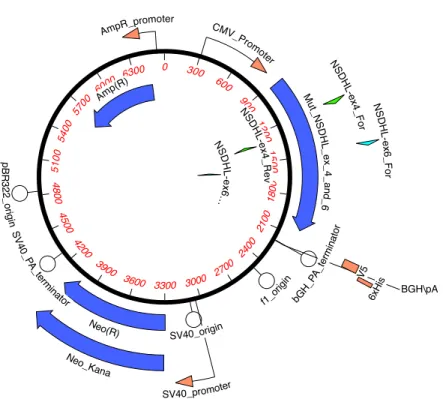
Plasmid Construction
A plasmid was designed and subsequently constructed by Invitrogen GeneArt
Synthesis (Thermo Fisher Scientific, Regensburg, Germany) to include mutations of interest
A105V within Exon 4 and G>C = A182P, G>A = R199H, and G>A = G205S within Exon 6.
G>A = R199H was associated with a somatic mutation found in Verruciform xanthoma
tissues and the others have been reported as associated with CHILD Syndrome (Mehra et al.,
2005) (Figures 4, 5). The plasmid was cloned into the pcDNA3.1_V5-HistA_A122 vector
and the gene size was 1131 bp. 5µg of the lyophilized DNA was dissolved in 50µL of
distilled water. Transformation was accomplished with DH5-alpha competent cells (Thermo
Fisher Scientific, Durham, NC) according to manufacturer’s instructions. After overnight
incubation at 37ºC, the plasmid was recovered using the NucleoSpin Plasmid miniprep kit
(Macherey-Nagel GmbH & Co., Duren, Germany). The plasmid and its dilution to a
75ng/µL concentration were stored at -20ºC. The plasmid, which would include the
mutations of interest was constructed for comparison with test samples after spiking into a
control sample. This combination would serve as a positive control.
Polymerase Chain Reaction Amplification of Exons 4 and 6 of the NSDHL gene
Primer sets were designed to amplify mutations reported in the literature within exons
4 and 6 of the NSDHL gene (Eton Biosciences, Research Triangle Park, NC) (Mehra et al.,
2005). These include C>T = A105V within Exon 4 and G>C = A182P, G>A = R199H, G>A
= G205S, and C>T = Q210X within Exon 6. The primer sequences are 5’-CCA GCT CTG
AAA GGT GTA AAC ACA-3’ (sense) and 5’-CAA GTT TCA ATG ACA TTC TTG GTG
CC-3’ (antisense) for exon 4; 5’-A GTT CTG GGC GCC AAC GAT-3’ (sense) and 5’-CAA
TCA CGA ACT TCA TCT TGC CGT-3’ (antisense) for exon 6. We later designed an
additional primer – an intron 5 primer (5’ - GCA CTC TCT TGG CTT GGG – 3’ (sense)) -
mutation described in the literature for CHILD Syndrome, G>C = A182P (Mehra et al.,
2005). All primer pairs were verified via visualization by electrophoresis on 1.5% agarose
gels using HEK293T as a DNA template. The plasmid itself was verified via amplification
with the exon 4 (sense) and exon 6 (antisense) primer pairs (amplicon size of 409bp). The
expected size of polymerase chain reaction (PCR) amplicons with the test and controls
samples is 119 base pairs for exon 4 sense/antisense primers, 140 base pairs for exon 6
sense/antisense primers, and 206 base pairs for intron 5 (sense)/exon 6 antisense primers.
Each amplification was carried out in a Bio-Rad T100 Thermal Cycler (Bio-Rad
Laboratories, Hercules, CA) in a 50-µL PCR mixture containing: 600ng genomic DNA,
5µL(50µM) of each sense and antisense primer, 25µl Maxima Hot Start PCR Master Mix
(2X) (composition: 400µM dATP, 400µM dGTP, 400µM dCTP, 400µM dTTP, 4mM
magnesium chloride, Maxima Hot Start Taq DNA polymerase in 2X PCR buffer (Thermo
Fisher Scientific, Durham, NC)), and DNase/RNase-free distilled water.
The 20 test samples (designated T1-T20) and the 5 negative controls (designated
C1-C5) were amplified with exon 4 primer pairs, the exon 6 primer pairs, and the intron 5/exon 6
(antisense) primer pairs. The plasmid was amplified with exon 4 sense and exon 6 antisense
primers. As mentioned previously, one of the control samples (C5) spiked with the plasmid
(C5-PL) and amplified with the same primer pairs served a positive control. Thermocycling
conditions used were initial denaturation and hot start at 95°C for 15 minutes, 40 cycles
consisting of 30 seconds at 95°C, 30 seconds at 60°C, and then 1 minute at 72°C. Following
thermocycling, reactions were subjected to a 5-minute 72°C incubation. Polymerase chain
reaction amplicons were visualized by electrophoresis on 2.0% agarose gels and with Gel
corresponding to the aforementioned base pairs length (verified via molecular-weight size
marker) were incised from the agarose gels for purification.
PCR Purification and Sanger Sequencing of PCR Amplicons
The agarose gel sections containing the amplified PCR products were purified using
the NucleoSpin Gel and PCR Clean-up kit (Macherey-Nagel GmbH & Co., Duren,
Germany). DNA was sequenced using a DNA sequencer (model 377; Applied Biosystems,
Foster City, CA) as previously described (Eton Bioscience, Research Triangle Park, NC).
The DNA templates used for the sequencing were at the concentration of 10-30ng/µL.
Analysis of Sanger Sequencing
Reference sequences were obtained from GenBank (NCBI reference sequence
NM_015922.2) for the human NSDHL cDNA sequence and the mutant NSDHL cDNA
sequence, which contained the mutations of interest. Consensus alignments and analysis of
the sequencing results were completed using Molecular Biology 2.0 software (Benchling,
San Francisco, CA). Separate consensus alignments were used for the Exon 4 and Exon 6
amplicons. Any mutations were analyzed to determine whether any amino acid change (or
stop codon) resulted from the base pair change. Chromatograms were visualized using the
FinchTV (version 1.5.0) software. Consensus multiple sequence alignment figures were
created from relevant chromatograms using the Multalin software
RESULTS
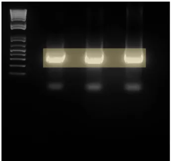
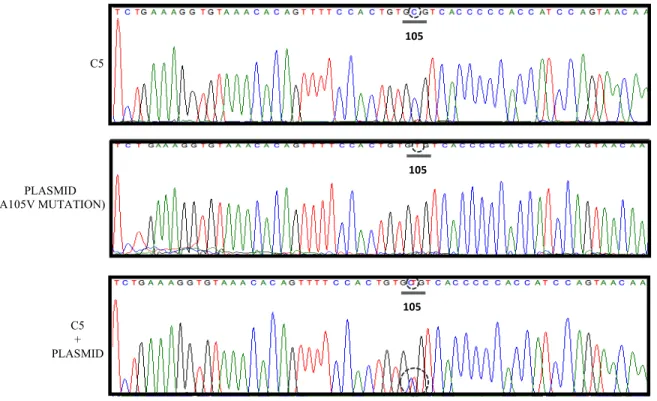
Successful PCR amplification of the three primer sets was confirmed with PCR
reactions using HEK293T genomic DNA as a template (Figure 6). Due to difficulties in
optimization of the extracted DNA, 16 samples from the original 20 tissue samples were
selected as test samples for this study (labeled T1-T16). DNA was successfully extracted
from all of the original five control samples (C1-C5). Confirmation that the plasmid
contained the designed mutations was completed with sequencing of PCR amplicons
(Figures 7, 8, 9). The plasmid was successfully spiked into a selected control (C5) tissue
sample with exon 4 sense and antisense primers (Figure 8) but was unsuccessful with the
exon 6 sense and antisense primer sets.
The mean age of the subjects from whom the oral VX samples were taken was 61.5
years old with a range of 15-92 years old (Table 1). The mean age of the control subjects
was 25 years old with a range of 10-39 years old (Table 2). In the test group, 68.8% of the
lesions were found in females (11 out of 16). In the control group, 3 of the 5 lesions were
from female subjects (60.0%). The most common clinical impressions submitted with
biopsies for the oral VX lesions were dysplasia, papilloma, and squamous cell carcinoma.
The clinical impressions of the control samples were overwhelming “mucocele.” This was
confirmed by the UNC Oral Pathology Laboratory and Biopsy Service. 12 of the 16 test
samples were listed as coming from the right side of patients (although the side of one of the
remaining samples was not specified and two were from the patients’ midline, encompassing
both left and right sides). Health history information was not available for any of the
subjects and it is unknown whether any of the subjects had a personal or family history of
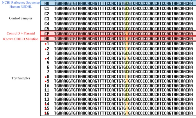
Eight of the oral VX lesions (50%) (T1, T2, T4, T8, T9, T14-T16) exhibited a
mutation in Exon 4 of the NSDHL gene that has been reported for CHILD Syndrome and
was one of the mutations of interest for this study (Figure 10). Two of these eight lesions
had a homozygous missense mutation at c.314 (C>T) at codon 105 (GCG > GTG) that
results in the replacement of the amino acid alanine by valine at the codon (A105V) in the
final protein product. The remaining six lesions had heterozygous missense mutations that
included the same nucleotide substitution as the homozygous mutations (GCG>GTG)
(Figures 8, 11). Neither the A105V mutation nor any other mutation was found in any of the
remaining 8 oral VX lesions or in any of the 5 control samples (Figure 10).
Two of the oral VX lesions (12.5%) exhibited two mutations in Exon 6 of the
NSDHL gene (Figure 12). One of the mutations has previously been reported for CHILD
Syndrome and the other was a mutation that was previously reported in the literature
associated with oral VX (Mehra et al., 2005). Both of these mutations were mutations of
interest for this study. Both T4 and T16 had a heterozygous missense mutation at c.613
(G>A) at codon 205 (GGC>AGC) that results in the replacement of the amino acid alanine
by serine at the codon (A205S) in the final protein product (Figure 13). This mutation was
one of our mutations of interest and is associated with CHILD Syndrome. Additionally, each
of the two lesions had a heterozygous missense mutation at c.596 (G>A) at codon 199
(CGC>CAC) that results in the replacement of the amino acid arginine by histidine at the
codon (R199H) in the final protein product (Figure 13). This mutation was previously
reported in two oral VX lesions but is not known to be associated with CHILD Syndrome
(Mehra et al., 2005). These same two oral VX lesions also had the mutations mentioned
mutation was found in any of the remaining 14 oral VX lesions or in any of the 5 control
samples (Figure 12). Additionally, neither of the other mutations of interest (A182P or
Q201X) was found in these tissue samples or any of the test or control samples (Figure 12).
A summary of the NSDHL mutational analyses is found in Table 3 and Table 4.
DISCUSSION
VX lesions are usually found in the oral mucosa, although extraoral locations,
including, most commonly, the anogenital areas have been reported. The lesion, regardless
of the location, has a distinct histopathological pattern of epithelial hyperplasia with
aggregates of lipid-laden macrophages (often referred to as “foam cells”) in the submucosa
or papillary dermis. While other lesions, including oral mucoceles, contain foamy
macrophages, VX is unique in that the cells are primarily found in the connective tissue.
Evidence suggests that the characteristic lesions found in CHILD Syndrome, with their
similar clinical and histological presentation, are strongly related to VX (Xu et al., 2015).
While the etiology of VX and foamy macrophage accumulation in the connective
tissue remains unresolved, two prevailing theories have been proposed. Zegarelli et al
suggested that epithelial degradation caused by a local irritant initiates formation of the
characteristic foam cells. The epithelium becomes entrapped with the crypts within the
stratified squamous epithelium and is not lost from the body. The entrapped epithelium then
degenerates, eventually forming lipids. The products of this breakdown elicit an
inflammatory response, explaining the neutrophil infiltrate that is often seen within the
epithelium and a mostly chronic cell infiltrate in the submucosa (Zegarelli et al., 1975). It is
cells might form. Additionally, Travis observed foam cell deposition in a multifocal VX in
the upper aerodigestive tract of a child, in an area of little epithelial degradation. Noting that
macrophages are known to produce a variety of growth factors (Nathan, 1987), it was
suggested that the foam cells might play a role in inducing epithelial hyperplasia. Therefore,
the accumulation of foam cells is the primary abnormality in the early lesion, and that
increasing epithelial hyperplasia and inflammation are secondary (Travis et al., 1989).
Cholesterol is a major precursor of steroid hormones influencing embryonic and
postnatal development and is vital to cell membranes. One of the later steps of its
biosynthesis involves a 3b-hydroxysteroid dehydrogenase discovered only within the last 15
years and encoded by the NSHDL gene. The protein is found within the membranes of
endoplasmic reticulum and on the surface of intracellular lipid storage droplets (Ohashi et al.,
2003). It has been reported that instead of just being a vessel for lipid storage, the droplets
themselves may be involved in the regulation of cholesterol biosynthesis. Normally, if an
adequate amount of lipid droplets is produced for the local environment, NSHDL migrates
from the endoplasmic reticulum to the lipid droplets and initiates a negative feedback to stop
producing lipid droplets. However, with the introduction of a missense mutation associated
with CHILD Syndrome (G205S, a mutation detected in our study) into the NSDHL gene, the
protein was no longer functional and the negative feedback was lost, resulting in an
increasing level of lipid droplets (Ohashi et al., 2003). This could account for the foam cells
that are hallmark for VX lesions.
Additionally, abnormalities in cholesterol synthesis could prevent the establishment
and upkeep of a complete cutaneous or epithelial barrier. It is believed that a non-functional
resulting lack of cholesterol, other sterols downstream of the block in biosynthesis, or by
accumulations of intermediate products upstream of the resultant of NSDHL expression
(Bornholdt et al., 2005).
Our study was inspired by a previous analysis by Mehra et al. in 2005 that concerned
the NSDHL gene in cutaneous VX lesions. The characteristic lesions in cutaneous VX and
CHILD syndrome have a similar appearance clinically. When evaluated histologically, they
both feature foamy macrophages within dermal papilla. CHILD Syndrome is caused by a
dysfunction of the NSDHL, caused by a mutation in the NSDHL gene. Therefore, Mehra et
al. group was looking for mutations in cutaneous VX lesions that had previously been
associated with CHILD Syndrome. To date, there have been over 20 mutations documented
in the NSHDL gene (Mi et al., 2015). When their study was conducted over a decade ago,
they focused on exons 4 and 6, as most of the documented cases involved mutations at these
locations. Specifically, they were looking for mutations at codons 105, 182, and 205, which
resulted in the amino acid changes A105V, A182P, and G205S associated with CHILD
Syndrome. They also analyzed for an amino acid change at codon 210, which results in the
amino acid change Q210X (a stop codon). While they were unable to find any mutations
matching those reported for CHILD Syndrome, they did discover a novel missense mutation
at codon 199 (R199H). We chose to do a similar study, but now looking for CHILD
Syndrome mutations in oral VX lesions. Additionally, we wanted to screen for the presence
of the somatic mutation that Mehra et al discovered (Mehra et al., 2005).
Early in the study, we encountered difficulty with successful extraction of the DNA
from the archived tissue samples. The tissue samples, derived from oral biopsies, were often
eventually had to exclude four tissue samples from the study as the concentration and purity
of the extracted DNA was poor and amplification with our primer sets was unsuccessful. We
also were unsuccessful in generating sequencing results with the amplicon from our exon 6
primers when spiking the constructed plasmid into a control sample. This was meant to
illustrate both the wild type and the mutation simultaneously and serve as positive control.
This was, however, successful with the exon 4 primer set and it did mirror the heterozygous
mutation that we discovered with 6 of the test samples.
In our test samples, eight samples (50%) displayed either a homozygous or
heterozygous mutation at codon 105 (GCG>GTG), resulting in a replacement of alanine with
valine in the final protein product (A105V). Two samples displayed a heterozygous mutation
at codon 205 (GGC>AGC), resulting in a replacement of alanine by serine in the final protein
product (G205S). These two different missense mutations are found in patients diagnosed
with CHILD Syndrome. These same two test samples also displayed a heterozygous
mutation at codon 199 (CGC>CAC), resulting in a replacement of arginine by histidine in the
final protein product (R199H). This is the same novel somatic missense mutation
encountered in the Mehra et al. study. It is notable that these two test samples are also
included in the eight test samples that had a mutation at codon 105. The other mutations of
interest for our study in exon 6 that had been previously reported with CHILD Syndrome
(A182P, Q210X) were not found.
A previous investigation compared the NSDHL gene from humans to similar
functional genes from other species (Bornholdt et al., 2005). When compared to other
functionally similar genes, point mutations present on NSDHL at codon positions 105, 182,
(Figure 14). It is predicted that these genetic mutations influence NAD(P) binding within
the active site of NSDHL. While disrupting substrate binding does not necessarily eliminate
catalytic activity, it will significantly reduce the catalytic efficiency of the enzyme. Without
a properly functioning enzyme, metabolism could be disrupted in a cell. Therefore, it is
predicted that these newly discovered mutations in the NSDHL gene contribute to a
reduction in in enzymatic activity of the NSDHL protein, possibly explaining their role in a
pathologic state.
Most of the mutations found in this study were heterozygous, while two homozygous
mutations were found in exon 4 of test samples 1 and 2. A comparison with known
mutations in CHILD Syndrome shows similar variability (König et al., 2000, Hummel et al.,
2003, Bittar et al., 2006). Patients diagnosed with CHILD Syndrome can display a wide
range of phenotypes ranging from very mild skin lesions to the classic manifestations of the
syndrome.
In humans, female cells carry two X-chromosomes and males carry an X and Y
chromosome. In the female, one of the X chromosomes is silenced at random with regard to
parental origin, a process termed X-inactivation, or lyonization. This randomization occurs
early in embryonic development and is maintained through all cell divisions. X-inactivation
results in every female consisting of a mosaic of two different cell populations (where either
the paternal or maternal derived chromosome is inactivated). (Deng et al., 2014). In
X-linked diseases caused by mutation, as is the case with CHILD Syndrome, males are usually
affected to a significantly greater degree, as they have only one X-chromosome. In males,
CHILD Syndrome is overwhelmingly male lethal. In our study, the only homozygous
seen in females. In CHILD Syndrome, heterozygous carriers for the disease can display mild
symptoms (Yu et al., 2018). This variability and the common clinical presentation primarily
on one side of the body (there seems to be a prevalence for the right side (Hummel et al.,
2003) has been explained by mosaicism by lyonization (Hashimoto et al., 1998).
However, even if an X-linked mutation results in the dysfunction or absence of a
protein, possibly affecting the growth or potentially causing the death of a particular cell, the
surrounding environment may provide compensating proteins or signals which may allow
masking, and function may be restored. Usually inactivation is random, but
X-chromosome inactivation skewing can occur, where the inactivation of one X X-chromosome is
favored over the other. Mouse models have skewing of X inactivation in the NSDHL gene as
adults (McLarren et al., 2010). Skewing can be caused by chance or directed by genes. In
addition, the selective pressure for skewing does not appear to be the same in all tissues or at
all developmental stages and skewing can be affected by aging. Many studies have found
that skewing is more prevalent in elderly women (Deng et al., 2014).
While CHILD Syndrome has usually presented as a sporadic manifestation, there are
eight documented cases of mother to daughter transmission. In one particular case, the
proband presented with the spectrum of CHILD Syndrome symptoms, yet her mother and
grandmother were only affected with a minor scaling lesion on one finger and toe,
respectively. Molecular analyses confirmed NSDHL mutations. The authors concluded that
the wide range of manifestations might be the result of either extreme lyonization or
genetically induced skewing of X chromosome inactivation. Furthermore, they deemed it
familial origin (Bittar et al, 2006). No familial aggregation of Verruciform xanthoma has
been reported.
The subjects with our mutations had ages ranging from 15-92 years old (mean age
61.5 years). Discounting Test Sample 16 from a 15-year old male patient, our range was
43-92 years old, with a mean age of 64.6 years. Furthermore, it is interesting that all of the
heterozygous mutations that we found in our test samples were derived from female patients,
except one (the 15-year old male). Both of the homozygous mutations were found in 67- and
89-year old men. Comparing our test subjects to CHILD Syndrome patients, male patients
are extremely rare (only one has been documented). In the reported male patient, he had an
otherwise normal karyotype, but was heterozygous for “wild-type” and mutant alleles in
cultured fibroblasts from affected cutaneous lesions. It has been proposed that the mutation
represented an early post-zygotic event.
A final observation was that, as is found with CHILD Syndrome, most of our lesions
with mutations were found specifically on the right side of the subject (5 out of 8, with two
of the other 8 samples presenting at the midline, and one from the left side) (Table 1).
While it is tempting to make direct comparisons between diagnosed CHILD
Syndrome patients and our oral VX patients, it is important to note that no germline testing
was conducted for any of the patients from whom we derived tissue samples.
CONCLUSIONS
We were able to demonstrate that missense mutations found in the NSHDL gene
known to be associated with CHILD Syndrome can be identified in oral VX lesions through
previous study that addressed cutaneous VX lesions, it is possible that additional exons of the
NSDHL gene may harbor additional identified CHILD Syndrome-associated mutations. Additional studies should include sequencing of additional exons of the NSDHL gene. In
addition, germline testing of affected patients may be beneficial. That we were able to
identify these mutations (and a previously reported somatic mutation unrelated to CHILD
Syndrome) may contribute to an etiologic theory based on genetics for VX. Further studies
may contribute to further efforts to evaluate for risk for NSHDL mutation associated
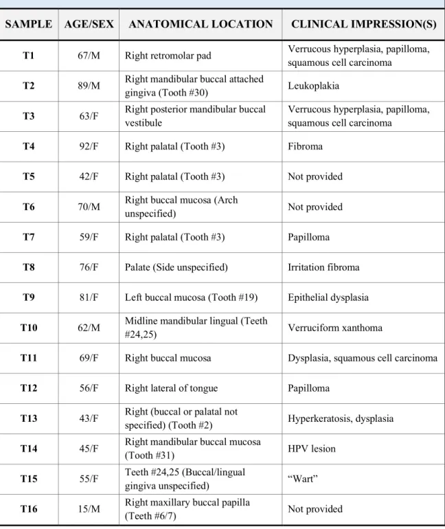
Table 1. Clinicopathologic Characteristics of Test Samples.
SAMPLE AGE/SEX ANATOMICAL LOCATION CLINICAL IMPRESSION(S)
T1 67/M Right retromolar pad Verrucous hyperplasia, papilloma,
squamous cell carcinoma
T2 89/M Right mandibular buccal attached
gingiva (Tooth #30) Leukoplakia
T3 63/F Right posterior mandibular buccal
vestibule
Verrucous hyperplasia, papilloma, squamous cell carcinoma
T4 92/F Right palatal (Tooth #3) Fibroma
T5 42/F Right palatal (Tooth #3) Not provided
T6 70/M Right buccal mucosa (Arch
unspecified) Not provided
T7 59/F Right palatal (Tooth #3) Papilloma
T8 76/F Palate (Side unspecified) Irritation fibroma
T9 81/F Left buccal mucosa (Tooth #19) Epithelial dysplasia
T10 62/M Midline mandibular lingual (Teeth
#24,25) Verruciform xanthoma
T11 69/F Right buccal mucosa Dysplasia, squamous cell carcinoma
T12 56/F Right lateral of tongue Papilloma
T13 43/F Right (buccal or palatal not
specified) (Tooth #2) Hyperkeratosis, dysplasia
T14 45/F Right mandibular buccal mucosa
(Tooth #31) HPV lesion
T15 55/F Teeth #24,25 (Buccal/lingual
gingiva unspecified) “Wart”
T16 15/M Right maxillary buccal papilla
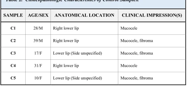
Table 2. Clinicopathologic Characteristics of Control Samples.
SAMPLE AGE/SEX ANATOMICAL LOCATION CLINICAL IMPRESSION(S)
C1 28/M Right lower lip Mucocele
C2 39/M Right lower lip Mucocele, fibroma
C3 17/F Lower lip (Side unspecified) Mucocele, fibroma
C4 31/F Right lower lip Mucocele
T abl e 3. S u m m ar y of NS DH L M u tat ion al A n al ys is in O ral VX Te st S am pl es . AS SOC IAT E D MU T A T IO N OR AL VX
No No -- No YES No -- -- -- No No -- -- -- -- No No No YES No
CH IL D SY N D R O M E YE S YE S
-- YES No YES -- -- -- YES YES -- -- -- -- YES YES YES No YES
RE SU L T A N T AA CH ANGE Al an in e > Va lin e Al an in e > Va lin e --Al an in e > Va lin e Ar gi ni ne > Hi st id in e Gl yc in e > Se ri ne -- -- --Al an in e > Va lin e Al an in e > Va lin e -- -- -- --Al an in e > Va lin e Al an in e > Va lin e Al an in e > Va lin e Ar gi ni ne > Hi st id in e Gl yc in e > Se ri ne TY PE OF MU T A T IO N (S ) C > T C > T --C > T G > A G >
A -- --
--C
>
T
C
>
T -- -- --
--C > T C > T C > T G > A G > A NUC L E OT IDE AF FE C T E D c. 31 4 c. 31 4 --c. 31 4 c. 59 6 c. 61 3 -- -- --c. 31 4 c. 31 4 -- -- -- --c. 31 4 c. 31 4 c. 31 4 c. 59 6 c. 61 3 MU T A T IO N
S EX
O
N
6
No No No YES No No No No No No No No No No No YES
EX O N 4 YE S YE S
No YES No No No YES YES No No No No YES YES YES
SA
M
PL
E
T abl e 4. S u m m ar y of NS DH L M u tat ion al A n al ys is in C on tr ol S am pl es . AS SOC IAT E D MU T A T IO N OR AL VX -- -- -- -- --CH IL D SY N D R O M E -- -- -- -- --RE SU L T A N T AA CH A N G E -- -- -- -- --TY PE OF MU T A T IO N (S ) -- -- -- -- --NUC L E OT IDE AF FE C T E D -- -- -- -- --MU T A T IO N
S EX
O
N
6
No No No No No
EX
O
N
4
No No No No No
SA
M
PL
E
Figure 4. NSDHL Mutations of Interest. Four known germline mutations associated with CHILD Syndrome in Exon 4 and Exon 6 of the NSDHL gene and one somatic mutation not known to be associated with CHILD Syndrome in Exon 6 discovered in the 2005 Mehra Study. Primer design noted.
Figure 5. Plasmid Engineered with Human HSDHL Mutant Template. Invitrogen GeneArt Synthesis, Germany.
0 300 600 900 120 0 1 50 0 18 00 210 0 2400 2700 3000 3300 3600 3900 42 00 45 00 48 00 5 10 0 540
0 5700 6000 6300 M ut_ N S D H L _ e x_ 4 _ a n d _ 6 Amp(R ) Neo(R) Neo _Kana p B R 3 2 2 _ o rig in CMV _Promo ter f1_o rigin bGH _PA _ter min ato r SV40_promoter S V 4 0_P A _term inator SV40_origin V5 AmpR_p romoter N SD H L -e x4_ R e v N S D H L -e x6 … N S D H L -e x4_ F or N S D H L -e x6 _ F o r 6xH is BGH\pA
100 200 300 400 500 600 700 800 900 1000 1100 1200 1300 1400 1500
NSDHL NSDHL
Exon 8 Exon 3
Exon 1
Exon 2 Exon 4
Exon 6 Exon 5
Exon 7
Exon 4_Rev Primer Exon 4_For Primer
Exon 6_Rev Primer Exon 6_For Primer C>T = A105V
G>C = A182P C>T = Q210X G>A = G205S G>A = R199H
Human NSDHLmut - NM_015922.2
Mehra Study mutation
CHILD Syndrome mutation
CHILD Syndrome mutation
Figure 6. Study Primer Pairs with HEK293T DNA to Confirm PCR Amplification. Expected size: 119bp NSDHL Exon 4 F/R; 140bp NSDHL Exon 6 F/R; 206bp NSDHL Intron 5F/Exon 6R.
Figure 7. Student Exon 4 sense/Exon 6 antisense Primers with Constructed Plasmid. Expected size: 409bp.
Figure 8. Sanger Sequencing, Exon 4 Amplicons. Controls.
Figure 9. Sanger Sequencing, Exon 6 Amplicons. Controls.
105 105 105
199 205 210
182
199 205 210
182 C5
PLASMID (A105V MUTATION)
C5 + PLASMID
C5
Figure 10. Consensus Multiple Sequence Alignment of Exon 4 Sanger Sequencing. Red circles indicate presence of mutation C > T (A105V) in test samples.
NCBI Reference Sequence Human NSDHL
Known CHILD Mutation Control 5 + Plasmid
Control Samples
Test Samples