Hepatitis C Virus Induces MDSCs-Like
Monocytes through TLR2/PI3K/AKT/STAT3
Signaling
Naicui Zhai1, Haijun Li1, Hongxiao Song1, Yang Yang1, An Cui1, Tianyang Li1, Junqi Niu2, Ian Nicholas Crispe1,3, Lishan Su1,4, Zhengkun Tu1,2*
1 Institute of Translational Medicine, the First Hospital of Jilin University, Changchun, China, 2 Department of Hepatobiliary and Pancreatic Diseases, the First Hospital of Jilin University, Changchun, China,
3 Department of Pathology, University of Washington, Seattle, Washington, United States of America, 4 Lineberger Comprehensive Cancer Center, School of Medicine, University of North Carolina at Chapel Hill, Chapel Hill, North Carolina, United States of America
Abstract
Background and Aims
Recent studies reveal the accumulation of myeloid derived suppressor cells (MDSCs) in human peripheral blood mononuclear cells (PBMCs) following HCV infection, which may facilitate and maintain HCV persistent infection. The mechanisms by which HCV induces MDSCs are poorly understood. In the present study, we investigated the mechanisms by which HCV induces MDSCs that lead to suppression of T cell proliferation and expansion of CD4+Foxp3+regulatory T cells.
Methods
Purified monocytes from healthy donors were cultured with HCV core protein (HCVc) or cell culture-derived HCV virions (HCVcc), and characterized the phenotype and function of these monocytes by flow cytometry, quantitative PCR, ELISA and western blot assays. In addition, peripheral blood from healthy donors and chronic HCV infected patients was col-lected, and MDSCs and CD4+CD25+CD127-regulatory T cells were analyzed by flow cytometry.
Results
Both HCVc and HCVcc induced expression of IDO1, PD-L1 and IL-10, and significantly down-regulated HLA-DR expression in human monocytes. HCVc-treated monocytes trig-gered CD4+Foxp3+Tregs expansion, and inhibited autologous CD4+T cell activation in an IDO1-dependent fashion. Our results showed that HCV virions or HCV core proteins induced MDSC-like suppressive monocytes via the TLR2/PI3K/AKT/STAT3 signaling path-way. Monocytes derived from patients with chronic HCV infection displayed MDSCs charac-teristics. Moreover, the percentages of CD14+MDSCs and CD4+CD25+CD127-Tregs in
a1111111111 a1111111111 a1111111111 a1111111111 a1111111111 OPEN ACCESS
Citation: Zhai N, Li H, Song H, Yang Y, Cui A, Li T,
et al. (2017) Hepatitis C Virus Induces MDSCs-Like Monocytes through TLR2/PI3K/AKT/STAT3 Signaling. PLoS ONE 12(1): e0170516. doi:10.1371/journal.pone.0170516
Editor: Eui-Cheol Shin, Korea Advanced Institute of
Science and Technology, REPUBLIC OF KOREA
Received: October 25, 2016 Accepted: December 14, 2016 Published: January 23, 2017
Copyright:©2017 Zhai et al. This is an open access article distributed under the terms of the
Creative Commons Attribution License, which permits unrestricted use, distribution, and reproduction in any medium, provided the original author and source are credited.
Data Availability Statement: All relevant data are
within the paper and its Supporting Information files.
Funding: This work was supported in part by
National Natural Science Foundation of China (81373143, 81571535, to ZT; 81501370, to NZ,
chronic HCV infected patients were significantly higher than healthy individuals, and the fre-quency of MDSCs correlated with CD4+CD25+CD127-Tregs.
Conclusions
HCV induced MDSC-like suppressive monocytes through TLR2/PI3K/AKT/STAT3 signaling pathway to induce CD4+Foxp3+regulatory T cells and inhibit autologous CD4+T cell activa-tion. It will be of interest to test whether antagonizing suppressive functions of MDSCs could enhance immune responses and virus control in chronic HCV infection.
Introduction
Hepatitis C virus (HCV) infection is a major public health problem. Up to 85% of HCV infec-tion become persistent, and may eventually lead to chronic liver diseases, including cirrhosis and hepatocellular carcinoma [1]. After infection is established, the interplay between the host and virus may lead to a unique disease progression pattern [2]. Many studies have demon-strated that the adaptive T cell immune response plays a major role both in controlling HCV infection and in contributing to hepatocellular damage [3]. The control of HCV infection requires a complex and coordinated interaction between innate and adaptive immune responses. However, the immune response to HCV is significantly impaired in patients with chronic infection. Although the recent development of direct acting antivirals (DAAs) improved HCV cure rates profoundly which targeted the HCV non-structural proteins [4], a prophylactic vaccine is not available. Therefore, a better understanding of the host immune response to HCV infection will be critical for HCV vaccine development.
It has been reported that HCV has evolved strategies to suppress the host immune response. HCV encodes several immunomodulatory proteins, such as HCV core protein (HCVc), envelop proteins and non-structural proteins to evade host immune defense. HCV-encoded proteins disable several innate immune signaling pathways by cleaving TRIF and MAVS [5]. Therefore it is important for improving therapy of HCV and for vaccine discovery to under-stand further the immune subversion mechanisms used by HCV.
The core protein is a major component of HCV nucleocapsids and shows various biological functions including immunomodulatory activity. It has been reported that HCVc can activate TLR2 in human monocytes, macrophages, Kupffer cells and regulatory T cells to induce an inflammatory cascade, including activation of IRAK-1 kinase, NF-kB, MAPK, and TNF-a pro-duction [6–10]. The HCVc protein also binds to and signals through the C1q complement receptor on macrophages and dendritic cells, leading to suppression of LPS-induced IL-12 expression [11]. In addition, HCVc blocks interferon signaling by inhibiting STAT-1 tran-scriptional activity [12], and activates STAT-3 transcriptional activity via an IL-6 autocrine pathway to impair inflammatory responses of monocytes/macrophage and dendritic cells [13].
Human monocytes circulate in the blood for 3–5 days, and are precursor cells of macro-phages and dendritic cells. The function of monocytes is determined by immune modulatory interactions with HCV after viral infection [14]. Myeloid-Derived Suppressor Cells (MDSCs) are immature circulating myeloid cells with suppressive capability to host immune responses [15,16]. They were initially defined as CD14+monocytes, which suppressed lymphocytes acti-vation [17,18]. The MDSC-mediated suppression of T cell function is partly attributed to their increased indoleamine 2,3-dioxygenase (IDO) activity. Chronic lymphocytic leukemia (CLL) cells induces IDOhiMDSCs from healthy donor monocytes, suggesting bidirectional crosstalks
Competing Interests: The authors have declared
between tumor cells and monocytes [19]. The IDO enzyme catabolizes tryptophan to kynure-nine. Recent studies have reported that the IDO1 expression in the liver of chronic HCV patients are up-regulated and their serum kynurenine/tryptophan ratio were increased [20,21]. IDO1 expression in the monocytes of chronic HCV patients is increased significantly but not in those from recovered patients [22]. However, signaling components involved in the up-regulation of IDO1 in monocytes of chronic HCV patients are so far largely unknown. More recent studies reveal that HCV induces the accumulation of MDSCs in human periph-eral blood mononuclear cells (PBMCs), which may facilitate and maintain HCV persistent infection [23,24]. The mechanisms by which HCV induces MDSCs remains to be investigated.
More recently, we reported that HCVc induces monocytes to differentiate into monocytic myeloid-derived suppressor cells [25]. To further explore the mechanism by which HCV induces MDSCs, in the present study we performed an analysis of the transcriptome at first, and found that HCVc induces an MDSCs-like gene expression profile in monocytes. We thus hypothesized that HCV employs HCVc to induce MDSCs-like monocytes, which inhibit T cells proliferation by inducing CD4+Foxp3+Tregs. Our results showed that both HCV virions and HCV core protein induced expression of IDO1, PD-L1 and IL-10, and significantly down-regulated HLA-DR expression in human monocytes. HCV-treated monocytes triggered CD4+Foxp3+Treg cell expansion, and inhibited autologous CD4+T cell activation via IDO1. We further found that HCV induced the MDSC-like suppressive monocytes through TLR2, PI3K, AKT and STAT3 signaling pathways. Finally, we validated that the levels of MDSCs and CD4+CD25+CD127-Tregs in chronic HCV infected patients were higher than those in healthy individuals, and that a significant correlation existed between the frequency of MDSCs and CD4+CD25+CD127-Tregs in patients with chronic HCV infection.
Materials and Methods
Ethics statement
The study was approved by the ethics committee of The First Hospital of Jilin University (No: 2015–215); written informed consent was obtained from all adult participants, and no children were involved in this study.
Samples
Twenty-four treatment-naive chronic HCV-infected patients and sixteen healthy individuals were enrolled in this study (S1 Table). Venous blood was withdrawn for serum and peripheral blood collection. The date range in which human subjects’ data/samples were collected was February 2015 to February 2016.
Reagents
Recombinant HCVc (aa 2–192 of the HCV polyprotein) andβ-galactosidase were purchased from Meridian Life Science, Inc. (Saco, ME, USA) and the purifications were greater than 95%; Human TLR2 antibody, PD-L1 antibody and 1-methyl-DL-tryptophan (1-MT) were purchased from R&D; Human IL-10 antibody was purchased from eBioscience; LY-294002 hydrochloride and Stattic were obtained from Sigma-Aldrich.
Detection of LPS contamination for HCVc
Cell culture of HCV virion (HCVcc)
Cell cultures of HCV (HCVcc) production were obtained in JFH-Huh7.5 cells as described previously, and Huh7.5 cells (provided by Apath, LLC, Brooklyn, NY) were used as control [26]. In brief, JFH-Huh7.5 cells were cultured in 75T flasks in DMEM complete medium for about 5 days. Then the supernatants were harvested, cleared from cellular debris by filtration (0.45μm) and concentrated by ultracentrifugation. HCV RNA levels in HCVcc were deter-mined using the COBAS AmpliPrep/COBAS TaqMan assay (Roche Molecular Diagnostics).
Cell isolation
PBMCs were isolated based on the density gradient-based Ficoll-Paque. CD14+monocytes and CD4+T cells were purified using magnetic beads (Miltenyi Biotec). The purities of mono-cytes and CD4+T cells were measured by flow cytometry and were always greater than 95%.
Cell culture
Purified monocytes were cultured with increasing concentrations of HCVc (0.1,1.0,10μg/ml) and HCVcc (104, 105, or 106copies/mL) in a 96 well plate containing 200μl complete RPMI 1640 medium (1×10⁶cells/well). Cells were collected for qRT-PCR and FACS analysis, and the supernatants were harvested for cytokine detection.
RNA Isolation, cDNA library construction and illumina deep sequencing
Purified monocytes from three healthy donors were treated with 10μg/ml HCVc for 10 hours in vitro. The following RNA extraction, cDNA library construction and deep sequencing on the Illumina sequencing platform (HiSeq™2500) were all completed in Shanghai OE Biotech Co., Ltd. (Shanghai, China).
Flow cytometry
Peripheral blood cells staining was performed within 5 hours after blood collection. The anti-bodies used for fresh peripheral blood staining are shown as below: anti-HLA-DR-PE-CY7, anti-CD11b-APC, anti-CD33-V450, anti-CD14-FITC, anti-CD127-BV421, anti-CD3-FITC, anti-CD4-PERCP and anti-CD25-PE (all from BD Biosciences, USA). Matched isotype control antibodies were used as negative controls. Directly labeled antibodies were added on total blood and incubated for 30 min at 4˚C in the dark, following by 10 minutes red blood cell lysis (BD Pharm Lyse™lysing solution). Pure monocytes were stained for their surface phenotype (anti-PD-L1-BV421, anti-HLA-DR-PE-CY7, BD Biosciences) and intracellular IDO expres-sion (anti-IDO-PE, ebioscience). BD LSRII Fortesa instrument was used to perform experi-ments and the data acquired were analyzed using FlowJo software.
Measurement of tryptophan and kynuridine concentration
Kynuridine (kyn) release and tryptophan (trp) degradation were measured by liquid chroma-tography-mass spectrometry (LC-MS, Qtrap5500). Standard kyn and trp were purchased from Sigma-Aldrich.
T cell proliferation assay
OKT3 (CD3 mAb, eBioscience) was precoated to plates overnight and anti-CD28 (5μg/ml) antibody was then added to co-cultures as well. After co-cultured for 5 days, when clumps were visible, cells were collected and stained with CD3-PE-CY7 and CD4-Percp anti-bodies and intracellular anti-Foxp3-PE antibody. The proliferating CD4+T cells and Foxp3 expression were characterized by flow cytometry. The supernatants were collected for IFN-γ ELISA assay. To investigate the involvement of selected molecules, blocking experiments were performed on day 0 of the co-culture by adding following inhibitor or antibodies: 1-MT (R&D, 0.5 mM), anti-IL-10 (eBioscience, 10μg/ml), and anti-PD-L1 (R&D, 10μg/ml). Control experiment were performed by using the isotype-matched mouse antibodies or vehicle media.
Cytokine measurement
IFN-γwas quantified in supernatants using an ELISA kit (R&D Systems), the concentration of IL-10 in cell culture supernatant using an IL-10 ELISA kit (ebioscience) according to the man-ufacturer’s recommended protocol.
RNA preparation and quantitative polymerase chain reaction
Total RNAs were extracted using RNeasymini kits (Qiagen), and cDNA were prepared using a cDNA Synthesis kit (Transgen). Messenger RNA quantifications were performed using real-time PCR (Roche SYBR Green). The specific primers used in our study are listed inS2 Table.
Western blot analysis
Peripheral blood CD14+monocytes were lysed in 1×RIPA buffer added with Protease/Phos-phatase Inhibitor Cocktail (Cell signaling technology, Beverly, MA). Protein was quantified using the Bio-Rad protein assay (Bio-Rad, Hercules, CA, USA) at 595 nm, and separated by SDS-PAGE on a 10% gel. After transferred to a Nitrocellulose membrane, the membrane was blocked in PBS supplemented with 5% Bovine Serum Albumin (BSA, Amresco) and then incu-bated with primary antibodies (1:1000) and the following secondary antibodies (1:2000), which were purchased from CST. The target proteins were assessed using ECL Plus reagent (GE Healthcare).
Statistical analysis
The data we analyzed were compared using unpaired t tests to determine the statistical signifi-cance. All statistical analysis were performed using GraphPad Prism 5 software.
Results
HCVc induces a distinctive gene expression profile in CD14
+monocytes
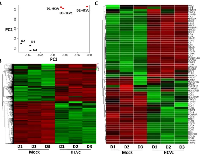
Our previous work reported that HCVc induced CD14+monocytes to differentiate into mono-cytic (Mo)-MDSCs [25]. We performed RNAseq to investigate if HCVc induced a distinctive gene expression profile in CD14+monocytes. Principal Component Analysis (PCA) showed that the gene expression profiles of HCVc-treated and mock monocytes clustered distinctively
(Fig 1A). Self-organizing heat-maps confirmed that HCVc-treated and mock monocytes
dis-played different gene expression profiles (Fig 1B). Among the most differentially expressed genes, we observed that HCVc induced monocytes a suppressive gene expression profile (i.e. up-regulation of IDO1, IDO2, CD274, IL-10, IL-10RB, SOCS2, SOCS3, and down-regulation of HLA-DR, IL-10RA, CD86), and a distinctive signaling pathway activation (i.e. up-regulation of STAT3, PIK3AP1 and down-regulation of STAT1, PIK3IP1), compared with controls
HCVc and HCVcc induce monocytes with suppressive characteristics in
vitro
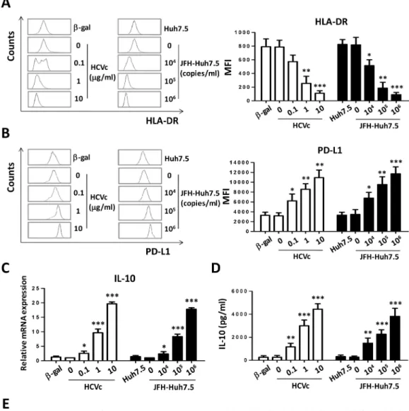
In order to strengthen the conclusion that HCVc induced in monocytes a suppressive gene expression profile, freshly purified monocytes from healthy HCV-negative blood donors were cultured for two days in the presence of increasing concentrations of HCV core protein (HCVc) or cell culture-derived HCV (HCVcc). Our results showed that the down-regulation of HLA-DR expression was induced by HCVc and HCVcc, with significant decrease at 1μg/ml HCVc (MFI 255±59, p = 0.0029) and 104copies/ml HCVcc (MFI 515±49, p = 0.0194) as com-pared with medium (MFI 787±57 or 818±63) (Fig 2A) as previously reported [23]. We also observed that increased PD-L1 expression, which is a key functional molecule in suppressing T cell proliferation and activation [27–30], was significantly induced by both of HCVc and HCVcc in a dose dependent manner (Fig 2B). Cells were collected at 10 hours for assay of IL-10 mRNA levels by qRT-PCR, the results showed that both HCVc and HCVcc caused a dose-dependent increase in IL-10 mRNA expression (Fig 2C) in monocytes, which was consistent
Fig 1. HCVc induces a distinctive gene expression profile in CD14+monocytes. Monocytes were purified from three healthy donors by CD14+microbeads, and cultured with HCVc for 12 hours. RNAseq was performed to analyze the change in transcriptomic profiles. (A) PCA plot representing differential clustering of monocytes treated with HCVc and controls based on mRNA expression profiles. (B) Self-organizing heat-map of the different expression genes with p-value<0.05 across all samples, showing different expression profiles in monocytes from HCVc-treated compared to controls. (C) The heat map shows the relative expression levels of 64 genes (row) of immune responses from monocytes and macrophages. The color scale indicates the degree of relative expression of immune genes (deep red, high expression; deep green, low expression). Abbreviation: RNAseq, RNA sequencing; PCA, Principal Component Analysis.
Fig 2. Both HCVc and HCVcc induce suppressive monocytes in vitro. Purified monocytes from healthy HCV-negative blood donors were cultured for two days in the presence of increasing concentrations of HCVc (0.1, 1.0, 10.0μg/ml) or HCVcc (104, 105,106HCV genome copies/ml). HLA-DR (A) and PD-L1 (B) expression was analyzed by flow cytometry. (C) The mRNA expression of IL-10 was determined by qRT-PCR. (D) IL-10 production was assayed with ELISA. (E) The surface marker expression profile of monocytes was analyzed by flow cytometry. One of five representative experiments is shown. MFI is normalized as the ratio of HCVc/mock and HCVcc/mock for surface marker expression profile. Abbreviation: MFI, Mean Fluorescence intensity.
with the transcriptomic profiles by RNAseq. The cell supernatants were collected at 48 hours, and IL-10 production was assayed by ELISA. Both HCVc and HCVcc induced a dose-depen-dent and significant increase of IL-10 production (Fig 2D). These data suggest that HCV may employ HCVc to induce IL-10 expression and production, as well as to up-regulate PD-L1, and down-regulate HLA-DR in monocytes.
To further characterize HCV-induced suppressive monocytes, we analyzed their surface marker expression profile. A representative result was shown inFig 2E. Using monocytes cul-tured with tissue culture medium alone (mock) as control, HCVc and HCVcc significantly induced HLA-DR down-regulation and PD-L1 up-regulation in monocytes as above. In addi-tion, a decreased expression level of CD11b and CD16, and an increased CD123 expression, were observed on monocytes cultured with either HCVc or HCVcc. There was no difference with respect to the expression level of CD33, CD54, CD40, CD80, CD86, B7H4, CD66b, CD62L, CD1a and CD163 on HCVc- and HCVcc-induced or control monocytes.
HCVc and HCVcc induce IDO1 expression in monocytes
IDO1 is the rate-limiting enzyme that catalyzes tryptophan (Trp) degradation through the kynurenine (Kyn) pathway [31]. Elevated tryptophan catabolism leads to Trp starvation of T cells, and subsequently to limit T cell proliferation and activation [32]. Moreover, IDO1 can also enhance immunosuppression mediated by regulatory T cells (Tregs) [33]. We further tested whether HCVc and HCVcc induce IDO1 expression to produce Kyn and degrade Trp in monocytes. Purified monocytes from healthy HCV-negative blood donors were cultured with HCVc or HCVcc. Cells were collected at 10 hours for measuring IDO1 mRNA levels by qRT-PCR. The result showed that both HCVc and HCVcc caused a dose-dependent increase in IDO1 mRNA expression in monocytes (Fig 3A). Intracellular staining of IDO1 proteins in monocytes showed that HCVc or HCVcc induced 5–10 fold up-regulation above control (Fig 3B, HCVc: 47.2%±1.3, HCVcc: 21.7%±1.5 vs control: 4.2%±0.9 or 3.9%±0.6). The cell culture supernatants were obtained and Kyn and Trp levels were examined by LC-MS. Both HCVc and HCVcc induced monocytes to release 10 fold higher Kyn and 2 fold lower Trp than con-trols (Fig 3C, 3D and 3E). These results indicate that HCV induced IDO1 expression in mono-cytes to catalyze Trp degradation and Kyn accumulation.
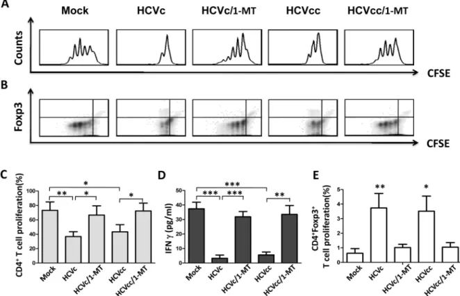
HCV-induced suppressive monocytes inhibit autologous CD4
+T cell
activation, associated with CD4
+Foxp3
+Treg expansion in an
IDO1-dependent manner
and HCVcc-treated monocytes (Fig 4B and 4E). However, anti-PD-L1 and anti-IL-10 antibod-ies showed no significant effect (S1 Fig). In addition, HCVc- and HCVcc-treated monocytes were co-cultured with autologous T cells, we found that HCVc- and HCVcc-treated mono-cytes also exhibited their suppressive ability on CD8+T cell proliferation (S2 Fig).
HCVc and HCVcc induce suppressive monocytes via TLR2/PI3K/AKT/
STAT3 signaling pathways
We have reported that HCVc induces TLR2-mediated activation of monocytes [10]. We thus tested if HCVc-induced suppressive monocytes were similarly mediated through engagement with TLR2. Purified monocytes were pretreated with different concentrations of anti-TLR2 antibody, and then treated with HCVc for two days. As expected, blockade of HCVc/TLR2 engagement with the anti-TLR2 antibody inhibited HCVc-induced expression of IDO1 (Fig 5A), PD-L1 (Fig 5C), and IL-10 (Fig 5D), and restored partially HLA-DR expression (Fig 5B).
To further identify the mechanism(s) responsible for HCVc- and HCVcc-induced suppres-sive monocytes, we examined the activation status of important signaling and transcription factors which influence monocyte activation and function following exposure to HCVc and HCVcc. STAT3 and AKT phosphorylation levels were increased following HCVc and HCVcc treatment, and LY2904002 (an inhibitor of PI3K) inhibited the HCVc- and HCVcc- induced STAT3 and AKT phosphorylation (Fig 6A). These results clearly indicate that HCVc- and
Fig 3. HCVc and HCVcc induce IDO1 expression, and produce kynurenine (Kyn) and degradate tryptophan (Trp) in monocytes. Purified monocytes from healthy HCV-negative blood donors were cultured with HCVc or HCVcc. Cells were collected at 10 hours for measuring IDO1 mRNA levels by qRT-PCR (A). Intracellular staining was performed for IDO1 expression (B). The cell culture supernatants were collected, and the concentrations of Kyn and Trp were examined by liquid chromatography-mass spectrometry. The concentrations of Kyn (C) and Trp (D) are shown, and E shows the ratio of Kyn/Trp. The bars represent the standard error of the mean. Abbreviation: Kyn, kynurenine; Trp, tryptophan; IDO1, indoleamine 2,3-dioxygenase 1.
HCVcc-mediated signaling requires the activation of PI3K/AKT for STAT3 phosphorylation. We further validated that the CD14+monocytes from HCV patients had higher levels of AKT and STAT3 phosphorylation than healthy controls (S3 Fig).
We next sought to determine whether HCVc- and HCVcc-mediated signaling pathway through PI3K/AKT/STAT3 resulted in monocytes with suppressive characteristics. Inhibitors of PI3K and STAT3 abolished HCVc-induced expression of IDO1 (Fig 6B), of IL-10 produc-tion (Fig 6C), and of PD-L1 up-regulation (Fig 6E), and restored expression of HLA-DR (Fig 6D). These results suggest that HCVc and HCVcc induce MDSC-like monocytes through the PI3K/AKT/STAT3 signaling pathway.
Peripheral monocytes from patients with chronic HCV infection display
MDSC-like suppressive characteristics, correlated with a significant
increase in CD14
+MDSCs and CD4
+Tregs in chronic HCV infected
patients compared to healthy individuals
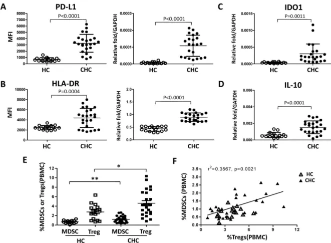
Peripheral blood mononuclear cells (PBMCs) were isolated from healthy individuals (n = 16) and chronic HCV infected patients (n = 24). We first analyzed PD-L1 and HLA-DR expression on peripheral blood monocytes of the two group individuals. Peripheral blood monocytes were identified by their size and granularity in forward and side scatter plots and CD14
Fig 4. HCVc and HCVcc induce suppressive monocytes to inhibit autologous CD4+T cell activation and expand CD4+Foxp3+Tregs in an IDO1-dependent manner. CFSE-labeled CD4+T cells were stimulated with pre-coated OKT3 (CD3 mAb) and CD28 antibody, and co-cultured with HCVc- and HCVcc-treated autologous monocytes in the absence or presence of 1-MT. Foxp3 expression and proliferation of the autologous CD4+T cell were evaluated by intracellular staining and flow
cytometry. One of five representative experiments is shown as histogram (A) and contour (B). The proliferation of CD4+T cells (C) and CD4+Foxp3+T cells (E) are statistically analyzed from five experiments. The supernatants were collected and assayed by ELISA for IFNγproduction (D). The bars represent the standard error of the mean. Abbreviation: 1-MT, 1-methyl-DL-tryptophan.
expression. The results were shown inFig 7A and 7B. The expression levels of PD-L1 and HLA-DR on monocytes from patients with HCV infection were significantly higher than those from healthy individuals (PD-L1 MFI: 3258±289 vs 690±71.3, p<0.0001; HLA-DR MFI: 4363±387.5 vs 2471±106.1, p = 0.0004).
We then purified peripheral blood monocytes from healthy controls and chronic HCV infected patients by using CD14 microbeads. The expression levels of HLA-DR, PD-L1, IL-10, and IDO1 were determined by qRT-PCR. We found that monocytes from chronic HCV infected patients expressed higher levels of IDO1 (p = 0.0011), PD-L1 (p<0.0001), HLA-DR (p<0.0001), and IL-10 (p<0.0001) than healthy controls (Fig 7A, 7B, 7C and 7D). Finally, we investigated the characteristics of monocytic MDSCs and CD4+CD25+CD127-Tregs in peripheral blood of patients with chronic HCV infection and healthy individuals. Peripheral blood from twenty-four treatment-naive chronic HCV-infected patients and sixteen healthy individuals were collected, and monocytic MDSCs and CD4+CD25+CD127-Tregs were identi-fied according to the gating strategy shown inS4 Fig. Patients with chronic HCV infection showed significant increases in the frequencies of MDSCs and CD4+CD25+CD127-Tregs compared to healthy individuals (Fig 7E), and a positive correlation existed between the fre-quency of MDSCs and that of CD4+CD25+CD127-Tregs (Fig 7F), as well as positive correla-tion of HCV RNA level or HCVc protein level with the percentages of CD14+MDSCs and CD4+CD25+CD127-Tregs in peripheral blood of chronic HCV patients was shown inS5 Fig.
Fig 5. HCVc induces suppressive monocytes through engagement with TLR2. Purified monocytes were pretreated with anti-TLR2 antibody (0.25 and 2.5μg/ml), and then stimulated with HCVc for two days. Cells were collected for the analysis of IDO1 (A), HLA-DR (B), and PD-L1 (C) expression by flow cytometry. The supernatants were collected for detecting IL-10 by ELISA (D).
These results support the hypothesis that HCV induces MDSCs-like monocytes that inhibit T cell proliferation by inducing or expanding CD4+Foxp3+Tregs.
Discussion
HCV viruses and core protein (HCVc) are detected in the blood of HCV infected patients [34,35]. Our previous study has shown that the serum level of HCVc is positively correlated with HCV RNA levels, indicating that HCVc concentration represents a stable and reliable marker of HCV viral replication [9]. It has been reported that HCVc is not only an HCV encoded RNA-binding viral capsid protein, but also an important protein involving in immu-nomodulation [36,37]. HCVc binds and signals through TLR2, leading to cytokine production and immune dysregulation of monocytes [7]. Our recent studies have reported that HCVc induces monocytes to differentiate into monocytic (Mo)-MDSCs [25]. In this study, we found that HCVc induced monocytes with a suppressive gene expression profile, including up-regu-lation of IDO1, CD274, IL-10, STAT3, PIK3AP1 and down-reguup-regu-lation of HLA-DR, CD86, STAT1, PIK3IP1 compared with controls. Therefore, we conclude that HCV exploited HCVc to induce suppressive monocytes. Our results showed here that both HCVc and HCVcc signif-icantly up-regulated PD-L1, IL-10 expression and production, and down-regulated HLA-DR expression on monocytes. In addition, decreased CD11b and CD16 expression levels were observed on monocytes treated with HCVc or HCVcc (Fig 2). Moreover, both HCVc and
Fig 6. PI3K and STAT3 are involved in HCVc- and HCVcc-induced suppressive monocytes. Purified monocytes from healthy HCV-negative blood donors were cultured with (10.0μg/ml) or HCVcc (106copies/ml) in the presence or absence of PI3K inhibitor (LY294002) or STAT3 inhibitor (stattic). Western blots were performed for AKT/AKT phosphorylation and STAT3/ STAT3 phosphorylation (A), and IDO1 expression (B).βactin was used as loading control. The supernatants were collected for IL-10 detection by ELISA (C). Cells were collected for the analysis of HLA-DR (D) and PD-L1 (E) expression by flow cytometry.
HCVcc induced IDO1 expression in monocytes to catalyze the Trp degradation and Kyn accu-mulation (Fig 3). Furthermore, we found that blockade of HCVc interaction with TLR2 inhib-ited HCVc-induced PD-L1 and IDO1 expression, and IL-10 production (Fig 5). These results indicate that HCV induces suppressive monocytes by HCVc engagement with TLR2 [10].
It has been reported that T cell immune function in chronic HCV infection is significantly impaired [38]. HCV exploits active immunosuppressive strategies to interfere with antigen-presenting cells, such as DC and macrophages to impair the function of adaptive immune effector cells [13]. Regulatory T cells (Tregs) are increased in chronic HCV infected patients, which may contribute to the sustained suppression of HCV-specific T-cell responses and per-sistant HCV infection. In 87 chronic HCV-infected patients, our recent study indicated that the level of HCVc showed a significant correlation with that of CD4+CD25+Foxp3+Tregs [9]. In this study, our results showed that both HCVcc- and HCVc-treated monocytes expanded CD4+Foxp3+Tregs, and inhibited autologous CD4+and CD8+T cell activation. Moreover, 1-methyl-DL-tryptophan (1-MT, a specific inhibitor of IDO1) suppressed CD4+Foxp3+T cell expansion triggered by HCVc- and HCVcc-treated monocytes, and abolished the ability of
Fig 7. Patients with chronic HCV infection display higher frequency of monocytic MDSCs and CD4+CD25+CD127-Tregs. PBMCs were isolated from healthy individuals (n = 16) and chronic hepatitis C patients (n = 24). PD-L1 (A) and HLA-DR (B) expression of monocytes was analyzed by flow cytometry. Monocytes from healthy individuals and patients with HCV infection were purified, and expression of IDO1 (C), PD-L1 (A), HLA-DR (B), and IL-10 (D) mRNA in monocytes was determined by qRT-PCR. Monocytic MDSCs and CD4+CD25+CD127-Tregs were characterized in peripheral blood of patients with chronic HCV infection and healthy individuals by flow cytometry. E shows statistical analysis, and F shows the correlation between the frequency of MDSCs and CD4+CD25+CD127-Tregs in chronic hepatitis C patients. Abbreviation: PBMCs, peripheral blood mononuclear cells; HC, healthy control; CHC, chronic hepatitis C patients.
HCVc- and HCVcc-treated monocytes to suppress CD4+T cell activation (Fig 4). Therefore, these results further demonstrate that HCV employed HCVc to increase CD4+CD25+Foxp3+ regulatory T cells and inhibit T cell immune response by inducing suppressive monocytes. Recent studies have demonstrated a crucial role of Indoleamine 2,3-dioxygenase (IDO) in the induction of immune tolerance during infection, pregnancy, transplantation, autoimmunity, and cancers [39,40]. IDO induces Tregs and inhibits T-cell activation through Trp starvation and/or the accumulation of Kyn [41,42]. Our results showed that both HCVc and HCVcc pro-moted monocyte-mediated Trp degradation and induced the accumulation of Kyn (Fig 3). An increased Kyn/Trp ratio in the cell culture supernatant of monocytes treated with HCVc or HCVcc was in accordance with the elevated IDO1 expression. The induction of IDO1 expres-sion is likely a strategy of HCV to suppress the host immunity and promote HCV persistence. Our study indicate that the IDO1 inhibitor 1-MT effectively restores CD4+T cell proliferation co-cultured with HCVc- or HCVcc-treated monocytes. These results are consistent with previ-ous studies, which report that HCV induces the accumulation of MDSCs leading to suppress-ing autologous T cells in an ROS-dependent manner [23]. MDSCs clearly work in more than one way, but our results imply that the use of a selective IDO1 inhibitor might be promising as a treatment of treating HCV infection.
Signal transducer and activator of transcription (STAT) protein 3 is important in the regu-lation of inflammatory responses by APCs. In monocytes, STAT3 exerts a critical function in limiting excessive inflammatory responses [43]. Our previous report demonstrated that the expression of PD-L1 (induced by TLR2 and TLR4) and TRAIL (induced by TLR3) on human Kupffer cells depended on phosphatidylinositol-3-kinase (PI3K) activity, revealing the cross talk between the MyD88 and toll-interleukin 1 receptor-domain-containing adapter-inducing IFN (TRIF) [7]. It has been reported that an important role for extracellular HCVc in the acti-vation of STAT3 in human monocytes depends upon the PI3K/AKT pathway [13]. In agree-ment with it, our results showed that both HCVc and HCVcc induced STAT3 and AKT phosphorylations in monocytes. We also observed that the peripheral monocytes from HCV patients had higher levels of AKT and STAT3 phosphorylation than healthy controls. In addi-tion, LY294002 (PI3K inhibitor) inhibited not only HCVc- and HCVcc-induced AKT phos-phorylations, but also STAT3 phosphorylations. We further found that HCVc-induced IDO1 expression and IL-10 production were inhibited by either PI3K or STAT3 inhibitors, and the inhibitors of PI3K and STAT3 also reversed the inhibition of HLA-DR expression by HCVc and HCVcc (Fig 6). A previous study has reported that PI3K-specific inhibition or shRNA knockdown diminishes IFNγ-induced IDO production [44]. These results suggest that HCV employs HCVc to induce MDSC-like suppressive monocytes through the TLR2/PI3K/AKT/ STAT3 signaling pathway.
Myeloid-derived suppressor cells (MDSCs) are characterized by an aberrant myeloid phe-notype and the ability to suppress immue responses. Human MDSCs are dichotomized into granulocytic and monocytic subsets. They exert their suppressive function through a plethora of mechanisms, including production of reactive oxygen species (ROS), immune-modulating cytokines (eg, IL-10), cyclooxygenase-2 (COX2), arginase-1 (ARG1), and indoleamine-2,3-dioxygenase (IDO) [48]. Based on our data and literatures, therefore, we suppose that HCVc and HCVcc induce monocytes differentiation to monocytic MDSCs, which express inhibitory molecules PD-L1, IDO1, IL-10, and suppress autologous CD4+T cell activation. Consistently, we observed in this study that the expression levels of PD-L1, IL-10 and IDO1 on monocytes from patients with HCV infection were significantly higher than those from healthy individuals. Finally, we characterized monocytic MDSCs and CD4+CD25+CD127 -Tregs in peripheral blood of patients with chronic HCV infection and healthy individuals according to their specific markers, and found that both of the frequencies of MDSCs and CD4+CD25+CD127-Tregs in chronic HCV infection were significantly higher than healthy individuals, which was consistent with some recent studies [24,49,50]. We also found the cor-relation between the frequency of MDSCs and CD4+CD25+CD127-Tregs (Fig 7), and the cor-relation of HCV RNA level or HCVc protein level with the percentages of CD14+MDSCs and CD4+CD25+CD127-Tregs in peripheral blood of chronic HCV patients, respectively.
In sum, our results indicate that HCV employed HCVc to induce MDSC-like monocytes via TLR2/PI3K/AKT/STAT3 signaling pathway. These MDSC-like monocytes inhibited CD4+T cell activation and expanded CD4+CD25+Foxp3+Tregs by IDO1-induced Trp degra-dation and the accumulation of Kyn. Moreover, we validated that peripheral monocytes from patients with chronic HCV infection displayed MDSCs characteristic in comparison to monocytes from healthy individuals, and the correlation between the frequency of MDSCs and CD4+CD25+CD127-Tregs in patients with chronic HCV infection. Taken together, our study identified the critical upstream signals for the induction of IDO1 in monocytes stimu-lated by hepatitis C virus that could be manipustimu-lated therapeutically to enhance or decrease their immunosuppressive function for HCV treatment. It will be of interest to test whether antagonizing suppressive functions of MDSCs could represent a mean for enhancing immune responses in chronic HCV infection.
Supporting Information
S1 Dataset. Mean Fluorescence intensity of PD-L1 on monocytes from healthy individuals and chronic hepatitis C patients analyzed by flow cytometry.
(XLSX)
S1 Fig. HCVc induce suppressive monocytes to inhibit autologous CD4+T cell prolifera-tion and expand CD4+Foxp3+Tregs in an IDO1-dependent manner, while PD-L1 and IL-10 show no significant effect.
(TIF)
S2 Fig. HCVc and HCVcc induce suppressive monocytes to inhibit autologous CD8+T cell proliferation. Purified monocytes from healthy HCV-negative blood donors were cultured
S3 Fig. Flow cytometric analysis of Akt (pS473) and Stat3 (pY705) levels in the peripheral CD14+monocytes of HCV patients. PBMCs were isolated from healthy individuals
(unshaded, n = 3) and treatment-naive chronic hepatitis C patients (shaded, n = 5). Cells were stained with anti-CD14-APC-Cy7 antibody, and then fixed for 10 min at 37˚C using BD Phosflow™Fix Buffer I (Cat. No. 557870), permeabilized in BD Phosflow™Perm Buffer III (Cat. No. 558050) on ice for 30 min. Cells were washed twice and stained with PE-CF594 Mouse Anti-Akt (pS473) antibody and Alexa Fluor 647 Mouse Anti-Stat3 (pY705) antibody for 30 min at room temperature. CD14+monocytes were analyzed for AKT phosphorylation (A) and STAT3 phosphorylation status (B). Abbreviation: PBMCs, peripheral blood mono-nuclear cells; HC, healthy control; CHC, chronic hepatitis C patients; MFI, Mean Fluores-cence intensity.
(TIF)
S4 Fig. Gating strategy for monocytic MDSCs and CD4+CD25+CD127-Tregs.
(TIF)
S5 Fig. Correlation of HCV RNA level or HCVc protein level with the percentages of CD14+MDSCs (A) and CD4+CD25+CD127-Tregs (B) in peripheral blood of 24 chronic HCV patients.
(TIF)
S6 Fig. Rough uncut image forFig 6.
(TIF)
S1 Table. Characteristics of hepatitis C patients and healthy controls.
(DOCX)
S2 Table. Primer sequences for qRT-PCR.
(DOCX)
Acknowledgments
The authors would like to thank all the patients and healthy donors for participation in our study, and all the staff for clinical data and specimen collection.
Author Contributions
Conceptualization: NZ INC LS ZT.
Formal analysis: NZ INC LS ZT.
Funding acquisition: NZ LS ZT.
Investigation: NZ HL HS YY AC TL.
Project administration: NZ ZT.
Resources: HL JN.
Writing – original draft: NZ ZT.
Writing – review & editing: NZ INC LS ZT.
References
2. Rouse BT, Sehrawat S. Immunity and immunopathology to viruses: what decides the outcome? Nat Rev Immunol 2010; 10:514–526. doi:10.1038/nri2802PMID:20577268
3. Bowen DG, Walker CM. Adaptive immune responses in acute and chronic hepatitis C virus infection. Nature 2005; 436:946–952. doi:10.1038/nature04079PMID:16107834
4. Pawlotsky JM. New hepatitis C therapies: the toolbox, strategies, and challenges. Gastroenterology 2014; 146:1176–1192. doi:10.1053/j.gastro.2014.03.003PMID:24631495
5. Ding Q, Cao X, Lu J, Huang B, Liu YJ, Kato N, et al. Hepatitis C virus NS4B blocks the interaction of STING and TBK1 to evade host innate immunity. J Hepatol 2013; 59:52–58. doi:10.1016/j.jhep.2013. 03.019PMID:23542348
6. Dolganiuc A, Norkina O, Kodys K, Catalano D, Bakis G, Marshall C, et al. Viral and host factors induce macrophage activation and loss of toll-like receptor tolerance in chronic HCV infection. Gastroenterol-ogy 2007; 133:1627–1636. doi:10.1053/j.gastro.2007.08.003PMID:17916356
7. Tu Z, Pierce RH, Kurtis J, Kuroki Y, Crispe IN, Orloff MS. Hepatitis C Virus Core Protein Subverts the Antiviral Activities of Human Kupffer Cells. Gastroenterology 2010; 138:305–314. doi:10.1053/j.gastro. 2009.09.009PMID:19769973
8. Hosomura N, Kono H, Tsuchiya M, Ishii K, Ogiku M, Matsuda M, et al. HCV-related proteins activate Kupffer cells isolated from human liver tissues. Dig Dis Sci 2011; 56:1057–1064. doi: 10.1007/s10620-010-1395-yPMID:20848204
9. Zhai N, Chi X, Li T, Song H, Li H, Jin X, et al. Hepatitis C virus core protein triggers expansion and acti-vation of CD4+CD25+ regulatory T cells in chronic hepatitis C patients. Cell Mol Immunol 2015; 12:743–749. doi:10.1038/cmi.2014.119PMID:25531392
10. Tu Z, Hamalainen-Laanaya HK, Nishitani C, Kuroki Y, Crispe IN, Orloff MS. HCV core and NS3 proteins manipulate human blood-derived dendritic cell development and promote Th 17 differentiation. Int Immunol 2012; 24:97–106. doi:10.1093/intimm/dxr104PMID:22190574
11. Waggoner SN, Cruise MW, Kassel R, Hahn YS. gC1q receptor ligation selectively down-regulates human IL-12 production through activation of the phosphoinositide 3-kinase pathway. J Immunol 2005; 175:4706–4714. PMID:16177118
12. Lin W, Kim SS, Yeung E, Kamegaya Y, Blackard JT, Kim KA, et al. Hepatitis C virus core protein blocks interferon signaling by interaction with the STAT1 SH2 domain. J Virol 2006; 80:9226–9235. doi:10. 1128/JVI.00459-06PMID:16940534
13. Tacke RS, Tosello-Trampont A, Nguyen V, Mullins DW, Hahn YS. Extracellular Hepatitis C Virus Core Protein Activates STAT3 in Human Monocytes/Macrophages/Dendritic Cells via an IL-6 Autocrine Path-way. J Biol Chem 2011; 286:10847–10855. doi:10.1074/jbc.M110.217653PMID:21282107
14. Ifrim DC, Quintin J, Joosten LA, Jacobs C, Jansen T, Jacobs L, et al. Trained Immunity or Tolerance: Opposing Functional Programs Induced in Human Monocytes after Engagement of Various Pattern Recognition Receptors. Clin Vaccine Immunol 2014; 21:534–545. doi:10.1128/CVI.00688-13PMID:
24521784
15. Nagaraj S, Nelson A, Youn JI, Cheng P, Quiceno D, Gabrilovich DI. Antigen-specific CD4(+) T cells reg-ulate function of myeloid-derived suppressor cells in cancer via retrograde MHC class II signaling. Can-cer Res 2012; 72:928–938. doi:10.1158/0008-5472.CAN-11-2863PMID:22237629
16. Wu J, Zhang R, Tang N, Gong Z, Zhou J, Chen Y, et al. Dopamine inhibits the function of Gr-1+CD115+ myeloid-derived suppressor cells through D1-like receptors and enhances anti-tumor immunity. J Leu-koc Biol 2015; 97:191–200. doi:10.1189/jlb.5A1113-626RRPMID:25341727
17. Mielcarek M, Martin PJ, Torok-Storb B. Suppression of Alloantigen-Induced T-Cell Proliferation by CD14+ Cells Derived From Granulocyte Colony-Stimulating Factor–Mobilized Peripheral Blood Mono-nuclear Cells. Blood 1997; 89:1629–1634. PMID:9057645
18. Ino K, Bierman PJ, Varney ML, Heimann DG, Kuszynski CA, Walker SA, et al. Monocyte activation by an oral immunomodulator (bestatin) in lymphoma patients following autologous bone marrow transplan-tation. Cancer Immunol Immunother 1996; 43: 206–212. PMID:9003465
19. Jitschin R, Braun M, Bu¨ttner M, Dettmer-Wilde K, Bricks J, Berger J, et al. CLL-cells induce IDOhi CD14+HLA-DRlo myeloid-derived suppressor cells that inhibit T-cell responses and promote TRegs. Blood 2014; 124:750–760. doi:10.1182/blood-2013-12-546416PMID:24850760
20. Larrea E, Riezu-Boj JI, Gil-Guerrero L, Casares N, Aldabe R, Sarobe P, et al. Upregulation of indolea-mine 2,3-dioxygenase in hepatitis C virus infection. J Virol 2007; 81:3662–3666. doi:10.1128/JVI. 02248-06PMID:17229698
21. Higashitani K, Kanto T, Kuroda S, Yoshio S, Matsubara T, Kakita N, et al. Association of enhanced activity of indoleamine 2,3-dioxygenase in dendritic cells with the induction of regulatory T cells in chronic hepatitis C infection. J Gastroenterol 2013; 48:660–670. doi:10.1007/s00535-012-0667-z
22. Schulz S, Landi A, Garg R, Wilson JA, van Drunen Littel-van den Hurk S. Indolamine 2,3-dioxygenase expression by monocytes and dendritic cell populations in hepatitis C patients. Clin Exp Immunol 2015; 180:484–498. doi:10.1111/cei.12586PMID:25605587
23. Tacke RS, Lee HC, Goh C, Courtney J, Polyak SJ, Rosen HR, et al. Myeloid suppressor cells induced by hepatitis C virus suppress T-cell responses through the production of reactive oxygen species. Hepatology 2012; 55:343–353. doi:10.1002/hep.24700PMID:21953144
24. Zeng QL, Yang B, Sun HQ, Feng GH, Jin L, Zou ZS, et al. Myeloid-Derived Suppressor Cells Are Asso-ciated with Viral Persistence and Downregulation of TCRζChain Expression on CD8(+) T Cells in Chronic Hepatitis C Patients. Mol Cells 2014; 37:66–73. doi:10.14348/molcells.2014.2282PMID:
24552712
25. Pang X, Song H, Zhang Q, Tu Z, Niu J. Hepatitis C virus regulates the production of monocytic myeloid-derived suppressor cells from peripheral blood mononuclear cells through PI3K pathway and autocrine signaling. Clin Immunol 2016; 164:57–64. PMID:26821305
26. Zhong J, Gastaminza P, Cheng G, Kapadia S, Kato T, Burton DR, et al. Robust hepatitis C virus infec-tion in vitro. Proc Natl Acad Sci U S A 2005; 102:9294–9299. doi:10.1073/pnas.0503596102PMID:
15939869
27. Shi L, Chen S, Yang L, Li Y. The role of PD-1 and PD-L1 in T-cell immune suppression in patients with hematological malignancies. J Hematol Oncol 2013; 6:74. doi:10.1186/1756-8722-6-74PMID:
24283718
28. Francisco LM, Salinas VH, Brown KE, Vanguri VK, Freeman GJ, Kuchroo VK, et al. PD-L1 regulates the development, maintenance, and function of induced regulatory T cells. J Exp Med 2009; 206:3015– 3029. doi:10.1084/jem.20090847PMID:20008522
29. Shi F, Shi M, Zeng Z, Qi RZ, Liu ZW, Zhang JY, et al. PD-1 and PD-L1 upregulation promotes CD8+ T-cell apoptosis and postoperative recurrence in hepatoT-cellular carcinoma patients. Int J Cancer 2011; 128:887–896. doi:10.1002/ijc.25397PMID:20473887
30. Brusa D, Serra S, Coscia M, Rossi D, D’Arena G, Laurenti L, et al. The PD-1/PD-L1 Axis Contributes To T-Cell Dysfunction In Chronic Lymphocytic Leukemia. Haematologica 2013; 98:953–963. doi:10.3324/ haematol.2012.077537PMID:23300177
31. Yasui H, Takai K, Yoshida R, Hayaishi O. Interferon enhances tryptophan metabolism by inducing pul-monary indoleamine 2,3-dioxygenase: its possible occurrence in cancer patients. Proc Natl Acad Sci U S A 1986; 83:6622–6626. PMID:2428037
32. Mellor AL, Munn D, Chandler P, Keskin D, Johnson T, Marshall B, et al. Tryptophan catabolism and T cell responses. Adv Exp Med Biol 2003; 527:27–35. PMID:15206713
33. Munn DH, Mellor AL. Indoleamine 2,3-dioxygenase and tumor-induced tolerance. J Clin Invest 2007; 117:1147–1154. doi:10.1172/JCI31178PMID:17476344
34. Maillard P, Krawczynski K, Nitkiewicz J, Bronnert C, Sidorkiewicz M, Gounon P, et al. Nonenveloped Nucleocapsids of Hepatitis C Virus in the Serum of Infected Patients. J Virol 2001; 75:8240–8250. doi:
10.1128/JVI.75.17.8240-8250.2001PMID:11483769
35. Masalova OV, Atanadze SN, Samokhvalov EI, Petrakova NV, Kalinina TI, Smirnov VD, et al. Detection of hepatitis C virus core protein circulating within different virus particle populations. J Med Virol 1998; 55:1–6. PMID:9580878
36. Sundstro¨m S, Ota S, Dimberg LY, Masucci MG, Bergqvist A. Hepatitis C virus core protein induces an anergic state characterized by decreased interleukin-2 production and perturbation of mitogen-acti-vated protein kinase responses. J Virol 2005; 79:2230–2239. doi:10.1128/JVI.79.4.2230-2239.2005
PMID:15681425
37. Eisen-Vandervelde AL, Waggoner SN, Yao ZQ, Cale EM, Hahn CS, Hahn YS. Hepatitis C Virus Core Selectively Suppresses Interleukin-12 Synthesis in Human Macrophages by Interfering with AP-1 Acti-vation. J Biol Chem 2004; 279:43479–43486. doi:10.1074/jbc.M407640200PMID:15292184
38. Soguero C, Joo M, Chianese-Bullock KA, Nguyen DT, Tung K, Hahn YS. Hepatitis C Virus Core Protein Leads to Immune Suppression and Liver Damage in a Transgenic Murine Model. J Virol 2002; 76:9345–9354. doi:10.1128/JVI.76.18.9345-9354.2002PMID:12186917
39. Boasso A, Herbeuval JP, Hardy AW, Anderson SA, Dolan MJ, Fuchs D, et al. HIV inhibits CD4+ T-cell proliferation by inducing indoleamine 2,3-dioxygenase in plasmacytoid dendritic cells. Blood 2007; 109:3351–3359. doi:10.1182/blood-2006-07-034785PMID:17158233
41. Munn DH, Sharma MD, Lee JR, Jhaver KG, Johnson TS, Keskin DB, et al. Potential Regulatory Func-tion of Human Dendritic Cells Expressing Indoleamine 2,3-Dioxygenase. Science 2002; 297:1867– 1870. doi:10.1126/science.1073514PMID:12228717
42. Ju¨rgens B, Hainz U, Fuchs D, Felzmann T, Heitger A. Interferon-gamma-triggered indoleamine 2,3-dioxygenase competence in human monocyte-derived dendritic cells induces regulatory activity in allo-geneic T cells. Blood 2009; 114:3235–3243. doi:10.1182/blood-2008-12-195073PMID:19625705
43. Kortylewski M, Yu H. Role of Stat3 in suppressing anti-tumor immunity. Curr Opin Immunol 2008; 20:228–233. doi:10.1016/j.coi.2008.03.010PMID:18479894
44. Mounayar M, Kefaloyianni E, Smith B, Solhjou Z, Maarouf OH, Azzi J, et al. PI3kαand STAT1 Interplay Regulates Human Mesenchymal Stem Cell Immune Polarization. Stem Cells 2015; 33:1892–1901. doi:10.1002/stem.1986PMID:25753288
45. Zheng J, Liang H, Xu C, Xu Q, Zhang T, Shen T, et al. An unbalanced PD-L1/CD86 ratio in CD14(++) CD16(+) monocytes is correlated with HCV viremia during chronic HCV infection. Cell Mol Immunol 2014; 11:294–304. doi:10.1038/cmi.2013.70PMID:24531620
46. Jeong HY, Lee YJ, Seo SK, Lee SW, Park SJ, Lee JN, et al. Blocking of monocyte-associated B7-H1 (CD274) enhances HCV-specific T cell immunity in chronic hepatitis C infection. J Leukoc Biol 2008; 83:755–764. doi:10.1189/jlb.0307168PMID:18086898
47. Frumento G, Rotondo R, Tonetti M, Damonte G, Benatti U, Ferrara GB. Tryptophan-derived Catabolites Are Responsible for Inhibition of T and Natural Killer Cell Proliferation Induced by Indoleamine 2,3-Diox-ygenase. J Exp Med 2002; 196:459–468. doi:10.1084/jem.20020121PMID:12186838
48. Talmadge JE, Gabrilovich DI. History of myeloid-derived suppressor cells. Nat Rev Cancer 2013; 13:739–752. doi:10.1038/nrc3581PMID:24060865
49. Vasquez-Dunddel D, Pan F, Zeng Q, Gorbounov M, Albesiano E, Fu J, et al. STAT3 regulates argi-nase-I in myeloid-derived suppressor cells from cancer patients. J Clin Invest 2013; 123:1580–1589. doi:10.1172/JCI60083PMID:23454751