PHARMACOLOGICALREVIEWS Pharmacol Rev 68:1074–1109, October 2016 Copyright © 2016 by The Author(s)
This is an open access article distributed under the CC BY-NC Attribution 4.0 International license.
ASSOCIATE EDITOR: LESLIE A. MORROW
Adolescent Alcohol Exposure Persistently Impacts
Adult Neurobiology and Behavior
Fulton T. Crews, Ryan P. Vetreno, Margaret A. Broadwater, and Donita L. Robinson
Bowles Center for Alcohol Studies (F.T.C., R.P.V., M.A.B., D.L.R.), Department of Psychiatry (F.T.C., D.L.R.), and Department of Pharmacology (F.T.C.), School of Medicine, University of North Carolina, Chapel Hill, North Carolina
Abstract . . . 1074
I. Introduction . . . 1075
II. Brain Maturation and Adolescence . . . 1076
III. Adolescent Alcohol Sensitivity. . . 1078
A. Developmental Insensitivity to Ethanol . . . 1078
B. Developmental Sensitivity to Ethanol. . . 1078
IV. Adolescents Binge Drink . . . 1079
V. Modeling Adolescent Alcohol Drinking. . . 1081
VI. Lock-In—Persistence of an Adolescent Phenotype in Adulthood, Including an Adolescent-Typical Response to Ethanol . . . 1082
VII. AIE Increases Ethanol Self-Administration in Adulthood . . . 1083
VIII. AIE Results in Decreased Behavioral Flexibility . . . 1083
A. Flexibility in Spatial Tasks . . . 1083
B. Flexibility on Operant Tasks . . . 1085
IX. Adolescent Alcohol Effects on Anxiety and Negative Affective Behavior . . . 1085
A. Rodent Models of Anxiety . . . 1085
B. Anxiety or Disinhibition? . . . 1086
C. Rodent Models of Social Anxiety . . . 1086
X. Adolescent Alcohol-Induced Neuroimmune Gene Induction. . . 1087
XI. Brain Electroencephalography and Sleep . . . 1089
XII. Cholinergic System Development and Effects of AIE . . . 1091
XIII. Monoamine System Development and Effects of AIE. . . 1092
A. Dopamine . . . 1092
B. 5-HT . . . 1094
XIV. Hippocampal Development and Effects of AIE . . . 1095
A. Neurogenesis in Development and Adulthood . . . 1096
B. Long-Lasting Loss of Neurogenesis after AIE . . . 1097
XV. Adolescent Alcohol-Induced Epigenetic Alterations in Gene Expression. . . 1099
XVI. Conclusions. . . 1100
References . . . 1103
Abstract——Adolescence is a developmental period when physical and cognitive abilities are optimized, when social skills are consolidated, and when sexual-ity, adolescent behaviors, and frontal cortical functions mature to adult levels. Adolescents also have unique responses to alcohol compared with adults, being less
sensitive to ethanol sedative–motor responses that most likely contribute to binge drinking and blackouts. Population studies find that an early age of drinking onset correlates with increased lifetime risks for the development of alcohol dependence, violence, and injuries. Brain synapses, myelination, and neural
This work was supported in part by the National Institutes of Health, National Institute on Alcoholism and Alcohol Abuse [Grants AA019767, AA11605, AA007573, and AA021040], Neurobiology of Adolescent Drinking in Adulthood Consortium [Grants AA020023 and AA020024], and National Institute on Alcoholism and Alcohol Abuse Training Grant AA007573.
Address correspondence to:Dr. Fulton T. Crews, University of North Carolina School of Medicine, Bowles Center for Alcohol Studies, 1021 Thurston Bowles Building CB 7178, Chapel Hill, NC 27599-7178. E-mail: [email protected]
dx.doi.org/10.1124/pr.115.012138.
circuits mature in adolescence to adult levels in parallel with increased reflection on the consequence of actions and reduced impulsivity and thrill seeking. Alcohol binge drinking could alter human development, but variations in genetics, peer groups, family structure, early life experiences, and the emergence of psychopathology in humans confound studies. As adolescence is common to mammalian species, preclinical models of binge drinking provide insight into the direct impact of alcohol on adolescent development. This review relates human findings to basic science studies, particularly the pre-clinical studies of the Neurobiology of Adolescent Drinking in Adulthood (NADIA) Consortium. These studies focus on persistent adult changes in neurobiology and behavior following adolescent intermittent ethanol
(AIE), a model of underage drinking. NADIA studies and others find that AIE results in the following: increases in adult alcohol drinking, disinhibition, and social anxiety; altered adult synapses, cognition, and sleep; reduced adult neurogenesis, cholinergic, and serotonergic neurons; and increased neuroimmune gene expression and epigenetic modifiers of gene expression. Many of these effects are specific to adolescents and not found in parallel adult studies. AIE can cause a persistence of adolescent-like synaptic physiology, behavior, and sensitivity to alcohol into adulthood. Together, these findings support the hypothesis that adolescent binge drinking leads to long-lasting changes in the adult brain that increase risks of adult psychopathology, particularly for alcohol dependence.
I. Introduction
Adolescence is a period of developmental transition, encompassing physical, mental, emotional, and social aspects. The development of both physical and inter-personal skills required to successfully integrate into society is essential for living in groups, and these skills improve through adolescence to adult levels. In addi-tion, adolescence is a time when talents, reasoning, and other abilities are formed. Adolescence in humans and other social animals is characterized by high expression of risk taking, exploration, novelty and sensation seek-ing, social interaction, and play behavior that contrib-utes to this transition. Recent discoveries using human brain imaging provide strong evidence that these characteristics are linked to maturation of brain struc-ture (Lenroot and Giedd, 2006; Bava and Tapert, 2010). Although much of development involves programmed sequences of change in gene expression related to cellular differentiation and protein expression, experience and environment during adolescence also contribute to life-long adult abilities and characteristics. Nutrition, alco-hol exposure, and multiple other environmental factors are known to impact both prenatal and postnatal phys-ical development.
Adolescent development of abilities, social skills, and other complex processes is difficult to define and quantitate. However, in general, training and acquisi-tion of skills in adolescence are important for developing both highly-skilled human and animal individuals. Training during adolescence improves abilities involv-ing cognition, like playinvolv-ing chess or traininvolv-ing to be a guide dog, as well as physical abilities. Training at all ages improves performance, but the improvement is often much faster and greater during adolescence. During
adolescence, physical abilities improve in parallel to the development of self-control, consideration of future con-sequences, planning, and socialization skills, and even-tually reductions in risk taking and sensation seeking. Frontal cortical synaptic refinement and increased myelination in adolescence most likely contribute to mat-urational changes in reasoning, goal setting, impulse con-trol, and evaluation of consequences. Other adolescent brain changes include increased hippocampal neurogenesis, maturation of brain regulatory neurotransmitters (e.g., their receptors and transporters), as well as hormonal maturation during puberty. Each of these maturation processes is driven by innate programming that responds to environmental stimuli. Adolescent development is com-mon to humans and rodents, allowing controlled pre-clinical studies to focus on those environmental factors that create resilience or risk for long-lasting changes in adult characteristics.
The complex interactions of nature and nurture, intermixed with adolescent resilience and sensitivities, confound discernment of what characteristics are highly sensitive versus insensitive to environment. Many mental disorders emerge during adolescence, perhaps due to genetically programmed dysfunctional develop-ment, environmental disruption of developmental pro-grams, or more likely a combination of both (Paus et al., 2008; Davidson et al., 2015). In humans, family struc-ture and socioeconomic status, adolescent choice of peer group, and other individual selections create unique environments that confound a clear understanding of their impact on maturation of adult characteristics and skills. Animal studies have the advantage of control over environmental and genetic factors and can better elucidate the impact of specific environmental events on adolescent development. This review presents findings
that support adolescence as a unique period of brain maturation that is characterized by increased vulnera-bility to binge alcohol-induced alterations in brain maturation and adult neurobiology due to the distinct adolescent responses to alcohol relative to adults. Pre-clinical studies from the Neurobiology of Adolescent Drinking in Adulthood (NADIA) Consortium, funded by the National Institute of Alcohol Abuse and Alcoholism, are presented and related to human findings when possible. Together, they support the hypothesis that adolescent binge drinking produces long-lasting effects in the brain that increase the risk for the development of psychopathology in adulthood, including alcohol-use disorders.
The adolescent period is marked by behavioral and hormonal changes that are common across species. Adolescents are highly tuned to the environment and peers, and adolescence is a critical period of social development and integration into society. In the rat, the adolescent period has been conservatively demar-cated as postnatal day (P) 28–P42 (Spear, 2000), although some have suggested a more liberal range from P21 to P60 (Laviola et al., 2003). More recently, the adolescent period has been divided into early (P25– P42) and late (P43–P55) adolescence in rats, with the early and late periods corresponding to approximately 10–18 and 18–25 years of age in humans, respectively (Spear, 2015). Puberty, the hormonal and physiologic change associated with sexual maturation, takes place within the broader adolescent period. Although there are species-specific behavioral and hormonal responses, adolescence and puberty are general developmental periods that are shared across mammalian species. As in humans, complete pubertal maturation of the rat occurs earlier in females than males (approximately P36 and P44, respectively) (Vetter-O’Hagen and Spear, 2012). Importantly, adolescent-typical behavioral char-acteristics are also conserved across species, such as increased reward and sensation seeking, social interac-tions with peers, and risk taking, and reduced responses to aversive stimuli, which are all observed during adolescence, even beyond the peripubertal period (for review, see Spear, 2000, 2011). For instance, increased time spent engaging in social behaviors is common in human adolescents (e.g., increased communication with peers) (Csikszentmihalyi et al., 1977; Steinberg, 1989) as well as in adolescent rodents and nonhuman pri-mates (e.g., increased levels of play and affiliative behaviors, such as huddling and grooming) (Ehardt and Bernstein, 1987). In rodents, increased social interactions influence food choices (Galef, 1977) and sexual and aggressive behaviors (Fagen, 1976; Smith, 1982). Rodent adolescents also find peers (Douglas et al., 2004) and novelty (Douglas et al., 2003) more rewarding than adults do. These adolescent-typical char-acteristics are important during the transition from dependence to independence. These characteristics also
result in increased possibility of environmental exposures and influences. As discussed below, the recent discovery of epigenetic mechanisms under environmental regula-tion may represent a significant porregula-tion of the genetic aspects of adolescent maturation. Adolescent high novelty-seeking and risk-taking behaviors contribute to the in-creased propensity for experimentation and initiation of drug and alcohol use during this developmental period. Furthermore, the ability to learn and acquire new skills or habits can combine with initiation of drug use to in-crease the risk of long-lasting adult pathology. Given that adolescence is a unique period of brain and behavioral development that is highly sensitive to environmental influences, clinical and preclinical studies focused on adolescent development to understand what factors best promote individual success for all in the community are of great importance.
II. Brain Maturation and Adolescence
and Blumcke, 1994; Celio et al., 1998; Frischknecht et al., 2009; see Coleman et al., 2014). Thus, adolescent brain maturation involves neuronal and glial signal-ing that regulates synapses, particularly interneuron– projection neuron synaptic fields that are tuned during development to more stable and less plastic adult brain synapses.
Synapses are functional elements of the brain that are very small—most are less than 0.1mM3—whereas brains are 1012–1014times that size (e.g., human brain is about 1200 cm3and adult rat brain about 2100 mm3) (Oguz et al., 2013). Interestingly, overall brain struc-ture changes during adolescence, with decreases in gray matter and increases in white matter shown in both human (Giedd et al.,1999; Gogtay et al., 2004; Bava et al., 2010) and rodent studies (e.g., Oguz et al., 2013; Mengler et al., 2014). These changes are far larger than can be explained by changes in synapses, and they are thought to be associated with the processes of synaptic pruning, extracellular matrix formation, and increased myelination. The developmental trajectory of brain regional volumes in humans has been studied (Giedd et al., 1996; Sowell et al., 1999; Gogtay et al., 2006; Demaster and Ghetti, 2013) and is generally similar to that found in rats (Calabrese et al., 2013; Oguz, et al., 2013). For instance, subcortical limbic structures, such as the hippocampus and amygdala, mature during adolescence in humans (Giedd, et al., 1996; Sowell et al., 1999; Suzuki et al., 2005; Gogtay et al., 2006; Uematsu et al., 2012; Demaster and Ghetti, 2013) at a relatively faster pace than the prefrontal cortex (PFC) (see Casey et al., 2005 for review). The PFC is the last structure to mature, and development of PFC structural and functional connectivity continues into late adoles-cence and early adulthood in humans (Lebel et al., 2008; Petanjek et al., 2011) and rodents (Cunningham et al., 2002; Markham et al., 2007). An immature PFC, along with more developed limbic regions, may lead to an imbalance or disruption of top-down control, which is thought to underlie particular adolescent-typical be-havior such as impulsivity and risk taking (Andersen and Teicher, 2008; Casey et al., 2008; Ernst and Fudge, 2009; Casey and Jones, 2010). PFC development and connectivity parallel the appearance of adult executive functions.
Late youth and adolescence are also when mental diseases commonly emerge (Paus et al., 2008; Davidson, et al., 2015), with some clearly related to alterations in the patterns of gray and white matter that exemplify the adult brain (Giedd, 2004). Indeed, white matter structures mature hierarchically and become more organized in parallel with the development of cognitive faculties (Asato et al., 2006; Lenroot and Giedd, 2006; Bava and Tapert, 2010). Myelin increases efficient neural transmission throughout the brain, and it is thought to contribute to the enhanced brain-regional connectivity, processing speed, and cognitive function
that occur during childhood and adolescence (Casey et al., 2008). In a study of 885 individuals between 3 and 20 years of age, magnetic resonance imaging brain scans accurately distinguished biologic age, primarily by using diffusivity indices of white matter maturation (Brown et al., 2012). Recent studies have related the development of white matter along an accumbofrontal tract connecting the orbitofrontal cortex (OFC) and nucleus accumbens to the maturation of developmental models of decision making (Karlsgodt et al., 2015). Exercise, as assessed by fitness among adolescents, is associated with increased white matter microstructure and frontal and motor fiber connectivity, consistent with the postulate that environment and experience impact white matter development and connectivity (Herting et al., 2014). In rats, whole brain volume increases by approximately 20% from P28 to P80 (that is, from early adolescence to young adulthood), whereas white matter, including the corpus callosum and exter-nal capsule, increases by about 30% (Oguz et al., 2013). In rats, there are maturational changes in corpus callosum anisotropy found with diffusion tensor imag-ing (Vetreno et al., 2016a), and diffusion tensor imagimag-ing has been used to detect anisotropic changes in the human adolescent brain that are consistent with in-creased myelination (Zhu et al., 2012).
2010 for review). Neuromodulatory neurotransmitters integrate GABAergic interneuronal and glutamatergic pyramidal neuronal firing, synchronizing firing and connectivity. Thus, both human and animal studies are consistent with adolescence being a critical period of frontal cortical activity-dependent plasticity. Fur-thermore, it is thought that adolescent frontal cortical integration underlies the maturation of adult emotion and reasoning. As PFC circuits mature, reflections on long-term consequences start to guide behavior, an important adult characteristic that may blunt the impulsive thrill seeking that is often seen during adolescence.
III. Adolescent Alcohol Sensitivity
A. Developmental Insensitivity to Ethanol
Numerous studies have found that adolescents are less sensitive to certain adverse effects of ethanol relative to adults (see Spear, 2011, 2014; Novier et al., 2015 for review), perhaps contributing to a propensity for adolescents to binge drink (Johnston et al., 2015). [TheNational Institute of Alcohol Abuse and Alcoholism definition of binge drinking is 4+or 5+drinks in a row for females or males, respectively, or achieving blood ethanol concentrations (BECs) of greater than 0.08 g/dL.] For example, adolescent rats are generally less sensitive to ethanol-induced sedative/hypnotic effects (Moy et al., 1998; Silveri and Spear, 1998; Draski et al., 2001), social inhibition at high ethanol doses (Varlinskaya and Spear, 2002), motor impairment (Hollstedt et al., 1980; Silveri and Spear, 2001; White et al., 2002a), conditioned taste aversion (Anderson et al., 2010; Schramm-Sapyta et al., 2010), and acute ethanol withdrawal (i.e., hangover) (Doremus et al., 2003; Varlinskaya and Spear, 2004; Doremus-Fitzwater and Spear, 2007). Thus, adolescents are less sensitive to several factors that may serve as feedback cues to limit alcohol consumption. A low sedative response to alcohol is a risk factor for development of alcohol-use disorder in humans (Schuckit et al., 2004) and is an adolescent characteristic that crosses species (Spear, 2011). Fur-thermore, low sensitivity to the perception of alcohol, as measured by the Subjective High Assessment Scale, has been established as one of the most significant risk factors for the development of heavy drinking and alcoholism (Schuckit et al., 2014). Studies relating blood alcohol to behavior have suggested that adolescent humans are less sensitive than adults (Day et al., 2013), although this is more clearly established in animal studies (Spear, 2014). Another index of alcohol sensitivity may be the amount of alcohol consumed, and studies find that both adolescent humans and rodents consume about twice as much as adults (Spear, 2014). Although the mechanisms of adolescent low alcohol sedative response or tolerance-like ethanol responses are not known, adolescent binge drinking in humans is
predictive of adult alcohol-use disorders (for review, see Patrick and Schulenberg, 2013), and studies in rodents that control for genetic and environmental differences find adolescents are less sensitive to alcohol sedative/-hypnotic effects (Silveri and Spear, 1998; Spear, 2014) and adolescent alcohol exposure of rats leads to long-lasting changes in adult rats that support hypotheses on long-lasting changes in adult human brain due to adolescent drinking.
The mechanisms underlying age-specific ethanol sensitivity are not fully understood, but one possibility is that adolescents are less susceptible to many ethanol effects because they metabolize ethanol faster. Al-though some studies have found that rodent adoles-cents metabolize ethanol slightly faster than adults (Hollstedt et al., 1980; Brasser and Spear, 2002), this is not a consistent finding (Kelly et al., 1987; Silveri and Spear, 2000). Furthermore, enhanced sensitivity to certain ethanol effects observed in adolescents (detailed below) argues against metabolic rate being the primary mechanism for age-related differences in ethanol sen-sitivity. Lastly, several studies have directly compared developmental responses to various ethanol concentra-tions in vitro when metabolism is not a factor (e.g., Swartzwelder et al., 1995a,b; Li et al., 2003). Another potential mechanism is that the functional properties of the neural systems underlying ethanol responses are fundamentally different between adolescents and adults. As suggested by Spear (2014), altered sensitivity to ethanol during adolescence may be due to age-related differences in excitatory glutamate [particularly at N-methyl-D-aspartate (NMDA) receptors], inhibitory GABAergic, and modulatory opioid systems. Relative immaturity of these neurotransmitter systems, which are directly targeted by alcohol, may alter brain excitatory–inhibitory balance during adolescence, per-haps contributing to age-related differences in ethanol effects (for review, see Spear and Varlinskaya, 2005; Spear, 2014). For example, adolescent rats differ from adults in electrophysiological properties, with reduced sensitivity to GABA type A (GABAA) receptor-mediated inhibition in hippocampus (Li et al., 2003, 2006; Yan et al., 2010; but see Yan et al., 2009), yet enhanced sensitivity to ethanol-induced inhibition of NMDA-mediated excitatory postsynaptic currents (Swartzwelder et al., 1995a). Thus, altered responsivity of these neuro-transmitter systems during adolescence may underlie differential alcohol sensitivity, perhaps increasing risks of excessive drinking. However, additional research is needed to clearly define the unique aspects of the adoles-cent response to alcohol.
B. Developmental Sensitivity to Ethanol
an effect not observed in adult rats (Varlinskaya and Spear, 2002, 2006), and greater ethanol-mediated re-inforcement than adults (Pautassi et al., 2008). In-creased sensitivity to the positive and/or reinforcing effects of ethanol may promote alcohol intake, although some would argue that elevated alcohol consumption is due to decreased sensitivity to the rewarding effects in adults (e.g., Koob and Le Moal, 1997). In animal and human studies, multiple factors impact behavior, mak-ing unequivocal conclusions on reinforcement difficult (for review, see Stephens et al., 2010). In the case of adolescent alcohol consumption, humans (SAMHSA, 2006) and rodents (Brunell and Spear, 2005; Doremus et al., 2005; Vetter et al., 2007) have been reported to consume up to 3 times more ethanol than adults, which may be related to altered ethanol sensitivity.
Adolescents are also more sensitive to some memory-impairing effects of ethanol. For example, adolescent rats show greater memory impairment than adults when assessed on the Morris water maze and in discrimination tasks (Markwiese et al., 1998; Land and Spear, 2004), but the opposite is observed in fear conditioning, another learning and memory paradigm; specifically, adolescent rats are less sensitive to memory-disrupting effects of ethanol (Land and Spear, 2004; Broadwater and Spear, 2013b). Also, people in their early 20s have been found to be more sensitive to the effects of ethanol on multiple memory tasks than those in their late 20s; however, tolerance due to prolonged alcohol use in the older age group cannot be definitively ruled out in this study (Acheson et al., 1998). When measuring the hippocampal electrophysi-ological response in adolescent rats relative to adults, ethanol more potently inhibits adolescent NMDA receptor-mediated synaptic activity (Swartzwelder et al., 1995a) and the induction of long-term potentia-tion (Swartzwelder et al., 1995b), perhaps contributing to enhanced sensitivity to memory-impairing effects of ethanol during adolescence. Adolescent rats are also more sensitive to frontal cortical brain damage in binge-ethanol models (Crews et al., 2000), consistent with the hypothesis that developing brain regions are more sensitive to ethanol toxicity than mature brain regions. Although not assessed in the aforementioned studies, others have reported that adolescents do not show higher brain or blood ethanol concentrations compared with adults. Ethanol is typically administered at doses relative to body weight to account for the large differ-ences in body mass between adolescent and adult rodents, but it distributes preferentially into watery, nonfatty tissues (Kalant, 1971). Body composition changes across the life span, and factors that might contribute to adolescent–adult distribution of ethanol include decreases in water content in lean tissue as well as increases in percentage of body fat from adolescence into adulthood (for review in humans, see Veldhuis et al., 2005). Consistent with an increase in percentage
of body fat, adult rodents tend to have higher blood ethanol concentrations and a more prolonged ethanol clearance relative to adolescents (Doremus et al., 2003), making the possibility of higher ethanol exposure contributing to enhanced sensitivity to cognitive effects of ethanol during adolescence unlikely. Taken together, these findings suggest that adolescents are more sensi-tive to some effects of ethanol than adults, perhaps due to enhanced sensitivity of NMDA-mediated ethanol responses.
IV. Adolescents Binge Drink
Differing from the adult and alcoholic patterns of daily, heavy drinking, adolescents generally drink in social groups on weekends. Moreover, human and rodent adolescents drink about 2–3 times more alcohol than adults per drinking occasion (SAMHSA, 2006; Doremus et al., 2005). Adolescent binge drinking is a problem in many countries. The percentage of students in 2003 who reported being drunk 10 times or more in the last year were 40% in Denmark, 25% in the United Kingdom, and 8% in the United States (Andersson et al., 2002). In the United States 2014 Monitoring the Future Survey, 11%, 30%, and 50% of 8th, 10th, and 12th graders reported having been drunk in their lifetime, and 19% of 12th graders reported binge drinking (5+ drinks in a row) within the past 2 weeks (Johnston et al., 2015). Binge drinking peaks between the ages of 18 and 25 years of age, with males reporting binge drinking four to five times per month (2003 National Survey on Drug Use and Health). In fact, many adolescents drink more, as 1 in 10 high school seniors reported drinking 10 or more drinks in a row, and 5.6% of high school seniors reported consuming 15 or more drinks in a row (Patrick et al., 2013). Longitudinal studies of adolescent and young adult men and women (ages 18 and 24) find that 15–20% report 15–20 maximum drinks per occa-sion in the 6 months prior to each follow-up (Schuckit et al., 2014). The low sensitivity to alcohol sedation, combined with high risk taking and social reward seeking, most likely contributes to the extreme heavy drinking found in some adolescents.
2005). Among a sample of US college students, 51% report having experienced an alcohol-related blackout —40% within the last year and 9% within the past 2 weeks (White et al., 2002b). In another study that determined maximum drinks per occasion in subjects from ages 18 to 24, most subjects endorsed 5 as maximum, but about 15–20% endorsed 15–22 drinks as maximum per occasion (Schuckit et al., 2014), which would produce very high BECs. Magnetic resonance imaging studies find lower GABA in frontal cortex in 18-to 24-year-old binge drinkers compared with light drinkers, and binge drinkers with blackouts addition-ally had lower levels of frontal cortical glutamate (Silveri et al., 2014). In rats, equivalent binge models induce significantly more frontal cortical damage in adolescents than in adults (Crews et al., 2000). Thus, alcohol-related blackouts are common among human adolescents, and rat studies find the adolescent-maturing frontal cortex is uniquely sensitive to damage from binge-drinking levels of alcohol.
A lasting impact of adolescent binge drinking is suggested by associations of age of drinking onset with a number of lifelong risks. Adolescents who start drinking before 15 years of age are 4 times more likely to develop alcohol dependence in their lifetime than those who start drinking after 20 years of age (Grant and Dawson, 1997). A young age of drinking onset is also associated with increased risk for lifetime violence and fights and injuries associated with alcohol use (Grant and Dawson, 1997; Sher and Gotham, 1999; DeWit et al., 2000; Dawson et al., 2008; Hingson et al., 2009). Individual genotype and/or personality factors (such as sensation seeking) most likely contribute to early drinking, although peer use and alcohol-abusing par-ents are environmental factors that also contribute to an earlier onset of alcohol and substance use (Siqueira and Smith, 2015). Population studies of 9- to 20-year-old individuals find that a 10% delay in age of drinking initiation leads to a 35% decrease in subsequent alcohol consumption (Pedersen and Skrondal, 1998). For exam-ple, individuals who started drinking before age 13 con-sumed an average of 7 L alcohol/yr, whereas those who started after age 17 consumed 3.8 L/yr, suggesting that delaying onset of alcohol use can markedly reduce later alcohol consumption (Pedersen and Skrondal, 1998). Twin studies of 10- to 28-year-old subjects also find that early drinking increases risks for alcohol dependence, and that the risk for development of alcohol dependence declines by 21% for each additional year that drinking onset is delayed (Prescott and Kendler, 1999). More-over, these authors find females to have higher risks than males from early drinking, and they attributed risks to familial factors related to genetics (Prescott and Kendler, 1999). Other studies have linked drinking onset and increased risks of alcohol dependence to familial density of alcoholism, extroversion, event-related brain potentials, and high posture sway (Hill
and Shen 2002), supporting genetic components. More recent studies on familial factors have proposed that alcohol may promote unique induction of genes in adolescents that underlies the strong familial associa-tions with an early age of drinking onset (Agrawal et al., 2009). Another recent study found that youth sipping alcohol in the 6th grade, often at home with parents, greatly increased the chances of getting drunk and drinking heavily by 9th grade when compared with nonsippers, even controlling for temperament and other behavioral problems (Jackson et al., 2015), suggesting an environmental familial influence. Thus, the strong familial contribution to early onset drinking and risks of alcohol dependence include both genetic and environ-mental components that are hard to untangle.
As mentioned earlier, extreme binge drinking of 10– 15 or more drinks in a row was reported among 5–10% of 12th graders in the past 2 weeks (Patrick et al., 2013). This may represent a group that is at particularly high risk of later alcohol problems (Patrick and Schulenberg, 2013). Regardless, the high prevalence of alcohol binge drinking among school children indicates that many are drinkers (Table 1). Large longitudinal population stud-ies find that the younger the age of drinking onset, the greater the prevalence of lifetime alcohol dependence. When these are combined with assessments of adoles-cent drinking, they support the idea that a large percentage of those who develop alcohol-use disorder do so, in part, due to adolescent binge drinking. However, other confounding factors are the adolescent emergence of conduct disorder or antisocial personal-ities that may identify themselves with early onset of alcohol drinking and that later develop into alcohol dependence. Alternatively, heavy binge drinking might disrupt adolescent brain development, altering matu-ration in complex ways. One study (White et al., 2011) following boys from 8 to 18 years of age found that impulsivity generally declined with increasing age, as mentioned above. Among a subgroup with intermediate impulsivity, heavy drinking at age 14 increased impul-sivity at 15, but not older ages. However, continued heavy drinking at 14, 15, and 16 increased impulsivity within the binge group at each age, although both binging and nonbinging individuals showed decreased impulsivity with increasing age (White et al., 2011). These longitudinal findings indicate that the emergence of specific personality traits, such as impulsiveness, thrill seeking, and anxiety, are all adolescent traits, as well as traits associated with risk for alcohol depen-dence, and that there may be a bidirectional influence between alcohol use and the expression of these traits. Along these lines, impulsivity among university stu-dents has been found to predict the quantity of alcohol consumed per month (Caswell et al., 2016).
cannot model all aspects of alcoholism (Leeman et al., 2010; Stephens et al., 2010), there are many similarities between animal and human alcohol use. For example, impulsivity is greater in adolescent human binge drinkers and mice with high alcohol consumption (Sanchez-Roige et al., 2014a). Recent studies have also indicated that alcohol can change gene expression through epigenetic mechanisms in a manner that is inherited, representing an environmental alcohol-induced genetic change that was previously unexpected (see Pandey et al., 2015). Indeed, mouse studies find that exposure to alcohol epigenetically alters neuroen-docrine and immune gene expression for at least three generations (Sarkar, 2016). Studies in rhesus monkeys have found that drinking in young adulthood strongly disposes individuals toward heavy drinking in adult-hood, and this effect is independent of the sociocultural factors present in humans (Helms et al., 2014). Fur-thermore, studies in mice (Alfonso-Loeches and Guerri, 2011) and rats (Alaux-Cantin et al., 2013) have found that adolescent exposure to alcohol increases later voluntary alcohol drinking. These findings and those described below support the hypothesis that the age of drinking onset contributes to risks of alcohol depen-dence later in life at least in part via biologic conse-quences of alcohol exposure.
V. Modeling Adolescent Alcohol Drinking
Human alcohol abuse and dependence (Leeman et al., 2010), as well as sensitivity to alcohol response (Crabbe et al., 2010), can be difficult to model in rats and mice. Humans will drink far more alcohol by choice than rodents, although alcohol drinking preference, positive reinforcement, and negative reinforcement can be mod-eled in animals. Furthermore, components of alcohol dependence, alcoholic liver disease, and fetal alcohol syndrome are modeled by exposing animals to alcohol via various routes of administration, including ethanol vapor chambers, intragastric gavage, and i.p. injections, all of which can be used to reach high BECs like those
associated with human binge drinking and blackouts. Models of adult alcohol abuse and alcohol dependence often involve long-lasting alcohol exposures, but human adolescent drinking is not typically characterized by continuous daily drinking. Generally, adolescent drink-ing is heavy bdrink-inge drinkdrink-ing separated by periods of abstinence, as it often involves social events clustered around weekends or holidays when alcohol is available. Due to commonalities of adolescent development across mammalian species (as described above), we can use animal models to explore the impact of heavy binge drinking during adolescence on the maturation of adult characteristics. Adolescent intermittent ethanol (AIE) exposure is a model that incorporates adolescent age with intermittent ethanol administration, most commonly 2 days of ethanol exposure followed by 2 days off (no exposure). Although all ethanol exposure regi-mens (vapor, gavage, i.p.) are compared with an appro-priate vehicle control exposure, there is the potential for high levels of ethanol to be aversive. Guerri and colleagues first used this model (Pascual et al., 2007), and others have adopted it to investigate adolescent underage drinking in preclinical studies (e.g., Pascual et al., 2009; Vetreno and Crews, 2012; Alaux-Cantin et al., 2013; Ehlers et al., 2013b; Coleman et al., 2014). Some studies directly compare adolescent and adult responses, exposing adolescents to AIE and adults to an identical adult chronic intermittent ethanol (CIE) exposure, and this AIE-to-CIE comparison provides insight into adolescent-specific maturational or age-dependent responses. A major focus of the NADIA Consortium is on AIE-induced changes in behavior and physiology that persist into adulthood. The AIE models used by the NADIA Consortium encompass the adolescent period, include intermittent exposure, and achieve binge-like BECs (.0.10 g/dL). Below we de-scribe studies largely from the NADIA Consortium finding that AIE leads to a persistent increase in neuroimmune gene expression, loss of cholinergic and other neuronal markers, reduced neurogenesis and brain-derived neurotrophic factor (BDNF), as well as TABLE 1
The prevalence of lifetime adult alcohol use disorders is related to age of alcohol drinking onset
The value in the last column is the percentage of the population with lifetime alcohol dependence (AD) related to adolescent drinking. It is calculated from the percentage having been drunk (Johnston et al., 2015), the prevalence of lifetime alcoholism related to age of initiation of drinking (Grant and Dawson, 1997), assuming having been drunk would be considered initiation of drinking. The last column calculates the prevalence of lifetime alcohol dependence related to adolescent drinking as a percentage of whole population studies of the prevalence of alcohol dependence in the United States (Hasin et al., 2007). These estimates suggest about one third to three quarters of alcohol dependence in the United States could be related to adolescent drinking.
Adolescent Age Adolescent Prevalence of“Having Been Drunk”a Prevalence of Lifetime ADby Age of Drinking Onsetb
Prevalence of Lifetime Alcoholism Related to Having Been Drunk at Various Agesb
US Lifetime Prevalence of Alcohol Dependencec(12% of All
Ages in US Population)
% of Each Grade % that AD Calculated % of Population % of AD Due to Adolescent
Drinking Age
Grade 8: 13–14 years old 11 38 4.2 35
Grade 10: 15–16 years old 30 30 9.0 75
Grade 12: 17–18 years old 50 17 8.5 71
persistence of adolescent-like responses to alcohol in adulthood, increased adult anxiety, increased adult alcohol drinking, and epigenetic signaling—all of which suggest that heavy binge drinking in adolescence has long-lasting effects on adult brain and behavior.
VI. Lock-In—Persistence of an Adolescent Phenotype in Adulthood, Including an Adolescent-Typical Response to Ethanol
Several preclinical studies have supported the hy-pothesis of a lock-in effect: that is, the idea that adolescent-typical ethanol sensitivities are retained into adulthood following a history of AIE (see Spear and Swartzwelder, 2014 for review). As mentioned earlier, adolescents are less sensitive to certain adverse effects of ethanol. Interestingly, several studies have found a similar adolescent-typical attenuated ethanol sensitivity in adults exposed to AIE, such as decreased sensitivity to ethanol-induced motor impairment (White et al., 2002a), conditioned taste aversion (Diaz-Granados and Graham, 2007; Sherrill et al., 2011; Saalfield and Spear, 2015), social inhibition (Varlinskaya et al., 2014), acute withdrawal (Boutros et al., 2014), and sedative/hypnotic effects (Matthews et al., 2008; Quoilin et al., 2012). The rewarding effects of ethanol may also be enhanced in adulthood after adolescent ethanol exposure, with evidence for greater motivation to consume ethanol on an operant task (Alaux-Cantin et al., 2013) and increased ethanol-induced social facilitation (Varlinskaya et al., 2014). Just as in adolescence, the maintenance of these adolescent-like phenotypes may allow and/or promote greater ethanol consumption in adulthood by attenuat-ing sensitivity to adverse effects of ethanol and enhanc-ing sensitivity to rewardenhanc-ing effects. Indeed, evidence is mounting to suggest that adolescent alcohol exposure in rats increases alcohol intake in adulthood (Pascual et al., 2009; Maldonado-Devincci et al., 2010; Gilpin et al., 2012; Alaux-Cantin et al., 2013; Milivojevic and Covault, 2013); this is described in more detail below.
Other long-lasting effects of adolescent ethanol expo-sure that appear to lock in an adolescent-like phenotype are, for example, a lack of an event-related potential response to ethanol (Ehlers et al., 2014a), increases in impulsivity (although this effect was unmasked after re-exposure to a chronic ethanol procedure in adult-hood) (Mejia-Toiber et al., 2014), and greater risk preference (Boutros et al., 2014; Sanchez-Roige et al., 2014a,b; Schindler et al., 2014). Adults with a history of AIE also show adolescent-like increases in sensitivity to the deleterious effects of acute ethanol, such as impair-ment in hippocampal-dependent memory (White et al., 2000; Broadwater and Spear, 2013b; Risher et al., 2013), and there is evidence of an immature pattern of learning in a fear-conditioning paradigm (Broadwater and Spear, 2014a). Thus, adolescent ethanol exposure
produces a variety of long-lasting consequences, many of which are reminiscent of adolescent-like ethanol responses.
Although the mechanisms of AIE-induced changes in ethanol responses are poorly understood, Spear and Swartzwelder (2014) propose that synaptic maturation of excitatory and inhibitory balance may be altered after adolescent ethanol, thereby contributing to the reten-tion of an adolescent-like phenotype in adulthood. For example, persistent alterations in GABAA subunit expression have been observed after adolescent ethanol (Centanni et al., 2014; Risher et al., 2015), a receptor system that undergoes considerable reorganization during adolescence (Yu et al., 2006). Furthermore, there is evidence for enhanced sensitivity of GABAergic tonic current (Fleming et al., 2012) and increased propensity for induction of long-term potentiation (LTP) at lower levels of stimulation in the adult CA1 region of the hippocampus (Risher et al., 2015) after AIE. This lowered threshold for hippocampal LTP induction is indicative of an AIE-induced hyperplastic state across the hippocampal circuit, leading to interference in mem-ory processes, and is reminiscent of an adolescent-like hyperexcitability, at least in the hippocampus. AIE exposure also alters adult dendritic spine density in amygdala and hippocampus in a manner consistent with blunted synaptic maturation, although the precise findings differ across brain regions, perhaps due to differences in stage of development. In hippocampus, AIE-exposed adult rats showed an increased number of dendritic spines, typical of immaturity as well as LTP sensitization (Risher et al., 2015). In amygdala, AIE caused a decrease in dendritic spine density in adulthood that was associated with decreased expres-sion of BDNF and increased anxiety-like behavior and alcohol drinking (Pandey et al., 2015). The differences in projection neurons and interneurons as well as the development of synapses in these various brain regions require additional studies. However, as mentioned above, cortical maturation involves changes in inter-neuron GABAergic synapses regulating pyramidal neuronal inputs, with immature synapses being asso-ciated with a low alcohol response. Glial extracellular matrix deposition appears to stabilize synaptic struc-ture and reduce plasticity, and AIE was found to increase frontal cortical extracellular matrix proteins (Coleman et al., 2011). Thus, it is possible that AIE-induced extracellular matrix deposition and/or micro-glial priming would stabilize immature synapses, resulting in the persistence of adolescent-like responses in adulthood, although more studies are needed to test this hypothesis.
response. Acute ethanol challenges increase cFos and egr1 expression in PFC, amygdala, nucleus accumbens, and ventral tegmental area of adult rats (Liu and Crews, 2015). However, adults with a history of AIE have a markedly reduced expression of immediate early genes in response to ethanol challenge in the PFC (both prelimbic and OFC portions; Fig. 1), and the adult neuronal response in the amygdala is slightly blunted by AIE. In contrast, the nucleus accumbens, a brain region associated with reward and reinforcement, shows an exaggerated cFos neuronal activation to ethanol challenge after AIE. These data support the interpretation that adolescent binge drinking (i.e., AIE) results in increased activation of reward circuitry and inactivation of frontal cortical executive functions dur-ing adult bdur-inge ethanol, even after long periods of abstinence. Together, these findings indicate that AIE alters adult brain responses to ethanol as well as other adolescent-typical characteristics in a manner consis-tent with increased risks of alcoholism.
VII. AIE Increases Ethanol Self-Administration in Adulthood
Human studies report that earlier initiation of alcohol drinking is associated with an increased likelihood of developing an alcohol-use disorder across the life span (Grant and Dawson, 1997; DeWit et al., 2000). Pre-clinical models of binge AIE have also revealed in-creased voluntary ethanol drinking in adulthood in rodents (Pascual et al., 2009; Alaux-Cantin et al., 2013; Broadwater et al., 2013c; Gass et al., 2014; Pandey et al., 2015). When assessed by two-bottle, free-choice drinking with increasing ethanol concentra-tions (3%, 7%, and 9% every 3 days) beginning in adulthood, an i.p. AIE exposure led to a twofold increase in voluntary ethanol self-administration in male Sprague–Dawley rats (Pandey et al., 2015). Similarly, Alaux-Cantin et al. (2013) found that early (P30–P43), but not late (P45–P58), i.p. AIE exposure to male Sprague–Dawley rats increased voluntary ethanol con-sumption and preference in adulthood by approxi-mately 75%, also assessed by two-bottle, free-choice drinking. In the same study, increasing the ethanol concentration (i.e., from 10% to 20% ethanol) and limiting the two-bottle choice to every-other-day access led to a larger, twofold increase in drinking and greater escalation of ethanol intake in adulthood. Finally, assessment of operant self-administration of 10% etha-nol in adulthood revealed an approximate 70% increase in ethanol intake. These AIE-exposed adults also dis-played a higher breakpoint across progressive ratio sessions, indicating that AIE-exposed rats will expend more effort to obtain ethanol. In another study, expo-sure of male Long–Evans rats to AIE vapor inhalation (P28–P42) increased ethanol intake by approximately 30% in adulthood when assessed via operant
self-administration (Gass et al., 2014). Interestingly, these AIE-exposed rats later required approximately 33% more sessions to extinguish the learned ethanol-seeking behavior (Gass et al., 2014). In an adolescent self-administration model involving sole-source 10% ethanol in a sweet solution (0.125% saccharin/3% sucrose) for 30 minutes from P28 to P42, adult Sprague–Dawley rats increased voluntary consumption of sweetened ethanol by approximately 30%, but not consumption of 20% ethanol, relative to control subjects drinking the sweet-only solution (Broadwater et al., 2013c). A caveat of this study, however, was that control rats exposed to the sweet-only solution during adoles-cence drank relatively more sweet-only solution in adulthood, indicating greater adolescent responding for all rewards as well as the exposure effect increasing familiarity—the adult rats preferred whatever solution they experienced in adolescence. In another study (Pascual et al., 2009), male Wistar rats with a history of i.p. AIE exposure (P25–P38) that were assessed in adulthood on a two-bottle, free-choice model with 10% ethanol every other day for 10 days exhibited a twofold increase in both ethanol preference and resulting BECs in adulthood, and AIE-exposed adults continued to drink more ethanol than controls during a subsequent limited access to ethanol (1-hour access to 10% ethanol at the end of the light phase). Taken together, these rodent studies are consistent with human data and support the hypothesis that early initiation of binge drinking during adolescence increases ethanol seeking and drinking in adulthood, contributing to the develop-ment of alcohol-use disorders later in life.
VIII. AIE Results in Decreased Behavioral Flexibility
Behavioral flexibility refers to the ability to change a previously learned reinforced behavioral response to a new response in light of changing task demands or reinforcement. In a practical sense, behavioral flexibility may represent the ability to adjust to the responsibilities of emerging independence and parenthood. A consistent finding of the NADIA Consortium is that AIE exposure leads to impairments in behavioral flexibility in adult-hood. In the section that follows, the long-term effects of AIE exposure on behavioral flexibility will be reviewed.
A. Flexibility in Spatial Tasks
and the rodent uses spatial cues to locate the escape box. These tasks are ideal for assessing not only spatial learning, but also behavioral response to a subsequent challenge, such as moving the platform or escape hole, that would require a flexible strategy. Several studies have shown that AIE exposure does not affect spatial learning in adult mice (Coleman et al., 2011, 2014) or rats (Vetreno and Crews, 2012; Acheson et al., 2013) when assessed on the Morris water maze or the Barnes maze. Similarly, AIE exposure does not alter acquisi-tion of a radial arm maze or operant task (Risher et al., 2013). However, when the learned location of the escape platform or hole is moved, adult AIE-treated mice and rats require significantly more trials to learn the new location or rule (Coleman et al., 2011, 2014; Vetreno and Crews, 2012). Perseveration of previously learned be-haviors or difficulties breaking previously learned
alterations in PFC synaptic maturation, increased per-severation, and blunted ability to adapt to changes in the environment.
B. Flexibility on Operant Tasks
Instrumental conditioning involves training an ani-mal to perform a specific action (such as a lever press or nose poke) to obtain a reward, which reinforces the operant action. Several studies have determined that AIE exposure does not alter acquisition of operant self-administration of a reward (Semenova, 2012; Risher et al., 2013; Gass et al., 2014; Mejia-Toiber et al., 2014; Boutros et al., 2016). It also does not change the preference for a large reward (Mejia-Toiber et al., 2014) or performance on a progressive ratio schedule (Gass et al., 2014). However, similar to AIE effects on spatial learning tasks, AIE deficits can emerge when the operant behavior is challenged, such as by changing the contingency between the operant and the reward. In a set-shifting study, Gass et al. (2014) trained rats to use a visual cue to determine which lever to press to receive a reward. Then they changed the rule so that the rat would use location cues and ignore the previously informative visual cue (i.e., set shifting). AIE exposure impacted learning this new rule—rats took longer to perform to criterion and made more errors than control rats (Gass et al., 2014). In a separate group of rats, Gass et al. (2014) trained rats to self-administer a 20% alcohol solution and found that AIE-exposed rats self-administered more alcohol than controls, similar to other reports (Alaux-Cantin et al., 2013). However, when the alcohol reward was withheld (i.e., extinction training), control rats learned to stop pressing the lever much faster than AIE-exposed rats (Gass et al., 2014). In humans, a similar resistance to extinction or absti-nence of alcohol drinking after adolescent binge drink-ing could increase alcohol consumption in adulthood, as well as make it more difficult for individuals to dis-continue drinking once initiated. Interestingly, the deficits in both set-shifting and extinction learning were reversed by treatment with the positive allosteric mGluR5 modulator 3-cyano-N-(1,3-diphenyl-1H-pyrazol-5-yl) benzamide, a putative cognitive-enhancing agent. The procognitive effect of 3-cyano-N-(1,3-diphenyl-1H-pyrazol-5-yl) benzamide may be due, in part, to its effects on the medial PFC (Fowler et al., 2013), a brain region particularly vulnerable to the neurotoxic effects of ado-lescent binge ethanol exposure (Crews et al., 2007). Thus, AIE disrupts frontal cortical control, increases repetitive habit-like responding, and reduces the ability to adapt to changes in reinforcement.
IX. Adolescent Alcohol Effects on Anxiety and Negative Affective Behavior
Adolescents can be highly emotional, with some suggesting that adolescents drink alcohol to enhance
positive emotional states (e.g., enhancement motives), which has been related to heavy drinking and is linked to certain adolescent personality characteristics, such as sensation seeking, low inhibitory control, and impul-sivity (Siqueira et al., 2015). Adolescents often exhibit high emotional and impulsive decision making, associ-ated with negative affective states and low distress tolerance (Ernst and Fudge, 2009), especially among teens who misuse alcohol or drugs (Clark et al., 2008). For example, among Caucasian adolescents, negative affect and low distress tolerance are associated with increased probability of alcohol use (Daughters et al., 2009). Furthermore, protracted heavy drinking may provoke negative affect (Brown et al., 1995; Liappas et al., 2002) and diminish problem-solving abilities (Brown et al., 2000; Goudriaan et al., 2007). Youth who engage in heavy episodic drinking have greater recent and lifetime alcohol consumption, more frequent alcohol-induced blackouts, and more withdrawal symp-toms, with all being associated with increases in negative affect (Winward et al., 2014). These studies are consistent with the hypothesis that binge levels of alcohol drinking during adolescence result in more negative affect in adulthood. Although emotional re-sponses are difficult to quantitate in animal models, multiple assessment methods of affect have been de-veloped to determine negative affect and/or anxiety-like behavior in rodents. In general, studies suggest that adolescent ethanol exposure induces long-lasting in-creases in adult negative affect, although there are some caveats to this conclusion.
A. Rodent Models of Anxiety
box, consistent with adolescent high anxiety-like behav-ior, but by late adolescence (P55) behavior is compara-ble to adult performance (Desikan et al., 2014). Acute ethanol is anxiolytic, and, similar to other ethanol responses, adolescent rats required a higher dose of alcohol to increase open arm times in the EPM than adult rats (Varlinskaya and Spear, 2002; Sakharkar et al., 2012, 2014; Pandey et al., 2015). When examining the long-term effects of adolescent alcohol, Sakharkar et al. (2016) found that AIE exposure led to increased anxiety-like behavior in adulthood, as indicated by a significant reduction from about 65% to 35% time spent exploring the illuminated compartment of the light–dark box. Likewise, AIE exposure of Sprague–Dawley rats resulted in heightened anxiety-like behavior in the EPM, specifically, a decrease in open arm entries from about 45% to 30% at 24 hours after AIE that persisted for at least 50 days (Pandey et al., 2015). In the open-field test, AIE-exposed mice exhibited reduced center exploration when assessed in adulthood (Coleman et al., 2014), and AIE-exposed rats displayed longer latencies to enter the center (i.e., thigmotaxis) when assessed over 100 days later (Vetreno et al., 2014). Consistent with the findings that AIE enhanced anx-iety in adulthood, other studies reported persistent increases in immobility in the Porsolt swim test. This test assesses the latency of the rodent to become immobile following placement into a cylinder of water and is a screen for antidepressant drugs, which increase the latency to immobility. Adult animals exposed to AIE exhibited both faster latency to immobility as well as more sinking episodes than controls (Slawecki et al., 2004; Ehlers et al., 2011).
B. Anxiety or Disinhibition?
As mentioned above, these common tests of anxiety measure the locomotion arising from the conflict of innate fear of brightly illuminated areas contrasted with the drive to explore novel environments. Conse-quently, these tests are known to vary across sites and can be confounded by impulsivity, poor behavioral control, and hyperactivity. In light of this, it may not be surprising that some studies have reported results that do not support enhanced anxiety when using the same tests. For example, Ehlers et al. (2013b) found that adult AIE-exposed animals exhibited shorter la-tencies to enter the light box as well as more vertical movements (rears) in the light compartment, which they interpreted as evidence that the AIE-exposed adult animals were more aroused and disinhibited. Other studies found that AIE exposure increases open arm time in the EPM in adulthood, suggesting arousal, disinhibition, and/or impulsivity, as well as anxiolytic responses (Ehlers et al., 2011; Gilpin et al., 2012; Gass et al., 2014). The interpretation of these data as disinhibition is supported by findings from the modified open-field conflict test. This test provides a measure of
disinhibition by assessing a rodent’s contact with a food pellet in the center of a brightly illuminated test chamber. Relative to control subjects, adult animals exposed to AIE spent significantly more time approach-ing and consumapproach-ing the food pellet, suggestive of dis-inhibitory behaviors (Ehlers et al., 2011). A potential mechanism for disinhibition could involve AIE-induced alterations in the maturation of the PFC. Indeed, Shah et al. (2004) found that inactivation of the PFC results in increased exploration of the open arms on the elevated plus maze.
Thus, anxiety and disinhibition appear to be con-founds in these tests of anxiety, and the assessments of AIE exposure most likely reflect relative effects be-tween these outcomes. One factor that may contribute to the disparate findings is the strain of rat, as rat strains are known to differ in baseline anxiety mea-sures. Specifically, some reports of AIE-induced anxiety in adulthood used Sprague–Dawley rats (Pandey et al., 2015; Sakharkar et al., 2016), whereas those reporting disinhibition or impulsivity used Long–Evans or Wistar rats (Ehlers et al., 2011; Gass et al., 2014), although AIE enhanced thigmotaxis (consistent with enhanced anxi-ety) in adulthood in Wistar rats (Vetreno et al., 2014). Another potential factor is the AIE regimen, as the studies reporting enhanced anxiety used bolus admin-istration routes (intragastric, i.p.) and those reporting disinhibition or anxiety applied the ethanol via vapor. A critical difference in these regimens is that the bolus administration will produce more dynamic BEC that rapidly rise and then fall, whereas vapor results in more stable, high BEC. Although all these routes achieve binge levels of alcohol, the different dynamics may shift the balance from enhanced anxiety to enhanced disin-hibition. Thus, evidence from multiple laboratories indicates that AIE can promote both anxiety and disinhibition, but the nature of rodent assessments prevents a clear determination of how AIE impacts these two traits.
C. Rodent Models of Social Anxiety
2003). In adolescent rats, low-dose acute ethanol chal-lenge (e.g., 0.50 g/kg) in familiar, nonanxiogenic environ-ments leads to increases in social behavior characterized by increased play fighting that is not observed in adults (Varlinskaya and Spear, 2002, 2006, 2007; Willey et al., 2009), which may be related to enhanced sensitivity to the rewarding effects of ethanol during adolescence (as discussed above). However, higher doses of ethanol (e.g., 1 g/kg) cause social inhibition, albeit to a lesser degree in adolescent relative to adult rats (Varlinskaya and Spear, 2002). These behavioral changes are not simple locomotor effects; the same doses of ethanol do not alter measures of nonspecific locomotion in novel test envi-ronments (Varlinskaya and Spear, 2002). Early AIE exposure (P25–P45) significantly decreases social pref-erence and social investigation in adult male but not female rats, indicating that AIE-induced social anxiety is sex-specific. Interestingly, this effect appears to be specific to early adolescence, as intermittent ethanol exposure during late adolescence (P45–P65) did not affect social measures in adulthood. Furthermore, a history of AIE, regardless of the timing of exposure, altered the adult male responses to an acute ethanol challenge—specifically, an alcohol challenge increased social investigation and play fighting displayed by AIE-exposed males that were reminiscent of behaviors typically observed during adolescence, an effect that was not observed in control-exposed rats (Varlinskaya et al., 2014). These data suggest that early adoles-cence, more than late adolesadoles-cence, is a critical period for establishment of age-appropriate social conse-quences in male rats.
X. Adolescent Alcohol-Induced Neuroimmune Gene Induction
As mentioned above, immune-signaling molecules and microglia, the brain monocyte-like cell, are involved in synaptic plasticity and brain development. Dur-ing brain development, microglia undergo dramatic changes in morphology, being rounded and amoeboid in the early postnatal period and attaining an adult-like morphology by approximately P20–P30 in rat cortex (Orłowski et al., 2003; Harry and Kraft, 2012). Immune-signaling molecules, such as TLRs, HMGB1, receptor for advanced glycation end products (RAGE), proinflammatory cytokines, and other immune-signaling molecules, contribute to brain develop-ment (Boulanger and Shatz, 2004; Barak et al., 2014). Although their precise developmental role is poorly understood, TLRs undergo dynamic changes in expres-sion during brain development (Kaul et al., 2012) and regulate neuroprogenitor cells (Barak et al., 2014). TLR and HMGB1 expression are increased in human develop-mental cerebral cortical dysplasia (Zurolo et al., 2011), consistent with involvement in cortical development. During maturation of rat PFC from late adolescence
(P56) to adulthood (P80), there is an age-associated reduction in expression of immune-signaling receptors (TLR3, TLR4, and RAGE) that parallels the maturational loss of cholinergic and other neurotransmitter receptors (Vetreno and Crews, 2012; Vetreno et al., 2013). In contrast, HMGB1 shows a developmental increase in expression in PFC during maturation (Vetreno and Crews, 2012). There are also developmental increases and subunit changes in GABA and glutamate receptors that most likely reflect maturation of synapses, as discussed above. Interestingly, studies in mice find that microglia play an important role in maturation of brain synapses and function (Paolicelli et al., 2011; Paolicelli and Gross, 2011). Brain neuronal development involves overproduction of neurons and synapses that are later pruned, and elimination of nonintegrated neurons and silent synapses is associated with improved brain function (Paolicelli et al., 2011) and brain regional connectivity (Paolicelli and Gross, 2011).
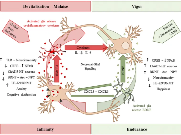
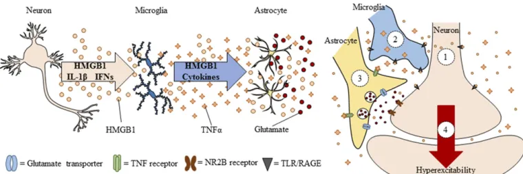
Neuroimmune signals and HMGB1 activate micro-glia as well as release glutamate from astrocytes (Pedrazzi et al., 2006). Signaling between neurons, microglia, and astrocytes contributes to synaptic exci-tation (Fig. 2). Neuronal exciexci-tation can release HMGB1 from neurons, activating microglia, and astrocytes that in turn increase synaptic glutamate and other mole-cules to impact synaptic signaling. Moreover, alcohol activates microglia and astrocytes (Guerri and Pascual, 2010) through neuroimmune signaling, possibly via HMGB1 release from neurons (Zou and Crews, 2012). Postmortem brains of humans with alcohol-use disorder exhibited elevated microglial markers (He and Crews, 2007) and increased expression of HMGB1, TLR2, TLR3, and TLR4 (Crews et al., 2013), as well as proinflammatory cytokines and other neuroimmune-signaling molecules (Crews and Vetreno, 2016). A recent study reported that heavy binge-drinking ado-lescents have increased blood cytokines (Ward, et al., 2014). These results and others have led to the hypoth-esis that ethanol induces neuroimmune-signaling mol-ecules and microglial activation, and that this induction in adolescence disrupts synaptic maturation.
(discussed below), as well as to more specific insults to key neurotransmitter systems during adolescent maturation (Crews and Boettiger, 2009; Vetreno et al., 2014).
Although this review highlights HMGB1–TLR4 sig-naling, there are multiple other proinflammatory genes and proteins increased after AIE exposure in the rat, many of which we have also observed in postmortem brains of individuals with alcohol-use disorder. Our first human brain studies looked at microglia and the proinflammatory cytokine monocyte chemoattractant protein-1 (MCP-1; CC chemokine ligand 2), which is the cytokine induced most robustly by ethanol among those measured in brain slice cultures (Crews et al., 2006a; Zou and Crews, 2010). We found that postmortem brains from subjects with a history of alcohol-use disorder contain increased levels of MCP-1 protein and the microglial marker Iba-1 in hippocampus, ventral tegmental area, nucleus accumbens, and amyg-dala (He and Crews, 2007). In later studies, we focused on the OFC, a component of the PFC, and determined that postmortem alcoholic OFC has more expression of HMGB1 as well as TLRs and RAGE (Crews et al., 2013; Vetreno et al., 2013). We also observed increased interleukin (IL)-1B inflammasome markers in post-mortem alcoholic hippocampus that could contribute to loss of neurogenesis (Zou and Crews, 2012). In addition, NADPH-oxidase is increased in human alco-holic OFC (Qin et al., 2013), consistent with increased oxidative stress, as found in the mouse brain after ethanol exposure (Qin et al., 2013). These findings show
that neuroimmune-signaling pathways are upregulated in alcohol-use disorder, which may be an important aspect of the neurobiology of the disease (Fig. 3). Indeed, work from the Harris laboratory found that activation of the innate immune system increases alcohol consump-tion in mice (Blednov et al., 2011). Studies by multiple laboratories find that TLR, HMGB1, and other neuroimmune-signaling molecules are increased by alcohol and/or alter responses and preference for drink-ing alcohol, suggestdrink-ing a bidirectional relationship be-tween neuroimmune signaling and alcohol drinking.
As adolescent drinking is known to increase the risk of developing alcohol dependence during one’s lifetime, we investigated the relationship between alcohol drink-ing and neuroimmune gene expression across control and alcoholic postmortem brains (Vetreno et al., 2013). Interestingly, two forms of correlations were found linking neuroimmune gene expression to alcohol con-sumption and alcoholism. First, we found that HMGB1– TLR4 expression in OFC was negatively correlated with age of drinking onset—that is, expression was higher in individuals who initiated alcohol use early. Second, total lifetime alcohol consumption across groups was posi-tively correlated with OFC expression of HMGB1, TLR4, TLR3, TLR2, and RAGE. This persistent relationship between cumulative alcohol use and HMGB1 and TLR gene induction in brain provides support to the hy-pothesis that alcohol-induced neuroimmune signaling results in long-term changes in brain function and neurodegeneration.
The critical role of neuroimmune gene induction in the persistent effects of adolescent alcohol exposure on neurobiology is stongly supported by Guerri’s studies in both rats (Pascual et al., 2014) and mice (Alfonso-Loeches and Guerri, 2011). AIE exposure in rodents insults PFC, hippocampus, cerebellum, white matter, as well as cognition and reward. Guerri’s laboratory finds that alcohol exposure increases neuroimmune protein expression, as assessed by both in vitro and in vivo methods. Guerri’s studies describe adolescent alcohol-induced changes in the dopaminergic system, white matter, and myelination, as well as synaptic and epigenetic factors, all of which may contribute to changes in adult alcohol reinforcement, anxiety, and cognition dysfunction, and other behaviors consistent with alcohol addiction (e.g., Pascual et al., 2007, 2009, 2012, 2014; Montesinos et al., 2015, 2016). Multiple studies have found that transgenic mice lacking TLR4 do not show adolescent brain neuroimmune gene in-duction following adolescent alcohol exposure (Montesinos et al., 2015, 2016; Alfonso-Loeches et al., 2016). Fur-thermore, these mice lacking TLR4 do not show the
changes in anxiety, alcohol drinking, cognitive dysfunc-tion, reduced myelinadysfunc-tion, glial activadysfunc-tion, glutamate, and GABA receptor protein expression or epigenetic marker expression typically found following AIE treat-ment of control mice. Taken together, these studies support the hypotheses that the long-lasting pathology associated with adolescent alcohol abuse is linked to alcohol-induced neuroimmune activation and its result-ing pathologic changes in brain.
XI. Brain Electroencephalography and Sleep
Brain function can be assessed using electroenceph-alography (EEG), an electrophysiological method that records the electrical activity across the brain to evaluate function. EEG rhythmic activity or event-related poten-tials (ERP) that measure brain responses to a specific sensory, motor, or cognitive event can be studied in both rats and humans to investigate how the brain processes sensory information (Handy, 2005). The P300 or P3 component of the ERP is an electrophysiological mea-sure commonly studied in both humans and rats (Bauer Fig. 3. Innate immune-signaling cascades and evidence for upregulation in brain following AIE exposure. A simplified schematic of the TLR and RAGE signaling cascades. Stimulation of TLRs and RAGE with their endogenous agonist HMGB1 and other inflammagens [e.g., lipopolysaccharide (LPS)] leads to the generation of proinflammatory oxidases and reactive oxygen species (ROS) and downstream activation of NF-kB. Nuclear translocation of NF-kB leads to the secretion of proinflammatory gene expression, innate immune gene induction, cell death, and addiction-like behaviors. AP-1, activator protein-1; CD14, cluster of differentiation 14; ERK, extracellular signal-regulated kinase; IKK, inhibitor of nuclear factor
and Hesselbrock, 1999; Porjesz et al., 2005; Ehlers and Criado, 2010). The P3 is a positive potential that occurs approximately 300 ms after unexpected and task-relevant sounds or lights (Gratton et al., 1988). In humans, the amplitude and latency of the visual P3 reduce across adolescence until stabilizing in early adulthood (Hill and Shen, 2002). Adolescent humans and rats have higher amplitude and longer auditory P3 latency compared with adults of their species (Polich et al., 1990; Ehlers and Criado, 2010). A low P3 amplitude in youth with a family history of alcohol-ism has been suggested to represent impaired in-hibitory regulation or disinhibition, possibly due to a developmental delay (Hill and Shen, 2002; Bauer and Hesselbrock, 2003; Berman et al., 2006; Tremere and Pinaud, 2006). Studies of young adult southwestern California Native Americans with a history of adoles-cent binge drinking reported that low P3 amplitude was related to ethanol dependence (Criado and Ehlers, 2007; Ehlers et al., 2007). Similarly, rats exposed to AIE for 10 days (P30–P40) and assessed as adults 6– 7 weeks after ethanol exposure display a reduced P3 ERP amplitude in the dorsal hippocampus (Criado and Ehlers, 2007; Ehlers et al., 2007). Adults tend to have increased ERP amplitude as compared with adoles-cents. The reduced hippocampal ERP amplitude follow-ing AIE exposure is consistent with disruption of hippocampal maturation of function (Ehlers and Criado, 2010). Additional studies are needed to determine how the lasting changes in ERP may be related to alterations in hippocampal neurogenesis, cholinergic signals, gluta-mate excitatory synapses, and/or other AIE-induced changes in adult hippocampus.
The effect of ethanol challenge on ERP responses in adult rats is also altered by AIE treatment. Similar to humans, adolescent rats (P32) have longer latency P3 components compared with adults. In rats, a dose-dependent increase in the latency of the P3 auditory ERP was observed after ethanol (1.5 and 3.0 g/kg) in both adolescents and adults. In adult rats (P99), the change in P3 latency due to ethanol challenge was smaller in rats with a history of AIE compared with age-matched controls not exposed to ethanol during adolescence (Ehlers et al., 2014a). These findings are consistent with other AIE findings supporting long-lasting decreases in adult response sensitivity to etha-nol and retention of the adolescent phenotype (Ehlers et al., 2014a). These P3 ERP studies support the hypothesis that AIE alters brain information processing in adulthood, particularly after ethanol challenge, in a manner that reflects behavioral disinhibition and per-sistence of adolescent-like responses to ethanol.
The EEG also assesses rhythmic neural activity, with rhythmic activity divided into frequency bands known as alpha (8–15 Hz), beta (16–31 Hz), theta (4–7 Hz), and delta (,4 Hz) (Ehlers and Criado, 2010). Event-related oscillations (EROs) within and between different brain
regions are thought to reflect neural networks and can provide insight into brain maturation in both humans and rodents (Ehlers et al., 2014b). Higher ERO en-ergy and lower synchrony are found in adolescent humans and rats as compared with their adult coun-terparts. During early adolescence, humans have higher ERO energy in all frequency ranges (alpha, beta, theta, delta) across cortical regions compared with adults. Similarly, early adolescent rats have higher ERO energy in all frequency ranges in parietal cortex and in all frequencies except beta in frontal cortex as compared with adult rats. Early adolescent humans and rats also have lower synchrony within and across cortical regions (Ehlers et al., 2014b). EEG under wake and sleep conditions undergoes large changes in char-acteristic amplitude and frequency during adolescence. For example, EEG amplitude and frequency of the posterior alpha rhythm are increased in the adolescent brain. Slower waves in the waking EEG also decline across adolescence (Niedermeyer and Lopes da Silva, 1999; Ehlers and Criado, 2010). These findings are consistent with adolescent remodeling of the brain to increase brain regional connectivity, decrease ERO energy, and increase synchrony during maturation of local and regional neurocircuits in both rats and humans. Interestingly, the adult EEG theta response to acute ethanol following AIE was blunted in parietal cortex (Ehlers et al., 2013a). Thus, similar to the P3 ERP studies described above, adolescent waking EEG is less sensitive to ethanol than adult responses, and AIE blunts the sensitivity of waking EEG to ethanol chal-lenge in adult rats.