The handle
http://hdl.handle.net/1887/42846 h
olds various files of this Leiden University
dissertation.
Author
: Wiekmeijer A.S.
human T-cell development
©2016 A. Wiekmeijer, Oegstgeest, The Netherlands ISBN: 978-94-028-0254-2
Development of human T-cell progenitors after transplantation of human hematopoietic stem cells in severe immunodeficient mice mirrors human T-cell development, which is difficult to study in vivo. Studying human T-cell development in these transplanted mice, represented by the reflection of the mountain in the lake, provides insight (by looking over the bushes) into the real situation (the mountain itself).
Printed by: Ipskamp Printing Cover: Anna-Sophia Wiekmeijer
Lay-out: Tara Kinneging, Persoonlijk Proefschrift
The research described in this thesis was performed at the department of Immunohematology and Bloodtransfusion at the Leiden University Medical Center, The Netherlands. The work de-scribed in this thesis was financially supported by KiKa (Children Cancer Free).
Financial support for the publication of this thesis was kindly provided by BD Biosciences
human T-cell development
Proefschrift
ter verkrijging van
de graad van Doctor aan de Universiteit Leiden,
op gezag van Rector Magnificus prof.mr. C.J.J.M. Stolker,
volgens besluit van het College voor Promoties
te verdedigen op donderdag
8 september 2016 klokke 15.00
uur
door
Anna-Sophia Wiekmeijer
geboren te Haarlem
Co-promotor:
Dr. K. Pike-Overzet
Promotiecommissie: Prof. dr. F. Koning
Prof. dr. A. Thrasher (University College London)
Dr. M. van der Burg (Erasmus Universiteit Rotterdam)
Chapter 1
General Introduction 7
Chapter 2
Sustained engraftment of cryopreserved human bone marrow CD34+ cells
in young adult NSG mice 37
Chapter 3
Identification of checkpoints in human T-cell development using severe combined
immunodeficiency stem cells 53
Chapter 4
Identification of a novel type of T-B+-SCID with a late double positive thymic arrest 75
Chapter 5
Development of a diverse human T cell repertoire despite severe restriction of
hematopoietic clonality in the thymus 93
Chapter 6
Overexpression of LMO2 causes aberrant human T-cell development in vivo by
three distinct cellular mechanisms 117
Chapter 7
General Discussion 145
Chapter 8
English summary 165
Nederlandse samenvatting 169
Dankwoord 175
Curriculum Vitae 177
Chapter 1
1
first step in the transplantation procedure, the cells of the immune system in the patient are often depleted by chemotherapy, which is called conditioning, and then the patient will receive donor-derived HSC that can engraft and develop a new healthy immune system. HSCs can be isolated from different sources; BM, mobilized peripheral blood and umbilical cord blood, all of which are used in the clinic for transplantation.
HSCs are rare cells that are difficult to characterize precisely by marker expression alone. The most robust criterion to determine true stem cell potential is the ability to provide long-term repopulation of an entire host with all hematopoietic lineages. In mice, this is often assessed by performing transplantations in secondary recipients to determine self-renewal capacity. For human HSCs this is, of course, not feasible in a clinical setting.
When an HSC is transplanted it can migrate to the BM of the recipient, which in most but not all patients had been depleted of autologous cells before transplantation by irradiation or chemotherapeutics. The BM niche contains many different stromal cells providing a favorable milieu for the HSC, which in turn will undergo the process of both self-renewal and differentiation6. Long-term repopulating (LT)-HSCs give rise to short-term repopulating
(ST)-HSCs giving rise to multi-potent progenitors (MPPs), each being more restricted in their potential to self-renew and their multi-lineage differentiation potential7. A lot of
studies have been performed to determine the phenotype of the LT-HSC, most of which have been performed in mice. The field of human HSC knowledge lags behind compared to that of mice due to absence of appropriate models to study both self-renewal and multi- lineage differentiation.
In a clinical setting, the CD34+ fraction is used for transplantation as these cells can be isolated
in a good laboratory practice (GLP) setting. However, already in 1997 it was described that the phenotype of HSCs could be further refined to CD34+CD38- containing a frequency
of 1 in 617 cells with true HSC potential, defined by the capacity to repopulate a NOD/Scid mouse8. Thereafter, it was shown that this cell fraction can be divided in 3 groups based on
the expression of both CD90 and CD45RA. The Lin-CD34+CD38-CD90+CD45RA- cell population
isolated from umbilical cord blood was demonstrated to have multi-lineage BM engraftment potential when 10 cells were transplanted9. This cell population could be further subdivided by
CD49f discrimination of which the CD49f+ population contained a frequency of LT-HSC of 1 in
10.5 cells10. This illustrates that currently we are not yet able to identify the one cell phenotype
that is most primitive and contains the highest long term repopulating capacity. Currently, the human HSC is described to be most enriched within the Lin-CD34+CD38-CD45RA-CD90+CD49f+
population followed by the MPP that has lost expression of both CD90 and CD49f11 (Fig. 1).
From the MPP two cell types branch off; the CD34+CD38-CD45RA+CD90- MLP (multi-lymphoid
progenitor) that can give rise to NK-, B- and T cells, and the Lin-CD34+CD38+CD45RA-CD135+ CMP
(common myeloid progenitor) that can give rise to the megakaryocytic-erythroid progenitor (MEP) and granulocyte-monocyte progenitor (GMP)12. The MLP has also been named common
lymphoid progenitor (CLP), which comes from many studies on hematopoiesis in the mouse. In humans this has been studied less extensively. Cells from the myeloid lineage, erythrocytes and granulocytes are progeny from the last two progenitor types. Also on the gene expression “As part of a normal day, most people will flush a toilet, open a door, or drink from a water
fountain without even thinking about it – or about the lurking pathogens poised to infect us. We are afforded this luxury, because of our immune system, which responds rapidly and specifically to just about anything thrown at it.” (from: Editor’s Summary of Gaspar et al. 2011, Sci Transl Med)1
As illustrated by the quote above, our immune system protects us every day from pathogens that are present in the environment. When the immune system is compromised, infections cannot be cleared which can lead to severe illness. Development of the different cells from the immune system is tightly regulated by expression of many genes. Deficiencies or deregulation of these genes can have severe consequences that heavily impact on normal life. In this thesis, the effect of lower or absent expression of genes and overexpression of genes on the development of the human immune system is described using severe combined immunodeficiency (SCID) and T-cell acute lymphoblastic leukemia (T-ALL) as examples. Therefore, the development of lymphoid cells from hematopoietic stem cells (HSCs) is discussed in this introduction together with the consequences of genetic aberrancies affecting these processes.
Hematopoiesis
The blood in our body consists of many different cell types. HSCs, which reside in the bone marrow (BM), are able to produce all the different cells present in our blood system, including platelets, red blood cells and white blood cells. This involves a highly controlled process of both renewal, to maintain the pool of HSCs, and differentiation. The process of both self-renewal and differentiation is coordinated by many signaling pathways, such as Notch2, Wnt3, 4, BMP5 and several others. Aberrancies in genes and their expression, either congenital or
acquired, can influence these processes, eventually leading to arrests in development or to the development of hematological malignancies.
Under normal circumstances, HSCs give rise to all white blood cells, including both innate and adaptive immune cells. The innate immune system is already present at birth and is a non-specific defense against pathogens and therefore is able to respond quickly. It is comprised of different cells types, including mast cells, macrophages, neutrophils, eosinophils, dendritic cells and natural killer (NK) cells. The cells of the adaptive immune system, comprised of B cells and T cells, are also present at birth as the cells from the innate immune system. However, cells from the adaptive immune system respond in an antigen-specific manner. These cells express receptors specific for antigens and upon antigen encounter they will proliferate but also form memory cells. These memory cells are able to respond quicker upon a second encounter with the same antigen; a regimen that is made use of by vaccination, thereby providing protection against the pathogen. The adaptive immune system is only found in vertebrates.
1
receptors cannot bind and therefore the NK cell will be activated. Activating receptors are able to recognize infected cells and often activation needs to occur via recognition with more than one activating receptors except for CD16.
Rearrangement of antigen specific receptors
Unlike NK cells that express both activating and inhibitory receptors for recognition of pathogens or allogeneic cells, B cells and T cells have antigen-specific receptors. A high degree of diversity is created by V(D)J recombination of the receptor loci. Both the T-cell receptor (TCR) and immunoglobulin (Ig) loci contain variable (V) and joining (J) segments, some also contain diversity (D) segments, and through recombination of these segments a high diversity is generated to be able to recognize many different antigens. The Ig heavy chain contains V, D and J segments, while the Ig kappa and Ig lambda chain only contain V and J segments. An Ig molecule, also known as B-cell receptor (BCR) contains 2 heavy chains together with 2 light chains, either kappa or lambda. For the TCR, the loci encoding the delta (TRD) and beta (TRB) chain contain V, D and J segments and the alpha (TRA) and gamma (TRG) loci contain only V and J segments. A TCR is either composed of a TCRγ chain paired with TCRδ or of a TCRα chain paired with TCRβ.
Rearrangements of Ig and TCR loci take place in a highly ordered fashion. First, a D segment rearranges to a J segment which is then followed by rearrangement of a V segment to DJ. When D segments are not present within the locus, V rearranges directly to a J segment. Recombination activating gene (RAG) proteins (RAG1 and RAG2) make double strand breaks (DSB) at the recombination signal sequences (RSSs) between the V (D) and J segments. These DSBs are recognized by a complex of DNA-dependent protein kinase catalytic subunit (DNAPKcs) together with KU70 and KU80, which can phosphorylate Artemis which then opens the coding joints that are left after RAG mediated enzymatic DNA cleavage18. Another complex
involving X-ray repair cross-complementing protein 4 (XRCC4), DNA ligase IV (LIG4) and XRCC4-like factor (XLF) is needed for ligation in order to complete recombination.
B cells
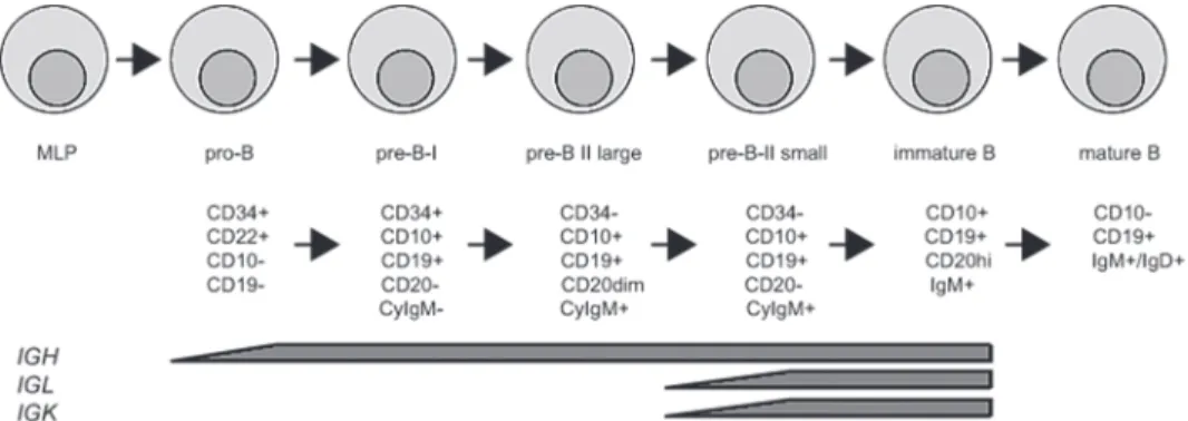
As NK cells, B cells develop in the BM up to an immature stage after which they migrate to peripheral lymphoid organs, such as spleen and lymph nodes, to further mature. In BM aspirates the different stages of development can be characterized using flow cytometry19.
Using this approach, patients suffering from B-cell deficiencies, for instance SCID, can be identified by determining arrests in B-cell development. During development the Ig heavy chain and the kappa and lambda light chain loci are rearranged to generate a functional and diverse repertoire. Rearrangement of IGH starts in the pro-B cell stage and in the small pre-B-II stage the IGK and IGL loci are rearranged20 (Fig. 2). In Pax5 deficient mice, it was demonstrated that
expression of Pax5 is needed for rearrangement of the IGH locus and is considered as a B-cell level there is a separation between lymphoid fate and a myeloid fate at the MLP stage13. Many
of the transcription factors that are important in HSCs are known to be causative of leukemia when deregulated, for example RUNX1, MLL, SCL/TAL1 and LMO27, 14. From the progenitors
onwards, most of the lineages develop within the BM, except for the T cells that need the specialized environment provided by the thymus. Hereafter, the focus will be on lymphocytes that have developed from the MLP.
Figure 1: Overview of hematopoiesis. Schematic depiction of hematopoiesis starting from the
long-term reconstituting hematopoietic stem cell (LT-HSC) that gives rise to the multi-potent progenitor (MPP). From the MPP onwards two branches diverge; the common myeloid progenitor (CMP) and the common lymphoid progenitor (CLP)/multi-lymphoid progenitor (MLP). The CMP gives rise to both the megakaryocytic-erythroid progenitor (MEP) and granulocyte-monocyte progenitor (GMP). Myeloid dendritic cells (mDC), monocytes and granulocytes develop from the GMP, while erythrocytes and megakaryocytes develop from the MEP. The MLP gives rise to natural killer (NK) cells, B cells, T cells and plasmacytoid dendritic cells (pDC). This is according to the classical model of hematopoiesis as studied extensively in the mouse, the alternative model suggests a less strict separation of lineages but more a loss of potential during development.
NK cells
NK cells are part of the innate immune system and develop in the BM but can further mature in other lymphoid organs. They need interleukin (IL)-15 for their survival and proliferation. In addition, NK cells can be found in the thymus15 and they share a common progenitor with the
developing T cells16. Mature NK cells in humans are characterized by the expression of CD56
1
as it has been postulated that developing T cells “prime” the thymic environment making it more receptive27. The first wave of thymus seeding cells, as described in mice, might have this
same function allowing for better seeding and differentiation of the second wave comprised of more multipotent cells. Robust and rapid outgrowth of T cells after HSC transplantations can still be problematic28 and ways to improve this might benefit clinical outcome.
In humans, three phenotypes of TSPs have been proposed; a CD34hiCD45RAhiCD7+ phenotype29, 30, Lin-CD34+CD10+CD24-31 and cells characterized as Lin-CD34+CD10-CD45RA+CD62Lhi 32. Haddad
et al. did show that the CD34hiCD45RAhiCD7+ cells they identified were able to migrate into
thymic lobes in an ex vivo culture system. These cells could be differentiated from CD34+
cells isolated from cordblood using the in vitro OP9-DL1 coculture system and were able to engraft thymi of immunodeficient mice33. The TSP phenotype identified by Six et al., however,
do not express CD7 and have capacity to develop into B cells, NK cells and T cells using in vitro coculture systems31. Furthermore, in the human thymus cells positive for the expression
of CD34 but negative for the expression of CD7 can be found, and it was demonstrated that CD34+ cells upregulate CD7 only after 4 days of coculture on OP9-DL133. This argues against
CD7+ cells as being the most immature cells in present in the thymus; however, it might be
that the thymus can also be seeded by multiple populations. The cells identified by Kohn et al. are negative for the expression of CD7 but they did not succeed in transplanting these cells in immunodeficient mice to monitor for thymus seeding and engraftment. Therefore, the nature of the TSP in humans remains controversial.
The subsequent steps of T-cell development and commitment and the phenotypes associated with these processes have been extensively described in the mouse26, 34. There are many
similarities between T-cell development in mice and men, as in both species it starts in de CD4
-CD8- double negative (DN) compartment and also the CD4+CD8+ double positive (DP) stage is
comparable after which cells become either CD4+ single positive (SP) or CD8+ SP (Fig. 3). The
SP cells have undergone positive selection, to select for thymocytes of which their TCR can bind major histocompatibility complex (MHC, in humans also called human leukocyte antigen - HLA), and negative selection, to eliminate cells that recognize self-antigens. These mature T cells are now ready to emigrate to the peripheral blood.
However, differences also exist between T-cell development in mice and men. Between the DN and DP stage lies the immature single positive (ISP) stage that is CD8+ in mice and
CD4+ in humans, but in both species there is no expression of CD3 or a TCR35. In mice, the
DN compartment can be further subdivided in DN1-4 based on the expression of CD44 and CD2536 and further subdivision of these DN compartments has been described too26, 34. The DN
compartment of human developing T-cell progenitors can also be subdivided, but different markers have been described to do so; such as CD34 in combination with CD38 and CD1a37,
CD1a and CD538 and CD733, 39. During T-cell development the TCR loci are rearranged, most cells
will eventually become a TCRαβ+ T cell, and in mice the point of β-selection is at the DN3 stage.
At this point the progenitor needs to have rearranged the TRB locus in a way that it produces a functional β-chain, otherwise the progenitor cannot progress in its development and will eventually die38. In humans, it has been debated where the point of β-selection exactly resides;
commitment factor as it is required for B-cell development and suppresses the development of other lineages21. B cells that have in-frame rearrangement of the heavy chain and one of the
light chains, either κ or λ, are able to express a BCR and will be positively selected. Naïve B cells in the periphery can be recognized by expression of both IgM and IgD. First the immature B cell expresses the IgM chain but by alternative splicing of the heavy chain transcript it will also express IgD when becoming a naïve B cell. Immunoglobulins can be either membrane bound to serve as BCR or can be secreted as antibodies. When a B cell recognizes antigen through the BCR it can either become a natural effector B cell or it will migrate to a germinal center. Here it can encounter a T cell recognizing the same antigen which will than provide the T cell help needed for class switch recombination of the heavy chain to change isotype to IgG, IgA or IgE22. Different isotypes confer different effector functions while still recognizing the same
antigen. During this process the naïve B cell will become a memory B cell characterized by upregulation of CD2723, which in its turn can give rise to plasma cells that can produce large
amounts of antibody.
Figure 2: Human B cell development. B cells develop in the bone marrow from the multi-lymphoid
progenitor (MLP). Indicated are the different stages of B cell development, the markers that can be used to identify these stages and the stages at which recombination of immunoglobulin (Ig) loci is ongoing. Adapted from thesis of H. IJspeert and thesis of K. Pike-Overzet.
T cells
T cells develop within the thymus providing a specialized environment consisting of the appropriate cytokines and specialized stromal cells24. Thymus seeding progenitors (TSP) that
have developed from the HSC in the BM, migrate via the peripheral blood to the thymus. Both the precise phenotype of this cell and the signals that drive its migration to the thymus are still controversial. It has been estimated that there are only 10-50 cells migrating to the thymus each day25, which makes it hard to detect and isolate these cells. In mice, studies
have been performed to identify the early thymic progenitor (ETP) by careful flow cytometric analysis and isolation of murine thymi. It was found that the fetal thymus is seeded in two waves by distinct progenitors, first by a T-lineage restricted progenitor and later by a more multipotent progenitor26. Also in the setting of HSC transplantations it has been proposed that
1
the TRB locus will start rearranging and the precursor will develop along the TCRαβ lineage. γδ T cells do not recognize MHC-restricted antigens but do recognize CD1, which can present lipids50 and are frequently found in gut and skin51.
Severe Combined Immunodeficiency
SCID is a subset of primary immunodeficiency (PID) that is characterized by a deficiency in (functional) T cells with an incidence ranging from 1-2 per 100,000 live births52-55. Patients
often present within their first year of life with a failure to thrive and recurrent infections52, 56. Different forms of SCID exist in which the deficiency in T cells can be accompanied by a
deficiency in B cells or NK cells or both57, 58. As SCID patients lack an adaptive immune response
they present with opportunistic infections and a failure to thrive, which is often diagnosed within their first year of life. The different phenotypes of SCID are caused by the differences in mutations and affected genes that are causative of SCID (Table 1). Currently, around 16 genes have been identified59 but there are still cases remaining of affected children without a known
genetic cause. In a cohort studied by Gaspar et al.60 there were 20 out of 117 patients (17%)
without a molecular diagnosis, this percentage might be different in other cohorts as presence of certain types of SCID can vary between geographic regions. The different types of SCID are categorized in two different groups based on the presence or absence of B cells: T-B- SCID and
T-B+ SCID61. Both groups encompass patients with presence or absence of circulating NK cells,
depending on the genetic aberrancy.
Table 1: Overview of SCID-causing genes.
T-B- ADA, AK2
RAG1, RAG2, Artemis (DCLRE1C), DNA-PKcs (PRKDC), LIG4, XLF
T-B+ IL2RG, JAK3
IL7RA, CD45 (PTPRC), CD3D, CD3E, CD3Z (CD247), CORO1A
Severe combined immunodeficiency (SCID) is characterized by a deficiency of (functional) T cells that can be accompanied by a deficiency in B cells or NK cells or both. Indicated are the different phenotypes of SCID and the genes, when mutated, can cause this type of deficiency. Included genes were based on criteria described by Bousfiha et al.59.
T
-B
-SCID
T-B-NK- SCID is caused by genes that are involved in cell metabolism, such as ADA62, PNP63, 64 or AK265,
the latter leading to a disease also called reticular dysgenesis. As the phenotype caused by PNP deficiency is less profound these patients are often classified as combined immunodeficiency (CID) instead of SCID66. Deficiency in ADA, encoding adenosine deaminase, results in the
accumulation of 2’-deoxyadenosine, which will be converted to deoxyadenosinetriphosphate (dATP). The dATP is the primary cause of lymphotoxicity as demonstrated in mice67. Using FTOC,
it was demonstrated that inhibition of ADA in human thymocytes does results in accumulation of intracellular dATP which leads to apoptosis68. ADA-SCID patients can be treated by BM
it has been ascribed to the DP stage40, the ISP stage41-43 and to the CD1a+ DN3 stage35. The
current opinion is that, as in mice, it resides in the DN3 population35, 37.
Figure 3: Human T cell development. T cells develop within the specialized environment provided by
the thymus. The thymus seeding progenitor (TSP) seeds the thymus and develops into the immature thymocytes. Indicated are the stages of rearrangement of T cell receptor (TR) loci and markers to identify different stages. DN; double negative (CD4-CD8-), ISP; immature single positive, DP; double positive
(CD4+CD8+), SP; single positive. Adapted from thesis of K. Pike-Overzet and Dik et al.37.
After successful development and selection of the progenitors in the thymus they emigrate as mature T cells to the peripheral lymphoid organs and help protect us against pathogens. These mature CD4+ and CD8+ T cells have a TCR comprised of an α and a β chain. CD8+ T cells are also
called cytotoxic T cells and recognize antigens that are presented by MHC class-I to fight virus infected cells and it has been demonstrated that they can be reactive towards tumor cells44.
CD4+ T cells recognize MHC class-II restricted antigens and are also called T helper cells. These
cells help other white blood cells to fight pathogens, for example they are needed for class switching of B cells. The different subtypes of T helper cells secrete different cytokines after antigen recognition to aid in distinct immune responses. Another type of CD4+ T cells is the
regulatory T cell that has a high expression of CD2545. Regulatory T cells, which can develop in
the thymus during the DP stage46, help to dampen the immune response and play important
roles in autoimmunity and cancer; with autoimmunity they do not respond adequate enough to dampen immune responses and T cells attack autologous cells, in the case of cancer the cancer cells induce regulatory T cells to dampen the T cell response against the tumor47.
There are also T cells that have a TCR comprised of a γ and a δ chain (γδ T cells). Less is known about the development and function of these cells especially when compared to TCRαβ+ T
1
effects on B cells are difficult to study in knockout mice for Il2rg or Il7ra as these mice also suffer from a B-cell deficiency which is opposed to the phenotype observed in patients84, 85.
Furthermore, this demonstrates differences in lymphoid development between mice and men. Another type of SCID only involves deficiency specific for T cells resulting in a T-B+NK+
phenotype. Most often this is caused by a mutation in the IL-7 receptor α chain (IL7RA), which, together with IL-2Rγ, makes up the receptor for IL-7 that is needed early in T-cell development86, 87. Furthermore, mutations in CD45 (encoded by PTPRC)88, 89, the molecule that
marks lymphocytes and in Coronin 1a (CORO1A), which is important in thymic egress90 have
been found in SCID patients only deficient in T cells. Mutations in the kinase ZAP-70 lead to a deficiency in peripheral CD8+ T cells, while CD4+ T cells are present but fail to respond to
TCR stimulation, do not produce IL-2 and have reduced tyrosine phosphorylation91. Of most
SCID patients it is unknown at which stage the arrest in T-cell development resides as thymic biopsies are not routinely performed. For the ZAP-70 deficiency it has been demonstrated using immunohistochemistry of thymic biopsies that DP cells were present in the cortex but no CD8+ SP cells were present, showing a CD8 specific block at the DP stage91. For SCID caused
by mutations in the CD3 δ chain (CD3D) the developmental arrest has been described and assigned to the ISP to DP transition92 although by a different group ascribed to the DN stage,
but in that study the presence of ISP cells was not determined93. The CD3 complex, which
associates with the TCR, exists of different subunits next to the δ chain; the γ chain (CD3G), ε chain (CD3E) and ζ chain (CD3Z). Mutations in all of these different CD3 chains have been described in T-B+NK+ SCID patients92, 94, 95. Patients with mutations in CD3G, CORO1A and ZAP70
have also been classified as CID66.
For SCID caused by various mutations it is known where the arrest in B-cell development reside as this can be determined by flow cytometric analyses and repertoire analysis of the immunoglobulin loci in bone marrow aspirates 19, 74, 76. On the T cell side the arrests are only
known for mutations in ZAP70 and CD3D91-93; for other mutations this remains unknown as
thymus biopsies are not routinely taken. To further study the effects of mutations causing SCID different knockout mouse models have been made and studied84, 85, 96-100. As already mentioned,
murine and human T-cell development show similarities but also many discrepancies. This is also illustrated in many of these mouse models, as for instance, Il7r-/- mice are deficient in both
T cells and B cells85 while in humans only T cells are absent86 and in Il2rg-/- mice the arrest in
T-cell development is less strict84. This underscores the need for better models to study T-cell
arrests for human SCID.
Gene therapy for SCID
SCID is fatal if left untreated. For a long time, the only treatment for SCID was a BM transplant, although ADA-SCID patients can benefit from ERT using bovine PEG-ADA. However, ERT is very costly as it involves lifelong administration and requires appropriate monitoring69. Depending
on the donor, the extent of human leukocyte antigen (HLA) matching and type of SCID can lead transplantation, enzyme replacement therapy (ERT) using bovine PEG-ADA or gene therapy69.
Mutations in purine nucleoside phosphatase (PNP) that affect the enzymatic activity can cause the same phenotype as ADA-deficiency70. However, they can have variable B-cell function.
PNP catalyzes the conversion of both inosine and deoxyinosine to hypoxanthine and of both guanosine and deoxyguanosine to guanine. The intracellular accumulation of deoxyguanosine triphosphate is believed to be toxic for lymphocytes and blocks cell division63. Patients suffering
from reticular dysgenesis have one of the most severe forms of SCID as they, in addition to the absence of lymphocytes, also do not have granulocytes. AK2 (adenylate kinase 2) is involved in mitochondrial oxidative phosphorylation and its deficiency can be compensated for in many cells types by AK1. In the mononuclear cell fraction from the BM, no expression of AK1 was detected whereby this could not compensate the AK2 deficiency and the disease manifests so profound in the immune system65. Furthermore, it was demonstrated that restoration of AK2
expression in bone marrow cells restores development towards neutrophils, thereby, showing the causative effect of AK2 deficiency71.
Mutations in genes involved in V(D)J recombination of TCR and immunoglobulins lead to SCID characterized as T-B-NK+. Genes that have been identified include: RAG1 or RAG272, Artemis
(encoded by DCLRE1C)73, LIG474, XLF (also known as Cernunnos and Non-homologous end-joining
factor 1, NHEJ1)75 and DNA-PKcs (encoded by PRKDC)76. As with PNP deficiency, mutations in LIG4
and XLF can also be classified as CID instead of SCID66. As described, these genes are needed
during V(D)J recombination of TCR and immunoglobulin loci. When one of all these genes is mutated this can lead to defective recombination, which leads to absence of expression of a functional TCR in T cells or immunoglobulins in B cells and because these receptors are needed to proceed during T-cell and B-cell development, defects in recombination will result in immunodeficiency.
T
-B
+SCID
Mutations in the interleukin-2 receptor gamma chain (IL2RG)77 and Janus kinase 3 (JAK3)78, 79
can be causative of T-B+NK- SCID, which is the most prevalent form of SCID80. IL2RG encodes
the common gamma chain (γc), which is involved in IL-2, -4, -7, -9,-15 and -21 signaling81. The
gene is located on the X chromosome, thereby mainly affecting boys and also known as X-linked SCID or X-SCID. JAK3 is downstream of IL2RG on the cytoplasmic part of the receptor complex and thereby deficiencies in both these genes result in a comparable phenotype. As humans that only lack IL-2 had normal T-cell and NK-cell development82, it is thought that the
absence of signaling through IL -7 and IL-15 are causative for the deficiency in both T cells and NK cells, respectively, as these cytokines are known to be important early in development of these lymphocytes. Furthermore, it has been observed that in T-B+NK- SCID patients also the
1
SIN lentiviral vectors
After the occurrence of severe adverse events in the gene therapy trials based on the γ-retroviral vector, new vectors have been studied that do not rely on the strong viral LTR for expression of the transgene. These new vectors were based on human immunodeficiency virus type 1 (HIV-1), which is a lentivirus also belonging to the family of retroviridae. Effectiveness of this system, encompassing a split-genome packaging design using 3 plasmids, was shown by Naldini et al.117. Furthermore, they demonstrated that lentivirus is far more effective in transducing
non-dividing cells than the MLV-based counterpart. This is especially of use for transduction of HSCs, as the otherwise needed proliferation might affect their pluripotentency. Thereafter this system was modified to the so-called third generation lentivirus vector system. A split-genome packaging design of 4 plasmids was used to increase safety and viral genes that are important for virus replication were removed118. Furthermore, an additional modification was made that
removes the U3 region from the 3’ LTR, thereby decreasing the transactivational activity it can have on nearby genes119. These self-inactivating (SIN) lentiviruses have been used widely
for preclinical120-126 and clinical127-130 gene therapy studies. Besides being able to transduce
non-proliferating cells, lentiviruses also have a favorable integration pattern when compared to γ-retroviruses. Gamma retroviruses have a tendency to integrate more frequently around the transcription start site of a gene and thereby the risk of dysregulation is higher109, 131. The more
random integration pattern, together with the removal of the U3 region from the LTR region, make these third generation SIN lentiviruses safer than the MLV-based γ-retroviral vectors. Indeed, no development of leukemia has been observed using the lentiviral vectors127, 128, 132,
which have now been widely implemented. Only one clonal dominance with integration near HMG2A has been observed in a trial for β-thallasemia, but no leukemia development has been documented130. Furthermore, when integration sites of both vectors were compared it was
observed that integrations detected for lentiviral vectors have a safer integration profile133, 134.
So far, SIN lentiviral vectors have been proven safe in clinical trials.
SIN γ-retroviral vectors
Next to SIN lentiviruses, SIN γ-retroviral vectors have been created. These also have a deletion of the U3 region in the LTR and need an internal promoter to drive expression of the transgene135.
The SIN design eliminates, as with the lentiviral vectors, the enhancer activities on neighboring genes. However, a concern might be that their integration pattern is comparable to the MLV-based γ-retroviral vectors136. These vectors have been developed and tested in preclinical
models for CGD, WAS and X-linked SCID, which demonstrated efficacy135, 137, 138. Trials using SIN
γ-retroviral vectors for X-linked SCID using the elongation factor 1α short (EFS) promoter are currently ongoing139-141. Recently, it was reported that gene therapy using a SIN γ-retroviral
vector in which IL2RG expression is driven by the EFS promoter was effective in 7 out of 9 treated patients142. One patient died of an adenoviral infection before T-cell reconstitution.
This study demonstrated initial safety of the SIN γ-retroviral vector, as no leukemias were observed, and the efficacy of gene therapy using a γ-retroviral vector expressing IL2RG. It to differences in outcome. Transplantation of HSC from a related genetically matched donor
results in a 10-year survival of 84%101. For a related phenotypically identical and an unrelated
donor the survival is 64% and 66% respectively, and for a related HLA-mismatched this is only 54%101. For monogenic disease, such as SCID, gene therapy might be a successful treatment
option, especially when an HLA-matched donor is not available.
The advantage is that autologous HSCs are used and therefore the risk of graft rejection or graft-versus-host-disease is very low. Furthermore, gene corrected cells will, most likely, have a selective growth advantage over their non-transduced counterparts that suffer from a developmental arrest102. Several gene therapy trials have been conducted for different types
of SCID, starting in the early 2000s103-105. In short, blood stem cells are harvested from the
BM of the patient, in which a correct copy of the affected gene is introduced ex vivo using a crippled virus for gene delivery after which the cells are given back to the patient. These initial studies used a mouse leukemia virus (MLV)-based γ-retroviral vector to drive expression of the transgene by the long terminal repeat (LTR). A retrovirus reverse transcribes its RNA into DNA which then uses the enzyme integrase to integrate the DNA into the host genome. Due to this integration, the gene will be passed on to every daughter cell. Initially, there was restoration of functionality and patients demonstrated presence of cells from different lymphoid cell lineages in their peripheral blood together with improved immune functionality. Patients were able to go home and live in a normal environment and had normal growth and development. Unfortunately, thereafter it was reported that 4 patients in the X-linked SCID trial conducted in Paris and 1 patient in the London X-linked SCID trial out of the 20 total treated patients developed T-ALL106-108. In the trial for ADA-SCID, that had used a similar vector, no T-ALL development was
observed109 and also not in another ADA-SCID gene therapy trial conducted in London110. The
T-ALLs in the X-linked SCID trials did result from insertional mutagenesis leading to ectopic expression of oncogenes. Insertional mutagenesis was also detected in gene therapy trials for X-linked chronic granulatomous disease (CGD)111 and Wiskott-Aldrich Syndrome (WAS)112
leading to development of myelodysplasia and leukemia, respectively. Below, the mechanisms of insertional mutagenesis and T-ALL development will be described in more detail. Due to the occurrence of these adverse side effects, new viral vectors were designed for delivery of the transgene (reviewed in 113, 114).
The 5 patients that developed a T-ALL due to insertional mutagenesis were treated with chemotherapy, which cured the leukemia in 4 patients. Unfortunately, one patient did not survive106. No insertional mutagenesis related T-ALL developed in both gene therapy trials for
ADA-SCID although integrations near LMO2 were detected, which lead to T-ALL development in the X-linked SCID trials109. In 2010, it was reported that for gene therapy trials using a
MLV-based γ-retroviral vector 18 out of 20 X-linked SCID patients treated with gene therapy and all 27 treated ADA-SCID patients are alive115. In addition, 17 X-linked SCID patients and 19 ADA-SCID
1
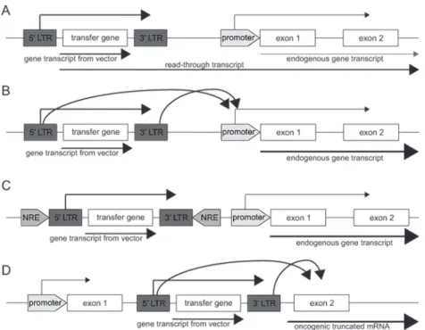
Figure 4: Different mechanisms of insertional mutagenesis leading to aberrant expression of endogenous genes. It is needed to have integration of the vector in the DNA in order to have expression of the
therapeutic gene in all offspring from a cell. This can, however, lead to overexpression of genes which might lead to the development of leukemia. Different mechanisms leading to aberrant expression are:
A) integration of the vector in front of an oncogene leading to a long read-through transcript, B) enhancer
effects of the viral long terminal repeat (LTR) on an oncogene, C) disruption of a negative regulatory element (NRE) thereby leading to aberrant expression of an oncogene and D) integration within a gene thereby generating a truncated constitutively active form. Adapted from Staal et al.114.
It has been demonstrated that the expression of LMO2, TAL1 and LYL1 is high in human CD34+
cells and increases when stimulated with cytokines used in the X-linked SCID gene therapy trials144. This was confirmed by the data from Schwarzwaelder et al.131 that postulated that
many of the integrations that they found in X-linked SCID patients were in genes that are active in CD34+ cells. Indeed LMO2, TAL1 and LYL1 were frequently hit by insertional mutagenesis in
the patients that developed T-ALL as outlined above. However, it has been suggested that the constitutive expression of IL2RG might be causative of the observed T-ALLs, as it was observed that overexpression of IL2RG in a murine X-linked SCID model led to the development of thymomas145. In response to this, it was demonstrated that the overexpression of IL2RG in
human CD34+ cells did not lead to aberrant T-cell development in vitro culture systems, while
overexpression of LMO2 did144, 146. Furthermore, LMO2, TAL1 and LYL1 are known oncogenes
associated with human T-ALL147, while IL2RG is not. Intriguingly, integrations near LMO2
were also found in patients in the ADA-SCID gene therapy trial, but here it did not lead to development of T-ALL148.
needs to be noted that the follow-up time in this report was too short to conclusively say that the SIN retroviral vector was safer than the full LTR homolog.
Insertional mutagenesis
During the gene therapy trials performed for X-linked SCID using the MLV-based γ-retroviral vectors T-ALL was observed in 5 out of 20 patients106-108. The same phenomenon was observed
in a gene therapy trial for CGD111 and in a WAS gene therapy trial112 using comparable vectors.
In the CGD trial this led to the development of myelodysplasia in 2 out of 2 treated patients and in the WAS trial 1 patient developed AML, 4 patients developed T-ALL and 2 patients developed T-ALL with secondary AML. The development of these leukemias was caused by integration of the therapeutic vector in the DNA, resulting in dysregulation of surrounding genes, which is called insertional mutagenesis. The γ-retroviral vector preferentially integrates in transcription start sites (TSS) and transcriptionally active regions143. Indeed, in 5 patients
that were treated with gene therapy for X-linked SCID in London, which did not have T-ALL, it was found that a high percentage of integrations was located around the TSS and a higher percentage than expected was located in common integrations sites (CIS)131. In addition, many
integration sites were found near genes that are transcriptionally active in CD34+ cells, which
is the cell type used for transduction in these trials. There are several mechanisms that can underlie insertional mutagenesis; a long read-through transcript could be generated from the viral gene including a nearby gene, the LTR could have enhancer effects on the promoter from nearby genes, integration of the vector could potentially disrupt a negative regulatory element and integration of the vector within a gene could generate a truncated constitutively active form of the gene (Fig. 4)114.
In the X-linked SCID trials in London an integration upstream of LIM-domain only 2 (LMO2) was found108, while in Paris two patients had integrations near the TSS of LMO2107, one patient
near the TSS of cyclin D2 (CCND2) and one patient harbored integrations in both LMO2 and BMI1 polycomb ring finger oncogene (BMI1)106. Furthermore, it was demonstrated that these
genes were highly expressed in the T-ALLs. In the CGD trial the two treated patients developed myelodysplasia due to insertional mutagenesis in the MDS1-EVI1 locus, which led to higher expression of ecotropic viral integration site 1 (EVI1)111. Insertions within or near LMO2 were
found in all 6 T-ALL cases which in 2 patients was combined with an integration upstream of TAL1 (T-cell acute lymphocytic leukemia 1) and in another patient combined with an integration near the TSS of LYL1 (lymphoblastic leukemia derived sequence 1) in the WAS trial (Table 2)112.
1
T-cell acute lymphoblastic leukemia (T-ALL)
Overexpression of oncogenes or deletions in tumor suppressor genes can lead to the development of cancer. Often more consecutive mutations are needed for a cell to become cancerous as described in the multiple-hit model149, 150 and as also has been observed for the
T-ALLs that developed in the gene therapy trials. Overexpression of proto-oncogenes or loss of tumor-suppressor genes can lead to aberrant T-cell development and if more genetic lesions are acquired this can lead to T-ALL. The leukemic cells will then migrate to lymphoid organs such as peripheral blood, BM and spleen. The symptoms associated with T-ALL are a result of the increase in white blood cells in the blood of the patient, which decreases the number of red blood cells per mm3 and can lead to anemia, dizziness and fever.
T-ALL can present with different phenotypes mirroring different stages of T-cell development, which is caused by the genes that are affected or aberrantly expressed. Different clusters have been described using these expression profiles and phenotypes, which demonstrated that T-ALLs from different clusters have different prognoses and sometimes need different treatment regimens. NOTCH1 has a prominent role in T-cell development and activating mutations in this gene have been found in more than 50% of T-ALL cases151. Activation of NOTCH1
can also be caused by genomic rearrangements. Rearrangements are often detected in T-ALL cells and they mostly involve translocation of one of the TCR loci to an oncogene, which, in addition to NOTCH1, has been described for LYL1, LMO1, LMO2, TAL1 (SCL), TAL2, HOX11 (TLX1), HOX11L2 (TLX3), LCK, CCND2, and BHLHB1152. The promoter or enhancer sequences from the
TCR loci will then drive the expression of the oncogenes. Most of these genes are expressed in HSC and in the early stages of T-cell development and are downregulated thereafter. The translocations keep these genes constitutively active whereby thymocytes retain a high proliferative potential and arrest in development until they acquire additional mutations that can lead to T-ALL.
Two types of mutations can be distinguished in T-ALL; type A, which are also called driver mutations, and type B mutations, which are also known as helper mutations. Type A mutations can be grouped in 7 different clusters; TAL/LMO, TLX1, TLX3, LYL1147, 153, HOXA42, NKX2.1/NKX2.2
and MEF2C154. The TAL/LMO cluster is phenotypically characterized as late cortical by the
expression of TCRαβ and CD3147. NKX2.1/NKX2.2 is biologically related with TLX1 T-ALL, both
having an arrest at the cortical stage characterized by CD1 expression147, 154 while the TLX3
cluster is more mature compared to TLX142. The T-ALLs characterized by high MEF2C expression
have a very immature phenotype with expression of CD34 and the myeloid markers CD13 and/or CD33 and are also called ETP T-ALL154. The T-ALLs in the MEF2C cluster also have high
expression of LYL1 and LMO2. High expression of LMO2 was also found in the LYL1 cluster in other studies while the latter cluster is characterized as immature42, 147, 155. Type B mutations
include, amongst others, mutations in NOTCH1, CDKN2A, CDKN2B, FBWX7, IL7R and RAS153, 156. To determine overexpression of genes in T-ALL it is necessary to have the right control
samples from the corresponding developmental stage as some of the oncogenes have a high expression level during early stages of normal T-cell development157. Otherwise overexpression
can be falsely claimed and T-ALLs will be assigned to a different cluster with different prognosis and treatment.
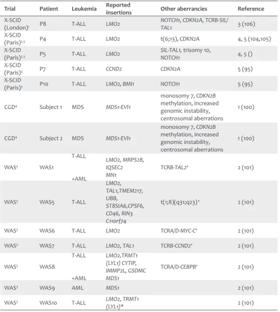
Table 2: Overview of leukemias that developed from insertional mutagenesis in different gene therapy trials.
Trial Patient Leukemia Reported insertions Other aberrancies Reference
X-SCID
(London)1 P8 T-ALL LMO2
NOTCH1, CDKN2A, TCRB-SIL/
TAL1 3 (106)
X-SCID
(Paris)2,3 P4 T-ALL LMO2 t(6;13), CDKN2A 4, 5 (104,105)
X-SCID
(Paris)2,3 P5 T-ALL LMO2 SIL-TAL1, trisomy 10, NOTCH1 4, 5 ()
X-SCID
(Paris)3 P7 T-ALL CCND2 CDKN2A 5 (95)
X-SCID
(Paris)3 P10 T-ALL LMO2, BMI1 NOTCH1 5 (95)
CGD4 Subject 1 MDS MDS1-EVI1
monosomy 7, CDKN2B methylation, increased genomic instability, centrosomal aberrations
1 (100)
CGD4 Subject 2 MDS MDS1-EVI1
monosomy 7, CDKN2B methylation, increased genomic instability, centrosomal aberrations
1 (100)
WAS5 WAS1
T-ALL
+AML
LMO2, MRPS28, IQSEC2
MN1 TCRB-TAL2
¥ 2 (101)
WAS5 WAS5 T-ALL
LMO2, TAL1,TMEM217, UBB, ST8SIA6,CPSF6, CD46, RIN3 C11orf74
t(1;8)(q31;q23) ¥ 2 (101)
WAS5 WAS6 T-ALL LMO2 TCRA/D-MYC-C¥ 2 (101)
WAS5 WAS7 T-ALL LMO2, TAL1 TCRB-CCND2¥ 2 (101)
WAS5 WAS8
T-ALL +AML LMO2,TRMT1 (LYL1) CYTIP, IMMP2L, GSDMC MDS1
TCRA/D-CEBPB¥ 2 (101)
WAS5 WAS9 AML MDS1 2 (101)
WAS5 WAS10 T-ALL LMO2, TRMT1
(LYL1)* 2 (101)
The columns of the table indicate the trial (the type of immunodeficiency for which the trial was performed), the number of the patient that did develop leukemia, the type of leukemia, the genes in which insertions were found and other aberrancies that were detected in leukemic cells. X-SCID; X-linked SCID, CGD; X-linked chronic granulatomous disease, WAS; Wiskott-Aldrich Syndrome, T-ALL; T-cell acute lymphoblastic leukemia, AML; acute myeloid leukemia. * analysis of WAS10 was still ongoing at time of publication of manuscript by Braun et al.112, ¥ detailed data obtained from karyotyping can be found in manuscript of Braun et al.112, 1 patient
has been described by Howe et al.108, 2 patients have been described by Hacein-Bey-Abina et al.107, 3 patients
have been described by Hacein-Bey-Abina et al.106, 4 patients have been described by Stein et al.111, 5 patients
1
a ubiquitously expressed protein, and interaction of both molecules inhibits phagocytosis. NSG mice show increased repopulation levels as compared to other humanized mouse models probably due to this polymorphism of SIRPα.
Besides allowing higher engraftment of human cells, the NOG and NSG mice also sustain human T-cell development in their thymus172, 173. It was shown that the developed lymphocytes were
functional too, as demonstrated by production of cytokines, antibodies and HLA-restricted immune responses173-175. Engraftment capacity and lineage differentiation of expanded HSC
have been studied in the NSG model176 and the model can even be used to study the phenotype
and functionality of human LT-HSC by performing single cell transplantations in NSG mice10.
Because the NSG mice allow for the development of different cell types of the human immune system after transplantation of human HSC, even at limiting, doses, this model offers a great potential for the studies of normal and pathological human T-cell development. Furthermore, the NSG mice are useful in testing preclinical gene therapy approaches120, since the viral doses
needed and efficacy might be different between murine and human cells.
Outline of this thesis
Both in SCID and in the T-ALLs that developed due to LMO2 insertional mutagenesis there are problems in T-cell development. In this thesis the results of studying human T-cell development are described. The overall aim of the research described in this thesis is to study normal and pathological forms of human T-cell development in an in vivo setting to obtain insight into the regulation of human T-cell development. First, we have optimized the NSG humanized mouse model to allow for robust T-cell development from cryopreserved BM derived human HSCs. This model has been used for the other studies and is described in chapter 2. In chapter 3 we describe the results obtained after transplantation of SCID-patient derived HSC in the NSG xenograft model in order to study the stages of developmental arrest for different types of SCID. Furthermore, the results gave new insights in human T-cell development. There are still patients suffering from SCID with an unknown genetic cause. One of these we have studied in chapter 4 to determine whether this patient suffered from a cell intrinsic defect or a niche problem. Here, we could identify the block in T-cell development and identify this patient as being a true SCID. With exome sequencing we identified a potential underlying genetic cause. By the use of cellular barcoding, we have studied the number of clones that seed the thymus and the restriction during T-cell development as described in chapter 5. With SCID, the absent expression of a gene causes the problem in T-cell development, however, overexpression of a gene can also results in arrests in development. In chapter 6 we have studied the effects of LMO2 overexpression, as was observed in the T-ALLs that developed in several gene therapy trials, and to determine the mechanisms that underlie the T-ALL development caused by LMO2. The data from the different chapters are put in perspective to each other and existing literature in chapter 7 together with suggestions for further research.
Humanized mouse model
It is difficult to study human T-cell development, especially in an in vivo system. Many studies on T-cell development have been performed in mice, where the thymus can be removed and studied and furthermore transgenic mice can be created to study the effects of genes on T-cell development. However, human and mouse T-cell development are quite similar but also differ in many aspects35, 37, 48, 158. As mentioned, markers to describe the DN compartments and
ISP stage differ between mice and men. Ways to study human T-cell development are largely descriptive, including gene expression analysis and phenotyping by flow cytometry. The only models to study functionality and differentiation of human T-cell progenitors is the OP9-DL1 model159, 160 and fetal thymic organ cultures (FTOC)161. Advantages of these models are the
ability to study kinetics and follow differentiation over time, which cannot be done on ex vivo thymi. The disadvantage of these models is that it is an in vitro situation with supplemented cytokines and therefore might be different from the physiological situation. Furthermore, in OP9-DL1 cocultures the 3D architectural structure is lost, which is still present in FTOC cultures. Humanized mice are a better alternative for studies on human T-cell development, as this offers the possibility to study the development in an in vivo setting. Development of humanized mice started with the description of the spontaneous Prkdcscid mutation in CB17 mice in 1983162
and the observation that these mice could be engrafted with different types of human hematopoietic cells although no functional human immune system was generated (reviewed by Shultz et al.163). These CB17-scid mice were then crossed with the non-obese diabetic (NOD)
mice to create NOD/Scid mice; mice that allowed a higher level of engraftment of human cells. For a long period these NOD/Scid mice have been used extensively to study human HSCs and their differentiation towards different lineages164. These mice do not have endogenous B cells
and T cells due to the mutation in Prkdc and therefore human HSC are less frequently rejected and able to engraft. These mice have been used widely for e.g. HSC expansion protocols165 and
gene therapy approaches166. Unfortunately, these mice do not allow for development of human
T cells. In 2004, human CD34+ cells that were derived from cordblood were intrahepatically
injected into newborn Rag2-/- γ
c-/- mice167. These mice developed a human immune system,
which was demonstrated to be functional too. In 2002 and 2005, new mouse models have been described that were an adaptation of the earlier NOD/Scid model; by crossing it with an Il2rg-/- (Il2rg encodes the γ
c chain) mouse to generate NOD/Shi-scidIl2rg-/- (NOG)168 or NOD/
LtSz-scidIl2rg-/- (NSG)169 mice, respectively. These mice do not have NK-, B- and T cells whereby
they are not able to reject human cells and these can engraft with higher efficiency than in the NOD/Scid model. The difference between both mouse strains is a small difference in the NOD background and in the type of Il2rg mutation, the NOG mice have a truncated form, which might be able to bind and capture cytokine, while the NSG mice carry a null mutation. Both mice have been compared side by side with the NOD/Scid model; both demonstrated higher engraftment of human lymphoid cells170. In addition, the NSG mice showed a higher
1
20. van Zelm, M.C., et al. Ig gene rearrangement steps are initiated in early human precursor B cell subsets and correlate with specific transcription factor expression. J Immunol 2005, 175(9): 5912-5922.
21. Nutt, S.L., Thevenin, C., Busslinger, M. Essential functions of Pax-5 (BSAP) in pro-B cell development. Immunobiology 1997, 198(1-3): 227-235.
22. Klein, U., et al. Transcriptional analysis of the B cell germinal center reaction. Proc Natl Acad Sci U S A 2003, 100(5): 2639-2644.
23. van Zelm, M.C., Szczepanski, T., van der Burg, M., van Dongen, J.J. Replication history of B lymphocytes reveals homeostatic proliferation and extensive antigen-induced B cell expansion. J Exp Med 2007, 204(3): 645-655.
24. Rothenberg, E.V. Transcriptional control of early T and B cell developmental choices. Annu Rev Immunol 2014, 32: 283-321.
25. Zlotoff, D.A., Bhandoola, A. Hematopoietic progenitor migration to the adult thymus. Ann N Y Acad Sci 2011, 1217: 122-138.
26. Ramond, C., et al. Two waves of distinct hematopoietic progenitor cells colonize the fetal thymus. Nat Immunol 2014, 15(1): 27-35.
27. Reimann, C., et al. Human T-lymphoid progenitors generated in a feeder-cell-free Delta-like-4 culture system promote T-cell reconstitution in NOD/SCID/gammac(-/-) mice. Stem Cells 2012, 30(8): 1771-1780.
28. Danby, R., Rocha, V. Improving engraftment and immune reconstitution in umbilical cord blood transplantation. Front Immunol 2014, 5: 68.
29. Haddad, R., et al. Molecular characterization of early human T/NK and B-lymphoid progenitor cells in umbilical cord blood. Blood 2004, 104(13): 3918-3926.
30. Haddad, R., et al. Dynamics of thymus-colonizing cells during human development. Immunity 2006,
24(2): 217-230.
31. Six, E.M., et al. A human postnatal lymphoid progenitor capable of circulating and seeding the thymus. J Exp Med 2007, 204(13): 3085-3093.
32. Kohn, L.A., et al. Lymphoid priming in human bone marrow begins before expression of CD10 with upregulation of L-selectin. Nat Immunol 2012, 13(10): 963-971.
33. Awong, G., et al. Characterization in vitro and engraftment potential in vivo of human progenitor T cells generated from hematopoietic stem cells. Blood 2009, 114(5): 972-982.
34. Tan, C., et al. Ten-color flow cytometry reveals distinct patterns of expression of CD124 and CD126 by developing thymocytes. BMC Immunol 2011, 12: 36.
35. Weerkamp, F., Pike-Overzet, K., Staal, F.J. T-sing progenitors to commit. Trends Immunol 2006, 27(3): 125-131.
36. Godfrey, D.I., Kennedy, J., Suda, T., Zlotnik, A. A developmental pathway involving four phenotypically and functionally distinct subsets of CD3-CD4-CD8- triple-negative adult mouse thymocytes defined by CD44 and CD25 expression. J Immunol 1993, 150(10): 4244-4252.
37. Dik, W.A., et al. New insights on human T cell development by quantitative T cell receptor gene rearrangement studies and gene expression profiling. J Exp Med 2005, 201(11): 1715-1723.
38. Blom, B., et al. TCR gene rearrangements and expression of the pre-T cell receptor complex during human T-cell differentiation. Blood 1999, 93(9): 3033-3043.
References
1. Gaspar, H.B., et al. Long-term persistence of a polyclonal T cell repertoire after gene therapy for X-linked severe combined immunodeficiency. Sci Transl Med 2011, 3(97): 97ra79.
2. Bigas, A., Espinosa, L. Hematopoietic stem cells: to be or Notch to be. Blood 2012, 119(14): 3226-3235.
3. Luis, T.C., Ichii, M., Brugman, M.H., Kincade, P., Staal, F.J. Wnt signaling strength regulates normal hematopoiesis and its deregulation is involved in leukemia development. Leukemia 2012, 26(3): 414-421.
4. Luis, T.C., et al. Canonical wnt signaling regulates hematopoiesis in a dosage-dependent fashion. Cell Stem Cell 2011, 9(4): 345-356.
5. Soderberg, S.S., Karlsson, G., Karlsson, S. Complex and context dependent regulation of hematopoiesis by TGF-beta superfamily signaling. Ann N Y Acad Sci 2009, 1176: 55-69.
6. Trumpp, A., Essers, M., Wilson, A. Awakening dormant haematopoietic stem cells. Nat Rev Immunol 2010, 10(3): 201-209.
7. Orkin, S.H., Zon, L.I. Hematopoiesis: an evolving paradigm for stem cell biology. Cell 2008, 132(4): 631-644.
8. Bhatia, M., Wang, J.C., Kapp, U., Bonnet, D., Dick, J.E. Purification of primitive human hematopoietic cells capable of repopulating immune-deficient mice. Proc Natl Acad Sci U S A 1997, 94(10): 5320-5325.
9. Majeti, R., Park, C.Y., Weissman, I.L. Identification of a hierarchy of multipotent hematopoietic progenitors in human cord blood. Cell Stem Cell 2007, 1(6): 635-645.
10. Notta, F., et al. Isolation of single human hematopoietic stem cells capable of long-term multilineage engraftment. Science 2011, 333(6039): 218-221.
11. van Galen, P., et al. Reduced lymphoid lineage priming promotes human hematopoietic stem cell expansion. Cell Stem Cell 2014, 14(1): 94-106.
12. Doulatov, S., et al. Revised map of the human progenitor hierarchy shows the origin of macrophages and dendritic cells in early lymphoid development. Nat Immunol 2010, 11(7): 585-593.
13. Laurenti, E., et al. The transcriptional architecture of early human hematopoiesis identifies multilevel control of lymphoid commitment. Nat Immunol 2013, 14(7): 756-763.
14. Sive, J.I., Gottgens, B. Transcriptional network control of normal and leukaemic haematopoiesis. Exp Cell Res 2014.
15. Spits, H., et al. Early stages in the development of human T, natural killer and thymic dendritic cells. Immunol Rev 1998, 165: 75-86.
16. Vargas, C.L., Poursine-Laurent, J., Yang, L., Yokoyama, W.M. Development of thymic NK cells from double negative 1 thymocyte precursors. Blood 2011, 118(13): 3570-3578.
17. Lanier, L.L. Up on the tightrope: natural killer cell activation and inhibition. Nat Immunol 2008, 9(5): 495-502.
18. Ma, Y., Pannicke, U., Schwarz, K., Lieber, M.R. Hairpin opening and overhang processing by an Artemis/ DNA-dependent protein kinase complex in nonhomologous end joining and V(D)J recombination. Cell 2002, 108(6): 781-794.