© 2020 by the Serbian Biological Society How to cite this article: İmir NG. MicroRNA-29a plays a prominent role in 173 PRIMA-1Met-induced apoptosis in ovarian cancer cells. Arch Biol Sci.
2020;72(2):173-9.
MicroRNA-29a plays a prominent role in PRIMA-1
Met-induced apoptosis in ovarian
cancer cells
Nilüfer Gülmen İmir1,2
1Department of Biology Education, Faculty of Education, University of Akdeniz, Antalya, Turkey 2Department of Biology, Life Sciences Institute, University of Akdeniz, Antalya, Turkey
*Corresponding author: [email protected]
Received: November 28, 2019; Revised: February 12, 2020; Accepted: March 10, 2020; Published online: March 18, 2020
Abstract: The structural analog of the small 2,2-bis(hydroxymethyl)-1-azabicyclo[2,2,2]octan-3-one molecule named
PRIMA-1Metfor “p53 reactivation and induction of massive apoptosis” has been shown to inhibit cell growth and induce
apoptosis in human tumor cells by restoring the tumor suppressor function of tumor protein p53. In several microRNA (miRNA) profiling studies related to ovarian cancer, different miRNAs associated with PRIMA-1Met have been reported,
but miRNAs related to PRIMA-1Met-induced apoptosis remain unclear. This study was designed to explain the potential
mechanism of PRIMA-1-induced apoptosis. According to the MTSassay and fluorescence-activated cell sorting (FACS) analysis results, PRIMA-1Met induced a significant decrease in cell viability and an increase in apoptosis in both A2780 and
Caov-3 cells, regardless of p53 status. PRIMA-1Met upregulated miRNA-29a in both cell lines. To determine the effect of
miRNA-29a on PRIMA-1Met-induced apoptosis, A2780 and Caov-3 cells were transfected with miRNA-29a inhibitor. After
treatment with PRIMA-1Met, cell viability increased and apoptosis decreased in the transfected cells. The results of this study
suggest that miRNA-29a potentially regulates PRIMA-1Met-induced apoptosis in ovarian cancer cells.
Keywords: apoptosis; miRNA29a; ovarian cancer; PRIMA-1Met
INTRODUCTION
The P53 tumor suppressor protein is a transcription factor that suppresses tumor growth by regulating many target genes involved in central cellular processes such as transcription, DNA repair, genomic stability, aging, cell cycle control and apoptosis [1,2]. It has also become a target for mechanism-based anticancer drug discovery because p53 mutations are found in more than 50% of human cancers [3-7]. In recent years, attempts have been made to use p53 as a target for cancer treatment, and various small molecules have been identified as capable of reversing the oncogenic properties of mutant p53 [8,9]. Among these small molecules, the most studied are PRIMA-1 and the
methylated derivative PRIMA-1Met[10,11]. PRIMA-1Met
was shown to inhibit cell proliferation and to induce apoptosis in malignant cell cultures containing dif-ferent p53 mutations and in some xenograft animal tumors by restoration of the p53 tumor suppressor function [12-16].
identified altered expression of miRNAs in response
to PRIMA-1Met treatment in ovarian cancer cell lines
with mutant and wild-typep53; we demonstrated that miRNA-29a was among the most regulated miRNAs
in response to PRIMA-1Met treatment, however, it was
not shown whether PRIMA-1Met induces apoptosis by
regulating specific miRNA expression. Therefore, in this study a potential role of miRNA-29a for
PRIMA-1Met-induced apoptosis has been investigated.
MATERIALS AND METHODS
Cell culture and reagents
The human ovarian cancer cell line Caov-3 (mutant p53) was obtained from the American Type Culture Collection (ATCC, USA). A2780 cell line (wild-type p53) was purchased from the Sigma (Sigma, USA). Cells were grown in Dulbecco’s Modified Eagle Medium (DMEM, Gibco, USA) with 10% fetal bovine serum and 1% penicillin/streptomycin in a humidified incubator
(37ºC, 5% CO
2). PRIMA-1Met (purchased from Santa
Cruz, USA)was diluted in Ultra Pure water (Sigma, USA) and was used for cellular treatments.
Cell viability analysis
Cell viability in PRIMA-1Met treated and untreated
(control) cells was assessed using a Cell Titer 96 aque-ous nonradioactive cell proliferation assay (Promega,
USA). The cells were seeded at 1×10⁴ cells/well in 200
µL complete medium in a 96-well plate and allowed to attach for 24 h [29]. Subsequently, A2780 (p53-wild type) and Caov-3 (p53-mutant) cells were treated
with PRIMA-1Met at doses of 20 µM and 40 µM,
re-spectively, for 48 h [30]. At the end of the incubation time, 20 µL of MTS[3-(4,5-dimethylthiazol-2-yl)- 5-(3-carboxymethoxyphenyl)-2-(4-sulfophenyl)-2H-tetrazolium]/PMS (phenazine methosulfate) solution was added to each well for 4 h. The absorbance at 490 nm was measured in a microplate reader (Thermo Labsystems Multiskan Spectrum, Thermo Lab Systems, USA), using wells without cells as background. The sample readings were calculated by subtracting the average of background absorbances. All experiments were performed at least four times.
Apoptosis assay
A2780 and Caov-3 cells were treated with PRIMA-1Met
at doses of 20 µM and 40 µM, respectively, for 48 h. Apoptotic cells were determined using the Annexin V-FITC apoptosis kit (BD Biosciences, USA). All procedures were performed according to the
manu-facturer’s protocol. Briefly, 1x10⁶ cells were rinsed with
a binding buffer and were suspended in 200 µL of the same buffer. Then 5 µL of Annexin V and 5 µL of propidium iodide were added to the suspension. The cells were incubated for 15min at room temperature in the dark and then assayed by FACS.
Quantitative real-time PCR
Total RNA, including miRNAs, was isolated from the cells using the miRNA Easy kit (Qiagen, Germany) ac-cording to the manufacturer instructions, and was then quantified with a spectrophotometer (Thermo Fisher Scientific, Inc., USA) at wavelengths of 230, 260, and 280 nm. For miRNA analysis, samples of isolated RNA were reverse-transcribed into cDNA using anmiScript II RT kit (Qiagen, Germany). Briefly, the RT master mix included the following: 5 µL template RNA (250 ng), 4 µL 5xmiScriptHiSpec buffer, 2 µL 10x nucleic mix, 7 µL RNase free water and 2 µL miScript reverse transcriptase mix up to a total volume of 20 µL. The reaction mixture was centrifuged, placed on ice and
then incubated at 37ºC for 60 min and 95ºC for 5 min.
cDNA samples were transferred to a -20ºC freezer. The
expression of miRNA-29a was examined in the two cell
lines after exposure to PRIMA-1Met using the miScript
SYBR Green PCR kit (Qiagen, Germany) according to the manufacturer’s protocol. Briefly, the PCR amplifi-cation was conducted in a 25-µL reaction using a 2X Quanti Tect SYBR Green PCR master mix (12.5 µL), 10xmiScript Universal primer (2.5µL), 10xmiScript miRNA-29a-spesific primer (2.5µL), RNase free water (5.5 µL) and 10 ng template cDNA (2 µL). Real-time PCR was performed in a 96-well plate using a Step One Plus™ Real-Time PCR System (Thermo Fisher
Scientific, Inc., USA) at 95ºC for 15 min, followed by
40 cycles at 94ºC for 15 s, 55ºC for 30 s and 70ºC for
30 s. The expression of miRNA-29a was calculated relative to small nucleolar RNA (SNORD)72, and
fold-changes were calculated by the 2-ΔΔCt method [31].
Inhibition of miRNA-29a
To test the efficacy of miRNA-29a on PRIMA-1Met
-induced apoptosis in ovarian cancer cells A2780 and
Caov-3,cells (4× 10⁴) were transfected with either 10
nM of mirVana inhibitor of miRNA-29a (Invitrogen, USA) or 10 nM of the negative control, mirVana miRNA inhibitor (Invitrogen, USA) by the lipofectamine RNAi MAX transfection method (Invitrogen, USA) according to the manufacturer’s protocol. After 24 h incubation, the medium containing lipofectamine RNAi MAX reagent was removed and normal DMEM was added.
The transfected cells were treated with PRIMA-1Met
and incubated for additional 48 h, and cell viability and apoptosis were evaluated in the transfected cells.
Statistical analysis
All values are presented as the mean±standard devia-tion. The Student’s t-test was used for comparison of the two groups. Analyses were performed with Graph Pad In Stat v.10.0 software (GraphPad Software, Inc., USA). P<0.001 was considered statistically significant. Sigma Plot v.10.0 were used for illustration.
RESULTS
PRIMA-1Met causes a significant decrease in cell viability and an increase in apoptosis in ovarian cancer cells
To examine the effect of PRIMA-1Met on apoptotic cell
death in ovarian cancer cells, the A2780 (p53-wild type)
and Caov-3 (p53-mutant) cell lines were treated with
20 µM and 40 µM PRIMA-1Met, respectively, and cell
viability was determined using the MTS assay after
48 h of treatment. It was determined thatPRIMA-1Met
was significantly cytotoxic for both cell lines, indepen-dently of p53 status (p<0.001). After the treatment with
PRIMA-1Met, the Caov-3 and A2780 cells had 44.75%
and 42.75% viable cells, respectively (Fig.1A).These results were confirmed by FACS analysis. In Caov-3 cells, 13% of the cells in the control group and 64.6%
of the cells in the PRIMA-1Met-treated group were
apoptotic, whereas in A2780 cells, the apoptotic cell
percentages in the control and PRIMA-1Met-treated
group were respectively 10.8 and 57.8% (Fig. 1B).
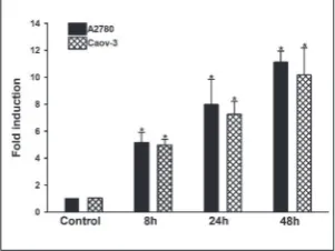
PRIMA-1Met induces miRNA-29a expression in ovarian cancer cells
Previously we observed by miRNA PCR array that miRNA-29a was among the most expressed miRNAs
afterPRIMA-1Metexposure in both cell lines (A2780
and Caov-3).To further confirm the miRNA array data, miRNA expression was examined in the two
cell lines after treatment to PRIMA-1Met. For this, the
cell lines were treated with PRIMA-1Metat 20 µM and
40 µM doses (A2780 and Caov-3, respectively), and miRNA-29a expression at 8, 24 and 48 h was evaluated using miScript microRNA PCR analysis. As shown
in Fig. 2, PRIMA-1Metraised miRNA-29a expression
Fig. 1. Analysis of cell viability and apoptosis after PRIMA-1Met treatment. A2780 (20µM) and
Caov-3 (40 µM) cells were treated with PRIMA-1Met, and the MTS assay was performed to
determine cell viability 48h post-treatment (A). Cells treated with PRIMA-1Met for 48 h were
stained with the Annexin V-FITC apoptosis kit and analyzed by FACS (B). Each experiment was repeated four times. The results are presented as the mean±SD. * indicates P<0.001.
Fig. 2. PRIMA-1Met upregulates expression of
miRNA29a in ovarian cancer cells. RT-qPCR analysis was used to evaluate the expression of miRNA29a. The cells were treated with PRIMA-1Met for 8, 24 and 48 h. RT-qPCR
was carried out in a 96-well plate using a Step One Real Time-PCR instrument. Fold induction was calculated by the 2-ΔΔCt method.
after 48 h treatment 11.12- and 10.16-fold in A2780 and Caov-3 cells, respectively, when compared with untreated controls (p<0.001).
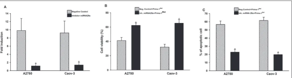
Inhibition of miRNA-29a reduces PRIMA-1Met -induced apoptosis
As mentioned, PRIMA-1Met increases miRNA-29a
expression in A2780 and Caov-3 cells. To investigate
whether miRNA-29a regulates PRIMA-1Met-induced
apoptosis, A2780 and Caov-3 cells were transfected with mirVana, the mir-29a inhibitor and negative control. A significant reduction in miRNA-29a expression in cells transfected with mirVana mir-29a inhibitor was indicative of successful transfection (Fig.3A). After 24
h of PRIMA-1Met treatment, cell viability in transfected
cells was measured as previously described. As shown
in Fig.3B, after treatment with PRIMA-1 Met, cell
vi-ability increased in cells transfected with the miRNA-29a inhibitor when compared to cells transfected with the negative control in both cell lines (p<0.001).This result showed that miRNA-29a plays an important role
in sensitizing ovarian cancer cells to PRIMA-1Met. To
examine in more detail whether miRNA-29a regulates
PRIMA-1Met-induced apoptosis, cells transfected with
miRNA-29a inhibitor were treated with PRIMA-1Met
for 24 h, and apoptosis was evaluated by FACS analysis (Fig.3C). The apoptosis results were also consistent with viability. Compared to cells transfected with inhibitor miRNA and cells transfected with its negative control, cell viability increased from 42% to 62.4% in A2780 cells and from 32% to 65.4% in Caov-3 cells. As expected,
inhibition of miRNA-29a reduced PRIMA-1-induced apoptosis, namely; the proportion of apoptotic cells in cells transfected with inhibitor miRNA-29a decreased to 22.8% and 19.8%, in A2780 and Caov-3 cells, re-spectively. These results, when evaluated together, showed that miRNA-29a could play a prominent role
in the regulation of PRIMA-1Met-induced apoptosis in
ovarian cancer cells.
DISCUSSION
Recently, different strategies have been developed to use P53 as a target in cancer treatment. Identifica-tion of various small molecules that can reverse the oncogenic properties of mutant p53 is among these
strategies[9,32]. PRIMA-1Met is a methylated
deriva-tive of one of these low molecular weight compounds,
PRIMA-1[33]. Studies have shown that PRIMA-1Met is
more effective in cancer cells expressing mutant p53 than wild-type p53 or null cells [16,34], while several
studies have shown that PRIMA-1Met exhibits cytotoxic
effects independent of the p53 mutation status[35-38].
It was reported that PRIMA-1Met has sufficient activity
to suppress growth in epithelial ovarian cancer cells, regardless of the mutation status of p53 [30].PRIMA-1 was shown to trigger apoptosis by altering the expres-sion of p53 downstream genes such as p53 upregulated modulator of apoptosis (PUMA), phorbol-12-myristate-13-acetate-induced protein 1 (NOXA) and apoptosis regulator BAX, also known as bcl-2-like protein 4 in the mutant p53 bladder cancer cell line [39]. PRIMA-1 induces apoptosis by promoting the activation of
Fig. 3.Inhibition of miRNA-29a reduces PRIMA-1Met-induced apoptosis. A2780 and Caov-3 cells were transfected with the mirVana
miRNA inhibitor to inhibit the expression of miRNA-29a. Transfected cells were treated with PRIMA-1Met for 24 h.RT-qPCR was used
to confirm the specific knockdown of miRNA-29a in the transfected cells (A). After treatment with PRIMA-1Met, cell viability was
proapoptotic signaling pathways and inhibiting JNK cell viability in mutant p53 breast cancer cells [40].In
this study, PRIMA-1Met was shown to cause a significant
reduction in cell viability and increased apoptosis in ovarian cancer cells carrying both wild-type p53 and mutant p53. There is no precise information about
the mechanism of PRIMA-1Met-induced apoptosis,
especially in ovarian cancer. The fact that PRIMA-1Met
is effective in both mutated and unmutated ovarian cancer cells can only be explained by p53-independent mechanisms of action [41].
MiRNAs act in a variety of physiological and bio-logical processes, such as cell proliferation, differentia-tion, and hematopoiesis, by regulating the expression of multiple target genes [42]. Recently, dysregulation of miRNAs has been associated with cancer initiation and progression. The role of miRNAs in apoptosis is not fully understood, but there is evidence that miR-NAs are important in this process [43]. For example, when the role of miR-34 family members in regulating PRIMA-1 induced apoptosis was investigated [44], it was reported that PRIMA-1 upregulated miR-34a in mutant p53 human lung cancer cells, which suggests that PRIMA-1 upregulates miR-34a to induce apoptosis in lung cancer cells. Investigation of the role of miRNA-29a
in response to PRIMA-1Met in multiple myeloma cells
revealed that miRNA-29a plays an important role in
sensitizing the cells to PRIMA-1Met [42]. Comparison
of miRNA expression levels among PRIMA-1-treated and untreated groups in a lung tumor model in trans-genic mice expressing human mutant p53showed that 9 miRNAs changed at least 6-fold after PRIMA-1 treatment compared to the control group; these are 194, 1937a, 1937b, 34c, 192, miR-1949, miR-2135, miR-3472, miR-712 and miR-1931. However, thus far, it has not been determined which
miRNAs regulate PRIMA-1Met-induced apoptosis in
ovarian cancer cells. In the present study, we showed that miRNA-29a expression was increased by
PRIMA-1Met treatment in A2780 and Caov-3 ovarian cancer cells
carrying wild-type p53 and mutant p53, respectively. These results indicate that miRNA-29a is associated with
PRIMA-1Met-induced apoptosis. Thus, cells transfected
with the mirVana miRNA-29a inhibitor were treated
with PRIMA-1Met, and cell viability and apoptosis were
analyzed. The reduction of apoptosis in these transfected cells provides important evidence that miRNA-29a plays
a prominent role in PRIMA-1Met-induced apoptosis.
The increase in cell viability in these transfected cells supports the importance of miRNA-29a in PRIMA-1
Met-induced apoptosis.
CONCLUSION
The results of this study show that the mechanisms of
action of PRIMA-1Met differ in part from those
previ-ously described. In the present report, the antitumor
activity of PRIMA-1Met was shown to be independent
of p53 mutation status in ovarian cancer cells and that apoptosis was induced by increasing miRNA-29a.
Accordingly, PRIMA-1Met should be examined as a
potential anti-cancer agent in in vivo and clinical tri-als. Further studies are needed to examine the role of
miRNA-29a in the mechanism of PRIMA-1Met-induced
apoptosis in more detail, including the apoptotic genes that it regulates.
Funding: This work was supported by Akdeniz University Scientific Research Projects Coordination Unit (FBA-2017-2656).
Acknowledgements: All experiments were carried out in the Cancer Molecular Biology Laboratory of the Biology Department of the Faculty of Science, Akdeniz University.
Conflict of interest disclosure: There is no conflict of interest.
REFERENCES
1. Harris CC. Structure and function of the p53 tumor suppres-sor gene: Clues for rational cancer therapeuetic strategies. J Natl Cancer Inst. 1996;88(20):1442-55.
2. Sullivan KD, Galbraith MD, Andrysik Z, Espinosa JM. Mechanisms of transcriptional regulation by p53. Cell Death Differ. 2018;25:133-43.
3. Teneriello MG, Ebina M, Linnoila R, Henry M, Nash JD, Park RC, Birrer MJ. p53 and Ki-ras gene mutations in epi-thelial ovarian neoplasms. Cancer Res. 1993;53:3103-08. 4. Mazare R, Pujol P, Maudelonde T, Jeanteur P, Theillet C.
p53 mutations in ovarian cancer: a late event? Oncogene. 1991;6:1685-90.
5. Eccles DM, Brett L, Lessells A, Gruber L, Lane D, Steel CM, Leonard RC. Overexpression of the p53 protein and allele loss at 17p13 in ovarian carcinoma. Br J Cancer. 1992;65:40-4. 6. Kohler MF, Kerns BJ, Humphrey PA, Marks JR, Bast RC,
Ber-chuck A. Mutation and overexpression of p53 in earlystage epithelial ovarian cancer. Obstet Gynecol. 1993;81:643-50. 7. Wang Z and Sun Y. Targeting p53 for novel anticancer
ther-apy. Transl Oncol. 2010;3(1):1-12.
9. Duffy MJ, Synnott NC, McGowan PM, Crown J, McGowan PM, Crown J, O’Connor D, Gallagher WM. p53 as a target for the treatment of cancer. Cancer Treat Rev. 2014;40(10):1153-60.
10. Bykov VJ, Issaeva N, Shilov A, Hultcrantz M, Pugacheva E, Chumakov P, Bergman J, Wiman KG, Selinova G. Restora-tion of the tumor suppressor funcRestora-tion to mutant p53 by a low molecular-weight compound. Nat Med. 2002;8:282-8. 11. Selinova G, Wiman KG. Reactivation of mutant p53:
molec-ular mechanisms and therapeutic potential. Oncogene. 2007;26(15):2243-54.
12. Bykov VJ, Zache N, Stridh H, Westman J, Bergman J, Seliva-nova G, Wiman KG. PRIMA-1MET synergizes with cisplatin to induce tumor cell apoptosis. Oncogene. 2005;24:3484-91. 13. Shi H, Lambert JM, Hautefeuille A, Bykov VJ, Wiman KG, Hainaut P, Caron de Fromentel C. In vitro and in vivo cytotoxic effects of PRIMA-1 on hepatocellular carcinoma cells express-ing mutant p53ser249. Carcinogenesis. 2008;29:1428-34. 14. Zache N, Lambert JM, Wiman KG, Bykov VJ.
PRIMA-1MET inhibits growth of mouse tumors carrying mutant p53. Cell Oncol. 2008;30:411-8.
15. Liang Y, Besch-Williford C, Hyder SM. PRIMA-1 inhibits growth of breast cancer cells by re-activating mutant p53 protein. Int J Oncol. 2009;35:1015-23.
16. Zandi R, Selivanova G, Christensen CL, Gerds TA, Wil-lumsen BM, Poulsen HS. PRIMA-1Met/APR-246 induces apoptosis and tumor growth delay in small cell lung cancer expressing mutant p53. Clin Cancer Res. 2011;17:2830-41. 17. Oliveto S, Mancino M, Manfrini N, Biffo S. Role of microR-NAs in translation regulation and cancer. World J Biol Chem. 2017;8(1):45-56.
18. Zhang B, Pan X, Cobb GP, Anderson TA. MicroRNAs as oncogenes and tumor suppressors. Dev Biol. 2007;302:1-12. 19. Kumar MS, Lu J, Mercer KL, Golub TR, Jacks T. Impaired microRNA processing enhances cellular transformation and tumorigenesis. Nat Genet. 2007;39:673-7.
20. Yang D, Sun Y, Hu L, Zheng H, Ji P, Zhao Y, Reynolds S, Cheng H, Rupaimoole R, Cogdell D, Nykter M, Broaddus R, Rodriguez-Aguayo C, Lopez-Berestein G, Liu J, Shmu-levich I, Sood AK, Chen K, Zhang W. Intregrated analyses identify a master microRNA regulatory network for the mesenchymal subtype in serous ovarian cancer. Cancer Cell. 2013;23:186-99.
21. Iorio MV, Visone R, Di Leva G, Donati V, Petrocca F, Casa-lini P, Taccioli C, VoCasa-linia S, Liu CG, Alder H, Calin GA, Menard S, Croce CM. MicroRNA signatures in human ovar-ian cancer. Cancer Res. 2007;67:8699-707.
22. Yang N, Kaur S, Volinia S, Greshock J, Lassus H, Hasegawa K, Liang S, Leminen A, Deng S, Smith L, Johnstone CN, Chen XM, Liu CG, Huang Q, Katsaros D, Calin GA, Weber BL, Bützow R, Croce CM, Coukos G, Zhang L. MicroRNA microarray identifies Let-7i as a novel biomarker anti-ther-apeutic target in human epithelial ovarian cancer. Cancer Res. 2008;68:10307-14.
23. Dahiya N, Sherman-Baust CA, Wang TL, Davidson B, Shih IM, Zhang Y, Wood W, Becker KG, Morin PJ. MicroRNA expression and identification of putative miRNA targets in ovarian cancer. PLoS One. 2008;3(6):e2436.
24. Nam EJ, Yoon H, Kim SW, Kim H, Kim YT, Kim JH, Kim JW, Kim S. MicroRNA expression profiles in serous ovarian carcinoma. Clin Cancer Res. 2008;14:2690-95.
25. Dahiya N and Morin PJ. MicroRNAs in ovarian carcinomas. Endocr Relat Cancer. 2010;17:77-89.
26. Mateescu B, Batista L, Cardon M, Gruosso T, de Feraudy Yi Mariani O, Nicolas A, Meyniel JP, Cottu P, Sastre-Garau X, Mechta-Grigoriou F. miR-141 and miR-200a act on ovarian tumorigenesis by controlling oxidative stress response. Nat Med. 2011;17:1627-35.
27. Yeh YM, Chuang CM, Chao KC, Wang LH. MicroRNA-138 suppresses ovarian cancer cell invasion and metastasis by targeting SOX4 and HIF-1a. Int J Cancer. 2013;133:867-78. 28. Park SY, Lee JH, Ha M, Nam JW, Kim VN. miR-29 miRNAs
activate p53 by targeting p85 alpha and CDC42. Nat Struct Mol Biol. 2009;16(1):23-9.
29. Aydemir EA, Oz ES, Gokturk RS, Ozkan G, Fiskin K. Glyc-yrrhizaflavescens subsp. antalyensis exerts antiproliferative effects on melanoma cells via altering TNF-α and IFN-α levels. Food Chem Toxicol. 2011;49(4):820-8.
30. Livak KJ and Schmittgen TD. Analysis of relative gene expres sion data using real-time quantitative PCR and the 2(-delta delta C(T)) method. Methods. 2001;25:402-8. 31. Parrales A and Iwakuma T. Targeting oncogenic mutant p53
for cancer therapy. Front Oncol. 2015;5:288.
32. Bykov VJ, Issaeva N, Selivanova G, Wiman KG. Mutant p53-dependent growth suppression distinguishes PRIMA-1 from known anticancer drugs: a statistical analysis of infor-mation in the National Cancer Institute database. Carcino-genesis. 2002;23:2011-8.
33. Izetti P, Hautefeuille A, Abujamra AL, de Farias CB, Gia-comazzi J, Alemar B, Lenz G, Roesler R, Schwartsmann G, Osvaldt AB, Hainaut P, Ashton-Prolla P. PRIMA-1, a mutant p53 reactivator, induces apoptosis and enhances chemother-apeutic cytotoxicity in pancreatic cancer cell lines. Invest New Drugs. 2014;32:783-94.
34. Russo D, Ottaggio L, Foggetti G, Masini M, Masiello P, Fronza G, Menichini P. PRIMA-1 induces autophagy in cancer cells carrying mutant or wild type p53. BiochimBio-physActa. 2013;1833:1904-13.
35. Aryee DN, Niedan S, Ban J, Schwentner R, Muehlbacher K, Kauer M, Kofler R, Kovar H. Variability in functional p53 reactivation by PRIMA-1(Met)/APR-246 in Ewing sarcoma. Br J Cancer. 2013;109:2696-704.
36. Tessoulin B, Descamps G, Moreau P, Maïga S, Lodé L, Godon C, Marionneau-Lambot S, Oullier T, Le Gouill S, Amiot M, Pellat-Deceunynck C. PRIMA-1Met induces myeloma cell death independent of p53 by impairing the GSH/ROS balance. Blood. 2014;124:1626-36.
37. Li XL, Zhou J, Chan ZL, Chooi JYi Chen ZR, Chang WJ. PRIMA-1met (APR-246) inhibits growth of colorectal can-cer cells with different p53 status through distinct mecha-nisms. Oncotarget. 2015;6(34):36689-99.
39. Piantino c, Reis ST, Viana NI, Silva IA, Morais DR, Antunes AA, Dip N, Srougi M, Leite KR. PRIMA-1 induces apoptosis in bladder cancer cell lines by activating p53. Clinics (Sao Paulo). 2013;68(3):297-303.
40. Wang T, Lee K, Rehman A, Daoud SS. PRIMA-1 induces apoptosis by inhibiting JNK signaling but promoting the activation of Bax. Biochem Biophys Res Commun. 2007;352(1):203-12.
41. Lambert JM, Moshfegh A, Hainaut P, Wiman KG, Bykov VJ. Mutant p53 reactivation by PRIMA-1MET induces mul-tiple signaling pathways converging on apoptosis. Oncogene. 2010;29(9):1329-38.
42. Saha MN, Abdi J, Yang Y, Chang H. miRNA-29a as a tumor suppressor mediates PRIMA-1Met-induced anti-myeloma activity by targeting c-Myc. Oncotarget. 2016;7(6):7149-60. 43. Othman N and Nagoor NH. The Role of microRNAs in the
Regulation of Apoptosis in Lung Cancer and Its Application in Cancer Treatment. Biomed Res Int. 2014;2014(Special issue):1-19.