551
© 2018 by the Serbian Biological Society How to cite this article: Savicka M, Petjukevičs A, Batjuka A, Škute N. Impact of moderate heat stress on the biochemical and physiological responses of the invasive waterweed Elodea canadensis (Michx. 1803). Arch Biol Sci. 2018;70(3):551-7.
Impact of moderate heat stress on the biochemical and physiological responses of the
invasive waterweed
Elodea canadensis
(Michx. 1803)
Marina Savicka*, Aleksandrs Petjukevičs, Anna Batjuka and Nataļja Škute
Department of Ecology, Institute of Life Sciences and Technologies, Daugavpils University, Parades str. 1A, Daugavpils,
Latvia, LV-5401
*Corresponding author: [email protected]
Received: January 19, 2018; Revised: April 10, 2018; Accepted: April 10, 2018; Published online: April 18, 2018
Abstract: Global warming can negatively affect freshwater macrophytes. However, the degree to which freshwater plants can survive after long-term or short-term warming and the underlying molecular mechanisms are not fully understood. The aim of our study was to analyze the responses of an invasive plant to moderate heat stress (HS). Biochemical and physi-ological stress responses to experimental warming (30±1.0°C/25±1.0°C, day/night) were assessed in the invasive waterweed
Elodea canadensis. The effect of the moderate HS on the macrophyte was evaluated through changes in the total protein
content, catalase activity, lipid peroxidation, cellular membrane permeability by electrolyte leakage and the concentrations of carotenoids and photosynthetic pigments. Catalase activity and carotenoid concentrations increased significantly (p<0.01) in comparison to the control. A significant increase (p<0.05) in malondialdehyde concentration was observed. However, at the same time there was a persistent low level of electrolyte leakage in heat-treated plants as compared to the control. The results demonstrated that moderate HS improved membrane stability and increased the concentration of photosynthetic pigments and antioxidant activity in E. canadensis shoots. Moderate alterations in temperature may favorably affect the physiology and growth of the invasive macrophyte E. canadensis. It is reasonable to expect that warming could lead to a gradual change in E. canadensis distribution and to changes in composition of freshwater ecosystems.
Key words: Elodea canadensis; antioxidants; moderate heat stress; malondialdehyde; electrolyte leakage
IntRoduCtIon
Global warming can promote the growth of invasive aquatic macrophytes [1,2]. Macrophytes play an im-portant role in aquatic ecosystems (as primary pro-ducers, sources of habitats and refuges) and it is essen-tial to study them in the context of the effects of global warming [3]. Elevated water temperature can severely affect submerged macrophytes, as the magnitude and intensity of temperature fluctuations and the toler-ability of plant species determine the severity of these effects [4]. Plant responses to high temperature vary with the degree and duration of temperature stress and plant type because the increase in temperature affects plant growth and development. Plant distri-bution varies with the temperature range in which they can grow. For example, the growth temperature ranges from 15 to 25°C for Egeria densa Planch., from 10 to 25°C for Elodea canadensis, from 10 to 25°C
for Lagarosiphon major (Ridl.) Moss ex Wager, and from 8 to 36°C for Hydrilla verticillata (L. f.) Royle [3,5]. A temperature increase will be favorable for an aquatic plant species that has a high temperature threshold value, as observed in invasive submerged macrophytes such as Hydrilla verticillata, Myriophyl-lum spicatum L. [6]. However, the temperature range for different aquatic macrophytes tends to be narrower than that for terrestrial plants, although many aquatic macrophytes can exist in shallow waters (<1 m depth) where seasonal and diurnal temperature fluctuations are much larger [7,8].
se-ries of enzymatic and non-enzymatic detoxification mechanisms. ROS in plant tissues can initiate lipid peroxidation (LP) that damages cell membranes and is considered to be the most important mechanism of tis-sue damage [9]. Enzymatic detoxification mechanisms that principally minimize cellular levels of superoxide radicals (O2−) and hydrogen peroxide (H
2O2) include
the production of antioxidant enzymes such as catalase (CAT), peroxidase (POD), ascorbate peroxidase (APX) and superoxide dismutase (SOD) [10]. The non-en-zymatic antioxidant system includes vitamins (ascor-bic acid and tocopherols), glutathione, carotenoids, phenols, etc., which are among the key antioxidants involved in scavenging toxic ROS [11]. Carotenoids are pigments found in plants and microorganisms. Carot-enoids play a key role in the photosynthetic reaction center where they are involved in mechanisms regulat-ing photoprotection against auto-oxidation [12]. Ca-rotenoids are non-enzymatic lipophilic antioxidants that at sufficiently high concentrations protect lipids from peroxidative damage [13].
Heat-induced oxidative stress has been widely discussed for terrestrial vegetation [14-16]. However, less attention has been paid to heat-induced oxida-tive stress and antioxidant system activity in aquatic macrophytes. There are a few cases where the stress-induced oxidative reactions in aquatic macrophytes have been measured, and most of these studies have focused on the biochemical and physiological chang-es under different strchang-essors such as hypoxia, salinity, heavy metals [17-21].
Submerged macrophytes play a key role in biodi-versity and water transparency in shallow freshwater ecosystems and they can be exposed to pronounced temperature fluctuations [22]. E. canadensis is a pe-rennial invasive plant in natural aquatic ecosystems in Eurasia and is common in eutrophic waters [1]. E. canadensis forms thick stands, outcompeting native biodiversity. If the climate gets warmer, it is likely that E. canadensis will spread further [23]. E. canadensis is well-studied from various aspects, in-cluding invasion success and competitive ability, its impact on native aquatic communities [24] and on responses to environmental variables [25], and on morphological changes in response to temperature changes [26]. However, it is important to mention that there is almost no information about the influence of
temperature on oxidative changes and activity of the antioxidant system in E. canadensis exposed to mod-erate HS. In this study we examined the influence of increases in water temperature on the physiological and biochemical changes in the submerged invasive macrophyte E. canadensis.
MAteRIAls And Methods
high temperature treatments under controlled conditions
The experimental material (E. canadensis plants) was collected from the nearest lake. The plants were rinsed with clean water to remove debris and the at-tached algae were separated using forceps. The cleaned plants were cultured in glass aquaria under laborato-ry conditions. The experiment was conducted under controlled temperature in climate chambers (POL-EKO, Poland). All the treatments were subjected to a photoperiod regime of 8 h dark and 16 h light with a light intensity of 50 µmol photons m-2s-1. The air
tem-perature in the climate chamber was maintained at 18±1.0°C/15±1.0°C (day/night) for the control plants, and at 30±1.0°C/25±1.0°C (day/night) in order to sim-ulate moderate HS. The short-term effects of moderate HS were determined 24 h after exposure to HS for 7 days. E. canadensis samples from the control and heat-treated plants were collected 2, 3, 4 and 7 days after the initiation of high temperature treatment, and one day after the high temperature treatment was finished.
Plant tissue preparation for protein assay
Fresh plant samples (~300 mg) were extracted with ice cold 50 mM phosphate buffer (pH 7.8). Polyvinylpyr-rolidone (PVP) was added to the extraction to mask the effects of phenolic compounds in the plant tissues. The extractions were centrifuged at 5 000 ×g at 4°C for 15 min, and the supernatant was kept at -80°C until further analysis.
Protein determination
determination of catalase activity
The method of Aebi [27] was used to measure catalase (CAT) (EC 1.11.1.6) activity. The decomposition of H2O2 was measured as the decrease in absorbance at 240 nm. Fifty mM phosphate buffer (pH 7.8) and 10 mM H2O2 were used in the reaction solution.
determination of electrolyte leakage
The electrolyte leakage (EL) in E. canadensis shoots was determined as described [28]. The EL was calcu-lated from the following formula: EL(%)=(Cbeforeboiling -CH2O))/(Cafterboiling-CH2O )×100, with C the conductivity of the solution before and after boiling.
Measurement of lipid peroxidation
The malondialdehyde (MDA) content was deter-mined by the thiobarbituric acid reaction (TBARS) as described [29]. The concentration of MDA was es-timated by subtracting the non-specific absorption at 600 nm from the absorption at 532 nm using a molar extinction coefficient 155 mM-1cm−1.
determination of photosynthetic pigments
The concentrations of chlorophyll (Chla, Chlb, Chla+b) and total carotenoids were determined spec-trophotometrically in 200 mg of the plant material ground in a pre-chilled mortar in the presence of 5 mL of 80% acetone (v/v). After complete extraction, the mixture was centrifuged twice, and the volume adjusted to 10 mL with cold acetone. The absorbance of the extract was read at 663, 646, and 470 nm (Var-ian, USA) and pigment concentrations were calculated according to Lichtenthaler [30].
statistical analysis
The data were expressed as the mean±standard devia-tion and were analyzed statistically using Microsoft Excel 2010. The data are presented after taking into consideration the standard deviation (SD) of three replicates. The results were analyzed by one-way ANOVA to identify significant differences between the groups, and their significance levels (p<0.05) were determined.
Results
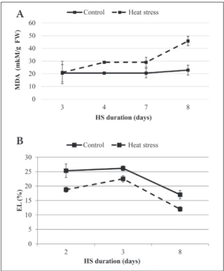
The results revealed that the biochemical and physi-ological changes in E. canadensis shoots were sig-nificantly affected depending on HS duration. The measurement of MDA and EL as parameters of oxida-tive stress showed significant differences between the heat-treated and control plants. The concentration of MDA was significantly higher (p<0.05) in the heat-treated plants (Fig. 1A), while a significant decrease (p<0.05) in EL was observed in the heat-treated plants compared to the control (Fig. 1B); on day 2 after expo-sure to stress it was decreased by 26% and it reached 29% at the end of the investigated period compared to the control.
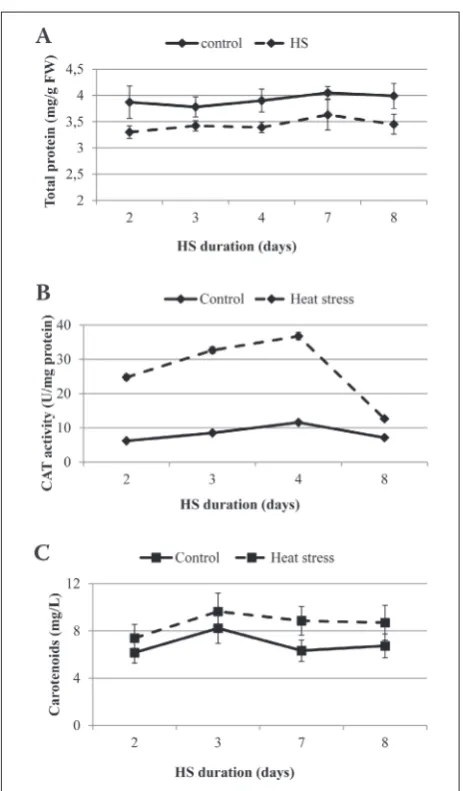
The change in the total protein content in E. ca-nadensis shoots during moderate HS and on the day after the stress treatment as compared to the control are shown in Fig. 2A. A statistically significant
crease (p≤0.001) in the total protein content by 14% was observed after the HS treatment. CAT under HS also exhibited significant differences when compared to the control (Fig. 2B). However, a 4-fold increase (p≤0.001) in CAT activity during the HS was observed during the investigated period compared to the con-trol, despite the significant decrease in total protein. Lipid damage, expressed as the increase in MDA in the cell membrane, was increased two-fold a day after the HS treatment was finished (Fig. 1A, p<0.05), whereas the activity of CAT significantly decreased (Fig. 2B).
Parallel with the high activity of CAT, the mod-erate heat stress altered the carotenoid content in E. canadensis leaves. The carotenoid content was signifi-cantly increased in plants exposed to HS. The changes in total carotenoids in E. canadensis leaves during and after exposure to HS as compared to the controls are shown in Fig. 2C. The total carotenoids increased dur-ing the investigated period: on day 2 after exposure to HS the carotenoid content increased by 20% and it reached 40% at the end of the investigated period when compared to the control (p<0.01) and remained at the same level for a day after the HS (Fig. 2C).
Fig. 3 shows the relationships between the du-ration of HS and Chl a, Chl b and Chl a+b in E. canadensis leaves (p<0.01). Chlorophyll a and b in-creased gradually under moderate HS (p<0.01). The concentration of chlorophyll a increased by 12% while a 2-fold increase (p≤0.01) in chlorophyll b was ob-served at the end of the investigated period in com-parison to the control.
dIsCussIon
Elevated temperatures can affect the growth and de-velopment of aquatic macrophytes, leading to varia-tions in their distribution patterns and competitive advantage in the natural environment [31]. HS can induce oxidative stress through peroxidation of mem-brane lipids and disruption of cell memmem-brane stability by protein denaturation [32]. Therefore, under
differ-Fig. 2. The effect of duration of moderate HS (30±1.0°C/25±1.0°C, day/night) on the total protein content (mg/g fresh weight (FW)) (A), CAT activity (B), and carotenoid concentrations (C) in E. canadensis (solid line – control, dashed line – heat stress (HS)). The bars indicate standard deviations (n=3); missing deviation bars indicate that they are smaller than the symbol. Days 2, 3, 4, and 7 – HS treatment; day 8 – the day after the HS treatment.
ent environmental stresses EL and the concentration of MDA in plants played a key role in oxidative dam-age [33]. The products of lipid peroxidation are com-monly used to assess oxidative injury of membrane lipids [28]. In the present study, the concentration of MDA as an expression of lipid peroxidation, was in-creased by the high temperature treatment. Inin-creased MDA due to HS in terrestrial plants has been reported [15,29,34]. There are no data available on the changes of MDA concentrations in aquatic macrophytes after HS. However, at the same time there was a constantly low level of EL in the heat-treated plants. It was ob-served [35] that moderate HS (below 45°C) decreased the EL of date palm, but a temperature above 53°C was lethal because EL was increased by more than 50%. The low level of EL might be due to the increased con-centration of carotenoids and increased CAT activity in E. canadensis plants that reduced the generation of free radicals and lowered lipid peroxidation. Increased CAT activity was observed despite the reduced total protein content. Obviously, the plants exposed to mild or moderate HS exhibited an increase in activity of the enzymatic antioxidant systems. Similar results were observed in plants exposed to other moderate environmental stresses, such as water [36,37] and salt stress [20].
In stress conditions, the ROS concentration is elevated to damaging levels in mitochondria, chloro-plasts and peroxisomes when compared to the level produced during normal growth conditions [38]. Elimination of ROS is mainly achieved by antioxidants such as CAT and carotenoids. Carotenoids influence many plant processes, and as antioxidants they can protect photosynthetic organisms against oxidative stresses and serve as modulators of membrane mi-croviscosity [39]. These functions reduce the effects of changing temperatures and light intensity, thus maintaining plant development during environmental stress. The synthesis of carotenoids was increased in E. canadensis under moderate HS, perhaps because these compounds acted as antioxidants which minimized the oxidative damage induced by HS [12,13].
In general, moderate short-term HS might be favorable for the growth and development of E. ca-nadensis because of increased antioxidant activity (CAT) and carotenoid levels, membrane stability (EL),
and elevated concentrations of photosynthetic pig-ments (p<0.01). It was reported [3] that an increase in water temperature to 23°C significantly favored the growth E. canadensis and Egeria densa. Our re-sults agree with those reported previously [40] which showed that moderate HS (30°C) led to increased an-tioxidant system activity and concentrations of photo-synthetic pigments in Elodea nuttallii Planch. St. John. However, increased ROS accumulation was observed at a higher temperature (35°C) for E. nuttallii; as a result, it suppressed the competitive ability of the plant [40]. Similar results were observed after moderate salt stress [20] and exposure to low concentrations of urea [41] in E. densa.
Growth enhancement, measured as an increase in shoot length, plant height, leaf surface area and biomass production, has been reported in different submerged and emergent macrophytes exposed to temperature 3-7°C above the ambient temperature [2]. Enhanced growth could also be attributed to the increase in physiological activities such as respiration and photosynthesis [42]. Thus, the obtained results supplement the existing data on the effect of global warming on the growth and development of aquatic plants.
Our results show that the moderate HS leads to improved membrane stability and increased concen-trations of photosynthetic pigments and antioxidant activity in E. canadensis shoots. Therefore, moderate alterations in temperature may favorably affect the physiology and growth of the invasive macrophyte E. canadensis. It is reasonable to expect that warming can gradually lead to a change in distribution of E. canadensis, which could substantially change ecosys-tem structure.
Acknowledgements: This study was supported by the National Research Program 2014-2017 “EVIDEnT” sub-project 1.4. “Func-tioning of food-webs”.
Author contributions: Marina Savicka participated in all phas-es of the rphas-esearch and in writing the manuscript; Aleksandrs Petjukevičs and Anna Batjuka performed the laboratory mea-surements; Nataļja Škute contributed to the drafting and writing of the manuscript.
ReFeRenCes
1. Zhang X, Odgaard R, Olesen B, Lauridsen TL, Liboriussen L, Søndergaard M, Liu Z, Jeppesen E. Warming shows differen-tial effects on late-season growth and competitive capacity of
Elodea canadensis and Potamogeton crispus in shallow lakes. Inland Waters. 2015;5:421-32.
2. Hossain K, Yadav S, Quaik S, Pant G, Maruthi AY, Ismail N. Vulnerabilities of macrophytes distribution due to climate change. Theor Appl Climatol. 2017;129:1123-32.
3. Silveira MJ, Thiébaut G. Impact of climate warming on plant growth varied according to the season. Limnologica. 2017;65:4-9.
4. Wahid A, Gelani S, Ashraf M, Foolad MR. Heat tolerance in plants: an overview. Environ Exp Bot. 2007;61:199-223. 5. Rybicki NB, Carter V. Light and temperature effects on the
growth of wild celery and hydrilla. J Aquat Plant Manag. 2002;40:92-9.
6. Hughes L. Biological consequences of global warming: Is the signal already apparent. Trends Ecol Evol. 2000;15:56-61. 7. Jacobs AFG, Jetten TH, Lucassen DC, Heusinkveld BJ,
Nieveen JP. Diurnal temperature fluctuations in a natural shallow water body. Agric For Meteorol. 1997;88(1-4):269-77. 8. Jacobs AFG, Heusinkveld BG, Kraai A, Paaijmans, KP. Diur-nal temperature fluctuations in an artificial small shallow water body. Int J Biometeorol. 2008;52(4), 271-80.
9. Mittler R, Finka A, Goloubinoff P. How do plants feel the heat? Trends Biochem Sci. 2012;37(3):118-25.
10. Sairam R, Tyagi A. Physiology and molecular biology of salin-ity stress tolerance in plants. Curr Sci. 2004;86(3):407-21. 11. Kasote DM, Katyare SS, Hegde MV, Bae H. Significance of
antioxidant potential of plants and its relevance to therapeu-tic applications. Int J Biol Sci. 2015;11(8):982-91.
12. Gururani MA, Venkatesh J, Tran LSP. Regulation of photo-synthesis during abiotic stress-induced photoinhibition. Mol Plant. 2015;8(9):1304-20.
13. Sharma P, Jha AB, Dubey RS, Pessarakli M. Reactive oxy-gen species, oxidative damage, and antioxidative defense mechanism in plants under stressful conditions. J Bot. 2012;2012:217037.
14. Anderson JA, Padhye SR. Protein aggregation, radical scav-enging capacity, and stability of hydrogen peroxide defence systems in heatstressed vinca and sweet pea leaves. J Am Soc Hortic Sci. 2004;129:54-9.
15. Savicka M, Škute N. Some morphological, physiological and biochemical characteristics of wheat seedling Triticum aesti-vum L. organs after high-temperature treatment. Ekologija. 2012;58(1):9-21.
16. Kipp E, Boyle M. The effects of heat stress on reactive oxy-gen species production and chlorophyll concentration in
Arabidopsis thaliana. Res Plant Sci. 2013;1:20-3.
17. Nayek S, Gupta S, Saha R. Effects of metal stress on bio-chemical response of some aquatic macrophytes growing along an industrial waste discharge channel. J Plant Interact. 2010;5(2):91-9.
18. Netten JJC, Van der Heide T, Smolders AJP. Interactive effects of pH, temperature and light during ammonia toxicity events in Elodea Canadensis. Chem Ecol. 2013;29(5):448-58.
19. Petjukevičs A, Batjuka A, Škute N. The impact of differ-ent levels of sodium chloride on the quantitative changes of chlorophyll and carotenoids in chloroplasts of Elodea canadensis (Michx. 1803). Biologija. 2015;61(1):34-41. 20. Parveen M, Asaeda T, Rashid MH. Biochemical
adapta-tions of four submerged macrophytes under combined exposure to hypoxia and hydrogen sulphide. PLoS ONE. 2017;12(8):e0182691
21. Gaya KS, Mathew L, Ramesh Babu MG. An assessment of heavy metal accumulation capacity of five aquatic macrophytes and biochemical response. Int J Adv Res. 2017;5(10):839-47.
22. Jeppesen E, Søndergaard M, Jensen JP. Climate warming and regime shifts in lake food webs - some comments. Limnol Oceanogr. 2003;48:1346-49.
23. Grīnberga L, Priede A. Elodea canadensis Michx. in Latvia. Acta Biol Univ Daugavp. 2010;10(1):43-50.
24. Josefsson M, Andersson B. The environmental consequences of alien species in the Swedish lakes Mälaren, Hjälmaren, Vänern and Vättern. AMBIO. 2002;30(8):514-21.
25. Pagano AM, Titus JE. Submersed macrophyte growth at low pH: contrasting responses of three species to dissolved inorganic carbon enrichment and sediment type. Aquat Bot. 2004;79:65-74.
26. Madsen TV, Brix H. Growth, phytosynthesis and acclimation by two submerged macrophytes in relation to temperature. Oecologia. 1997;110: 320-7.
27. Aebi H. Catalase in vitro. Methods Enzymol. 1984;105:121-26. 28. Guo Z, Ou W, Lu S, Zhong Q. Differential responses of
anti-oxidative system to chilling and drought in four rice cultivars differing in sensitivity. Plant Physiol Biochem. 2006;44:828-36.
29. Ali MB, Hahn EJ, Paek KY. Effects of light intensities on antioxidant enzymes and malondialdehyde content during short-term acclimatization on micro-propagated Phalaenop-sis plantlet. Environ Exp Bot. 2005;54(2):109-20.
30. Lichtenthaler HK. Chlorophylls and carotenoids: pigments of photosynthetic biomembranes. Methods Enzymol. 1987;148:350-82.
31. Pilon J, Santamaría L. Seasonal acclimation in the photo-synthetic and respiratory temperature responses of three submerged freshwater macrophyte species. New Phytologist. 2001;151(3):659-70.
32. Hasanuzzaman M, Nahar K, Alam MM, Roychowdhury R, Fujita M. Physiological, biochemical, and molecular mechanisms of heat stress tolerance in plants. Int J Mol Sci. 2013;14(5):9643-84.
33. Siddiqui MH, Al-Whaibi MH, Sakran AM, Ali HM, Basalah MO, Faisal M, Alatar A, Al-Amri AA. Calcium-induced amelioration of boron toxicity in radish. J Plant Growth Regul. 2013;32:61-71.
34. Han Y, Fan S, Zhang Q, Wang Y. Effect of heat stress on the MDA, proline and soluble sugar content in leaf lettuce seedlings. Agric Sci. 2013;4(5b):112-15.
36. Toscano S, Farieri E, Ferrante A, Romano D. Physiologi-cal and biochemiPhysiologi-cal responses in two ornamental shrubs to drought stress. Front Plant Sci. 2016;7:645.
37. Schweiggert RM, Ziegler JU, Metwali EMR, Almaghrabi OA, Kadasa NM, Carle R. Carotenoids in mature green and ripe red fruits of tomato (Solanum lycopersicum L.) grown under different levels of irrigation. Arch Biol Sci. 2017;69(2):305-14. 38. Tripathy BC, Oelmüller R. Reactive oxygen species gen-eration and signalling in plants. Plant Signal Behav. 2012;7(12):1621-33.
39. Zakar T, Laczko-Dobos H, Toth TN, Gombos Z. Carot-enoids assist in cyanobacterial photosystem II assembly and function. Front Plant Sci. 2016;7:295.
40. Chalanika De Silva HC, Asaeda T. Effects of heat stress on growth, photosynthetic pigments, oxidative damage and competitive capacity of three submerged macrophytes. J Plant Interact. 2017;12(1):228-36.
41. Maleva M, Borisova G, Chukina N, Nekrasova G, Prasad MN. Influence of exogenous urea on photosynthetic pig-ments, (14)CO 2 uptake, and urease activity in Elodea densa
- environmental implications. Environ Sci Pollut Res Int. 2013;20(9):6172-7.