BIMAXILLARY ORTHOGNATHIC SURGERY AND SLEEP DISORDERED BREATHING OUTCOMES
Jason M. Scherer
A thesis submitted to the faculty at the University of North Carolina at Chapel Hill in partial fulfillment of the requirements for the degree of Master of Science in the School of Dentistry
(Orthodontics).
Chapel Hill 2015
Approved by:
Ceib Phillips
Rose Sheats
ABSTRACT
Jason M. Scherer: Bimaxillary Orthognathic Surgery and Sleep Disordered Breathing Outcomes (Under the direction of Ceib Phillips)
Introduction: Sleep disordered breathing (SDB) is a serious condition associated with
increased morbidity and mortality. Studies vary on whether bimaxillary orthognathic surgery
(BOS) increases the risk of SDB, and no study has assessed impact on sleep-related quality of
life (QofL). The objective was to assess whether BOS patients are at an increased risk for SDB
and/or a reduction in QofL compared to a control treated with orthodontics-only. Methods: The
two groups were asked to complete three sleep questionnaires: The Berlin Questionnaire,
Functional Outcomes of Sleep-10 (FOSQ-10), and Epworth Sleepiness Scale (ESS). Results:
There was no significant difference between the BOS and orthodontic-only groups in the Berlin
or FOSQ-10 questionnaires. According to the ESS, there was significantly less daytime
sleepiness in the BOS group. Conclusions: The results suggest that BOS patients are at no
greater risk for SDB and/or reduction in sleep-related QofL compared to patients treated with
orthodontics alone.
ACKNOWLEDGEMENTS
Thank you to my committee members, Dr. Phillips, Dr. Sheats, and Dr. Turvey, for your
expertise, guidance, and advice throughout my project. Thank you to Debbie Price and David
Best for their help with data gathering and statistical analysis. Thank you to the Dental
Foundation of North Carolina for their Masters Research Grant. Thank you to my wife and
TABLE OF CONTENTS
LIST OF TABLES ... vi
LITERATURE REVIEW ...1
Introduction and Epidemiology of Sleep Disordered Breathing...1
Bimaxillary Surgery and Airway ...4
Bimaxillary Surgery and Sleep Apnea Risk ...5
Bimaxillary Surgery and Sleep-Related Quality of Life...7
Conclusion ...9
REFERENCES ...10
BIMAXILLARY ORTHOGNATHIC SURGERY AND SLEEP DISORDERED BREATHING OUTCOMES ...15
Introduction ...15
Methods...17
Results ...20
Discussion ...21
Conclusions ...25
Tables ...27
LIST OF TABLES
Table 1 - Descriptive Statistics for Study Participants ...27
Table 2 - Berlin Questionnaire Results ...27
Table 3 - Functional Outcomes of Sleep Questionnaire-10 Results ...28
LITERATURE REVIEW
Introduction and Epidemiology of Sleep Disordered Breathing
Sleep disordered breathing (SDB) is regarded as a spectrum of diseases involving
increased upper airway resistance during sleep and includes snoring, upper airway resistance
syndrome, and obstructive sleep apnea (OSA).1 Individuals with SDB can progress, in severity, from snoring to OSA with increased airway collapse over time. OSA is the most severe form of
SDB and is characterized by the recurrent narrowing and obstruction of the pharyngeal airway
during sleep. OSA and other forms of SDB have been reported to increase the risk of morbidity
and mortality through their association with diabetes, hypertension, cardiovascular disease, and
cerebrovascular disease.2-7 It is thought that 93% of women and 82% of men with moderate to severe OSA remain undiagnosed.8 The gold standard for diagnosing SDB, including OSA, is overnight polysomnography (PSG).9 Through recording physiological and breathing functions during sleep, PSG measurements are used identify and classify the severity of SDB.
OSA is classified in terms of apneas and hypopneas. An apnea is defined as the cessation
of breathing for at least 10 seconds, while a hypopnea is defined as a reduction in airflow and
decrease in oxyhemoglobin saturation ending with an arousal from sleep.10 The apnea-hypopnea index (AHI), which measures apneas and hypopneas per hour of sleep, is used to classify
severity of OSA. Severity classifications can vary in the literature, but the most common OSA
The prevalence of OSA in the United States and abroad has been increasing since
population studies were conducted. A widely cited population study by Young et al.,11 using polysomnography data from the Wisconsin Sleep Cohort Study in the 1990’s, found the
prevalence of OSA in adults to be between 2% and 4%.11 A follow-up study from 2007-2010 using the same cohort found the overall prevalence of OSA in adults to be estimated at 26%.12 Increases in obesity are cited as a major reason for the increased prevalence of OSA.
Obesity is a common clinical finding for patients with OSA and is estimated to be found
in more than 60% of patients referred for sleep studies.13 Excess body weight is consistently recognized as the greatest risk factor for the development of OSA, and multiple longitudinal
studies have shown that body mass index (BMI) increases can lead to the development of
moderate to severe OSA.14,15 Due to local fat deposition, the upper airway can become more sensitive to collapse in overweight individuals. Being overweight and obese is a strong causal
factor for OSA development, and with obesity becoming a major epidemic in the United States
and abroad, the prevalence of OSA will almost certainly increase accordingly.
Population studies have consistently identified men as having a greater prevalence for
OSA.8,11,12,16 Sex differences in the anatomical and functional aspects of the upper airway have been thought to attribute to the increased prevalence of OSA among males.17 Hormonal
influences also appear to have a role in OSA risk. For example, post-menopausal women were
found to be at a significantly higher risk for OSA than premenopausal women and
post-menopausal women on hormone replacement therapy.16 This is consistent with the finding of a stronger relationship between increased age and SDB in women than in men.12 Population studies have shown OSA prevalence to increase with age in both men and women.16,18
chronic sleep-related complaint.19 The risk of OSA with increasing age, however, may not be of significance until middle age. The popular risk assessment questionnaire, the STOP-Bang, uses
age 50 as a threshold for increased OSA risk.20
The majority of population studies examining OSA prevalence have looked at Caucasians
subjects. Recently, disease prevalence in other racial and ethnic groups has been explored.
Studies have shown that OSA prevalence in Asian populations is similar to that of Western
societies even though Asians are less overweight than western populations. Moreover, OSA
severity has been shown to be greater in Asian populations.21 In studies with African-American subjects, OSA prevalence in adults is comparable to that of other racial groups.22 However, in older (≥65) and younger (≤25) age groups, the prevalence and severity of OSA among
African-Americans has been found to be greater.23 Data from the Sleep Heart Health Study indicates that snoring is more common in Hispanics than in whites.24 Increased prevalence of OSA in non-white populations may be attributed to the higher prevalence of comorbid medical conditions,
including obesity.
Craniofacial anatomy differences can also affect and individual’s risk for OSA. Soft and
hard tissue variations can alter the upper airway and increase the risk of its collapse during sleep.
Anatomical structures that have been found to increase the risk of OSA are tonsillar hypertrophy,
enlarged tongue or soft palate, maxillary and mandibular retrusion, inferiorly positioned hyoid
bone, and a reduction in posterior airway space.25 A review of the literature noted that mandibular body length demonstrated a significant association with OSA.26 Detectable craniofacial abnormalities are clinically important in identifying patients that could be
considered high risk for OSA. Moreover, jaw surgeries that alter the soft and hard tissues of the
Bimaxillary Surgery and Airway
Mandibular setback surgery, either alone or in conjunction with maxillary advancement,
is a surgical treatment option for patients with skeletal class III malocclusions. This type of
malocclusion is characterized by either mandibular prognathism, maxillary deficiency, or a
combination of both. Several studies have suggested that patients may develop OSA after
mandibular setback surgery due to a narrowing of the posterior airway space (PAS).27-29 In a recent systematic review of cephalometric and cone-beam computed tomography (CBCT)
studies on setback surgery and airway, the authors concluded that there is moderate evidence that
isolated mandibular setback surgery leads to a decrease in oropharyngeal airway volume after
surgery.30 Follow-up studies of a year or greater have also shown a continued decrease in upper and middle airway dimension over time.31,32
Due to concerns about airway reduction and unfavorable facial profile esthetics, many
surgeons in the United States are doing fewer isolated mandibular setbacks. Less than 10% of
class III surgery patients are receiving isolated setbacks, while approximately 40% undergo
bimaxillary orthognathic surgery (combination of mandibular setback and maxillary
advancement); the other half receive maxillary advancement surgery alone.33 With the growing preference for bimaxillary orthognathic surgery (BOS) over isolated setbacks, many studies have
looked at its effect on the airway. Changes in airway measurements after orthognathic surgery
have traditionally been calculated from 2-dimensional (2D) lateral cephalograms, but recently,
3-dimesional (3D) studies using CBCT imaging are becoming the norm. A recent study by Sears
et al. identified a lack of correlation between 2D and 3D airway size and volume measurements
plane, which cannot be visualized from 2D imaging, is the most relevant plane because it is
perpendicular to the airflow.35 Even with the increase in 3D airway studies, the effect of BOS on airway volume is still not clear. Some CBCT studies found an overall decrease in airway volume
after BOS,36-39 but others found an increase,40 or even no change.41,42
Although studies have reported an association between reduced airway volume and the
risk for sleep disordered breathing, threshold limits for airway size have not been established for
the development or severity of SDB. Moreover, airway studies are becoming less concerned
about the change in total airway volume and instead focusing on the link between SDB and the
narrowest cross-sectional area of the airway where the obstruction occurs.40 The size and shape of the airway may also have an impact on the presence and severity of OSA. Abramson et al.
found that the presence of OSA was associated with an increase in airway length and decreasing
lateral/anteroposterior dimension ratio.43 They suggested that small decreases in the cross-sectional airway along an increased airway length may magnify a patient’s airway resistance.43
Bimaxillary Surgery and Sleep Apnea Risk
Even if BOS leads to a decrease in airway volume, the risk for developing OSA after
surgery has not been sufficiently explored. Studies are limited, and conclusions vary regarding
the extent to which BOS leads to SDB confirmed by polysomnography.40,44,45 In the PSG study of Foltán et al,44 BOS was found to worsen respiratory parameters with significant decreases in oxygen saturation (SpO2) and nasal airflow measured before and after (mean 8.5 months)
surgery. However, in a different PSG study, Hasebe et al45 was unable to detect significant
differences in SDB or changes in SpO2 or AHI in patients 6 months after BOS. The investigators
surgery. Turnbull and Battagel46 compared overnight pulse oximetry and respiratory noises before and after BOS and found no significant changes despite identifying a reduction in the
retrolingual airway diameter in all patients. In a recent PSG study by Gokce et al.,40 sleep quality and efficiency improved significantly after BOS (mean 1.4yrs) with significant increases
in SpO2 and decreases in AHI.
Although the diagnostic gold standard for assessing OSA is overnight polysomnography
(PSG), validated questionnaires are frequently used as convenient and cost-effective screening
tools for OSA.9 The Berlin Questionnaire is a validated survey that scores subjects as “high risk” or “low risk” for OSA.47 In a study done in a primary care setting, the Berlin was found to predict an AHI>5 with a sensitivity of 0.86, a specificity of 0.77, and a positive predictive value
of 0.89.48 In a recent systematic review of validated OSA screening questionnaires, the Berlin had a pooled sensitivity and specificity of, 0.77 and 0.74, respectively.49 Encisco et al found that subjects having a “high risk” score on the Berlin are 5.8 times more likely to have OSA than
subjects with no risk.50 The Berlin Questionnaire is composed of 10 questions divided among 3 symptom categories: snoring, daytime sleepiness, and obesity/hypertension. Patients with
frequent and persistent symptoms in any 2 of the 3 categories are considered at high risk for
OSA. At least 2 affirmative answers in either the snoring or daytime sleepiness categories is
confirmation of the presence of that symptom. For the obesity/hypertension category, an answer
of “yes” to having hypertension or a body mass index (BMI) of >30kg/m2
is considered a
positive score. In a national sleep poll of 1506 people, the Berlin Questionnaire found 19% of
participating adults to meet the criteria for high risk of OSA.51
yes/no questions on Snoring, Tiredness during the day, Observing cessation of breathing during
sleep, and high blood Pressure.20 The STOP-Bang questionnaire is an alternative scoring model incorporating BMI, age, neck circumference, and gender. A patient is considered to be at high
risk for OSA if they answer “yes” to three or more items. In a systematic review of OSA
questionnaires by Abrishami et al.,49 the STOP-Bang questionnaire had the highest sensitivity for predicting moderate and severe OSA at .0.93 and 1.0, respectively. It was also reported in the
review that the STOP and STOP-Bang had the highest methodological validity and easy-to-use
features.
Bimaxillary Surgery and Sleep-Related Quality of Life
A number of studies have examined quality of life subsequent to jaw surgery for
dentofacial deformities,52 however, no studies were identified that explored the impact of Class III jaw surgeries on sleep-related quality of life. While objective measures of SDB have
traditionally been reported in the literature, quality of life assessments are increasingly being
recognized as an important outcome variable as well.53,54 PSG values such as AHI and SpO2 are good objective measures of SDB risk but do not address patients’ perception of quality of life.
Disease-specific quality of life questionnaires have been developed to assess how sleep
disorders affect quality of life. The Functional Outcomes of Sleep Questionnaire (FOSQ-30) is a
valid and reliable 30-item questionnaire that is considered to be the gold standard in assessing
the impact of sleepiness on quality of life.55 The FOSQ-10 is a shorter version of the original FOSQ-30 and has been shown to be easier to use and to reach the same statistical conclusions as
the longer version regarding comparisons in sleep-related quality of life between normal controls
subscales: general productivity, activity level, vigilance, social outcome, and intimacy and sexual
relationships.55 Each question is scored from 1-4, with 1 indicating “yes, extreme difficulty” and 4 being “no difficulty” performing the activities in question. A score of 0 is applied to the
question if the subject marks not applicable or if there is a missing response. A mean-weighted
item score is computed for those subscales with more than one item, and the total score is then
derived by calculating the mean of the subscale scores and multiplying that mean by five. Total
scores range from 5-20 with lower values suggesting poorer sleep-related quality of life.
According to the authors, a total score greater than or equal to 18 shows a normal functional
status.55
The Pittsburgh Sleep Quality Index (PSQI) is a validated sleep questionnaire used to
assess sleep quality during the previous month. It contains 19 questions that fall within 7 sleep
components: duration of sleep, sleep disturbance, sleep latency, habitual sleep efficiency, use of
sleep medicine, daytime dysfunction due to sleepiness, and overall sleep quality.57 Each component yields a score from 0 to 3, with 3 indicating the worst sleep quality. The sleep
component scores are summed to yield a global sleep quality score that ranges from 0 to 21 with
scores greater than 5 indicating poor sleep quality during the previous month.57
The Epworth Sleepiness Scale (ESS) assesses daytime sleepiness and is one of the most
widely used sleep assessment questionnaires in clinical settings. The scale can distinguish
between patients with excessive daytime sleepiness and normal subjects.58 Although the ESS has been found to have a low predictive value when used as a screening method for OSA,59-61 a study using participants from the Sleep Heart Health Study found excessive daytime sleepiness
ESS scores range from 0-24, and a score >10 (i.e. 11+) is considered indicative of excessive
daytime sleepiness.58 In a recent study evaluating ESS scores between OSA patients and non-OSA patients, the average values found were 10.94 and 7.73, respectively.61 Johns et al. has estimated that 10-20% of the general population has ESS scores >10.62
Conclusion
Sleep disordered breathing, including OSA, is a serious condition associated with
increased morbidity and mortality.2-7 Isolated mandibular setbacks are becoming rare in the United States due to both esthetic reasons and concerns over the risk of airway reduction
possibly leading to SDB. Studies on the effects of BOS on sleep function are limited and lead to
varying conclusions. Moreover, no study was identified that assessed patients’ perception of
sleep-related quality of life after BOS. With the prevalence of sleep disordered breathing (SDB)
known to increase with age and evidence suggesting continual decreases in airway space after
setback surgery, long-term follow up studies on BOS and SDB risk are needed.18,31,32,63 In addition, prospective research is needed to evaluate sleep-related quality of life before and after
BOS and to examine correlations between PSG data, sleep questionnaires, and 3D airway
parameters. The ability to more clearly identify an orthognathic surgery patient’s pre-surgical
REFERENCES
1. Panossian L, Daley J. Sleep-disordered breathing. Continuum (Minneap Minn). 2013;19(1 Sleep Disorders):86-103.
2. Young T, Finn L, Peppard PE, et al. Sleep disordered breathing and mortality: Eighteen- year follow-up of the Wisconsin Sleep Cohort. Sleep. 2008;31(8):1071-1078.
3. Peppard PE, Young T, Palta M, Skatrud J. Prospective study of the association between sleep-disordered breathing and hypertension. N Engl J Med. 2000;342(19):1378-1384.
4. Shahar E, Whitney CW, Redline S, et al. Sleep-disordered breathing and cardiovascular disease: Cross-sectional results of the sleep heart health study. Am J Respir Crit Care Med. 2001;163(1):19-25.
5. Peker Y, Carlson J, Hedner J. Increased incidence of coronary artery disease in sleep apnoea: A long-term follow-up. Eur Respir J. 2006;28(3):596-602.
6. Idris I, Hall AP, O'Reilly J, et al. Obstructive sleep apnoea in patients with type 2 diabetes: Aetiology and implications for clinical care. Diabetes Obes Metab. 2009;11(8):733-741.
7. Yaggi HK, Concato J, Kernan WN, Lichtman JH, Brass LM, Mohsenin V. Obstructive sleep apnea as a risk factor for stroke and death. N Engl J Med. 2005;353(19):2034-2041.
8. Young T, Evans L, Finn L, Palta M. Estimation of the clinically diagnosed proportion of sleep apnea syndrome in middle-aged men and women. Sleep. 1997;20(9):705-706.
9. Pang KP, Terris DJ. Screening for obstructive sleep apnea: An evidence-based analysis. Am J Otolaryngol. 2006;27(2):112-118.
10. Sleep-related breathing disorders in adults: Recommendations for syndrome definition and measurement techniques in clinical research. The report of an American academy of sleep medicine task force. Sleep. 1999;22(5):667-689.
11. Young T, Palta M, Dempsey J, Skatrud J, Weber S, Badr S. The occurrence of sleep-disordered breathing among middle-aged adults. N Engl J Med. 1993;328(17):1230-1235.
12. Peppard PE, Young T, Barnet JH, Palta M, Hagen EW, Hla KM. Increased prevalence of sleep-disordered breathing in adults. Am J Epidemiol. 2013;177(9):1006-1014.
13. Strohl KP, Redline S. Recognition of obstructive sleep apnea. Am J Respir Crit Care Med. 1996;154(2 Pt 1):279-289.
15. Newman AB, Foster G, Givelber R, Nieto FJ, Redline S, Young T. Progression and
regression of sleep-disordered breathing with changes in weight: The Sleep Heart Health Study. Arch Intern Med. 2005;165(20):2408-2413.
16. Bixler EO, Vgontzas AN, Lin HM, et al. Prevalence of sleep-disordered breathing in women: Effects of gender. Am J Respir Crit Care Med. 2001;163(3 Pt 1):608-613.
17. Jordan AS, McEvoy RD. Gender differences in sleep apnea: Epidemiology, clinical presentation and pathogenic mechanisms. Sleep Med Rev. 2003;7(5):377-389.
18. Bixler EO, Vgontzas AN, Ten Have T, Tyson K, Kales A. Effects of age on sleep apnea in men: I. prevalence and severity. Am J Respir Crit Care Med. 1998;157(1):144-148.
19. Foley DJ, Monjan AA, Brown SL, Simonsick EM, Wallace RB, Blazer DG. Sleep complaints among elderly persons: An epidemiologic study of three communities. Sleep. 1995;18(6):425-432.
20. Chung F, Yegneswaran B, Liao P, et al. STOP questionnaire: A tool to screen patients for obstructive sleep apnea. Anesthesiology. 2008;108(5):812-821.
21. Ong KC, Clerk AA. Comparison of the severity of sleep-disordered breathing in Asian and Caucasian patients seen at a sleep disorders center. Respir Med. 1998;92(6):843-848.
22. Redline S, Tishler PV, Hans MG, Tosteson TD, Strohl KP, Spry K. Racial differences in sleep-disordered breathing in African-Americans and Caucasians. Am J Respir Crit Care Med. 1997;155(1):186-192.
23. Ancoli-Israel S, Klauber MR, Stepnowsky C, Estline E, Chinn A, Fell R. Sleep-disordered breathing in African-American elderly. Am J Respir Crit Care Med. 1995;152(6 Pt 1):1946-1949.
24. O'Connor GT, Lind BK, Lee ET, et al. Variation in symptoms of sleep-disordered breathing with race and ethnicity: The sleep heart health study. Sleep. 2003;26(1):74-79.
25. Cistulli PA. Craniofacial abnormalities in obstructive sleep apnoea: Implications for treatment. Respirology. 1996;1(3):167-174.
26. Miles PG, Vig PS, Weyant RJ, Forrest TD, Rockette HE,Jr. Craniofacial structure and
obstructive sleep apnea syndrome--a qualitative analysis and meta-analysis of the literature. Am J
Orthod Dentofacial Orthop. 1996;109(2):163-172.
28. Liukkonen M, Vahatalo K, Peltomaki T, Tiekso J, Happonen RP. Effect of mandibular setback surgery on the posterior airway size. Int J Adult Orthodon Orthognath Surg. 2002;17(1):41-46.
29. Demetriades N, Chang DJ, Laskarides C, Papageorge M. Effects of mandibular
retropositioning, with or without maxillary advancement, on the oro-naso-pharyngeal airway and development of sleep-related breathing disorders. J Oral Maxillofac Surg. 2010;68(10):2431-2436.
30. Mattos CT, Vilani GNL, Sant’Anna EF, Ruellas ACO, Maia LC. Effects of orthognathic surgery on oropharyngeal airway: A meta-analysis. Int J Oral Maxillofac Surg.
2011;40(12):1347-1356.
31. Kawakami M, Yamamoto K, Fujimoto M, Ohgi K, Inoue M, Kirita T. Changes in tongue and hyoid positions, and posterior airway space following mandibular setback surgery. Journal of Cranio-Maxillofacial Surgery. 2005;33(2):107-110.
32. Eggensperger N, Smolka W, Iizuka T. Long-term changes of hyoid bone position and pharyngeal airway size following mandibular setback by sagittal split ramus osteotomy. J Craniomaxillofac Surg. 2005;33(2):111-117.
33. Busby BR, Bailey LJ, Proffit WR, Phillips C, White RP,Jr. Long-term stability of surgical class III treatment: A study of 5-year postsurgical results. Int J Adult Orthodon Orthognath Surg. 2002;17(3):159-170.
34. Sears CR, Miller AJ, Chang MK, Huang JC, Lee JS. Comparison of pharyngeal airway
changes on plain radiography and cone-beam computed tomography after orthognathic surgery. J Oral Maxillofac Surg. 2011;69(11):e385-94.
35. Isono S, Remmers JE, Tanaka A, Sho Y, Sato J, Nishino T. Anatomy of pharynx in patients with obstructive sleep apnea and in normal subjects. J Appl Physiol (1985). 1997;82(4):1319-1326.
36. Li YM, Liu JL, Zhao JL, Dai J, Wang L, Chen JW. Morphological changes in the pharyngeal airway of female skeletal class III patients following bimaxillary surgery: A cone beam
computed tomography evaluation. Int J Oral Maxillofac Surg. 2014;43(7):862-867.
37. Kim MA, Kim BR, Choi JY, Youn JK, Kim YJ, Park YH. Three-dimensional changes of the hyoid bone and airway volumes related to its relationship with horizontal anatomic planes after bimaxillary surgery in skeletal class III patients. Angle Orthod. 2013;83(4):623-629.
39. Degerliyurt K, Ueki K, Hashiba Y, Marukawa K, Nakagawa K, Yamamoto E. A comparative CT evaluation of pharyngeal airway changes in class III patients receiving bimaxillary surgery or mandibular setback surgery. Oral Surg Oral Med Oral Pathol Oral Radiol Endod.
2008;105(4):495-502.
40. Gokce SM, Gorgulu S, Gokce HS, Bengi AO, Karacayli U, Ors F. Evaluation of pharyngeal airway space changes after bimaxillary orthognathic surgery with a 3-dimensional simulation and modeling program. Am J Orthod Dentofacial Orthop. 2014;146(4):477-492.
41. Lee Y, Chun YS, Kang N, Kim M. Volumetric changes in the upper airway after bimaxillary surgery for skeletal class III malocclusions: A case series study using 3-dimensional cone-beam computed tomography. J Oral Maxillofac Surg. 2012;70(12):2867-2875.
42. Jakobsone G, Neimane L, Krumina G. Two- and three-dimensional evaluation of the upper airway after bimaxillary correction of class III malocclusion. Oral Surg Oral Med Oral Pathol Oral Radiol Endod. 2010;110(2):234-242.
43. Abramson Z, Susarla S, August M, Troulis M, Kaban L. Three-dimensional computed tomographic analysis of airway anatomy in patients with obstructive sleep apnea. J Oral Maxillofac Surg. 2010;68(2):354-362.
44. Foltan R, Hoffmannova J, Donev F, et al. The impact of Le Fort I advancement and bilateral sagittal split osteotomy setback on ventilation during sleep. Int J Oral Maxillofac Surg.
2009;38(10):1036-1040.
45. Hasebe D, Kobayashi T, Hasegawa M, et al. Changes in oropharyngeal airway and respiratory function during sleep after orthognathic surgery in patients with mandibular prognathism. Int J Oral Maxillofac Surg. 2011;40(6):584-592.
46. Turnbull NR, Battagel JM. The effects of orthognathic surgery on pharyngeal airway dimensions and quality of sleep. J Orthod. 2000;27(3):235-247.
47. Netzer NC, Stoohs RA, Netzer CM, Clark K, Strohl KP. Using the Berlin Questionnaire to identify patients at risk for the sleep apnea syndrome. Ann Intern Med. 1999;131(7):485-491.
48. Netzer NC, Hoegel JJ, Loube D, et al. Prevalence of symptoms and risk of sleep apnea in primary care. Chest. 2003;124(4):1406-1414.
49. Abrishami A, Khajehdehi A, Chung F. A systematic review of screening questionnaires for obstructive sleep apnea. Can J Anaesth. 2010;57(5):423-438.
50. Enciso R, Nguyen M, Shigeta Y, Ogawa T, Clark GT. Comparison of cone-beam CT
51. Hiestand DM, Britz P, Goldman M, Phillips B. Prevalence of symptoms and risk of sleep apnea in the US population: Results from the national Sleep Foundation Sleep in America 2005 poll. Chest. 2006;130(3):780-786.
52. Soh CL, Narayanan V. Quality of life assessment in patients with dentofacial deformity undergoing orthognathic surgery--a systematic review. Int J Oral Maxillofac Surg.
2013;42(8):974-980.
53. Baldwin CM, Griffith KA, Nieto FJ, O'Connor GT, Walsleben JA, Redline S. The
association of sleep-disordered breathing and sleep symptoms with quality of life in the sleep heart health study. Sleep. 2001;24(1):96-105.
54. Kushida CA, Littner MR, Hirshkowitz M, et al. Practice parameters for the use of continuous and bilevel positive airway pressure devices to treat adult patients with sleep-related breathing disorders. Sleep. 2006;29(3):375-380.
55. Weaver TE, Laizner AM, Evans LK, et al. An instrument to measure functional status outcomes for disorders of excessive sleepiness. Sleep. 1997;20(10):835-843.
56. Chasens ER, Ratcliffe SJ, Weaver TE. Development of the FOSQ-10: A short version of the functional outcomes of sleep questionnaire. Sleep. 2009;32(7):915-919.
57. Buysse DJ, Reynolds CF,3rd, Monk TH, Berman SR, Kupfer DJ. The Pittsburgh sleep quality index: A new instrument for psychiatric practice and research. Psychiatry Res. 1989;28(2):193-213.
58. Johns MW. A new method for measuring daytime sleepiness: The Epworth Sleepiness Scale. Sleep. 1991;14(6):540-545.
59. Chervin RD, Aldrich MS. The Epworth sleepiness scale may not reflect objective measures of sleepiness or sleep apnea. Neurology. 1999;52(1):125-131.
60. Rosenthal LD, Dolan DC. The Epworth Sleepiness Scale in the identification of obstructive sleep apnea. J Nerv Ment Dis. 2008;196(5):429-431.
61. Sil A, Barr G. Assessment of predictive ability of Epworth scoring in screening of patients with sleep apnoea. J Laryngol Otol. 2012;126(4):372-379.
62. Johns M, Hocking B. Daytime sleepiness and sleep habits of Australian workers. Sleep. 1997;20(10):844-849.
63. Duran J, Esnaola S, Rubio R, Iztueta A. Obstructive sleep apnea-hypopnea and related clinical features in a population-based sample of subjects aged 30 to 70 yr. Am J Respir Crit
BIMAXILLARY ORTHOGNATHIC SURGERY AND SLEEP DISORDERED BREATHING OUTCOMES
Introduction
Sleep disordered breathing (SDB) is regarded as a spectrum of diseases involving
increased upper airway resistance during sleep and includes snoring, upper airway resistance
syndrome, and obstructive sleep apnea (OSA).1 Individuals with SDB can progress, in severity, from snoring to OSA with increased airway collapse over time. OSA is characterized by the
recurrent narrowing and obstruction of the pharyngeal airway during sleep. OSA and other forms
of SDB have been reported to increase the risk of morbidity and mortality through the
association with diabetes, hypertension, cardiovascular disease, and cerebrovascular disease.2-4 With the prevalence of OSA among adults in the United States estimated at 26%,5 jaw surgeries that could alter the risk for OSA should be carefully evaluated.
Mandibular setback surgery, either alone or in conjunction with maxillary advancement,
is a surgical treatment option for patients with skeletal class III malocclusions. This type of
malocclusion is characterized by either mandibular prognathism, maxillary deficiency, or a
combination of both. Several studies have suggested that patients may develop OSA after
mandibular setback surgery due to a narrowing of the posterior airway space (PAS).6-8 In a recent systematic review of cephalometric and cone-beam computed tomography (CBCT) studies on
setback surgery and airway, the authors concluded that there is moderate evidence that isolated
Follow-up studies of a year or greater have also shown a continued decrease in upper and middle
airway dimension over time.10,11
Due to concerns about airway reduction and unfavorable facial profile esthetics, many
surgeons in the United States are doing fewer isolated mandibular setbacks. Less than 10% of
class III surgery patients are receiving isolated setbacks, while approximately 40% undergo
bimaxillary orthognathic surgery (combination of mandibular setback and maxillary
advancement); the other half receive maxillary advancement surgery alone.12 With the growing preference for bimaxillary orthognathic surgery (BOS), many recent studies have looked at its
effect on the airway. In recent CBCT studies on changes in airway volume after BOS, the effect
on the airway is still not clear. Some CBCT studies found an overall decrease in airway volume
after BOS,13-15 but others found an increase,16 or even no change.17,18
Although studies have reported an association between reduced airway volume and the
risk for sleep disordered breathing,19 threshold limits for airway size have not been established for the development of SDB. Even if BOS leads to a decrease in airway volume, the risk for
developing SDB after surgery has not been sufficiently explored. Studies are limited, and
conclusions vary, regarding the extent to which BOS leads to SDB confirmed by
polysomnography (PSG).16,20,21 PSG values are good objective measures of SDB risk, but they fail to address patients’ perception of sleep-related quality of life. No study was identified that
assessed patients’ perception of sleep-related quality of life after BOS.
With the prevalence of sleep disordered breathing known to increase with age and
evidence suggesting continual decreases in airway space after setback surgery, long-term follow
whether patients with skeletal class III malocclusions who underwent bimaxillary orthognathic
surgery are at an increased risk for OSA and/or a reduction in perceived sleep-related quality of
life compared to a group of non-surgical class III patients treated with orthodontics alone.
Methods
This study was approved by the Biomedical Institutional Review Board of the University
of North Carolina.
Subjects:
Bimaxillary Surgery: Two hundred sixty-two subjects with class III malocclusions who
had undergone bimaxillary orthognathic surgery at the University of North Carolina (UNC)
Memorial Hospital between 2003 and 2012 were identified from the UNC orthognathic surgery
database after accounting for inclusion and exclusion criteria. Subjects were included if they
were at least 1 year post-surgery, had current contact information, and were able to understand
and read English. The presence of a congenital syndrome led to exclusion from the study.
Orthodontic-Only Control: One hundred seventy-five patients with class III
malocclusions who were treated non-surgically in the UNC graduate orthodontic clinic and who
met the same inclusion and exclusion criteria as the surgery group were frequency matched to
the surgery group based on gender, age, and time since deband.
Each subject was mailed a packet which included a cover letter for informed consent, a
HIPAA authorization, an opt-out form, a set of questionnaires, and a business reply envelope.
Demographic data, information on OSA diagnosis or management since their class III treatment,
and responses to items on three questionnaires to assess OSA risk and quality of life were
be easily scanned, verified, and input into a SAS dataset for analysis. Non-responders were
mailed a second and, if necessary, a third packet at monthly intervals.
Questionnaires:
Subjects were asked to report age in years and months, gender (male/female), height in
feet and inches, weight in pounds, race/ethnicity, and information on previous OSA diagnosis or
treatment. Three sleep questionnaires (Berlin, Functional Outcomes of Sleep-10, and Epworth
Sleepiness Scale) were completed by participants in this study to assess OSA risk and
sleep-related quality of life. Although the diagnostic gold standard for assessing OSA is overnight
polysomnography (PSG), validated disease-specific questionnaires are frequently used as
convenient and cost-effective screening tools for OSA.23
The Berlin Questionnaire is a validated survey that scores subjects as “high risk” or “low
risk” for OSA.24
In a recent systematic review of validated OSA screening questionnaires, the
Berlin had a pooled sensitivity and specificity of, 77% and 74%, respectively.25 The Berlin Questionnaire is composed of 10 questions divided among 3 symptom categories: snoring,
daytime sleepiness, and obesity/hypertension. Patients with frequent and persistent symptoms in
any two of the three categories are considered at high risk for OSA. At least 2 affirmative
answers in either the snoring or daytime sleepiness categories is confirmation of the presence of
that symptom. For the obesity/hypertension category, an answer of “yes” to having hypertension
or a body mass index (BMI) of >30kg/m2 is considered a positive score. BMI was calculated from the self-reported height and weight.
The Functional Outcomes of Sleep Questionnaire (FOSQ-30) is a valid and reliable
on quality of life.26 The FOSQ-10 is a shorter version of the original FOSQ-30 and has been shown to be easier to use and to reach the same statistical conclusions as the longer version
regarding comparisons in sleep-related quality of life between normal controls and patients with
OSA.27 The FOSQ-10 assesses quality of life via 10 questions measuring 5 subscales: general productivity, activity level, vigilance, social outcome, and intimacy and sexual relationships.26 Total scores range from 5-20 with lower values suggesting poorer sleep-related quality of life.
The Epworth Sleepiness Scale (ESS) assesses daytime sleepiness and is one of the most
widely used sleep assessment questionnaires in clinical settings. Although the ESS has been
found to have a low predictive value when used as a screening method for OSA,28-30 a study using participants from the Sleep Heart Heath Study found excessive daytime sleepiness to be
strongly associated with reduced quality of life.31 The subject rates from 0-3 (0-never, 3- high) his/her chances of dozing off in eight situations that are often encountered in daily life. ESS
scores range from 0-24, and a score >10 (i.e. 11+) is considered indicative of excessive daytime
sleepiness.32
Statistical Analysis
All statistical analyses were conducted using SAS (SAS Institute Inc. Version 9.3 2011.
Cary, NC: SAS Institute Inc.)
The orthognathic surgery and orthodontic only groups were compared to assess
characteristic differences (age, sex, time since surgery/deband, race, BMI, diagnosis of OSA,
prescription for OSA treatment) and to assess whether the groups differed with respect to
perception of quality of life and risk for OSA. Descriptive and inferential statistics were used to
and a Cochran-Mantel-Haenszel row mean score test was used to compare continuous variables
between groups. The level of significance was set at 0.05.
Results
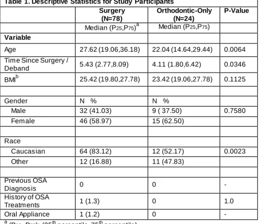
Of the 262 surgery subjects sent questionnaires, 78 patients responded (response rate of
29.77%). Surgery participants included 46 females and 32 males with a median age of 27.62
(19.06,36.18). They were all at least 2 years post-surgery with a median time since surgery of
5.43 (2.77,8.09) years. Twenty-four of the 175 subjects in the control group responded (response
rate of 13.71%). The control group consisted of 15 females and 9 males with a median age of
22.04 (14.64,29.44) years. They were all at least 1 year post deband (median time since deband
was 4.11 (1.80,6.42) years). The two groups were significantly different in median age (p<0.01),
time since surgery/deband (p<0.05), and race (p<0.01). Compared to the control group, the
surgery group was older, had a longer follow-up time, and was comprised of a higher percentage
of Caucasians. No statistical difference between gender and BMI was detected. One participant
in the surgery group acknowledged being treated with an oral appliance, but denied having a
previous OSA diagnosis. (Table 1)
The Berlin Questionnaire did not reveal any statistically significant difference in the OSA
risk assessment between the surgery and orthodontic-only groups nor were there any statistically
significant differences between groups in any of the symptom categories (Table 2). Overall,
8.97% of the surgery group and 16.67% of the orthodontic-only group were found to be at high
risk for OSA (Figure).
Analysis of the FOSQ-10 indicated no statistically significant difference between the
total FOSQ-10 score for the surgery and orthodontic-only with median total scores of 18.27
in any of the subscales: productivity, activity, vigilance, social outcomes, or intimacy and sexual
relations. (Table 3)
The difference in the Epworth Sleepiness Scale scores was significantly different
between the surgery and orthodontic-only groups (p<0.05). After excluding those with missing
data, the median ESS score for 76 of the BOS group was 6.30 (3.32,9.28) compared to 6.88
(2.41,11.35) for the orthodontic-only group. Both median scores, however, fell within the normal
range for daytime sleepiness. When assessed for the proportion of subjects who demonstrated
excessive daytime sleepiness, 10.53% of the BOS group and 20.83% of the orthodontic-only
group had an ESS total score >10. (Table 4)
Discussion
Sleep disordered breathing, including OSA, is a serious condition associated with
increased morbidity and mortality.3,4 Isolated mandibular setbacks are becoming rare in the United States due to both esthetic reasons and concerns over the risk of airway reduction
possibly leading to SDB. Studies on the effects of BOS on sleep function are limited and lead to
varying conclusions. In the PSG study of Foltán et al,20 BOS was found to worsen respiratory parameters with significant decreases in oxygen saturation (SpO2) and nasal airflow measured
before and after (mean 8.5 months) surgery. However, in a different PSG study, Hasebe et al21 was unable to detect significant differences in SDB or changes in SpO2 or Apnea Hypopnea Index (AHI) in patients 6 months after BOS. The investigators did note that 2 patients with very
large mandibular setbacks were diagnosed with mild OSA after surgery. Turnbull and Battagel33 compared overnight pulse oximetry and respiratory noises before and after BOS and found no
patients. In a recent PSG study by Gokce et al,16 sleep quality and efficiency improved
significantly after BOS (mean 1.4yrs) with significant increases in SpO2 and decreases in AHI.
One subject in the surgery group acknowledged having used an oral appliance for sleep
apnea, but denied having received a formal diagnosis. Either the patient failed to recall a
diagnosis or was provided the oral appliance in absence of an official diagnosis. The patient also
stated that it had been 2 years since the appliance was used. Since our resources did not allow for
overnight polysomnograms to definitively diagnose OSA in our subjects, we incorporated into
our study a widely used, validated sleep questionnaire, the Berlin Questionnaire, to estimate risk
for OSA to our two study groups.
Our findings of no significant difference in Berlin Questionnaire scores between the BOS
group and the orthodontic-only group is consistent with previous studies that were unable to
demonstrate an increased risk of SDB after BOS. The BOS group scores were also found to be
similar to recent reported OSA risk in population studies. For example, the Berlin Questionnaire
was used in a national sleep poll of 1506 people and 19% of participating adults were found to
meet the criteria for high risk of OSA.34 In our study, 8.97% of the surgery group was found to be at high risk for OSA.
While objective measures of SDB have traditionally been reported in the literature,
quality of life assessments are increasingly being recognized as an important outcome variable as
well.31,35 A number of studies have examined quality of life subsequent to jaw surgery for dentofacial deformities,36 however, no studies were identified that explored the impact of Class III jaw surgeries on sleep-related quality of life. In our study, we used two validated sleep
sleep-related quality of life. To our knowledge, this is the first study to assess patients’
perception of daytime sleepiness after BOS. With a median time post-surgery time of 5.43
(2.77,8.09) years, our study offered information on subjects with a longer follow up than any
previous study that measured sleep outcomes after BOS.
In a recent study evaluating ESS scores between OSA patients and non-OSA patients, the
average values found were 10.94 and 7.73, respectively.30 Although we found a statistically significant difference in ESS scores between our two groups (p<0.05), with the surgery group
having a lower median daytime sleepiness score, ESS scores in both groups fell within the
normal range. Both groups in our study were close to the reported non-OSA score of 7.73, with
the surgery group having a median ESS score of 6.3 (3.32,9.28) and the orthodontic-only group a
score of 6.88 (2.41,11.35). It has been estimated that 10-20% of the general population has ESS
scores >10.37 Our results were in that range with 10.53% of the BOS group and 20.83% of the orthodontic-only group having ESS scores >10. The significantly lower ESS score and lower
proportion of scores >10 in the surgery group suggest that BOS does not adversely impact
daytime sleepiness.
The FOSQ was developed to measure the impact of sleep on quality of life. Higher FOSQ
scores reflect better quality of life. In a previous FOSQ-10 study, patients with OSA had an
average score of 12.5 while non-OSA participants had an average score of 17.2. 27 In our BOS group, the FOSQ-10 score of 18.27 (16.41,20.13) compared favorably to the reported value in
the non-OSA patients. Thus, results from both the ESS and FOSQ-10 in our study suggest that
Class III bimaxillary surgery did not significantly affect the patients’ sleep-related quality of life
Study Limitations
The median age of both of our study groups was younger than we would have liked. Due
to the conversion in 2003 from paper charts to the Electronic Patient Record (EPR) at our
institution, we were limited in the time frame for which we had current contact information for
patients in the UNC surgery database. As such, the median age of both groups was <28 years and
may not reflect OSA outcome differences that may occur with increasing age.38 A well-known risk assessment questionnaire, the STOP-Bang, uses age 50 as a threshold for increased OSA
risk.39 If or how our groups differ after age 50 would be valuable information on clarifying whether BOS is associated with an increased risk of OSA. Although we attempted to frequency
match the age of the orthodontic-only group to the age of the surgery group respondents, the
median age of the surgery group was approximately five years older which one might have
speculated would have magnified a difference in OSA risk if it existed.
The increased follow-up time of approximately 1 year for the BOS group compared to the
orthodontic-only group is understandable because up to a year of orthodontic finishing remains
after surgery. We were not able to compare deband dates between groups because we did not
have access to the deband dates of the surgery group. The majority of the orthognathic surgery
patients seen at UNC have their orthodontic treatment carried out by local orthodontists.
The BMI used in this study was calculated from self-reported height and weight values.
Although the BMI was not significantly different between groups, any inaccuracies in BMI could
also have altered the scoring of the Berlin Questionnaire which uses BMI as one of its variables.
weight data from participants was not possible. The significantly more Caucasians in the surgery
group is consistent with the demographics of the surgery patients at UNC.
There was a significant difference in response rate between the BOS group and
orthodontic-only group with response rates of 29.77% and 13.71%, respectively. The BOS
subjects may have been more likely to participate in our study due to many having previously
agreed to participate in an ongoing surgery stability study at UNC. In addition, the BOS subjects
may have felt more of an obligation to participate because of the intense emotional and
psychological impact that comes from the profound positive changes in function and facial
esthetics after surgery.
Conclusions
To our knowledge, this study provides the longest follow-up information to date on the
effects of Class III bimaxillary orthognathic surgery (BOS) on sleep disordered breathing.
Moreover, this is the first study to assess sleep-related quality of life after BOS. The results of
this study suggest that young adults receiving this double jaw surgical procedure for the
correction of class III malocclusions are at no greater risk for OSA and/or reduction in
sleep-related quality of life compared to patients treated with orthodontics alone. Patients have been
shown to be at most risk for SDB if the mandible is setback significantly, preventing adaption to
their new respiratory position during sleep.21 Bimaxillary orthognathic surgery for Class III malocclusions may be able to limit the risk of SDB by minimizing the amount of mandibular
setback required and through compensating increases in the nasopharyngeal and velopharyngeal
questionnaires, and 3D airway parameters. The ability to more clearly identify an orthognathic
surgery patient’s pre-surgical risk of developing SDB is a goal that would guide surgeons and
benefit patients in the future.
Tables
Table 1. Descriptive Statistics for Study Participants Surgery
(N=78)
Orthodontic-Only (N=24)
P-Value
Median (P25,P75)a Median (P25,P75)
Variable
Age 27.62 (19.06,36.18) 22.04 (14.64,29.44) 0.0064 Time Since Surgery /
Deband 5.43 (2.77,8.09) 4.11 (1.80,6.42) 0.0346
BMIb 25.42 (19.80,27.78) 23.42 (19.06,27.78) 0.1125
Gender N % N %
Male 32 (41.03) 9 ( 37.50) 0.7580 Female 46 (58.97) 15 (62.50)
Race
Caucasian 64 (83.12) 12 (52.17) 0.0023 Other 12 (16.88) 11 (47.83)
Previous OSA
Diagnosis 0 0 -
History of OSA
Treatments 1 (1.3) 0 1.0
Oral Appliance 1 (1.2) 0 - a
(P25, P75): (25th percentile, 75th percentile). b
BMI: body mass index.
Table 2. Berlin Questionnaire Results
Surgery Orthodontic-Only P-Value
N % N %
Symptom Categories
Snoring
Positive 11 (14.10) 6 (25.00) 0.22 Negative 67 (85.90) 18 (75.00)
Daytime Sleepiness
Positive 17 (21.79) 6 (25) 0.74 Negative 61 (78.21) 18 (75.0)
Blood Pressure/BMI
Positive 15 (19.23) 2 (8.3) 0.34 Negative 63 (80.77) 22 (91.67)
Risk Assessment
Table 3. Functional Outcomes of Sleep Questionnaire-10 Results
Surgery Orthodontic-Only P-Value
Median (P25,P75) Median (P25,P75)
Subscale Scores
General Productivity Subscale 3.56 (3.05,4.07) 3.58 (3.0,4.16) 0.7401
Activity Level Subscale 3.47 (2.88,4.06) 3.57 (3.07,4.07) 0.6134
Vigilance Subscale 3.68 (3.24,4.12) 3.71 (3.11,4.31) 0.4702
Social Outcomes Subscale 3.85 (3.42,4.28) 3.83 (3.34,4.32) 0.8906 Intimacy and Sexual Relations
Subscale 3.69 (3.04,4.34) 3.47 (2.53,4.41) 0.0569
Total Scorea 18.27 (16.41,20.13) 18.13 (15.71,20.55) 0.9044
a
Total score is a mean-weighted item score.
Table 4. Epworth Sleepiness Scale (ESS) Results
Surgery Orthodontic-Only P-Values
Median (P25,P75) Median (P25,P75)
ESS Score 6.30 (3.32,9.28) 6.88 (2.41,11.35) 0.0492 N % N %
REFERENCES
1. Panossian L, Daley J. Sleep-disordered breathing. Continuum (Minneap Minn) 2013;19 1 Sleep Disorders:86-103.
2. Peppard PE, Young T, Palta M, Skatrud J. Prospective study of the association between sleep-disordered breathing and hypertension. N Engl J Med 2000;342:1378-1384.
3. Shahar E, Whitney CW, Redline S, et al. Sleep-disordered breathing and cardiovascular disease: Cross-sectional results of the sleep heart health study. Am J Respir Crit Care Med 2001;163:19-25.
4. Young T, Finn L, Peppard PE, et al. Sleep disordered breathing and mortality: Eighteen- year follow-up of the wisconsin sleep cohort. Sleep 2008;31:1071-1078.
5. Peppard PE, Young T, Barnet JH, Palta M, Hagen EW, Hla KM. Increased prevalence of sleep-disordered breathing in adults. Am J Epidemiol 2013;177:1006-1014.
6. Riley RW, Powell NB, Guilleminault C, Ware W. Obstructive sleep apnea syndrome following surgery for mandibular prognathism. J Oral Maxillofac Surg 1987;45:450-452.
7. Liukkonen M, Vahatalo K, Peltomaki T, Tiekso J, Happonen RP. Effect of mandibular setback surgery on the posterior airway size. Int J Adult Orthodon Orthognath Surg 2002;17:41-46.
8. Demetriades N, Chang DJ, Laskarides C, Papageorge M. Effects of mandibular
retropositioning, with or without maxillary advancement, on the oro-naso-pharyngeal airway and development of sleep-related breathing disorders. J Oral Maxillofac Surg 2010;68:2431-2436.
9. Mattos CT, Vilani GNL, Sant’Anna EF, Ruellas ACO, Maia LC. Effects of orthognathic surgery on oropharyngeal airway: A meta-analysis. Int J Oral Maxillofac Surg 2011;40:1347-1356.
10. Kawakami M, Yamamoto K, Fujimoto M, Ohgi K, Inoue M, Kirita T. Changes in tongue and hyoid positions, and posterior airway space following mandibular setback surgery. J
CranioMaxillofac Surg 2005;33:107-110.
11. Eggensperger N, Smolka W, Iizuka T. Long-term changes of hyoid bone position and pharyngeal airway size following mandibular setback by sagittal split ramus osteotomy. J
CranioMaxillofac Surg 2005;33:111-117.
13. Kim MA, Kim BR, Choi JY, Youn JK, Kim YJ, Park YH. Three-dimensional changes of the hyoid bone and airway volumes related to its relationship with horizontal anatomic planes after bimaxillary surgery in skeletal class III patients. Angle Orthod 2013;83:623-629.
14. Park SB, Kim YI, Son WS, Hwang DS, Cho BH. Cone-beam computed tomography evaluation of short- and long-term airway change and stability after orthognathic surgery in patients with class III skeletal deformities: Bimaxillary surgery and mandibular setback surgery. Int J Oral Maxillofac Surg 2012;41:87-93.
15. Degerliyurt K, Ueki K, Hashiba Y, Marukawa K, Nakagawa K, Yamamoto E. A comparative CT evaluation of pharyngeal airway changes in class III patients receiving bimaxillary surgery or mandibular setback surgery. Oral Surg Oral Med Oral Pathol Oral Radiol Endod 2008;105:495-502.
16. Gokce SM, Gorgulu S, Gokce HS, Bengi AO, Karacayli U, Ors F. Evaluation of pharyngeal airway space changes after bimaxillary orthognathic surgery with a 3-dimensional simulation and modeling program. Am J Orthod Dentofacial Orthop 2014;146:477-492.
17. Lee Y, Chun YS, Kang N, Kim M. Volumetric changes in the upper airway after bimaxillary surgery for skeletal class III malocclusions: A case series study using 3-dimensional cone-beam computed tomography. J Oral Maxillofac Surg 2012;70:2867-2875.
18. Jakobsone G, Neimane L, Krumina G. Two- and three-dimensional evaluation of the upper airway after bimaxillary correction of class III malocclusion. Oral Surg Oral Med Oral Pathol
Oral Radiol Endod 2010;110:234-242.
19. Enciso R, Nguyen M, Shigeta Y, Ogawa T, Clark GT. Comparison of cone-beam CT
parameters and sleep questionnaires in sleep apnea patients and control subjects. Oral Surg Oral
Med Oral Pathol Oral Radiol Endod 2010;109:285-293.
20. Foltan R, Hoffmannova J, Donev F, et al. The impact of Le Fort I advancement and bilateral sagittal split osteotomy setback on ventilation during sleep. Int J Oral Maxillofac Surg
2009;38:1036-1040.
21. Hasebe D, Kobayashi T, Hasegawa M, et al. Changes in oropharyngeal airway and respiratory function during sleep after orthognathic surgery in patients with mandibular prognathism. Int J Oral Maxillofac Surg 2011;40:584-592.
22. Bixler EO, Vgontzas AN, Ten Have T, Tyson K, Kales A. Effects of age on sleep apnea in men: I. prevalence and severity. Am J Respir Crit Care Med 1998;157:144-148.
23. Pang KP, Terris DJ. Screening for obstructive sleep apnea: An evidence-based analysis. Am J Otolaryngol 2006;27:112-118.
25. Abrishami A, Khajehdehi A, Chung F. A systematic review of screening questionnaires for obstructive sleep apnea. Can J Anaesth 2010;57:423-438.
26. Weaver TE, Laizner AM, Evans LK, et al. An instrument to measure functional status outcomes for disorders of excessive sleepiness. Sleep 1997;20:835-843.
27. Chasens ER, Ratcliffe SJ, Weaver TE. Development of the FOSQ-10: A short version of the Functional Outcomes of Sleep Questionnaire. Sleep 2009;32:915-919.
28. Chervin RD, Aldrich MS. The Epworth Sleepiness Scale may not reflect objective measures of sleepiness or sleep apnea. Neurology 1999;52:125-131.
29. Rosenthal LD, Dolan DC. The Epworth Sleepiness Scale in the identification of obstructive sleep apnea. J Nerv Ment Dis 2008;196:429-431.
30. Sil A, Barr G. Assessment of predictive ability of Epworth scoring in screening of patients with sleep apnoea. J Laryngol Otol 2012;126:372-379.
31. Baldwin CM, Griffith KA, Nieto FJ, O'Connor GT, Walsleben JA, Redline S. The
association of sleep-disordered breathing and sleep symptoms with quality of life in the Sleep Heart Health Study. Sleep 2001;24:96-105.
32. Johns MW. A new method for measuring daytime sleepiness: The Epworth Sleepiness Scale. Sleep 1991;14:540-545.
33. Turnbull NR, Battagel JM. The effects of orthognathic surgery on pharyngeal airway dimensions and quality of sleep. J Orthod 2000;27:235-247.
34. Hiestand DM, Britz P, Goldman M, Phillips B. Prevalence of symptoms and risk of sleep apnea in the US population: Results from the National Sleep Foundation Sleep in America 2005 Poll. Chest 2006;130:780-786.
35. Kushida CA, Littner MR, Hirshkowitz M, et al. Practice parameters for the use of continuous and bilevel positive airway pressure devices to treat adult patients with sleep-related breathing disorders. Sleep 2006;29:375-380.
36. Soh CL, Narayanan V. Quality of life assessment in patients with dentofacial deformity undergoing orthognathic surgery--a systematic review. Int J Oral Maxillofac Surg.
2013;42(8):974-980.
37. Johns M, Hocking B. Daytime sleepiness and sleep habits of Australian workers. Sleep 1997;20:844-849.
39. Chung F, Yegneswaran B, Liao P, et al. STOP questionnaire: A tool to screen patients for obstructive sleep apnea. Anesthesiology 2008;108:812-821.