THE ROLE OF HPV AND DNA METHYLATION IN THE DEVELOPMENT OF PRECANCEROUS CERVICAL LESIONS
Ayodele Gomih
A dissertation submitted to the faculty of the University of North Carolina at Chapel Hill in partial fulfillment of the requirements for the degree of Doctor of Philosophy in the
Department of Epidemiology in the Gillings School of Global Public Health.
Chapel Hill 2017
Approved by: Jennifer S. Smith Cathrine Hoyo
Michael G. Hudgens Kari E. North
ABSTRACT
Ayodele Gomih: The Role of HPV and DNA Methylation in the Development of Precancerous Cervical Lesions
(Under the direction of Jennifer S. Smith)
Biological factors associated with cervical intraepithelial neoplasia (CIN) development may be utilized to improve the efficiency of cervical cancer screening. The present study examined human papillomavirus (HPV) genotypes and DNA methylation of imprinted genes in a survival analysis, using two endpoints: CIN1 regression and progression to CIN2+.
The CIN Cohort Study (CINCS) was conducted at 10 Duke University clinics in North Carolina. Women ages 18-65 years with enrollment colposcopies following an abnormal Pap test had follow-up cytology/histology for 3 years with up to 5 visits. DNA was extracted from exfoliated cervical cells for methylation at differentially methylated regions (DMR) of imprinted genes and for HPV genotyping. Hazard ratios (HR) and 95% confidence intervals (CI) were calculated using Cox regression models to estimate the association between aberrant methylation and CIN1 regression (normal epithelia) versus persistence/progression (CIN1+). HRs/95% CIs were estimated to quantify genotype-specific risk of progression to CIN2+.
Of 1303 participants, 472 normal/CIN1 cases with HPV data and 151 CIN1 cases with
(HR=3.2,95% CI=1.5-7.2;HR=3.2,95% CI=1.3-7.6) versus non-16/51 infections. HPV-16/51 were most predictive of progression to CIN2+.
In the CIN1 regression analysis, one-third regressed to normal epithelium (n=53;35.1%). Median time-to-regression was 12.6 months (range:4.5-24.0). The probability of CIN1 regression was negatively correlated with increased methylation at IGF2AS CpG 5 (HR=0.42,95% CI=0.23-0.77;p=0.005) and PEG10 DMR (HR=0.78,95% CI=0.63-0.96;p=0.02).
ACKNOWLEDGEMENTS
I would like to thank you my advisor and dissertation committee chair, Jennifer S. Smith, for her guidance in and dedication to researching HPV and cervical cancer. I thank my committee members who also enriched my doctoral research: Michael Hudgens for his statistical expertise, Kari North for the extensive knowledge of genetic epidemiology, Wendy Brewster for providing meaningful clinical
direction and context for my research, and to Cathrine Hoyo for helping me navigate the blossoming field of epigenetics in epidemiology. To Cathrine and Susan Murphy, the Co-PIS of the CINCS Study, thank you for allowing me to be a part of this innovative project. A heartfelt thank you to CINCS participants for being willing to contribute their own personal experiences to helping others, as well as CINCS research team for facilitating the process. To all collaborators, thank you for your invaluable feedback on my manuscripts. I acknowledge the funding sources that supported this research—the National Institutes of Health (NIH) T32-5T32AI007001, R01CA142983, and R01CA142983-02S1. To Chris Wiesen and the Odum Institute: sincerely grateful to you for saving me from the SAS and R frustrations. I thank the EPID student services administrators Nancy, Carmen, Valerie, Jennifer, Chandra, Lisa, etc. fielding all
TABLE OF CONTENTS
LIST OF TABLES ... ix
LIST OF FIGURES ... xi
LIST OF ABBREVIATIONS ... xii
CHAPTER ONE: STATEMENT OF SPECIFIC AIMS ... 1
CHAPTER TWO: BACKGROUND AND SIGNIFICANCE ... 2
Burden of human papillomavirus and cervical cancer ... 2
HPV and Cervical Cancer Screening/Prevention ... 2
Methods for Cervical Cancer Screening ... 2
Epigenetics and Cervical Tumorigenesis ... 4
Epigenetics and Cancer/Disease ... 4
Genomic Imprinting Loss of Imprinting and Cancer ... 5
DNA Methylation Markers for Cervical Dysplasia ... 6
Influence of HPV and External Factors on DNA Methylation and Cancer ... 8
CHAPTER THREE: RESEARCH PLAN AND METHODS ... 10
Study Population ... 10
Recruitment and Study Follow-Up ... 10
Recruitment at the clinics... 10
Follow up of women after abnormal cytology diagnosis ... 11
Eligibility criteria ... 11
Data Collection ... 12
Cervical Cytology & Histology Specimens ... 12
DNA Methylation ... 13
Analytic Methods ... 15
CHAPTER FOUR: HIGH-RISK HUMAN PAPILLOMAVIRUS GENOTYPES IN THE PROGRESSION OF CERVICAL INTRAEPITHELIAL NEOPLASIA ... 20
Background ... 20
Methods ... 21
Results ... 26
Discussion ... 28
Tables and Figures ... 32
CHAPTER FIVE: DNA METHYLATION OF IMPRINTED GENE CONTROL REGIONS IN THE REGRESSION OF LOW-GRADE CERVICAL LESIONS ... 44
Background ... 44
Methods ... 46
Results ... 51
Discussion ... 53
Tables and Figures ... 56
CHAPTER SIX: DISCUSSION ... 65
Overview ... 65
Summary of Findings ... 65
Public Health Significance ... 66
APPENDIX ... 68
LIST OF TABLES
Table 2. 1 DNA Methylation of Imprinted Genes in Cancerous vs. Normal Cervical Tissue ... 7
Table 3. 1 Imprinted Gene Primers used in Sequenom analysis for DNA methylation ... 14 Table 3.2. 1 Progression to CIN2+ outcome, given enrollment CIN1 histology
and follow-up histology ... 15 Table 3.2. 2 Progression to CIN2+ outcome given enrollment normal histology
and follow-up histology... 16 Table 3.2. 3 Progression to CIN2+ outcome for enrollment CIN1 histology utilizing
follow-up cytology, given missing follow-up histology missing ... 16 Table 3.2. 4 Progression to CIN2+ outcome for enrollment normal histology
utilizing follow-up cytology, given missing follow-up histology ... 16 Table 3.3. 1 CIN1 regression outcome, given enrollment CIN1 histology and
follow-up histology ... 18 Table 3.3. 2 CIN1 regression outcome for enrollment CIN1 histology utilizing
follow-up cytology, given missing histology ... 18 Table 4. 1 Enrollment characteristics of 472 CINCS* colposcopy participants
with normal histology or low-grade CIN (CIN1)*, stratified by HPV† vaccination status ... 32 Table 4. 2 Distribution of single/multiple HPV* genotypes in 472 CINCS†
participants with normal histology/CIN1† at enrollment, stratified by HPV* vaccination status ... 34 Table 4. 3 Incidence rates of CIN2+*, stratified by HPV† genotype‡ infection
and vaccination history among 472 CINCS* participants over 3 years of follow-up ... 36 Table 4. 4 Incidence rates of CIN2+* by single HPV† infection, stratified by
vaccination status among 472 CINCS* participants over 3 years of follow-up ... 37 Table 4. 5 Risk of progression to CIN2+*, stratified by HPV† genotypes‡
among 472 CINCS* participants by HPV† vaccination history ... 40 Table 4. 6 Risk of progression to CIN2+*, stratified by risk groups for
high-risk HPV† infection‡ ... 42 Table 5. 1 Characteristics of 164 Women with CIN1 at enrollment in the
Table 5. 2 Imprinted gene DMR§ methylation distributions in 164 CIN1 patients at enrollment* ... 57 Table 5. 3 Analysis of CIN1 regression by imprinted gene DMR||/CpG site
among CINCS participants* ... 58 Table 5. 4 Sensitivity analysis: CIN1 regression by imprinted gene using
LIST OF FIGURES
Figure 2.1 Methylation at CpG islands at promoter regions controls gene
expression ... 5 Figure 3. 1 CINCS Study Visit Flow Chart ... 11 Figure 4. 1 3-year cumulative probability of CIN2+* progression in 194
unvaccinated and vaccinated CINCS women with single HPV† type
infection, stratified by risk group ... 43 Figure 5. 1 CINCS Study Flowchart ... 46 Figure 5.2. 1 Time to CIN1 regression for IGF2AS (at CpG 5), stratified
at median methylation percentage* ... 61 Figure 5.2. 2 Time to CIN1 regression for Kv DMR, stratified at median
methylation percentage* ... 62 Figure 5.2. 3 Time to CIN1 regression, stratified at median MEG3 DMR
methylation percentage* ... 63 Figure 5.2. 5 Time to CIN1 regression, stratified at median PEG10 DMR
LIST OF ABBREVIATIONS
AHR Adjusted hazard ratio AIC Akaike Information Criteria AIS Adenocarcinoma in situ
ASCCP American Society for Colposcopy and Cervical Pathology ASC-US Atypical squamous cells of undetermined significance ASC-H Atypical squamous cells, cannot exclude HSIL AGUS Atypical glandular cells of undetermined significance
AGUS-H Atypical glandular cells of undetermined significance, cannot exclude HSIL
CI Confidence interval
CIN Cervical intraepithelial neoplasia
CINCS Cervical Intraepithelial Neoplasia Cohort Study CKC cold knife conization
CPG Cytosine-phosphate-guanine DAG Directed acyclic graph DCRU Duke Clinical Research Unity DCTP Deoxycytidine triphosphate DMR Differentially methylated region DNA Deoxyribonucleic acid
DPC Duke Primary Care
DTTP Deoxythymidine triphosphate EDTA Ethylenediaminetetraacetic acid FDA Food & Drug Administration HPV Human papillomavirus
HR Hazard ratio
HSIL High-grade squamous intraepithelial lesion ICC Invasive cervical cancer
IQR Interquartile range LBC Liquid-based cytology
LEEP Loop electrosurgical excision procedure LRHPV Low-risk human papillomavirus
LSIL Low-grade squamous intraepithelial lesion
LSIL-H Low-grade squamous intraepithelial lesion, cannot exclude HSIL
ML Milliliter
MRNA Messenger ribonucleic acid NCE normal epithelium specimen
PAP Papanicolaou
PBR Peripheral benzodiazepine receptor PCR Polymerase chain reaction
RNA Ribonucleic acid
CHAPTER ONE: STATEMENT OF SPECIFIC AIMS
Cervical cancer is a highly preventable cancer by screening. In the United States, current screening guidelines recommend the use of high-risk human papillomavirus (HPV) detection with liquid-based cytology testing for women between the ages of 30 and 65 years for detection of high-grade cervical intraepithelial neoplasia (CIN), the precursor of cervical cancer [1]. All low-grade cytology and HPV-16/18-positive/cytology-negative cases are referred for immediate colposcopy [1]. The role of other non-16/18, high-risk HPV genotypes in clinical management of cervical lesions has not been well
characterized. The role of epigenetics in cervical carcinogenesis— specifically, DNA methylation of imprinted genes— may also serve as a potential biomarker for CIN progression. Novel biomarkers in screening can help further stratify risk of lesion progression and reduce the number of colposcopy
referrals, improving patient care and quality. To identify potential biomarkers in the development of CIN, the following research aims were addressed:
AIM 1: To examine the association between HPV infection by genotype and the progression of low-grade cervical epithelial neoplasia (CIN1 to CIN2+) or persistence (CIN1 to CIN1) of precancerous cervical lesions in the Cervical Intraepithelial Neoplasia Cohort Study (CINCS).
CHAPTER TWO: BACKGROUND AND SIGNIFICANCE Burden of human papillomavirus and cervical cancer
Human papillomavirus (HPV) is an extremely common sexually transmitted infection (STI), affecting nearly 79 million people in the United States (US), with approximately 14 million new
infections each year [2]. Of 100 known HPV genotypes, 40 can affect the genital area [3]. HPV infection is more prevalent among Black and Hispanics compared to Whites, especially in low-income populations [4, 5].
HPV infection is a necessary cause in cervical cancer development [6]. Fourteen high-risk oncogenic HPV types have been identified as the main etiologic agents in cervical cancer, in which types 16 and 18 alone cause 70% of all cases [7]. There are over 12,000 new HPV-related cervical cancer cases in the US each year [8]. In North America, 3 women per 100,000 die from cancer of the cervix [8].
The highest rates of cervical cancer in the US occur in the South; the incidence in North Carolina is comparable to overall cancer incidence nationwide (age-adjusted rate: 7.4 per 100,000). Furthermore, cervical cancer disproportionately affects racial and ethnic minorities relative Whites in the US, with higher prevalence of both HPV infection and cervical cancer in Blacks than Whites [4, 5]. While there are comparable screening rates, both incidence and mortality is higher among Blacks and Hispanics [9-11]. In NC, cancer incidence is more than 60% higher in Blacks and Hispanic women compared to White women [9, 12].
HPV and Cervical Cancer Screening/Prevention
Methods for Cervical Cancer Screening
cervical cancer (ICC) have been available for more than three decades. The Papanicolaou test, best known as a Pap smear test, is the original cytology-based method for detection of abnormal cervical cells.
Developed in 1941 by Dr. George Papanicolaou, the Pap test allowed for the identification of
precancerous and cancerous cells in cytology samples from vaginal aspirates [13]. The American Cancer Society endorsed the use of Pap tests in formal cervical screening settings in 1957.
The implementation of cytology-based screening and national screening and management guidelines designed by the American Society for Colposcopy and Cervical Pathology (ASCCP) have led to substantial decreases in the incidence and mortality of ICC in the United States [13, 14]. However, conventional Pap tests have variable sensitivity to detected high grade cervical lesions due to variation in sample and slide preparation [13, 15, 16]. Liquid-based cytology (LBC) screening methods were
developed to improve clinical performance within the past 17 years. In the US, 90% of Pap tests used to screen for cervical lesions are LBC-based methods such as ThinPrep (Cytyc Corp, Marlborough, MA), preferred over conventional Pap smear tests [13].
Overall, Pap tests have high specificity to distinguish non-diseased (>90%)[17]. However, detection by cytology alone requires a series of repeated Pap tests and/or unnecessary referral to colposcopy and potential surgical excision [15, 18-21]. While women with low-grade CIN— especially young women— are more likely to regress, women who have persistent infection with high-risk HPV (hrHPV) genotypes can lead to persistent dysplasia and progression to ICC [22]. Evidence demonstrates that high-risk HPV infection can better predict development of CIN3 and ICC [22, 23]. As a result, recent age-specific recommendations include the use of HPV testing in conjunction with cytology-based
screening for women over 30 years of age [22]. The Food and Drug Administration (FDA) approved the use of the cobas assay (Roche Diagnostics, Indianapolis, IN) for routine screening [22, 24]. Other companies are amidst developing and testing of other high-risk genotyping assays, such as APTIMA (Hologic, Marlborough, MA) and BD (Franklin Lakes, NJ).
16/18 are referred for immediate colposcopy, whereas positive tests for the other 12 hrHPV genotypes are referred for cytology and HPV-negative women follow routine screening guidelines[1, 24]. While HPV co-testing has increased the ability to detect high-risk women, improving the efficacy of cervical
screening methods can reduce the number of unnecessary screenings and associated cost, patient anxiety and issues with low adherence to follow-up recommendations. Therefore, it is important to further explore the impact of other oncogenic HPV types among women who have negative cytology and low-grade lesions that are more likely to progress to high-grade cervical lesions. With a better understanding of the sociodemographic determinants of CIN and ICC, cancer screening and management efforts may better address those groups at highest risk.
Additionally, recognizing the extent to which other oncogenic HPV (e.g. non-16/18) types lead to CIN progression is crucial to current prevention strategies. Further research in HPV typing in
carcinogenesis can inform vaccine development and improve coverage in preventing future transmission. Hence, this dissertation research aimed to determine whether infection with a particular HPV type varies the rate of progression to high-grade CIN or cervical cancer, and determine whether these rates differ among women of different racial/ethnic backgrounds in a multiracial cohort.
Epigenetics and Cervical Tumorigenesis
Epigenetics and Cancer/Disease
Genetic research has ensued for years in order to understand potential targets for disease prevention and clinical interventions. However, epigenetics of cancer has become an increasingly popular topic of interest for researchers. Epigenetics— the control of gene expression by external chemical
DNA methylation occurs at cytosine-phosphate-guanine (CpG) sites, which are concentrated in regions throughout the genome in “islands” [27]. These CpG rich regions are not typically methylated, and are situated at the 5’ end of regulatory regions for several genes, at promoters, that control gene expression [27]. Unmethylated CpG islands at gene promoters allow for the binding of transcription machinery and transcription of DNA into mRNA[28] (Figure 2.1). However, methylation of the promoter prevents transcription and subsequent gene expression[28] (Figure 2.1).
Figure 2.1 Methylation at CpG islands at promoter regions controls gene expression [29]
Genomic Imprinting Loss of Imprinting and Cancer
Imprinted genes typically exist in clusters, thus share common regulatory elements. These elements include differentially methylated regions (DMRs), which can be intergenic or intragenic and are critical in tissue-specific gene expression [31-33]. In mice, Igf2, H19, Zac, Mest, Peg3, Dlk1, Meg3, Grb10, Ndn, Cdkn1c and SLC38a4 appear to exhibit coordinated repression with growth deceleration [34]. In vitro experiments also showed genetic induction of several imprinted genes on different chromosomes related to the overexpression of another gene as a result of aberrant methylation [35]. As imprinted genes are susceptible to deregulation, they should be strongly considered as key mediators in cancer development.
Their role in cancer was first supported by their role in Beckwith-Wiedemann syndrome (BWS) caused by defects in imprinting on chromosome 15 with affected individuals predisposed to Wilms tumor and hepatoblastoma [26, 36]. Loss of imprinting also has occurred colorectal cancer with insulin growth factor-2 or IGF2 [37].
DNA Methylation Markers for Cervical Dysplasia
Because cancer progression occurs through the loss and/or gain of genetic function, genetic and epigenetic biomarkers for CIN2+ have received considerable attention in research aimed to distinguish women with benign infection from those requiring treatment over the past 15 years. Specifically, DNA methylation can influence transcription and expression of genetic factors involved in cervical cancer development without directly altering the underlying gene’s DNA sequence [25].
One proposed mechanism for CIN progression involves epigenetic modifications at gene promoter CpG islands that impact transcription, inhibiting expression of tumor suppressor genes. However, aberrant methylation of promoter regions has not been strongly associated with CIN
also been assessed as a potential pathway in the progression of cervical dysplasia [31, 40]. Specifically, integration of HPV-16 into the host genome has resulted in the expression of E6/E7 oncoprotein genes that in turn, results in overexpression of p53 and PBR tumor suppressing genes (citation).
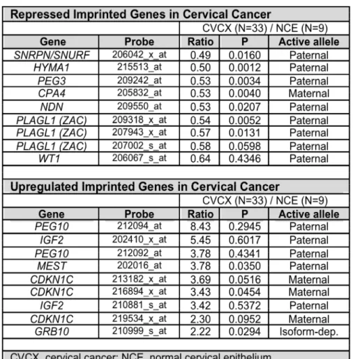
Hypermethylation of HPV-16 E6 and E7 oncogenes (which promote HPV virulence) may cause binding of DNA transcription factors that influence cancer development [31]. Further studies in humans have demonstrated differential changes in methylation at imprinting centers among women who had cervical intraepithelial and invasive cervical cancer. An analysis of publicly available microarray data found that expression of imprinted genes differed between specimens from ICCs compared to normal cervical epithelium (NCE) specimen (Table 2.1).
For example, paternally-expressed SNRPN/SNURF genes were suppressed compared to NCE samples; similarly, paternally-expressed PEG10 was overexpressed in comparison to normal cases. This finding supports that the expression profile of imprinted genes differs in cervical cancer cases compared to normal cases and thus these genes warrant further research in CIN progression.
Influence of HPV and External Factors on DNA Methylation and Cancer
Epigenetic changes to the host genome are typically attributed to external forces, such as the environment and lifestyle behaviors. Cofactors such as smoking, high parity, and oral contraceptive use may influence these epigenetic changes that lead to progression of cervical dysplasia. It is established that, in addition to persistent high-risk HPV infection, cigarette smoking, long term oral contraceptive use, and high parity are risk factors for cervical cancer and CIN [20]. These identified factors may also influence DNA methylation and therefore lead to loss of imprinting and potential functionality of both growth factors and tumor suppressor genes.
CHAPTER THREE: RESEARCH PLAN AND METHODS Study Population
The Cervical Intraepithelial Neoplasia Cohort Study (CINCS) is a 3-year prospective study that enrolled participants between 2010 to 2014 to determine whether epigenetic deregulation of known imprinted genes can be used to distinguish women more likely to progress to CIN2+.
The cohort includes 1303 women aged 18 years and older with abnormal cytology attending 10 Duke University and Duke Primary Care (DPC) clinics in Durham County, North Carolina. North Carolina is located in southeastern region of the USA, where cervical cancer is most prevalent.
Recruitment and Study Follow-Up
Recruitment at the clinics
Potential participants included women who were referred for colposcopy following an abnormal
cervical screening test. Prospective participants were identified by their physicians two weeks prior to a scheduled colposcopy appointment through the electronic clinic appointment logs of the Duke University
and community clinics. The attending physicians provided signed letters of invitation for study participation to mail to potential study participants prior to the colposcopy. Study invitation letters
explained the broad goals of the study and expectations of participants. The colposcopy patients were
instructed to call a toll-free number to register their intent to decline participation. During the scheduled
colposcopy appointment visit, the recruiting interviewer solicited participation directly from the
participant and administered the informed consent process. After consent, participants were given a
standardized questionnaire to ascertain demographic and clinical information. Further information was
collected on cervical dysplasia diagnosis through histopathological confirmation typically two weeks after
Follow up of women after abnormal cytology diagnosis
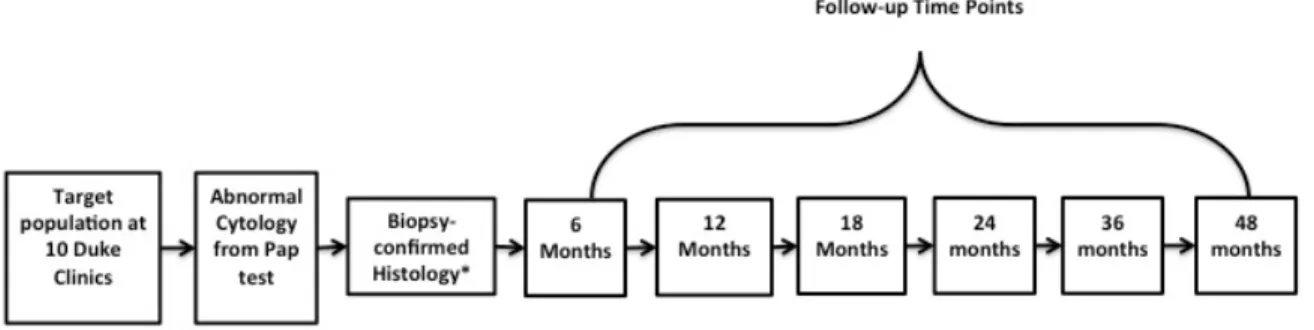
Study participants attended a clinic visit approximately every 6 months to 1 year for 3 years, following clinical practice guidelines for management of CIN (Figure 1). At each visit, clinic physicians conducted a cervical examination to ascertain information on any cytological/histological changes. A follow-up questionnaire was administered to ascertain information on any behavioral and clinical characteristics of the participant since enrollment.
Figure 3. 1 CINCS Study Visit Flow Chart
Eligibility criteria
Eligibility criteria for Aim 1
Women were required to have histopathologically-confirmed diagnosis of normal epithelial/CIN1 at their enrollment visit. Participants must have attended at least one follow-up visit following enrollment, at which they received either a Pap test and/or colposcopy with cervical biopsy and/or endocervical curettage. Participants without data on HPV status were excluded.
Eligibility criteria for Aim 2
Participants with a histopathology-confirmed CIN1 diagnosis at the enrollment visit were included. at their enrollment visit. Participants must have attended at least one follow-up visit following enrollment, at which they received either a Pap test and/or colposcopy with cervical biopsy and/or endocervical curettage. Participants without data on HPV status or imprinted gene methylation were excluded.
Data Collection
Cervical Cytology & Histology Specimens
During the Pap test at baseline, the clinic physician utilized a spatula and cytobrush to obtain
exfoliated cervical cells. Cervical specimens were suspended in a Thinprep vial containing proprietary
fluid with at least 50% methanol (Cytyc®, Malborough, MA, USA) for cytology confirmation. Following
abnormal cytology results, clinic physicians performed colposcopy-directed biopsy at baseline.
Information on cytology and lesion morphology, including size and location, were abstracted from patient
medical records. All specimens were tested for adequacy using the 2012 ASCCP guidelines. The
specimens were stored at 4°C prior to HPV testing.
All Pap test results were classified as one of the following by a pathologist according to 2012
Bethesda system [48]: i) negative/normal cytology; ii) low-grade squamous intraepithelial lesion (LSIL);
undetermined significance (AGUS); v) high-grade squamous intraepithelial lesion (HSIL); vi) low-grade
squamous intraepithelial lesion, cannot exclude HSIL (LSIL-H); or vii) atypical squamous cells cannot
exclude HSIL (ASC-H).
To ascertain the presence of CIN, cervical biopsies were adjudicated by a clinical pathologist.
CIN diagnoses were classified as follows: i) CIN-1; ii) CIN-2; iii) CIN-2/3; iv) CIN-3; or v) ICC.
HPV DNA testing
HPV typology was assessed using excess cervical tissue obtained from the enrollment biopsy.
ThinPrep® specimens and homogenized aliquoted biopsies were collected during the same baseline visit
and shipped to the University of Hawaii Cancer Center. Following DNA extraction, PGMY09/PGMY11
primers [49, 50] were used in PCR to target a 450-bp region of the HPV L1 genome. Amplification of the
human β-globin gene was included as an internal control for sample sufficiency. HPV-positive specimens
were subsequently genotyped by using the HPV Linear Array® (Roche Diagnostics, Branchburg, NJ,
USA). This assay is designed to detect 14 high-risk HPV types— 16, 18, 31, 33, 35, 39, 45, 51, 52, 56,
58, 59, 66 and 68— and low-risk HPV types— 6, 11, 26, 40, 42, 53, 54, 61, 62, 64, 67, 69, 70, 71, 72, 73,
81, 82, 83, 84.
DNA Methylation Nucleic acid extraction
stored at -80ºC until required. Methylation and host genetic analyses were performed by the Duke Epigenetics Group led by Drs. Murphy and Jirtle within their respective laboratories.
Methylation analysis
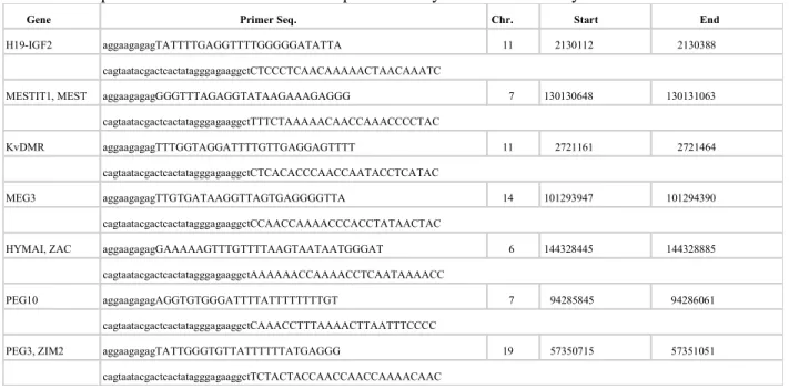
DNA methylation was measured at differentially methylated regions (DMRs) that regulate the imprinted genes IGF2 AS, IGF2/H19, PEG1/MEST, KV DMR, DLK1/MEG3, PLAG1/HYMA1, PEG10 and PEG3 DMR using the Sequenom MassARRAY platform with EpiTYPER analysis software. Primers were designed with EpiDesigner software which designs primers complementary to bisulfite treated DNA in regions without CpG nucleotides; it then adds a T7 promoter site to all forward primers (Table 3.1).
Table 3. 1 Imprinted Gene Primers used in Sequenom analysis for DNA methylation
After PCR amplification, unincorporated nucleotides were removed with shrimp alkaline phosphatase, and the PCR products were transcribed by T7 RNA & DNA polymerase with simultaneous cleavage by RNase A. Including either dCTP or dTTP in the transcription reaction restricts cleavage by RNase A to positions immediately 3’ of thymine or cytosine residues, respectively. The fragmented transcripts were spotted onto SpectroCHIPS for mass spectrometry analysis on the MassARRAY instrument. The fragments were identified by matching the molecular mass of detected particles to that expected from
Gene Primer Seq. Chr. Start End
H19-IGF2 aggaagagagTATTTTGAGGTTTTGGGGGATATTA 11 2130112 2130388
cagtaatacgactcactatagggagaaggctCTCCCTCAACAAAAACTAACAAATC
MESTIT1, MEST aggaagagagGGGTTTAGAGGTATAAGAAAGAGGG 7 130130648 130131063
cagtaatacgactcactatagggagaaggctTTTCTAAAAACAACCAAACCCCTAC
KvDMR aggaagagagTTTGGTAGGATTTTGTTGAGGAGTTTT 11 2721161 2721464
cagtaatacgactcactatagggagaaggctCTCACACCCAACCAATACCTCATAC
MEG3 aggaagagagTTGTGATAAGGTTAGTGAGGGGTTA 14 101293947 101294390
cagtaatacgactcactatagggagaaggctCCAACCAAAACCCACCTATAACTAC
HYMAI, ZAC aggaagagagGAAAAAGTTTGTTTTAAGTAATAATGGGAT 6 144328445 144328885
cagtaatacgactcactatagggagaaggctAAAAAACCAAAACCTCAATAAAACC
PEG10 aggaagagagAGGTGTGGGATTTTATTTTTTTTGT 7 94285845 94286061
cagtaatacgactcactatagggagaaggctCAAACCTTTAAAACTTAATTTCCCC
PEG3, ZIM2 aggaagagagTATTGGGTGTTATTTTTTATGAGGG 19 57350715 57351051
analysis of the reference sequence. The analysis software compares the relative amounts of methylated forms of each predicted fragment to quantify the methylated fraction.
Analytic Methods
Aim 1: To examine the association between HPV genotypes and the progression to CIN2+ among normal and low-grade CIN in the CINCS.
a.
Determine whether progression to CIN2+ varies by individual HPV status (positivity) or HPV type-grouping (high-risk HPV types compared to infection with low-risk HPV types).b.
Assess whether the association between HPV infection (positive vs. negative; high-risk vs.low-risk) and the progression to CIN2+ is modified by race, smoking, parity, or oral contraceptive use.
Time-to-progression analysis with Cox proportional hazards were used to examine the
relationship between HPV genotypes/risk group and progression to CIN2+. CIN outcome at follow-up was dichotomized as progression vs. persistence/regression.
We defined progression as a more severe CIN score (CIN-2+) at a follow-up visit; persistence as retention of the same diagnosis at a follow-up visit (CIN-1); and regression as a less severe CIN diagnosis.
Cytology results were used if the participants had missing histology data as a result of conservative clinical practice. A description of the outcome classification is described in Tables 3.2.1-3.2.4.
Table 3.2. 1 Progression to CIN2+ outcome, given enrollment CIN1 histology and follow-up histology Enrollment Histology Histology Follow-Up Status at Follow-Up
CIN1
Normal/Negative Regress
CIN1 Persist
CIN2 Progress
CIN2/3 Progress
CIN3 Progress
Table 3.2. 2 Progression to CIN2+ outcome given enrollment normal histology and follow-up histology Enrollment Histology Follow-Up Histology Status at Follow-Up
Normal
Normal/Negative Persist
CIN1 Non-Progress/
Persist*
CIN2 Progress
CIN2/3 Progress
CIN3 Progress
Cancer Progress
*CIN2+ is main endpoint in analyses, as it warrants clinical intervention, e.g. (LEEP, etc.) [1]
Table 3.2. 3 Progression to CIN2+ outcome for enrollment CIN1 histology utilizing follow-up cytology, given missing follow-up histology missing
Enrollment Histology Cytology Follow-Up Status at Follow-Up
CIN1
Normal/Negative Regress
LSIL Persist
AGUS-L Persist
ASCUS-L Persist
HSIL Progress
AGUS-H Progress
ASCUS-H Progress
ICC Progress
Table 3.2. 4 Progression to CIN2+ outcome for enrollment normal histology utilizing follow-up cytology, given missing follow-up histology
Enrollment Histology Follow-Up Cytology Status at Follow-Up
Normal
Normal/Negative Regress
LSIL Non-Progress/Persist*
AGUS-L Non-Progress/Persist*
ASCUS-L Non-Progress/Persist*
HSIL Progress
AGUS-H Progress
ASCUS-H Progress
Cancer (AIS/SCC) Progress
* CIN2+/HSIL+ is main endpoint in analyses, as it warrants immediate clinical intervention, e.g. (colposcopy/biopsy, etc.) [1]
We defined progression as a cytology result of HSIL, LSIL-H, or ASC-H at follow-up;
persistence as LSIL, ASCUS, or AGUS at follow-up; and regression as negative/normal cytology at
For women who received treatment (loop electrosurgical excision procedure, cold knife
conization, hysterectomy, etc.) at a follow-up visit, the histological diagnosis from the treatment specimen will be used as their follow-up diagnosis. Women who were treated for CIN were excluded from further data analyses after their treatment.
The outcome was dichotomized as the time to the first incidence or first progression of CIN from enrollment status. Person-time accumulated until progression to CIN2+ event, or at the last recorded follow-up visit. Administrative censoring occurred at 3 years’ post-study enrollment.
Univariate distribution of the 37 HPV types (6, 11, 16, 18, 26, 31, 33, 35, 39, 40, 42, 45, 51, 52, 53, 54, 56, 58, 59, 66, 61, 62, 64, 66, 67, 69, 70, 71, 72, 73, 81, 82, 83, 84) were examined individually,
and categorized as HPV types by “high-risk,” “low-risk,” and “no HPV infection.” Correlates of CIN/cervical cancer examined as follows:
• Age was examined as a continuous variable as well as a categorical variable
• Race was categorized as a binary variable of either “Black” and “(Non-Hispanic) White” • Current cigarette smoking was categorized as “Non-current smoker” vs. “Current smoker” • Parity was examined as a continuous variable and categorical variable
• Current Oral contraceptive use was categorized as “Non-current Use” and “Current Use”
• History of HPV Vaccination was categorized as “never received HPV vaccine” vs. “prior HPV
vaccination”
Potential confounders were identified a priori and placed into an elastic net Cox regression model [51][52]. Effect measure modification was also assessed using the Akaike Information Criterion (AIC) to determine the quality of model fit for interactions between covariates of interest and HPV type risk group.
The exposure of aberrant methylation was treated as a continuous variable and transformed by the interquartile range (IQR) for CIN1 regression modeling. Median methylation scores of multiple CpG loci for each imprinted gene were also calculated.
We also explored dichotomizing the outcome by establishing specific cutpoint for hyper- or hypo- methylation, given an expected value of 50% DNA methylation for monoallelically-expressed genes such as imprinted genes (as one allele is fully methylated while the other is completely
unmethylated). Methylation levels deviating by 5% from this expected value were considered. Based on the data, methylation was coded continuously for the analyses.
Regression of cervical lesions was defined as a diagnosis of negative/normal cytology at two
consecutive follow-up visits (Table 3.3.1 and 3.3.2).
Table 3.3. 1 CIN1 regression outcome, given enrollment CIN1 histology and follow-up histology Enrollment Histology Follow-Up Histology Outcome Status
at Follow-Up
CIN1
Normal/Negative Regress
CIN1 Persist
CIN2 Progress
CIN2/3 Progress
CIN3 Progress
Cancer Progress
Table 3.3. 2 CIN1 regression outcome for enrollment CIN1 histology utilizing follow-up cytology, given missing histology
Enrollment Histology Follow-Up Cytology Outcome Status at Follow-Up
CIN1
Normal/Negative Regress
LSIL Persist
AGUS-L Persist
ASCUS-L Persist
HSIL Progress
AGUS-H Progress
ASCUS-H Progress
Per conservative clinical practice, cytology results were utilized to determine regression status if the
participants had missing histology data. Persistence was defined as a diagnosis of low-grade histology
(CIN1) at follow-up or low-grade lesions during cytology testing (e.g. LSIL, ASC-US, AGUS-L).
Progression was defined as a follow-up histological diagnosis of CIN2+, or as development of high-grade
cytology, e.g. HSIL, LSIL-H, or ASC-H.
Women with a negative/normal screening at one follow-up time point were not considered to
have regressed for the main study analyses. For women who received treatment (LEEP, CKC,
cryotherapy, or hysterectomy) at a follow-up visit, the histological diagnosis from the pre-treatment specimen was utilized.
The covariates were examined as follows:
• Age will be examined as a continuous variable as well as a categorical variable (categories to be
explored)
• HPV Status will be dichotomized as “HPV-Positive” and “HPV-Negative”
• HrHPV Status will be dichotomized as “hrHPV-Positive” and “hrHPV-Negative (low-risk HPV
or no HPV infection)”
• Race was categorized as “Black”, “(Non-Hispanic) White”, and “Other”
• History of Oral contraceptive use will be categorized as “Never Use” vs. “Ever Use”
• Current cigarette smoking will be categorized as “Non-current smoker” vs. “Current smoker” • Parity will be examined as a continuous variable as well as a categorical variable.
CHAPTER FOUR: HIGH-RISK HUMAN PAPILLOMAVIRUS GENOTYPES IN THE PROGRESSION OF CERVICAL INTRAEPITHELIAL NEOPLASIA
Background
Over 250,000 women in the United States (US) are living with invasive cervical cancer (ICC), caused by oncogenic human papillomavirus HPV types [6, 53]. A total of 14 oncogenic or high-risk HPV (hrHPV) genotypes are causal factors for the development of ICC, which is preceded by high-grade cervical intraepithelial neoplasia (CIN2+)[54]. HPV genotypes 16 and 18 account for ~70% of cervical cancer cases, while hrHPV types 31, 33, 45, 52 and 58 account for approximately 15% of detected cases and 35, 59, 51, 56, 39, 68, 66 for the remaining 15% [7, 55, 56].
Cervical cancer is a highly preventable cancer by screening for cytology. In the US, annual cytology testing (Pap smear) has reduced morbidity and mortality from cervical cancer over the past several decades[57]. However, cytology testing alone has high specificity (93-98%), albeit relatively low sensitivity (11-28%) for CIN2+ detection[1, 17].
With the development of new screening technologies, the American Society of Colposcopy and Cervical Pathology (ASCCP) guidelines have evolved to improve detection of CIN2+ with the use of HPV testing [1]. Current ASCCP guidelines recommend liquid-based cytology (LBC) testing every 3-5 years (vs. annually), with HPV co-testing to improve the sensitivity of cytology to detect CIN2+ among women ages 30 and older[58]. HPV-16/18 positive women with negative cytology are referred for immediate colposcopy. For negative cytology cases who test positive for hrHPV excluding types 16/18 (“non-16/18 hrHPV-positive”), clinical management is less certain. Currently, follow-up
To minimize over-screening, new methods of HPV-testing at screening have been developed to include additional genotyping. As risk of CIN2+ due to non-16/18 types is not well characterized [1, 61], new screening assays may help in establishing risk stratification categories by hrHPV genotype for women who are hrHPV-positive with negative cytology. Additionally, low-grade cytological
abnormalities are currently referred for immediate colposcopy, with the exception of low-grade/HPV-negative cases[1]. Risk stratification by non-16/18 hrHPV types may also possibly further triage women with low-grade cytology results, ultimately reducing the number and frequency of colposcopies in CIN management.
With the advancement of new screening methods for cervical precancer, one must also consider the influence of HPV vaccination (bivalent, quadrivalent and 9-valent) on the distribution of HPV genotypes in the population. Any changes in prevalence due to immunity conferred from vaccination are important for contextualizing risk of CIN progression.
Improved risk management of patients who present with negative cytology, as well as low grade cervical abnormalities is essential to reducing the incidence of cervical cancer for all women. The use of hrHPV genotyping as a means of risk stratification would advance triaging methods for women who have low-grade cytology or are cytology-negative. This study aimed to investigate HPV genotypes in the development of cervical dysplasia. The objective of this study was to examine individual HPV types as predictors of progression to CIN2+ among negative and low-grade cytology patients in the Cervical Epithelial Neoplasia Cohort Study (CINCS).
Methods
Study population
comprised of a cohort of 1303 women who were referred for a colposcopy following an abnormal LBC result from a pelvic exam. Participants were eligible if they provided written consent, were new visitors to the clinic, 18-79 years old, and were English or Spanish speakers. We excluded women who had received previous treatment for cervical lesions—cold knife conization (CKC), electrosurgical excision procedure (LEEP), cryotherapy, or hysterectomy; or had moved out of the study area or did not intend to receive follow-up care at one of the 10 Duke clinics. Women with no lesion or with a CIN1 diagnosis at enrollment who had at least one follow-up visit were included in the present analyses. Approval was granted by the Institutional Review Boards at Duke University (Durham, NC, USA), North Carolina State University (Raleigh, NC, USA) and University of North Carolina (Chapel Hill, NC, USA).
Data collection and laboratory analyses
At enrollment, participants had a physician-directed cervical examination with a colposcopy-directed biopsy. Women diagnosed with normal/negative or CIN1 by colposcopic impression at enrollment were also included in the study, as prevalence of hrHPV was comparable to the hrHPV prevalence among those who underwent a biopsy and all women underwent colposcopy because of their initial abnormal cytology result. Study participants attended a clinical visit approximately every 6 months for the first two years, and at the end of 12 months for final third year, following clinical practice
guidelines for management of CIN. During each follow-up visit, all women underwent a LBC test. Given an abnormal cytology result, clinic physicians performed a colposcopic examination and obtained a
biopsy sample at follow-up visits if clinically necessary, according to physician’s best judgement and per
Ascertainment of Cervical Cytology and Histology
To conduct a LBC test, the clinic physician utilized a spatula and cytobrush to obtain exfoliated
cervical cells. Cervical specimens were suspended in a ThinPrep® vial containing proprietary fluid with at
least 50% methanol (Cytyc®, Malborough, MA, USA) for cytological assessment. All study pathologists
evaluated cytology according to Bethesda criteria [48]. The residual cervical exfoliated cell specimens
were stored at 4°C prior to HPV DNA testing.
Biopsy results were also reviewed and graded for severity by a pathologist at the study site. All
histological biopsy specimens were tested for adequacy using the 2002 and 2012 ASCCP guidelines.
Information on cytology and histology were abstracted from patient medical records.
HPV Testing and Typology
HPV typology was assessed using cervical exfoliated cells from the enrollment pelvic exam.
ThinPrep® specimens were collected during the same enrollment visit and sent to Johns Hopkins
University and the University of Hawaii Cancer Center for laboratory testing. Following DNA extraction,
PGMY09/PGMY11 primers [49, 50] were used in PCR to target a 450-bp region of the HPV L1 genome.
Amplification of the human β-globin gene was included as an internal control for sample sufficiency.
HPV-positive specimens were subsequently genotyped by using the HPV Linear Array® (Roche
Diagnostics, Branchburg, NJ, USA). This assay detects 37 HPV types— 6, 11, 16, 18, 26, 31, 33, 35, 39,
40, 42, 45, 51, 52, 53, 54, 56, 58, 59, 66, 61, 62, 64, 66, 67, 69, 70, 71, 72, 73, 81, 82, 83, 84, IS39 and
CP6108.
Statistical analyses
et al.): A) 16/31/18 (>5% risk of CIN3+); B) 33/58/52/45 (2-5% risk of CIN3+); and C) 39/68/35/51/59/56/66 (<2% risk of CIN3+) [63].
CIN progression was defined as a follow-up diagnosis of CIN2+ (CIN2, CIN2/3, CIN3, ICC) at a
follow-up visit. Cytology results were utilized to determine progression status if the participants had
missing histology data, per conservative clinical practice. A cytological diagnosis of high-grade squamous
epithelial lesions (HSIL), LSIL-H (LSIL, cannot exclude HSIL) or ASC-H (ASC, cannot exclude HSIL)
at follow-up was also considered a progression event. Persistence was defined as a diagnosis of low-grade
histology (CIN1) at follow-up or low-grade lesions during cytology testing (e.g. low-grade squamous
epithelial lesions or LSIL, atypical squamous cells of undetermined significance or ASC-US). Regression
was defined as a follow-up diagnosis of negative/normal cytology or histology. For women who received
treatment (LEEP, CKC, cryotherapy, or hysterectomy) at a follow-up visit, we utilized the histological diagnosis from the treatment specimen as their follow-up diagnosis.
We conducted bivariable analyses to assess the distribution of HPV genotypes and other risk factors of cervical dysplasia and cervical cancer among women with no CIN lesion and CIN1 cases and by HPV vaccination history (Tables 4.1 and 4.2). We compared differences by enrollment CIN diagnosis within vaccination status using the Chi-Square test and Fisher’s Exact test for stratum with less than 10 observations. The cumulative probabilities of progression to CIN2+ over 3 years were compared within HPV vaccination history (vaccinated vs. no vaccine) by enrollment diagnosis (no lesion vs. CIN1) using the Kaplan-Meier product-limit method and Log-rank test. Because probability estimates were similar regardless of enrollment diagnosis, subsequent analyses combined women with no lesion or CIN1 by their HPV vaccination status.
Incidence rates and 95% confidence intervals were estimated for each HPV genotype, infection with any hrHPV/lrHPV type, and hrHPV risk categories among all infections and among single type infections (Tables 4.3 and 4.4). The Kaplan-Meier product-limit method was used to estimate the cumulative probability of progression to CIN2+ by hrHPV risk categories among single genotype
over time by infection status. In the time-to-event analyses, Cox proportional hazards regression models were utilized to estimate unadjusted and adjusted hazard ratios (HR) and 95% confidence intervals (95% CIs) for the association between HPV genotypes and progression to CIN2+ by vaccination status (Tables 4.5 and 4.6). Time-to-progression was measured from the date of enrollment to the date of CIN2+ histological or HSIL+ cytological diagnosis, estimated in woman-months. Participants contributed woman-months up to the occurrence of progression or the date of the last attended clinical study visit.
Participants who received treatment during the study were right censored from further follow-up analyses at the date of procedure. Administrative censoring occurred at 3 years.
Confounders selected for the multivariable Cox regression model were determined a priori using conceptual models (directed acyclic graphs). Covariates considered for the analyses included continuous age at enrollment, HPV vaccination (yes vs. no), race/ethnicity (non-Hispanic White, Black/African-American, Other), current smoking status at enrollment (current vs. non-current), current oral contraceptive use at enrollment (current vs. non-current), and parity (continuous). A penalized Cox proportional hazards regression model was employed to determine the strongest predictors of progression to CIN2+ from a large set of variables [64]. Model parameter optimization for progression to CIN2+ was determined using the c060 package for extended inference with elastic net Cox models, allowing for the inclusion of highly correlated variables [52, 65]. The optimal alpha-level selected by the algorithm was α=0.012. We assessed for modification using Akaike Information Criteria for model fit and found
evidence of modification by race/ethnicity. Sensitivity analyses were conducted to assess for potential selection bias due to attrition. All statistical analyses were conducted using SAS version 9.4 (SAS Institute, Cary, NC) and R version 3.3.3 (Vienna, Austria).
Results
One quarter of CINCS participants with no lesion or CIN1 dropped out of the study after enrollment (N=159, 25.2%). There were no differences in characteristics between participants who dropped out after enrollment compared to those who remained in the longitudinal study (data not shown). Median age at enrollment among 472 women was 29.3 years (range: 19.7-64.7). One-fifth of CINCS participants had initiated HPV vaccination by their date of enrollment (22.9%, Table 4.1). Unvaccinated participants were 8 years older than those who were vaccinated (median age: 32.3 vs. 24.4 years), whereas two-thirds of vaccinated women were Non-Hispanic white relative to those with no prior vaccination (57.4% vs. 43.5%). Most women were non-current smokers regardless of vaccination history (81.5% unvaccinated, 86.1% vaccinated). A higher proportion of vaccinated women were current users of oral contraceptives compared to unvaccinated women (46.7% vs. 25.1%).
Over 80% of CINCS participants had infection with any HPV type (88.6%) (Table 4.2). Most women tested positive for more than one genotype (53.6%). Approximately 65% of women had infection with any hrHPV type and 43% with any lrHPV type. The prevalence of any hrHPV type among
unvaccinated women was slightly lower compared to women with HPV vaccination by enrollment (67.9% vs. 77.8%), however genotypes in the quadrivalent HPV vaccine (HPV-6, HPV-11, HPV-16, HPV-18) were less prevalent among vaccinated participants. Most participants were infected with multiple genotypes, regardless of vaccination status. The most frequently occurring hrHPV genotypes among single type infections were 16, 52, 35 in unvaccinated women, whereas genotypes 66, 51, and 16 occurred most frequently for multiple infections. Vaccinated participants were predominately infected with other single non-16/18 hrHPV genotypes, including 51, 66, 52. The most prevalent hrHPV genotypes in multiple infections among women with a history of HPV vaccination included 66, 39, 51. No women in the study had infection with lrHPV types 11 and 64.
unvaccinated participants (73%). Four of the 52 CIN2+ events occurred among patients who were HPV-negative at enrollment, whereas the most frequently occurring types among those who progressed were 16 (14 CIN2+ events) and 51 (10 CIN2+ events). Only 3 of the 10 CIN2+ patients with HPV-51 were co-infected with HPV-16/18. Median time-to-progression was 12.2 months (range: 0.8-36.0 months). Unvaccinated women progressed to CIN2+ within a shorter duration compared to those with a history of HPV vaccine (9.1 months vs. 12.2 months).
Over a 3-year period, the incidence rate of CIN2+ among unvaccinated HPV-positive
unvaccinated women was 4.3 per the 1000 woman-months (95% CI, 2.4-6.9; Table 4.3), whereas the rate of CIN2+ among vaccinated women was 5.7 per 1000 woman-months (95% CI: 2.1-12.6). Unvaccinated women infected with hrHPV genotypes 16, 33, 51, and 35 (inclusive of both single and multiple
infections) had the highest rates of CIN2+ over 3 years of follow-up. For vaccinated women, though the incidence rates of CIN2+ was highest among women infected with genotypes 33, 31, 45 and 16, there were less than 10 women exposed for each type. Among single infections, there were sparse data and events across vaccination status; however, rates of CIN2+ were highest among genotypes 66, 33, 51 among unvaccinated women and 31, 39, and 16 among vaccinated women (Table 4.4).
Unvaccinated participants infected with HPV-16 (single and multiple infection) experienced at least a 2-fold higher risk of progression to CIN2+ compared to those not infected with HPV-16 (adjusted HR or aHR, 2.5; 95% CI, 1.2-5.6; Table 4.5). The risk of progression to CIN2+ was highest among unvaccinated women infected with HPV-16 (aHR, 2.5; 95% CI: 1.2-5.6), followed by HPV-51 (aHR, 2.2; 95% CI, 0.9-4.9) and HPV-33 infection (aHR, 2.2; 95% CI: 0.5-9.3). High-risk genotypes HPV-16 and HPV-51 were most predictive progression to CIN2+ in CINCS women, with 3 times the risk of progression compared to women who did not have infection with either hrHPV type (HR: 3.2, 95% CI: 1.5-7.2). Using risk
probability of progression to CIN2+ was highest for infection with genotypes 16/31/18, followed by those infected with types 33/58/52/45 (Figure 4.1).
Discussion
This 3-year prospective study investigated progression to CIN2+ by HPV genotype among women in North Carolina with no CIN lesion and colposcopy-confirmed CIN1 at enrollment. The most prevalent hrHPV types among unvaccinated women were 66, 51, 16 in multiple infections and types 16, 52, 35 in single infections. The highest CIN2+ incidence rates among unvaccinated CINCS participants generally corresponded with prevalence estimates for any infection type, with the exception of high incidence in hrHPV-33 infection. However, incidence rates among single infections were highest among types 66, 33 and 51. In women with a history of HPV vaccination, there was decreased the prevalence of types 6, 11, 16, and 18, as expected. HrHPV types with the highest CIN2+ incidence rates (31, 39, 16 in single infections; 33, 31, and 45 in multiple infection) were not the most prevalent types (51, 66, 52/58 in single infections; 66, 39, 51 in multiple infections). HPV-16 and HPV-51 collectively were most
predictive of progression to CIN2+ among unvaccinated participants, whereas hrHPV types 33 and 42 were most predictive in vaccinated women. These findings increase the understanding of HPV
epidemiology and attribution of HPV genotypes in high-grade CIN progression, implicating potentially new screening criteria for clinical screening and management guidelines.
We assessed the predictability of hrHPV types for progression to CIN2+ individually and in three risk categories. Previous data suggest that collective assaying of hrHPV types could increase specificity of HPV testing while maintaining high sensitivity (>90%) to detect CIN2+ [66]. The present study findings, when stratified by absolute risk of CIN2/3+ [63] support that types 16, 31 and 18 may
16, 31, 18. Though the absolute risk of CIN2/3+ attributed to HPV-51 was 1.6%-9.0% in the BD Onclarity™ HPV Clinical Trial, our study may suggest that HPV-51, as well as established oncogenic genotype 16, are potentially predictive of progression to CIN2+ over a 3-year period.
Cross-sectional studies of HPV prevalence have attributed genotypes 16, 18 and 45 to
CIN2+/ICC [61, 67]. By contrast, a US-based screening population study showed that 16, 31, and 18 may indicate the highest risk of CIN2+ (20%, 10%, 6.6%, respectively) compared to other hrHPV types [68]. Our study, though a smaller sample size, provides useful information regarding the risk of progression to CIN2+ over time among negative and low-grade cases relative to case-control or prevalence study design. The current clinical guidelines for HPV co-testing with LBC recommend genotyping for hrHPV, and specifically for HPV 16/18, given a negative cytology result [1]. However, the predictive use of other hrHPV types in progression has not been well characterized for CIN management. It is imperative in the case of negative cytology, ASCUS and potentially LSIL results that the risk of CIN2/3+ be adequately quantified to avoid excessive follow-up and to continue improving efficacy of overall precancer screening.
The potential for genotypic interactions in multiple HPV infections may be addressed with the introduction of the recently FDA approved 9-valent (9v) vaccine, which protects against genotypes 6,11,16,18, 31, 33, 35, 52 and 58 [72]. Clinical trial data on the quadrivalent vaccine demonstrated cross-protective efficacy against CIN2+ associated with types 33, 31, 45, and 51 [73]. Though we did not observe high prevalence or attribution of progression to CIN2+ for all types included in the 9v vaccine in the CINCS cohort regardless of vaccination status, the inclusion of other hrHPV types in the latest iteration of the vaccine may show increased protection against infection with multiple and related genotypes and ultimately reduce CIN2+ risk.
To our knowledge, this study is among the few to prospectively examine progression to CIN2+ by HPV genotype who had normal epithelia or CIN1 following an abnormal cytology test. The use of longitudinal data to explore genotype-specific CIN2+ risk is advantageous compared to cross-sectional studies to quantify risk over time. The data on predictive HPV types were strengthened by the use of the elastic net Cox regression model for high-dimensional variable selection. In order to determine which combination of the genotypes may be most predictive of progression to CIN2+, the elastic net method allowed for the inclusion of correlated variables, as would be the case with HPV genotypes that are phylogenically-related.
Among potential limitations, we did not assess HPV infection by genotype at study follow-up, which would have improved the ability to examine persistent HPV infection and observe any genotypic changes in infection in relation to progression to CIN2+. Future work would be strengthened with
certain HPV genotypes in the vaccinated group, affecting the precision of HR estimates. However, it should be noted that fewer events among women with a history of HPV vaccination potentially demonstrates the efficacy of protection against development of CIN2+. The endpoint was defined as CIN2+, given the short duration of follow-up for the study. Observing CIN3+ events over a longer study duration would have strengthened our analyses, as a smaller proportion of CIN2+ cases progress to invasive cancer compared to CIN3+ cases [74]. Though the CIN3+ endpoint is more proximal to invasive cancer, there is clinical value in determining risk stratification earlier in the natural history of HPV-associated CIN.
In conclusion, the data support the need to further investigate the utility of non-16/18 hrHPV types for predicting progression to CIN2+ among women with negative or low-grade cytology/histology over time. Preventive strategies for CIN will benefit from increased knowledge of individual and
3
2
Tables and Figures
Table 4. 1 Enrollment characteristics of 472 CINCS* colposcopy participants with normal histology or low-grade CIN (CIN1)*, stratified by
HPV† vaccination status
Enrollment characteristic
No prior HPV† Vaccination(n=364)
N (%)
Prior HPV† Vaccination (n=108)
N (%)
N (%)
Normal N (%)
CIN1*
N (%) p-value|| N (%)
Normal N (%)
CIN1*
N (%) p-value||
Total 364 (77.1) 218 (59.9) 146 (40.1) 108 (22.9) 55 (50.9) 53 (49.1)
Age (years) Median (Range) 29.8 (20.1-64.7) 32.3 (20.5-64.7) 28.5
(20.1-64.4) 0.19
24.5 (19.7-39.9)
24.4 (20.9-35.2)
24.5
(19.7-39.9) 0.30
18-24 84 (23.1) 32 (14.7) 52 (35.6) 61 (56.5) 31 (56.4) 30 (56.6)
25-29 102 (28.0) 60 (27.5) 42 (28.8) 39 (36.1) 21 (38.2) 18 (34.0)
30-34 52 (14.3) 39 (17.9) 13 (8.9) 6 (5.6) 2 (3.6) 4 (7.5)
35+ 126 (34.6) 87 (39.9) 39 (26.7) 2 (1.8) 1 (1.8) 1 (1.9)
Race 0.39 0.80
Non-Hispanic White 158 (43.5) 98 (45.2) 60 (41.1) 62 (57.4) 34 (61.8) 28 (52.8)
Black 166 (45.7) 101 (46.5) 65 (44.5) 31 (28.7) 14 (25.5) 17 (32.1)
Other§ 39 (10.8) 18 (8.3) 21 (14.4) 15 (13.9) 7 (12.7) 8 (15.1)
Current Smoker‡ 0.78 1.0
No 296 (81.5) 173 (79.7) 123 (84.3) 93 (86.1) 46 (83.6) 47 (88.7)
Yes 67 (18.5) 44 (20.8) 23 (15.7) 15 (13.9) 9 (16.4) 6 (11.3)
Current Oral
Contraceptive Use‡ 0.10 0.15
No 248 (74.9) 144 (75.8) 104 (73.8) 56 (53.3) 29 (52.7) 27 (54.0)
Yes 83 (25.1) 46 (24.2) 37 (26.2) 49 (46.7) 26 (47.3) 23 (46.0)
Parity‡ 0.25 0.16
Nulliparous 155 (44.2) 90 (43.1) 65 (45.8) 85 (79.4) 47 (85.5) 38 (73.1)
3
3
Multiparous (2+) 100 (28.5) 58 (27.8) 42 (29.6) 6 (5.6) 3 (5.5) 3 (5.8)
High-risk HPV† 0.13 0.11
Negative 117 (32.1) 82 (37.6) 35 (24.0) 24 (22.2) 16 (29.1) 8 (15.1)
Positive 247 (67.9) 136 (62.4) 111 (76.0) 84 (77.8) 39 (70.9) 45 (84.9)
* CIN = cervical intraepithelial neoplasia; CINCS = Cervical Intraepithelial Neoplasia Cohort Study
† HPV = Human papillomavirus
‡ Numbers do not add up to the total sample size due to missing data
§ “Other” includes Hispanic/Asian/Pacific Islander/Native American/Multiracial
|| Chi-Square test p-value, comparing normal histology to low-grade CIN by vaccination status. Fisher’s Exact test p-value used for strata where
3
4
Table 4. 2 Distribution of single/multiple HPV* genotypes in 472 CINCS† participants with normal histology/CIN1† at enrollment, stratified by
HPV* vaccination status
HPV* Genotype
No Prior HPV* Vaccination (n=364) Prior HPV Vaccination* (n=108) HPV*-Positive N (%) Single N (%) Multiple N (%) HPV*-Positive N (%) Single N (%) Multiple n (%) Total Infected
n (%) 317 (87.1) 152 (41.8) 165 (45.3) 101 (93.5) 42 (38.9) 59 (54.6)
High-Risk
All types 247 (67.9) 109 (71.7) 138 (83.6) 84 (77.8) 34 (80.9) 50 (84.8)
16 40 (11.0) ‡ 16 (10.5) ‡ 24 (14.6) ‡ 10 (9.3) 3 (7.1) 7 (11.9)
18 22 (6.0) 0 (0.0) 6 (4.0) 1 (0.9) 0 (0.0) 1 (1.7)
31 22 (6.0) 8 (5.3) 14 (8.5) 2 (1.9) 1 (2.4) 1 (1.7)
33 7 (1.9) 1 (0.7) 6 (3.6) 2 (1.9) 0 (0.0) 2 (3.4)
35 20 (5.5) 11 (7.2) ‡ 9 (5.5) 4 (3.7) 1 (2.4) 3 (5.1)
39 29 (8.0) 7 (4.6) 22 (13.3) 12 (11.1) ‡ 2 (4.8) 10 (17.0) ‡
45 15 (4.1) 7 (4.6) 8 (4.9) 2 (1.9) 0 (0.0) 2 (3.4)
51 36 (9.9) ‡ 8 (5.3) 28 (17.0) ‡ 20 (18.5) ‡ 11 (26.2) ‡ 9 (15.3) ‡
52 33 (9.1 15 (9.9) ‡ 18 (10.9) 10 (9.3) 4 (9.5) ‡ 6 (10.2)
56 20 (5.5) 4 (2.6) 16 (9.7) 7 (6.5) 0 (0.0) 7 (11.9)
58 19 (5.0) 6 (4.0) 12 (7.3) 7 (6.5) 4 (9.5) ‡ 6 (10.2)
59 26 (7.1) 7 (4.6) 19 (11.5) 9 (8.3) 1 (2.4) 8 (13.6)
66 46 (12.6) ‡ 9 (5.9) 37 (22.4) ‡ 20 (18.5) ‡ 6 (14.3) ‡ 14 (23.7) ‡
68 16 (4.4) 4 (2.6) 12 (7.3) 4 (3.7) 1 (2.4) 3 (5.1)
Low-Risk
All types 169 (46.4) 40 (26.3) 129 (78.2) 54 (50.0) 8 (19.1) 46 (78.0)
6 13 (3.6) 5 (1.4) 8 (4.9) 0 (0.0) 0 (0.0) 0 (0.0)
11 0 (0.0) 0 (0.0) 0 (0.0) 0 (0.0) 0 (0.0) 0 (0.0)
26 3 (0.8) 1 (0.7) 2 (1.2) 0 (0.0) 0 (0.0) 0 (0.0)
40 8 (2.2) 1 (0.7) 7 (4.2) 2 (0.4) 0 (0.0) 2 (3.4)
42 13 (3.6) 1 (0.7) 12 (7.3) 3 (2.8) 0 (0.0) 3 (5.1)
53 35 (9.6) 14 (9.2) ‡ 21 (12.7) 17 (15.7) 3 (7.1) 14 (23.7)
3
5
55 13 (3.6) 2 (1.3) 11 (6.7) 1 (0.9) 0 (0.0) 1 (1.7)
61 22 (6.0) 3 (2.0) 19 (11.5) 5 (4.6) 0 (0.0) 5 (8.5)
62 29 (8.0) 2 (1.3) 27 (16.4) 13 (12.0) 3 (7.1) 10 (17.0)
64 0 (0.0) 0 (0.0) 0 (0.0) 0 (0.0) 0 (0.0) 0 (0.0)
67 6 (1.7) 1 (0.7) 5 (3.0) 2 (1.9) 1 (2.4) 1 (1.7)
69 1 (0.3) 0 (0.0) 1 (0.6) 0 (0.0) 0 (0.0) 0 (0.0)
70 16 (4.4) 5 (3.3) 11 (6.7) 4 (3.7) 0 (0.0) 4 (6.8)
71 2 (0.6) 0 (0.0) 2 (1.2) 0 (0.0) 0 (0.0) 0 (0.0)
72 4 (1.1) 0 (0.0) 4 (2.4) 0 (0.0) 0 (0.0) 0 (0.0)
73 12 (3.3) 1 (0.7) 1 (6.7) 3 (2.8) 0 (0.0) 3 (5.1)
81 18 (5.0) 0 (0.0) 18 (10.9) 2 (1.9) 0 (0.0) 2 (3.4)
82 8 (2.2) 1 (0.7) 7 (4.2) 1 (0.9) 0 (0.0) 1 (1.7)
83 7 (1.9) 1 (0.7) 6 (3.6) 7 (6.5) 0 (0.0) 7 (11.9)
84 23 (6.3) 0 (0.0) 23 (13.9) 5 (4.6) 1 (2.4) 4 (6.8)
* HPV = Human papillomavirus
† CINCS = Cervical Intraepithelial Neoplasia Cohort Study; CIN = cervical intraepithelial neoplasia
3
6
Table 4. 3 Incidence rates of CIN2+*, stratified by HPV† genotype‡ infection and vaccination history among 472 CINCS* participants over 3 years
of follow-up
No HPV† Vaccination (n=364) HPV† Vaccination (n=108)
HPV† Genotype
CIN2+* Events
Woman-Months§
Incidence Rate (95% CI)||
CIN2+* Events
Woman-Months||
Incidence Rate (95% CI)||
Total 34 7860 4.3 (3.0, 6.0) 14 2230 6.3 (3.6, 10.3) High-Risk
All 30 5428 5.5 (3.8, 7.8) 13 1729 7.5 (4.2, 12.5)
16/31/18¶ 14 1684 8.3 (4.7, 13.6) 4 201 2.0 (0.6, 4.8)
33/58/52/45¶ 6 1597 3.8 (1.5, 7.8) 5 482 1.0 (0.4, 2.3)
39/68/35/51/59/56/66¶ 10 3255 3.1 (1.6, 5.5) 4 1248 3.2 (1.0, 7.7)
16 10 891 11.2 (5.7, 20.0) 3 175 17.1 (4.4, 46.7)¶
18 2 535 3.7 (0.6, 12.4)¶ 0 20 0.0 (0.0)
31 2 407 4.9 (0.8, 16.2) 1 26 38.5 (1.9, 189.7)¶
33 2 182 11.0 (1.8, 36.3)¶ 1 18 55.6 (27.8, 274.0)¶
35 3 371 8.1 (2.1, 22.0)¶ 1 64 15.6 (0.7, 77.1)¶
39 4 592 6.8 (2.1, 16.3) 2 267 7.5 (1.3, 24.8)
45 1 361 2.8 (0.1, 13.7)¶ 1 41 24.4 (1.2, 120.3)¶
51 7 718 9.7 (4.3, 19.3) 2 342 5.8 (1.0, 19.3)¶
52 5 669 7.5 (2.7, 16.6) 1 218 4.6 (0.2, 22.6)¶
56 2 491 4,1 (0.7, 13.5) 2 186 10.8 (1.8, 35.5)¶
58 2 385 5.2 (0.9, 17.2) 3 222 13.5 (3.4, 36.8)¶
59 3 637 4.7 (1.2, 12.8) 0 154 0.0 (0.0)
66 4 954 4.2 (1.3, 10.1) 2 439 4.6 (0.8, 15.1)
68 1 376 2.7 (0.1, 13.1) 0 97 0.0 (0.0)
Low-Risk
All 17 3732 4.6 (2.7, 7.1) 6 1130 5.3 (2.2, 11.0)
6 2 275 7.3 (1.2, 24.0)¶ 0 -- 0.0 (0.0)
11 -- -- -- -- -- --
26 1 71 14.1 (0.7, 69.5)¶ -- -- --
40 0 154 0.0 (0.0) 0 53 0.0 (0.0)
42 2 313 6.4 (1.1, 21.1) 3 75 40.0 (10.1, 108.9)¶
53 4 824 4.9 (1.5, 11.7) 3 294 10.2 (2.6, 27.8)
54 0 237 0.0 (0.0) 1 74 13.5 (0.7, 66.7)¶
3
7
61 5 589 8.5 (3.1, 18.8) 1 74 13.5 (0.7, 66.7)¶
62 2 607 3.3 (0.6, 10.9) 0 221 0.0 (0.0)
64 -- -- -- -- -- --
67 1 149 6.7 (0.3, 33.1)¶ 0 53 0.0 (0.0)
69 0 24 0.0 (0.0) -- -- --
70 1 434 2.3 (0.1, 11.4) 0 92 0.0 (0.0))
71 0 72 0.0 (0.0) -- -- --
72 0 83 0.0 (0.0) -- -- --
73 1 217 4.6 (0.2, 22.7) 1 48 20.8 (1.0, 102.7)¶
81 0 475 0.0 (0.0) 0 56 0.0 (0.0)
82 1 231 4.3 (0.2, 21.4)¶ 0 11 0.0 (0.0)
83 1 170 5.9 (0.3, 29.0)¶ 2 154 13.0 (2.1, 42.9)¶
84 3 552 5.4 (1.4, 14.8) 0 135 0.0 (0.0)
* CINCS = Cervical Intraepithelial Neoplasia Cohort Study; CIN = cervical intraepithelial neoplasia
† HPV = Human papillomavirus
‡ Includes single and multiple genotype infections
§ Sum of woman-months for all women infected with noted genotype at enrollment at risk of developing CIN2+
|| IR = Incidence rate per 100 woman-months; 95% CI = 95% Confidence interval
¶ N<10 women “exposed” or infected with HPV type
![Figure 2.1 Methylation at CpG islands at promoter regions controls gene expression [29]](https://thumb-us.123doks.com/thumbv2/123dok_us/8306781.2200049/18.918.123.764.355.624/figure-methylation-cpg-islands-promoter-regions-controls-expression.webp)