CHARACTERIZATION AND APPLICATION OF HIGHLY SPECIFIC 2’-F RNA APTAMERS TARGETING A POTENTIALLY NOVEL PANCREATIC DUCTAL
ADENOCARCINOMA (PDAC) BIOMARKER(S)
Sarah Eileen Claypool
A dissertation submitted to the faculty at the University of North Carolina at Chapel Hill in partial fulfillment of the requirements for the degree of Doctor of Philosophy in the Department
of Pharmaceutical Science in the Eshelman School of Pharmacy (Chemical Biology and Medicinal Chemistry).
Chapel Hill 2015
ii
© 2015
iii
ABSTRACT
Sarah Eileen Claypool; Characterization and Application of Highly Specific 2’-F RNA Aptamers Targeting a Potentially Novel Pancreatic Ductal Adenocarcinoma (PDAC) Biomarker(s)
(Under the direction of Rihe Liu)
Pancreatic ductal adenocarcinoma (PDAC) has an extremely dismal 5-year survival rate of only 6%. Highly specific targeting ligands that can aid in early stage diagnosis and improved treatment are urgently needed. To address the challenge, cell-SELEX was used to develop a panel of partially modified, 2’-F RNA aptamers that highly selectively recognize pancreatic ductal adenocarcinoma cells. One of the best aptamers, termed 1502, was optimized to be the shortest target-binding motif that retains the target-binding specificity and affinity, and further chemically synthesized with various 3’ and 5’ functional groups for characterization and application. Using hyperthermia treatment mediated by gold nanoparticles targeted with this optimized aptamer, it was found that the aptamer recognizes all the eleven pancreatic cancer cell lines we have tested, but not normal pancreas, nor numerous non-pancreatic cancer cells.
iv
ACKNOWLEDGEMENTS
The last five years of my graduate student life have been a roller coaster of successes and failures, milestones and setbacks, and extensive development of my personal, professional, and scientific self. This dissertation thesis summarizes the scientific accomplishments of my
research, but doesn’t encompass the accomplishments that I’ve been able to achieve outside of the lab. Neither the lab-based nor personal and professional accomplishments could have been achieved without the support of numerous people. While this section is not long enough to include all those who have positively affected my time in graduate school, I will try my best to highlight the overwhelming love and support that I have received.
First I want to acknowledge my advisor, Dr. Rihe Liu. Rihe allowed me the opportunity to rotate and eventually join his lab without much prior experience in molecular biology. His lab is an interest driven environment, and with that came the support to explore new and ongoing stages of my project. Rihe has helped me learn how to think critically and understand the conceptual basis for everything that I performed on the bench. These skills have made me a better scientist, and will prove extremely useful in my future career.
v
challenging and constructive questions. I have greatly appreciated everything that he has done to lead my committee. I would also like to acknowledge Alex, Tim, Sam, and Zibo for their
expertise and support. Each of them has contributed to my thesis in a significant manner. I would like to acknowledge the Rihe Liu lab as well, both past and present. Dr. Hui (William) Chen was a postdoc in Rihe’s lab when I joined and had led my thesis project for two years. In that time, William was able to perform the PDAC-specific selection and initial
characterization of the selected RNA aptamers. While transitioning out of his postdoc in the lab, William offered me the opportunity to continue the great work that he had started. Without this opportunity, I would not be able to present the dissertation research that I have worked on for the last five years. In addition to William’s contributions, Ke (Tracey) Mu aided in the selection studies and became a good friend throughout our time in the Liu lab. Previous postdocs, Drs. Jianwei Zou and Dongwook Kim were tremendously generous with their time by sharing their knowledge and expertise. Beyond his assistance in the lab, Dongwook became a great friend and continues to offer his insight and advice to this day. Finally, I would like to acknowledge other students in the lab, Yingqui Zhou, Lu Zhang, and Adam Friedman. Yingqui and Lu provided a lot of support throughout the years and that support went a long way during the graduate program. I wish them the best in finishing their studies.
Adam Friedman, now Dr. Adam Friedman, came into the program and joined Rihe’s lab at the same time as I. Adam has been more than a co-worker and fellow graduate student to me. He has been one of my closest friends, someone that I can go to in times of happiness and
vi
became friends and I see that continuing for years to come. After sitting next to each other for the last 5 years, I already miss our daily conversations. I wish him the best in his career and know that he’ll be successful in any endeavor he takes on.
I would like to thank the UNC Eshelman School of Pharmacy, Division of Chemical Biology and Medicinal Chemistry, for accepting me into the program. I couldn’t be happier with my decision to come to UNC-Chapel Hill. There are a few administrators and faculty within the School of Pharmacy that I would like to acknowledge. First, Aaron Todd, for keeping his door open to myself and all other students. Aaron was a great supporter of the Graduate Student Organization, and I really appreciated everything that he has done for the program. I’d like to thank Drs. Bob Shrewsbury, Roy Hawke, Dihren Thakker, and Adam Persky for helping me through some of the tougher times of graduate school. Getting your doctorate is certainly a journey of ups and downs, and this faculty assisted me through this journey. Finally, I would like to acknowledge Dean Bob Blouin. Dean Blouin gave me the opportunity to develop my
leadership skills by including me on various committees and initiatives. His Student Leadership Advisory Board allowed for me to represent the graduate students in a manner that was truly impactful and valued.
Throughout the program, I expressed a continuous interest in translating my research and moving my career goals towards clinical studies. Dr. Lynn Dressler, currently at Mission Health, was a great supporter of my goals. Through countless conversations, Lynn had helped me
vii
Institutes. I would like to acknowledge both Drs. Watkins and Mosedale for offering me a postdoctoral fellowship at The Hamner. This opportunity allows me to pursue my clinical career goals and transition very easily out of my graduate program.
I would also like to greatly acknowledge the friends that I have made here in Chapel Hill over the last five years. To Julie, Josh, Kate, Catherine, Abhi, Mrudula, Jenni, Jared, James, Ericka, Dylan, Johannes, Kathleen, Theresa, and all of those in the Pharmaceutical Sciences program, thank you for being there for me and supporting me in more ways than you know. I couldn’t have asked for better friends.
I would like to give special recognition of one friend in particular, Dr. Susan Wolfkamp. I first met Susan during our recruitment weekend for CBMC. Little did I know that we would both being starting the doctorate program together in the fall. Susan took an alternative track by going into the PharmD program after our first year; however our friendship only became stronger. She has been there for my professional and personally, and I hope that I have done the same. We have been in each other’s weddings and have shared more with each other than most. I couldn’t have asked for a better, more supportive friend, who would do anything to help me or anyone she cares about. Thank you Susan for always being there.
viii
that. I love and appreciate them very much, and although I am 5 ½ hours away, I know that they would do anything for me, and I for them. James and Christina have also been a great support system over the years. Growing up with me wasn’t easy I am sure, but they did it well, and have become two of my greatest friends. They have continuously been there for me when I have needed someone to talk to or reminisce with. I was thrilled to have them be a part of my wedding recently and look forward to having them with me on my graduation day in December. I am very proud of both James and Christina, and am so very happy to have them both in my life.
In addition to my family that I was born into, I have been fortunate to have a family brought to me through my recent marriage. I would like to acknowledge Janet and Sam Thacker for taking me into their family early on with open arms. I truly feel like a daughter to you both and I cannot be happier to have you as in-laws. Also, I would like to acknowledge my sister-in-law and brother-in-sister-in-law, Kelly and Chris Abrecht and their children, Connor, Caitlin, and the little one on the way. You have become such special people in my life and I am very grateful for all of you.
ix
x
TABLE OF CONTENTS
ABSTRACT ... iii
ACKNOWLEDGEMENTS ... iv
TABLE OF CONTENTS ... x
CHAPTER I ... 1
INTRODUCTION ... 1
1.1 Background on Pancreatic Ductal Adenocarcinoma ... 1
1.2 Unmet Need with PDAC ... 3
1.3 Cell-SELEX Preliminary Data ... 4
1.4 Selecting Aptamer 1502 ... 11
1.5 Hypothesis, Aims, and Future Directions ... 14
1.5.a. Hypothesis... 14
1.5.b. Specific Aims ... 15
1.5.c. Ongoing Collaborations and Future Directions ... 17
1.6 Impact and Innovation ... 18
CHAPTER II ... 20
CHEMICAL SYNTHESIS AND CHARACTERIZATION OF APTAMER 1502 ... 20
2.1 Introduction ... 20
2.2 Results and Discussion ... 22
2.3 Concluding Remarks ... 47
2.4 Materials and Methods ... 48
CHAPTER III ... 61
TARGETED HYPERTHERMIA APPLICATION OF 1502 FUNCTIONALIZED GOLD NANOPARTICLES DEMONSTRATES THE SPECIFICITY OF 1502 TO PANCREATIC DUCTAL ADENOCARCINOMA ... 61
3.1 Introduction ... 61
xi
3.3. Concluding Remarks ... 84
3.3 Materials and Methods ... 86
CHAPTER IV ... 93
APPLICATION OF APTAMER 1502 FUNCTIONALIZED HYBRID LIPID-PLGA NANOPARTICLES FOR SELECTIVE CELL KILLING ... 93
4.1 Introduction ... 93
4.2 Results and Discussion ... 96
4.3. Concluding Remarks ... 117
4.4 Materials and Methods ... 119
CHAPTER V ... 125
COLLABORATIONS AND FUTURE DIRECTIONS ... 125
5.1 Introduction ... 125
5.2 Biomarker identification ... 126
5.3 In vivo delivery of 1502 and 1502 functionalized lipid-PLGA nanoparticle ... 137
1
CHAPTER I INTRODUCTION
1.1 Background on Pancreatic Ductal Adenocarcinoma
The pancreas is a gastrointestinal organ located behind the stomach and next to the spleen. This organ contains both endocrine and exocrine glands, both serving vital purposes in one’s body. The endocrine glands, which make up a smaller percentage of the cells in the pancreas (~5%), play an important role in making critical hormones, such as insulin and glucagon. These cells, when clustered together, are called the “islet of Langerhans”, and they release hormones into the blood, aiding in the regulation of sugar, among other things.
The exocrine glands synthesize pancreatic fluids that contain enzymes that help with food digestion in the intestines. These enzymes are released into small ducts that form together to become larger ducts, that empty into the pancreatic duct. This duct works with the bile duct to release the synthesized pancreatic fluids into the duodenum at the ampulla of Vater. These glands and ducts consist of the majority of the pancreatic cells, representing about 95%. Dysregulation or irregular growths within the exocrine glands of the pancreas would be
devastating to the proper function of the majority of the pancreas and affect other organs within the gastrointestinal system.
2
scans, which has helped to identify newly discovered benign pancreatic lesions. These include serous cystic neoplasms (SCNs), mucinous cystic neoplasms (MCNs), and intraductal papillary mucinous neoplasms (IPMNs). While SCNs are usually benign tumors, MCNs and IPMNs have the potential to become cancerous over time if not treated.
Additionally, there are rare cancerous tumors that can occur within or around the
pancreas. One of these types is solid pseudopapillary neoplasms (SPNs), a slow-growing tumor that usually occurs in young women. Surgery is the best treatment for SPNs, and fortunately, the outlook is favorable. Another rarer cancer related to the pancreas is ampullary cancer, which starts in the ampulla of Vater, where the bile duct and pancreatic duct come together and empty into the small intestine. With ampullary cancer, the tumors typically develop to form a blockage in the bile duct. This causes bile build up, resulting in jaundice and dark colored urine. These noticeable symptoms allow for ampullary cancer to be detected at an early stage, giving patients a fairly promising prognosis.
The exocrine cells and endocrine cells of the pancreas can both form tumors. It is critical to distinguish between these different pancreatic cancer types when determining the diagnostic tests, symptoms, risk factors, treatment, and prognosis. The endocrine tumors are much less common than the exocrine, representing only 4% of pancreatic cancer diagnoses. Cancers that can form within the exocrine cells include adenosquamous carcinomas, squamous cell
3
Exocrine tumors are by far the most common type of pancreatic cancer. Most patients that are diagnosed with pancreatic cancer has an exocrine type of cancer. Pancreatic ductal adenocarcinoma specifically is an adenocarcinoma that begins in the exocrine glands. About 95% of cancers that are exocrine are adenocarcinomas. These cancers usually begin in the ducts of the pancreas, however they can also develop from the cells that make the pancreatic enzymes, in which case they are considered acinar cell carcinomas. Pancreatic ductal adenocarcinoma (PDAC) is one of the worst human cancers, with diagnosis leading to an extremely poor chance of survival. In fact, only 7.2% of people with PDAC survive past five years of diagnosis.This dismal outlook is in part due to two major concerns regarding pancreatic cancer: a lack of effective therapies as well as poor early diagnostic tools1.
1.2 Unmet Need with PDAC
4
Unfortunately, when it comes to pancreatic cancer, this approach has proven very
difficult due to very few pancreatic cancer-specific biomarkers being proven and identified. Not to mention the effect that this has on discovering ligand-based biopharmaceuticals that can be used in targeted diagnosis and therapy 4,5. Therefore, there is an urgent need for isolating and identifying novel biomarkers specific for PDAC, to enable the development of targeted therapies and diagnostic tools.
1.3 Cell-SELEX Preliminary Data
To address this need, Dr. Hui Chen, a previous post-doc in the Liu lab, used the powerful directed molecular evolution technology to selectively isolate targeting ligands that sensitively and specifically bind to PDAC cells, and not normal cells, from a pool of nuclease-resistant RNA sequences with high diversity. All preliminary data in this chapter is attributed to Dr. Chen’s efforts. Ideally, using living pancreatic cancer cells to target the ligand development is the best method for determining the physiological interactions of the ligand and the cell-surface
biomarker. As a negative control, normal pancreas epithelial cells and other non-pancreatic cells can be used. This approach has a big advantage on using bioactive PDAC cell surface
biomarkers without need for target identification, expression and purification, a process that is very challenging and time-consuming, particularly for membrane-bound biomarkers that are often extensively glycosylated. The method of selection that we chose to use is known as cell-SELEX, a technology that has shown to be very successful in our previous studies.
5
peptides are less likely to bind a target with high affinity. In addition, most short peptides are quickly degraded by various proteases in plasma when applied under physiological conditions, even if they are PEGylated to minimize systemic clearance. One solution to the dilemma is to use nuclease-resistant nucleic acid molecules as targeting ligands. Aptamers are high affinity single stranded nucleic acid ligands (wild type RNA, DNA, or modified RNA), each specific for a given target molecule with high affinity and specificity. The term ‘aptamer’ was coined by the Szostak lab at Massachusetts General Hospital, where my thesis advisor, Dr. Liu, was well trained for the in vitro selection of functional macromolecular molecules 8. The Liu lab at UNC Chapel Hill has published a number of papers on the selection of functional macromolecules from combinatorial libraries with unusually high diversity using sophisticated directed in vitro selection technologies 9-13.
6
determined by its backbone composition. It was found that substitution of ribonucleotides with 2'-fluoro or 2'-O-Me nucleotides can greatly increase the plasma stability of an aptamer 15-17. Significantly, 2'-fluoro CTP and 2’-fluoro UTP can be incorporated into RNA molecules during in vitro transcription by using appropriate T7 RNA polymerase mutants 18-21. The Liu lab has developed and purified a series of T7 RNA polymerase mutants that allow for efficient
incorporation of different combinations of 2’-fluoro or 2’-OMe NTPs into RNAs. The resulting 2’-modified RNA aptamers are resistant to nucleases and highly stable in plasma, with in vitro half-lives in the 10 to 15 hour range. The plasma stability of an aptamer can be further enhanced during post-selection optimization by capping the 5’ and 3’ termini and incorporating non-nucleotide linkers 22.
When the putative cell surface biomarkers have unknown properties, interactions with other biomolecules, or roles in mediating downstream signaling pathways, as in pancreatic cancer, cell-SELEX is an ideal technology platform for the development of cancer-specific targeting ligands against such a putative biomarker. The conventional SELEX relies on using purified target molecule that has been well characterized. Since our knowledge on the targetable cell surface biomarkers for PDAC is extremely limited, we applied a method known as cell-SELEX, which allows the use of living cancer cells as targets. Compared to conventional SELEX, cell-SELEX has several major advantages7,23,24. First, the potential targetable PDAC
7
developing 2’-fluoro aptamers that highly specifically recognize and tightly bind to PDAC cells but not the closely-related normal pancreas cells. For the target PDAC cells, we initially used AsPC-1 cells. Figure 1.1 represents a schematic of our approach, using cell-SELEX to select for 2’-fluoro RNA aptamers specific to PDAC cells. To avoid enrichment of aptamers that recognize cell surface molecules that are present on both pancreatic cancer cells and normal pancreas cells, we used well characterized hTERT-HPNE and hTERT-HPDE normal pancreas cells for the counter selection.
Figure 1.1 Schematic of cell-SELEX.
8
polymerase mutant. Dr. Chen performed 15 rounds of cell-SELEX using the 2’-fluoro RNA library at 4ºC to facilitate the enrichment of surface receptor-bound aptamers. The selection stringency was gradually increased along with the selection progress by applying more extensive washing, using less number of PDAC cells, and shortening the incubation time. Negative
selection using normal pancreas cells was introduced in the 6th round and repeated every 2 rounds thereafter. The selection progress was monitored by comparing the cell-binding
9
Figure 1.2 Flow cytometry comparing the original selection pool to the 2nd, 7th, and 15th rounds of selection against AsPC-1 as well as the 15th round against HPNE (normal pancreas), Jurkat (acute T cell leukemia), and LNCaP (prostate adenocarcinoma) cells.
10
Figure 1.3 Cell-surface binding to AsPC-1 cells after 15 rounds of selection can be seen, with no cell-surface binding of HPNE cells. The library, when incubated with AsPC-1 or HPNE cells, does not have a cell-surface binding.
The cell-binding specificity of the selected aptamers was first examined using confocal microscopy. As illustrated in Figure 1.3, the aptamer pool isolated from round 15 bound PDAC cells very well, whereas the unselected original library showed minimal binding. The
11
Figure 1.4 At 4ºC, aptamers 1502, 1503, 1504 bound to the cell’s surface, however internalization of 15th round selected aptamers 1502, 1503, and 1504 at 37ºC can be seen. HPNE (control) cells, however, treated with aptamers at 37ºC, has no observed binding.
1.4 Selecting Aptamer 1502
12
1501 and aptamer 1502, which account for 14% and 19% of all cloned sequences, respectively, and together one third of the 15th round pool, were chosen for further analysis. In addition to their prevalence in the selected 15th round, these aptamers can recognize all of the pancreatic cancer cell lines that were tested. Mfold structural analysis showed that these two aptamers possess similar secondary structures with three stem-loop regions. The most notable differences between aptamer 1501 and aptamer 1502 are the base pairing at the 5’-end stem loop and the length of the linker between the stem-loop structures. There are two larger bulges in the 5’-end stem region of aptamer 1501, while only two mismatched bases in the 5’-end stem region of aptamer 1502. Also, the linker of aptamer 1502 is longer than that of aptamer 1501. These differences may lead to lower aptamer 1501 stability and potentially a weaker binding affinity. Based on these observations, Dr. Chen decided to use the three stem-loop structures as a foundation to truncate the aptamers.
13
Figure 1.5 Minimal binding motif study of 2’-F-RNA aptamer. Full length 1502 (A) and five truncated versions of 1502 (B)-(F) were tested for binding with flow cytometry. A scrambled sequence (C1) was used as a control in flow cytometry. Predicated secondary structures of full length 1502 and every truncated aptamer are listed beside flow cytometry results.
14
stem-loop of aptamer 1502 resulted in dramatically decreased binding affinity (Figure 1.5E), implying that the 3’-end stem-loop is critical for the target recognition of aptamer 1502. When tested by itself, however, the 3’-end stem-loop structure also lost target binding completely (Figure 1.5F). With these truncation results, Dr. Chen reasoned that the central stem-loop structure and 3’-end stem-loop structure are both indispensable for PDAC cell binding of aptamer 1502. Aptamer 1502 can be truncated by up to 31 nucleotides. This shortest aptamer (residues 32-82), containing only the two stem-loops (highlighted in colors), connected by the linker region, retains the PDAC cell-binding affinity and specificity. This length allowed us to chemically synthesize the PDAC-specific aptamer in a large quantity for further characterization and application (seen in Chapters 2-5).
1.5 Hypothesis, Aims, and Future Directions 1.5.a. Hypothesis
Pancreatic ductal adenocarcinoma (PDAC) is one of the most aggressive cancers with a very poor prognosis. One of the reasons that PDAC is so dismal is due to the lack of effective diagnostic tools or therapeutic drugs. It is therefore vital to identify biomarkers specific to PDAC cells as well as ligands that can strongly and selectively bind to these biomarkers. We propose that using a cell target-based selection approach, PDAC-specific ligands can be developed that will hold the promise of targeting fewer normal pancreas and non-pancreatic cells, reducing side effects, and improving the quality of patient’s lives.
My dissertation research focused on developing nuclease-resistant PDAC-specific RNA aptamers that were selected using a directed molecular evolution method known as cell-SELEX. The advantage of this method lies in the fact that there is no need for a protein target to be
15
partially modified 2’-fluoro aptamers that bind to the cell-surface of PDAC cells without affecting normal pancreas cells and non-pancreatic cells with high specificity can be
systematically synthesized and characterized; and that these aptamers can be further applied as targeting ligands for the development of novel diagnostic and therapeutic tools for pancreatic cancer. This project had three specific aims.
1.5.b. Specific Aims 1.5.b.i. Aim 1
Aim 1 involves the chemical synthesis and characterization of previously selected and truncated highly stable 2’-fluoro RNA aptamers that tightly and specifically bind and recognize PDAC cells but not normal pancreas cells. Previous studies performed in our lab have employed SELEX to identify 2’-fluoro partially modified aptamers that selectively bind to the cell-surface receptors of pancreatic ductal adenocarcinoma cancer (PDAC) cells, but not to normal pancreas cells. With four different types of PDAC cell lines used over 15 increasingly stringent rounds, this cell-SELEX study selected for 13 major classes of aptamers that selectively bind to surface receptors of PDAC cells. We chose to focus our studies on the most prevalent aptamer, termed 1502, for further large scale chemical synthesis, characterization, and application. Truncation of the 5’ and 3’ ends, done by Dr. Chen, allowed the length of aptamer 1502 to be only 52 nucleotides, from the original 83 nucleotide full length aptamer. Chemical synthesis of aptamer 1502 is possible at this truncated length, and can allow for efficient 3’ and 5’
modifications at a larger scale. There were several variations of aptamer 1502 synthesized that enabled further characterization and application of subsequent specific aims. In addition to quantitative and qualitative binding affinity characterization, we proposed to thoroughly
16
samples. Furthermore, aptamer 1502 was tested for its functional stability and compared to previously selected aptamers to confirm that its cell-surface target is a putative novel biomarker for pancreatic ductal adenocarcinoma.
1.5.b.ii. Aim 2
In Aim 2 of this research, PEGylated gold nanoparticles were functionalized with aptamer 1502 to further illustrate its ability to target and internalize into AsPC-1 (a PDAC cell line used in selection) and not HPNE cells. Targeted hyperthermia of PDAC cells was performed with an 800 nm IR laser to heat the internalized gold nanoparticles and kill the targeted cells exclusively. The targeted hyperthermia assay allows for the targeting capability and
internalization of 1502 to various cell lines to be tested with high sensitivity. Therefore, to further demonstrate the specificity of the selected aptamer against PDAC cell lines, 10 additional PDAC cell lines were tested, along with 3 liver cancer, 1 normal pancreas cell line, and 8 non-pancreatic cancer cell lines. This aim can further characterize the targeting capability and extreme specificity of RNA aptamer 1502.
1.5.b.iii. Aim 3
Aim 3’s mission was to deliver a therapeutic agent specifically to AsPC-1 cells without affecting normal pancreatic cells by functionalizing nanoparticles with aptamer 1502. The specificity of a ligand targeting cancer is critical to avoiding off-targeting side effects of
17
PDAC cells without affecting normal pancreatic cells, resulting in selective cell killing. In this aim, hybrid lipid-PLGA (poly-L-glycolic acid) nanoparticles were synthesized, purified, characterized and functionalized with aptamer 1502 that has a 5’-stearyl modification incorporated into the RNA during synthesis. These nanoparticles are biocompatible and can serve as the hydrophobic carriers of cytotoxic small molecule drugs, such as SN-38.
Functionalizing drug-carrying nanoparticles with optimized RNA aptamers will not only enable a more effective use of SN-38, but also facilitate targeted therapy of alternative desired
therapeutics that can treat pancreatic ductal adenocarcinoma.
1.5.c. Ongoing Collaborations and Future Directions
There are two major goals that are currently being studied and will continue beyond this dissertation research. One is to identify the putative novel aptamer-binding PDAC cell surface biomarker(s) that has great diagnostic and therapeutic potential. This will be done by using a combination of biochemical and proteomic approaches. One of the major reasons that there is an absence of diagnostic and therapeutic tools for pancreatic cancer is due to few number of
18
The second goal is to determine the effects that aptamer 1502 has on in vivo targeting against AsPC-1 tumors. We have begun these studies, using an orthotopic PDAC mouse model that we have been using in the Liu lab. Two different delivery platforms will be tested, both utilizing the targeting effects of aptamer 1502. The first platform is aptamer 1502 that is modified on the 5’ end with an amino group. This modification can be conjugated to NHS-DOTA and subsequently labeled with copper 64 for tracing with PET imaging. The second platform that we are currently testing is the 1502 functionalized lipid-PLGA nanoparticles. These nanoparticles can carry various hydrophobic dyes initially that can be visualized through IVIS imaging. The long term goal of this delivery system is to encapsulate a small molecule drug that is targeted to the PDAC tumor, allowing for a therapeutic effect against pancreatic ductal
adenocarcinoma.
1.6 Impact and Innovation
Given the lack of effective therapies and diagnosis for pancreatic cancer, there is a dire need for researchers to focus their studies on making advancements in these areas. My
19
there aren’t many pancreatic-cancer specific biomarkers known. We therefore have the capability to synthesize and characterize variations of aptamer 1502 that selectively targets a cell-surface biomarker(s) of pancreatic cancer cells in our first two aims and further apply this aptamer in aim 3. Furthermore, we are currently working on identifying this aptamer’s target which could be a novel biomarker of PDAC. The discovery of this cell-surface target would contribute
20
CHAPTER II
CHEMICAL SYNTHESIS AND CHARACTERIZATION OF APTAMER 1502
2.1 Introduction
Given the preliminary data from Dr. Chen, we felt that aptamer 1502 was an excellent candidate ligand to further characterize and apply in various cellular studies. We initially designed the necessary characterization and application studies that were necessary to translate this aptamer towards clinical studies. This guided our chemical synthesis plan using the ABI oligosynthesizer. Many researchers utilize the efficient enzymatic systems of PCR and in vitro transcription to synthesize RNA aptamers. Even given the partial 2’-F modifications on the pyrimidines, enzymatic synthesis can be done with a commercially available LAR T7
polymerase. For my extensive short-term and long-term application goals, however, chemical synthesis was the more efficient and cost-effective choice. With the truncated length of 52 nucleotides for 1502, it was feasible to synthesize the aptamer on nmol and even µmol scales. Chemical synthesis is often more reliable than enzymatic transcription for this shorter RNA aptamer length as well. This synthesis approach also allowed for facile 3’ or 5’ modification with various chemical entities, linkers, or florescent tags, which enabled further application and characterization to be done.
21
synthesized aptamers bind to the cell-surface of AsPC-1 but not HPNE cells, as seen in the enzymatically synthesized aptamers. We also designed variations of 1502 that had mutations in the loop regions of the aptamer, in aims of determining the region(s) of the aptamer that are critical for target binding. Finally, several versions of 1502 were designed with 5’ and 3’ functional groups that could be useful in applications, as seen in Chapters 3, 4, and 5.
Beyond the synthesis and initial characterization of aptamer 1502 and its binding affinity for the cell-surface receptor of PDAC cells, we wanted to be able to study the clinical potential of aptamer 1502. In this aim, we did that in two different studies. First, we were fortunate to have patient derived xenograft tissue samples from PDAC patients from our collaborator Dr. Jen Jen Yeh. These samples allowed for more clinically relevant testing to be done determining if aptamer 1502 can target pancreatic ductal adenocarcinoma patient tissue in addition to PDAC cell lines. Another important translational aspect of this first aim that was tested was the stability of the apatmer against nuclease degradation. One clinical concern of using RNA aptamers as targeting ligands is that wild type RNA isn’t very stable when exposed to nucleases, with an extremely short half-life of only a few minutes. The selected aptamer, 1502 is partially modified with 2’-F pyrimidines, which has shown to have an increased half-time when compared to wild type RNA25. Therefore, we wanted to test our synthesized aptamers, which have either 5’ or 3’ modifications, for stability against nuclease degradation to determine their translational potential when taken in vivo.
22
does not bind to the same cell-surface target as aptamers SQ-2 and M9-5. However, to confirm that the biomarker targeted by 1502 is different than the targets of these aptamers, studies were pursued in this aim. Even if the biomarker of 1502 was not identified in this dissertation research, it is important to know if it is the same or different from previously identified biomarkers of pancreatic ductal adenocarcinoma.
2.2 Results and Discussion
2.2.a. Chemical synthesis with an 394 ABI Oligosynthesizer
The RNA and DNA oligos were designed to have varying sequences or modifiers that served a purpose for characterization or appliation. The RNA aptamer “1502-original” was the original truncated sequence identified by Dr. Hui Chen in the selection that targets pancreatic ductal adenocarcinoma. Throughout this dissertation, this aptamer is also referred to as “1502”. It is important to note that the length of many of the synthesized RNA aptamers was 54
nucleotides, 2 nucleotides longer than the original 52mer, due to the addition of a dT on the 3’ end (to allow for a low volume synthesis column to be used) as well as a modifier on the 5’ end. These additional nucleotides had no effect on the binding of 1502 to PDAC. A scrambled sequence of this aptamer, “1502-scrambled”, was designed to be a non-binding RNA aptamer to PDAC, that shared the secondary structure of the 1502-original sequence. The structure was maintained by keeping consensus stem regions while changing the nucleotides in the loop regions, which are the regions of the oligo that we believe to be directly involved in the aptamer’s binding to its protein target. Additionally, a third variation of aptamer 1502 was
23
important to note that this design was a hypothesis, and was not validated in the confocal microscopy binding studies (see below).
There were a series of aptamers that were designed at mutant oligos, with mutations in all of the guanosines in the loop and bulge regions. Six mutants were designed, replacing the
guanosines with other nucleotides, so that the replacement did not change the structure of the oligo. These oligos were called, Mutant7-Sima”, Mutant11-Sima”,
“1502-Mutant24-Sima”, “1502-Mutant38-Sima”, “1502-Mutant41-Sima”, and “1502-Mutant45-Sima”. These oligos, labeled with flourescent Sima(hex) were tested with the aim of finding a sequence or sequences that didn’t bind to the cell surface of AsPC-1 cells. This could have helped to identify which region of the aptamer contributed to binding interactions with the target.
Unfortunately, none of the sequences diminished the binding of the aptamer to the cell surface of AsPC-1, as seen with confocal microscopy (following a similar prototocol to methods section 1.4c). Therefore, characterization utilizing the six mutant aptamers was not continued.
24
Also, the “1502-original-stearyl” and “1502-scrambled-stearyl” aptamers were applied in studies shown in Aim 3. Finally, the biotinylated version of 1502, with the biotin on the 3’ end, “Biotin-1502-original” was applied in the target identification studies that are discussed in Chapter 5.
DNA oligos that were designed included an antisense (44mer) of the 1502 RNA aptamer, as well as a DNA version of the 1502 RNA aptamer (53mer). The antisense was designed to aid in the target identification studies and the DNA version of 1502 was designed as a control sequence for application studies.
25
A) B) C)
D) E) F)
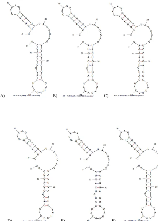
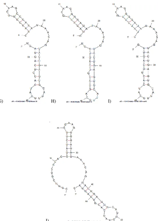
Figure 2.1 M-fold predicted secondary structures of RNA aptamers A) 1502-original B) 1502-scrambled (negative control) C) 1502-optimized D) 1502-mutant8 E) 1502-mutant12 and F) 1502-mutant25.
With Ethidium Bromide
26
G) H) I)
J)
27
K) L)
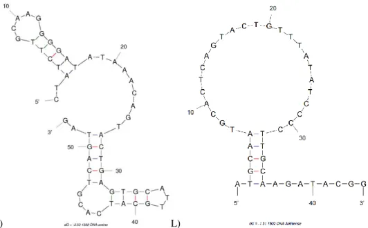
Figure 2.1 cont. M-fold predicted secondary structures of DNA aptamers K) 1502-original DNA and L) 1502 DNA antisense.
28
Figure2.2.a. A denaturing urea PAGE gel shows fractions (lanes 1-9)of Biotin-1502-original RNA with a low mw DNA ladder in lane 10, indicating the purity and length of the aptamer. This 1 µmol synthesis produced
approximately 365 nmol of RNA, or a 36.5% yield.
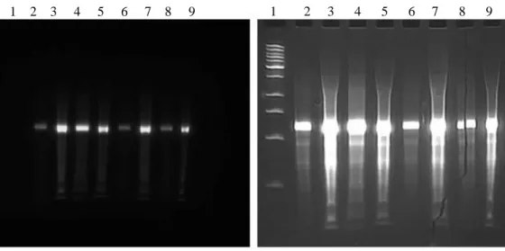
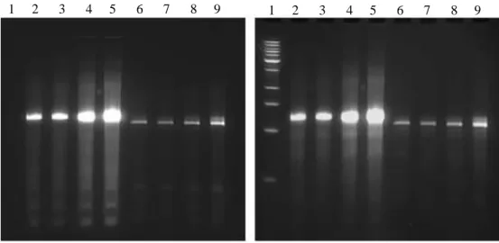
Figure2.2.b. Two denaturing urea PAGE gels shows fractions of 1502-original-RNA (lanes 2-9) and the low mw DNA ladder (lane 1), without (left) and with (right) ethidium bromide staining, indicating the presense of a the fluorescent Sima and the length of the aptamer. This 40 nmol synthesis produced approximately 19.5 nmol of RNA, or a 48.8% yield.
29
Figure 2.2.c. Two denaturing urea PAGE gels shows fractions of 1502-scrambled RNA (lanes 2-6) and Sima-1502-optimized RNA (lanes 7-10), without (left) and with (right) ethidium bromide staining, indicating the presense of a the fluorescent Sima and the length of the aptamer. These 40 nmol syntheses produced approximately 17.7 nmol of RNA for the Sima-1502-scrambled, or a 44.4% yield and approximately 40 nmol of the Sima-1502-optimized RNA, or a 100% yield.
Figure 2.2.d. Two denaturing urea PAGE gels shows fractions of 1502-Mutant8 RNA (lanes 2-4) and Sima-1502-Mutant12 RNA (lanes 5-8), without (left) and with (right) ethidium bromide staining, indicating the presense of a the fluorescent Sima and the length of the aptamer. These 40 nmol syntheses produced approximately 18.5 nmol of RNA for the Sima-1502-scrambled, or a 46.4% yield and approximately 39.2 nmol of the Sima-1502-optimized RNA, or a 98.0% yield.
1 2 3 4 5 6 7 8
1 2 3 4 5 6 7 8 9 10 1 2 3 4 5 6 7 8 9 10
30
Figure 2.2.e. Two denaturing urea PAGE gels shows fractions of 1502-Mutant25 RNA (lanes 2-5) and Sima-1502-Mutant39 RNA (lanes 6-9), without (left) and with (right) ethidium bromide staining, indicating the presense of a the fluorescent Sima and the length of the aptamer. These 40 nmol syntheses produced approximately 10.4 nmol of RNA for the Sima-1502-Mutant25, or a 26.0% yield and approximately 11.1 nmol of the Sima-1502-optimized RNA, or a 27.7% yield.
Figure 2.2.f. Two denaturing urea PAGE gels shows fractions of 1502-Mutant42 RNA (lanes 2-5) and Sima-1502-Mutant46 RNA (lanes 6-9), without (left) and with (right) ethidium bromide staining, indicating the presense of a the fluorescent Sima and the length of the aptamer. These 40 nmol syntheses produced approximately 28.1 nmol of RNA for the Sima-1502-Mutant25, or a 70.3% yield and approximately 13.3 nmol of the Sima-1502-optimized RNA, or a 33.2% yield.
1 2 3 4 5 6 7 8 9 1 2 3 4 5 6 7 8 9
31
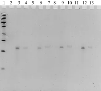
Figure 2.2.g. A denaturing urea PAGE gel shows fractions of 1502-original-alkyne RNA (lanes 1-10) with the low mw DNA ladder in lane 11, indicating the purity and length of the aptamer. This 200 nmol synthesis produced approximately 132.3 nmol of RNA for the 1502-original-alkyne, or a 66.1% yield.
Figure 2.2.h. A denaturing urea PAGE gel shows fractions of 1502-scrambled-alkyne RNA (lanes 2-13), with the low mw DNA ladder in lane 1, indicating the purity and length of the aptamer. This 40 nmol synthesis produced approximately32.4 nmol of RNA for the 1502-scrambled-alkyne, or a 81.1% yield.
1 2 3 4 5 6 7 8 9 10 11
32
Figure 2.2.i. A denaturing urea PAGE gel shows fractions of 1502-original-amino RNA (lanes 2-5), with the DNA low mw ladder in lane 1, indicating the purity and length of the aptamer. This 40 nmol synthesis produced
approximately 40 nmol of RNA for the 1502-original-alkyne, or a 100% yield.
Figure 2.2.j. A denaturing urea PAGE gel shows fractions of 1502-original-stearyl RNA (lanes 2-5), with the low mw DNA ladder in lane 1, indicating the purity and length of the aptamer. This 40 nmol synthesis produced approximately 31.7 nmol of RNA for the 1502-original-alkyne, or a 79.3% yield.
1 2 3 4 5
33
Figure 2.2.k. A denaturing urea PAGE gel shows fractions of 1502-ext-phosphoryl RNA (lanes 2-5), with the low mw DNA ladder in lane 1, indicating the purity and length of the aptamer. This 40 nmol synthesis produced approximately 36.2 nmol of RNA for the 1502-original-alkyne, or a 90.5% yield.
2.2.b. Qualitative binding of aptamer 1502 with confocal microscopy
To further analyze the cell surface receptor binding of the selected aptamers for future PDAC targeting applications, we utilized the chemically synthesized 1502 aptamers 1502 that contained a 5’ fluorescent Sima (hex) group to visualize binding (or non-binding) properties of the aptamers. These properties were first analyzed by confocal microscopy for a qualitative assessment of the aptamer binding to the cell surface of PDAC cells. Figure 2.3A illustrates that 1502-original-Sima, but not its scrambled version, shown in Figure 2.3B bound to the surface of AspC-1 cells when the binding was performed at 4°C for 30 min. No binding was observed when HPNE normal pancreas cells were used. When 1502-original-Sima was tested on HepG2 (liver
34
cancer) cells, a bright fluorescence was observed around the cell’s surface, similar, if not brighter in nature to 1502’s binding to AsPC-1 cells.
Figure 2.3A Binding of the 1502-original-Sima aptamer with AsPC-1 PDAC, HNPE normal pancreas, or HepG2 liver cancer cells visualized with confocal microscopy illustrates binding of the aptamer to AsPC-1 and HepG2.
35
1502-optimized-Sima was tested on AsPC-1 and HPNE cells to visualize binding comparative to original-Sima. An identical approach to the characterization of 1502-original-Sima and 1502-scrambled-Sima was taken to test the cell surface binding of this potentially optimized aptamer. Results (not shown) demonstrated binding to the cell surface of AsPC-1 and not HPNE cells, however the relative fluorescence was not brighter and therefore did not indicate an “optimized” aptamer. It was then determined that further characterization and application studies were to be done with the “original” selected sequence.
As described in the synthesis of the RNA aptamers, six mutant aptamers were
synthesized with a 5’ Sima group and tested for binding to AsPC-1 vs. HPNE cells. If one of the mutant aptamers observed diminished or a lack of binding to AsPC-1 cells, further binding studies were planned to study the interactions of aptamer 1502 to the potential target. The cell binding assay was done at 4ºC for 30 min, as done previously. The binding of these mutant sequences, data not shown, was not affected by the mutations that were designed into the sequences. Because of these results, no further studies were done with the six mutated 1502 sequences.
2.2.c. Quantitative binding of aptamer 1502 with flow cytometry
36
calculated on CellQuest software. These results confirm that the chemically synthesized aptamer 1502 binds strongly to the target cell line AsPC-1 and not the normal pancreas control cell line, HPNE. Additionally, the strong binding of HepG2 can be useful in identifying the unknown potentially novel target of aptamer 1502.
Figure 2.4 Mean fluorescent intensity (AU) was determined by flow cytometry when varying with concentrations (nM) of aptamer 1502 labeled with Sima were incubated with AsPC-1, HPNE, and HepG2 cells. AsPC-1 and HepG2 demonstrated binding of 125 nM± 4.95 nM and 60 nM ± 1.72 nM, with no measurable binding form HPNE.
2.2.d. Immunohistochemistry of 1502 with Tumor Tissues from Patient Derived Xenograft PDAC Mouse
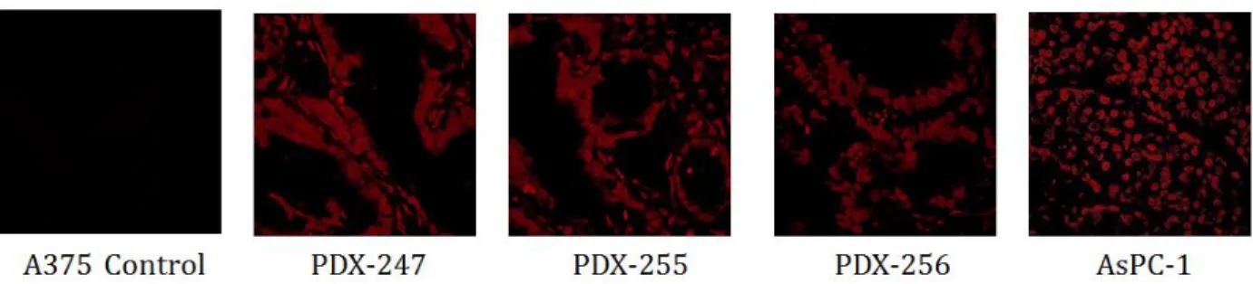
Prior to any meaningful translational applications, it is critical to examine whether the putative novel receptor that is recognized by 1502 is present on patient pancreatic cancer tissue. To address the question, we performed immunohistochemical studies by incubating TAMRA-labeled 1502 with tumor tissue samples from the Patient-Derived Xenograft (PDX) mice, in which different patient tumors had been engrafted as tumor fragments into immunocompromised mice. As shown in Figure 2.5, strong binding signal was observed in all the three tissue samples derived from different human patients, as well as in the AsPC-1 tumor tissue sample. These
-200.0 0.0 200.0 400.0 600.0 800.0 1000.0
0 50 100 150 200 250 300 350 400 450
M e an Fl u o re sce n t In te n si ty (AU )
Concentration of Sima-1502 (nM)
Flow Cytometry Binding of Sima-1502
to AsPC-1, HepG2, or HPNE Cells
AsPC-1
HPNE
HepG2 HepG2, Kd = 60 nM
AsPC-1, Kd = 125 nM
37
results suggest that the putative 1502-binding biomarker that is highly specific to PDAC cells is very likely present on patient PDAC tissue. The results also demonstrated that the TAMRA-labeled 1502 did not target the control A375 (malignant melanoma) tissue. While this sample set is small, it is an indication of the translational application of 1502 to target and identify not only PDAC cells, but PDAC patient tissue as well.
Figure 2.5 Immunohistochemistry studies on the binding of TAMRA-labeled 1502 to PDX tissue samples of pancreatic ductal adenocarcinoma.
2.2.e. Serum stability assay
38
1502-original-amino aptamer is further applied in Aim 2, and it is an aptamer that has
translational in vivo application, as seen in Chapter 5. It was predicted that this aptamer would have a similar stability to 1502-ext-phosphoryl.
The results of this assay can be seen in Figure 2.6. The 1502-ext-phosphoryl aptamer did degrade over time in both the 10% and 50% mouse serum, with less than 25% of the aptamer remaining after 24 h in 50% serum. This was to be expected for this aptamer. The 1502-original-amino aptamer, however, seemed to have an increased stability in 10 and 50% serum, with the only noticeable degradation occurring at 24 h in 50% serum. This was an unexpected result, although promising when considering utilizing the aptamer in vivo. Both aptamer’s assays were repeated 3 times to confirm the findings. It is believed that the 5’ amino modifier may have provided stability to the aptamer against nuclease degradation.
Figure 2.6 Preliminary results demonstrate that 1502-ext-phosphoryl degrades with 10 and 50% mouse serum, and is at least 75% degraded by 24 h in 50% serum. 1502-original-amino, however, does not appear to degrade in 10% serum and only exhibits slight degradation in 50% serum at 24 h. Lanes correspond to time of incubation in mouse serum; 1-0 min, 2-5 min, 3-2 h, 4-4 h, 5-8 h, 6-12 h, and 7-24 h.
39
researchers determined the target proteins of their aptamer to be cyclophilin B for RNA aptamer M9-5, and ALPPL-2 for aptamer SQ-2. Based on the differences in cell binding of these
aptamers against tested cell lines seen in the literature, there is an indication that aptamers M9-5 and SQ-2 have differing binding properties when compared to our selected 2’-F RNA aptamer, 1502. In Chapter 3, a comprehensive cell-binding study is done with 20+ cell lines to validate 1502’s specificity. However, because 1502’s target has yet to be identified, we would like to further confirm that 1502 does not bind to the same target as either aptamer M9-5 or SQ-2. With the M9-5 aptamer, there were technical difficulties in synthesizing the RNA aptamer by transcription. Both Klenow and PCR were attempted with various designed primers, however the RNA transcription was too low to be useful for labeling. Fortunately, I was able to enzymatically synthesize the full length SQ-2 aptamer, which had a non-functional 3’-end that was reported in the literature. The Lee group utilized this non-functional end for labeling with a tamra-antisense oligo, an approach that I took to label the SQ-2 aptamer. With a 3’-anitsense oligo labeled with tamra, I could anneal the SQ-2 RNA to the antisense for a fluorescent labeling of the aptamer, an approach also taken by the Lee group to demonstrate cellular binding.
Initially, a competition of 1502 against SQ-2 was done on AsPC-1 cells. In this
40
Figure 2.7 Competition study with Sima-1502 and unlabeled aptamer SQ-2 at 1:1(top images), 1:3 (middle images), and 1:6 (bottom images) molar ratios of 1502:SQ-2.
After labeling aptamer SQ-2 with the tamra-antisense oligo, I further demonstrated that the SQ-2 aptamer that I synthesized could bind to the cell lines tested in the paper, including ALPPL-2 expressing Capan-1, AsPC-1, and Panc-1 cell lines, without binding to low expressing HPNE, CFPAC-1, and MiaPaca-2 cells. Sima-labeled 1502 was tested on these same cell lines, which illustrated differences in binding. Figure 2.8a shows the binding similarities and more significantly, differences, between 1502 and SQ-2 with the tested PDAC and non-PDAC cell lines. The differences seen with these aptamers indicate that the aptamers likely bind to different targets.
Finally, it was important to additionally test cell lines that are known to have a high expression of ALPPL-2. The aim was to confirm that SQ-2 is functional and 1502 is not
functional against these cell lines. Although the ALPPL-2 protein is not completely understood, we were able to identify cell lines that have a higher expression of the protein through the Broad-Novartis Cancer Cell Line Encyclopedia 29. We found that the colorectal adenocarcinoma cell line, LoVo cells, have a relatively high mRNA expression that corresponds to ALPPL-2 expression. Additionally, a rectal adenocarcinoma cell line, SW837, also have a relatively high
a)
b)
41
mRNA expression according the Broad-Novartis Encyclopedia. We tested these cell lines with tamra-labeled SQ-2 and Sima-labeled 1502, data seen in Figure 2.8b. These results illustrate that 1502 does not bind to cell lines with a known higher expression of ALPPL-2, again suggesting that aptamer 1502 does not bind to ALPPL-2. The tamra-labeled SQ-2 aptamer demonstrated binding to both LoVo and SW837 cell lines; results that further supported the aptamer’s binding to ALPPL-2.
42
Figure 2.8.a. Aptamers SQ-2 (tamra labeled) and 1502 (sima labeled) at a 300 nM concentration, were tested against AsPC-1 cells at 4°C for 30 min. As expected from the literature and from our previous testing, binding was observed for SQ-2 and 1502. Cell only, the tamra-antisense oligo, and a fully modified non-functional labeled SQ-2 aptamer were used as controls and showed no binding to AsPC-1 cells.
43
Figure 2.8.c. Aptamers SQ-2 (tamra labeled) and 1502 (sima labeled) at a 300 nM concentration, were tested against Panc-1 cells at 4°C for 30 min. As expected from the literature and from our previous testing, binding was observed for SQ-2 and 1502. Cell only, the tamra-antisense oligo, and a fully modified non-functional labeled SQ-2 aptamer were used as controls and showed no binding to Panc-1 cells.
44
Figure 2.8.e. Aptamers SQ-2 (tamra labeled) and 1502 (sima labeled) at a 300 nM concentration, were tested against CFPAC-1 cells at 4°C for 30 min. As expected from the literature and from our previous testing, binding was observed 1502 but not for SQ-2, an indication of different cell surface targets. Cell only, the tamra-antisense oligo, and a fully modified non-functional labeled SQ-2 aptamer were used as controls and showed no binding to CFPAC-1 cells.
45
Figure 2.8.g. Aptamers SQ-2 (tamra labeled) and 1502 (sima labeled) at a 300 nM concentration, were tested against LoVo cells at 4°C for 30 min. The literature indicates that LoVo has a high ALPPL-2 expression and it was therefore expected that binding was observed for SQ-2. When tested, binding was not observed for 1502, however, suggesting that 1502 does not have a strong binding affinity for ALPPL-2. Cell only, the tamra-antisense oligo, and a fully modified non-functional labeled SQ-2 aptamer were used as controls and showed no binding to LoVo cells.
46
Figure 2.8.i. Aptamers SQ-2 (tamra labeled) and 1502 (sima labeled) at a 300 nM concentration, were tested against A549 cells at 4°C for 30 min. The literature indicates that A549 cells have a slightly elevated ALPPL-2 expression and it was therefore expected that there would be some binding observed for SQ-2. It is also suggested by the literature that A549 has a high expression of cyclophilin B. Aptamer 1502, as well as controls, cell only, the tamra-antisense oligo, and a fully modified non-functional labeled SQ-2 aptamer showed no binding to A549 cells, suggesting that cyclophilin B is not the unknown target of 1502.
47
By testing Sima-1502 against targeted PDAC cell lines, non-binding normal pancreas HPNE, ALPPL-2 positive non-PDAC cell lines, and cyclophilin B positive non-PDAC cell lines, and comparing the qualitative binding of 1502 to ALPPL-2 targeting aptamer SQ-2, the results suggest that aptamer 1502 does not target the PDAC biomarkers ALPPL-2 or cyclophilin B. Because the target of 1502 is still unknown, it was important to test for its binding to recently proven biomarkers of pancreatic adenocarcinoma. We believe that aptamer 1502 could be targeting a novel biomarker overexpressed specifically on PDAC cell lines, and that this target can be therefore very useful in further diagnostic and therapeutic development.
2.3 Concluding Remarks
This chapter highlights the extensive chemical synthesis and characterization of 2’-F RNA aptamer 1502. The synthesis was done on an ABI oligosynthesizer that was rebuilt and reprogrammed to facilitate low volume DNA and RNA solid-phase oligo synthesis. Cleavage, deprotection, and purification was customized and performed successfully for over 20 DNA and RNA variations of 1502, as seen by the PAGE denaturing gel analysis and quantification. The large scale synthesis and straight forward 3’ and 5’ modifications allowed for extensive
characterization and application of these aptamers in subsequent studies, something that would be much more difficult with enzymatic synthesis.
48
that 1502 may be binding to one of these surface proteins. Indeed, there was observable cell-surface binding of Sima-labeled 1502 to HepG2, seen with confocal microscopy and flow cytometry. These results will be useful when continuing the biomarker identification studies and was utilized in the targeted hyperthermia experiments, seen in Chapter 3.
2.4Materials and Methods
2.4.a. Chemical synthesis with an 394 ABI Oligosynthesizer
The optimized truncated RNA aptamers were chemically synthesized with an ABI 394 Oligosynthesizer (Table 2.1). These aptamers were modified on the 3’ or 5’ end with various linkers, fluorescent groups, and functional chemistries. A comprehensive list of these modifiers can be found in Table 2.2. To facilitate deprotection under relatively milder conditions, we used TBDMS-protected ultra-mild phosphoramidites and reagents (Chem Genes and Glen Research) for the oligosynthesis. Table 2.3 illustrates the full list of reagents used for both DNA and RNA synthesis. For DNA aptamers, the cycle used was a normal volume CE cycle, synthesizing at either a 40 nmol or 200 nmol scale. For the RNA aptamers synthesized, the cycle was programed to have longer coupling times with lower volumes of reagents needed, using low volume
49 Synthesized
Aptamers
Sequence (5’ to 3’) Synthesis Cycle Scale 1502-original-Sima (Hex) Sima-CUAUCUUGCAAGGGGAUAUAAACAGUAC UGAGUGCAUUGCAUCACGUCAGUAGdT LV40RNAA LL 40 nmol 1502-scrambled-Sima (Hex) Sima-CUAUCUUCUAGAGGGAUAUAAACAGUAC UGAGUGUGCCAGAUCACGUCAGUAGdT LV40RNAA LL 40 nmol 1502-optimized-Sima Sima- CUAUCUUGCAAGGGGAUAUAAACAGUAC UGAGUGCAUUUCAUCACGUCAGUAGdT
LV40RNAA LL 40 nmol 1502-mutated7-Sima Sima-CUAUCUUGCAAGGGGAUAUAAACAGUAC UGAGUGCAUUGCAUCACUUCAGUAGdT
LV40RNAA LL 40 nmol 1502-mutated11-Sima Sima- CUAUCUUGCAAGGGGAUAUAAACAGUAC UGAGUGCAUUGCAGCACGUCAGUAGdT
LV40RNAA LL 40 nmol 1502-mutated24-Sima Sima- CUAUCUUCUAGAGGGAUAUAAACAGUAC UGAGUGUGCCAGAUCACGUCAGUAGdT LV40RNAA LL 40 nmol 1502-mutated38-Sima Sima- CGGUCGCGCAAGGCGACCUAAACAGUCC UGAGUGCAUUGCAUCACGUCAGGAGdT LV40RNAA LL 40 nmol 1502-mutated41-Sima
Sima-CUAUCUUCCAAGGGGAUAUAAACAGUAC UGAGUGCAUUGCAUCACGUCAGUAGdT LV40RNAA LL 40 nmol 1502-mutated45-Sima Sima-
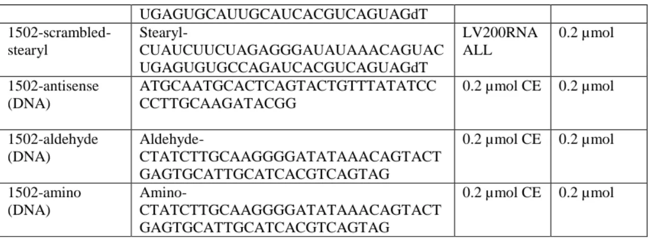
50 UGAGUGCAUUGCAUCACGUCAGUAGdT 1502-scrambled-stearyl Stearyl-CUAUCUUCUAGAGGGAUAUAAACAGUAC UGAGUGUGCCAGAUCACGUCAGUAGdT LV200RNA ALL 0.2 µmol 1502-antisense (DNA) ATGCAATGCACTCAGTACTGTTTATATCC CCTTGCAAGATACGG
0.2 µmol CE 0.2 µmol
1502-aldehyde (DNA)
Aldehyde-CTATCTTGCAAGGGGATATAAACAGTACT GAGTGCATTGCATCACGTCAGTAG
0.2 µmol CE 0.2 µmol
1502-amino (DNA)
Amino-CTATCTTGCAAGGGGATATAAACAGTACT GAGTGCATTGCATCACGTCAGTAG
0.2 µmol CE 0.2 µmol
51
Modifier Structure
Sima (hex) phosphoramidite
3’Biotin-TEG CPG
5’ –Amino-Modifier C6-PDA
Chemical Phosphorylation Reagent II
52
5'-Hexynyl Phosphoramidite
Stearyl Phosphoramidite
Table 2.2 Chemical modifiers syntheized on the 3’ and 5’ ends (Glen Research).
General Oligosynthesis Reagents DNA Oligosynthesis Reagents RNA Oligosynthesis Reagents
LV polystyrene columns (dT), (dG) dG-CE-phosphoramidite iPr-Pac-G-CE Phosphoramidite 0.02M I2 in THF/Pyridine/H2O dC-CE-phosphoramidite 2'-F-Ac-C-CE Phosphoramidite
Dichloromethane dT-CE-phosphoramidite 2'-F-U-CE Phosphoramidite
Acetonitrile 0.45 M Tetrazole in Acetonitrile 0.25M 5-Ethylthio-1H-tetrazole in Acetonitrile
Deblocking Mix (3% TCA/DCM) Cap Mix A
(THF/Pyridine/Ac2O)
Cap Mix A (THF/Pyridine/Pac20)
53
2.4.b. Cleavage, deprotection, and purification of aptamers
For the DNA control aptamers, the oligos were cleaved and base-deprotected with 30% ammonium hydroxide (Sigma) for 16 h at 55°C. The supernatant was dried and resuspended in TE with 50 mM NaCl.
The RNA oligos were cleaved and base-deprotected with a fresh 1:1 solution of 30% ammonium hydroxide and 40% methylamine (AMA) at room temperature for 2 h. The
supernatant was dried to a pellet, and the RNA was resuspended in 100 µL anhydrous DMSO. If necessary, the oligo was heated to 65°C for 5 min to get it into solution. To deprotect the 2’-silyl groups, triethylamine trihydrofluoride (TEA.3HF) was added to the sample and the mixture was heated to 65°C for 2.5 h. The deprotected oligo was desalted by a Nap 5 column or ethanol precipitation. The concentration was measured by NanoDrop and the aptamer length was
confirmed by a PAGE denaturing gel. The RNA aptamers were stored in DEPCtreated water at -20°C.
To purify the aptamers, the oligos with MMT on the 5’ end, were flushed through a GlenPak DNA cartridge (Glen Research) to remove abortive sequences, followed by cleavage of the MMT protecting group to result in a purified full-length oligo.
2.4.c. Cell culture
AsPC-1 (pancreatic ductal adenocarcinoma), PANC-1 (pancreatic ductal
adenocarcinoma), CAPAN-1 (pancreatic ductal adenocarcinoma), CFPAC-1 (pancreatic ductal adenocarcinoma), hTERT-HPNE (pancreatic ductal epithelial cell), LNCaP (prostate
54
adenocarcinoma), COLO 587 (pancreatic ductal adenocarcinoma), Mia-Paca-2 (pancreatic carcinoma), Hep3B (hepatocellular carcinoma), PC-3 (prostate adenocarcinoma), DU 145 (prostate carcinoma), A549 (lung carcinoma), SK-OV-3 (ovarian adenocarcinoma), MCF7 (breast adenocarcinoma), HT-29 (colorectal adenocarcinoma), A431 (epidermoid carcinoma), MCF7 (breast adenocarcinoma), SW837 (rectal adenocarcinoma), and LoVo (colon
adenocarcinoma) were purchased from the Tissue Culture Facility at Lineberger Cancer Center, UNC. These cells originated from the American Type Culture Collection (ATCC) cell repository and were therefore validated through this source. HepG2 (hepatocellular carcinoma) and Huh7 (heptatocelluar carcinoma) were acquired through a collaborator. Cell culture was maintained at 37°C and 5% CO2 in various mediums which included RPMI 1640, EMEM with NEAA, McCOY’s 5a, DMEM/F12, Leibovitz’s L-15, IMDM, F-12K, or DMEM medium supplemented with 10% heat-inactivated fetal bovine serum, FBS (GIBCO) and 100 units/ml penicillin– streptomycin (Cellgro), along with additional supplements when necessary.
2.4.d. Confocal microscopy
The binding of selected pools and individual aptamers to target cells was evaluated by fluorescence confocal imaging. Cells tested included AsPC-1, HPNE (control), and two additional liver cancer cell lines, HepG2 and Huh7. Cells were incubated with various
55
cell-adhesion solution (company) for examination with a confocal microscope. Fluorescence confocal imaging was performed on a Zeiss LSM 700 confocal microscope in the UNC School of Medicine Microscopy Services Laboratory. The objective used for imaging was a 40X oil-immersion objective.
2.4.e. Flow cytometry
To monitor the enrichment of aptamers along with the progress of SELEX and quantify the cell-binding of individual aptamers, Sima-labeled aptamer, Sima-original-1502, which is also termed 1502, was incubated with 1×10⁶ cells in 400 μL binding buffer at 4°C for 30 min. Similar to the confocal microscopy testing, the cell lines tested included AsPC-1, HPNE (control), and two additional liver cancer cell lines, HepG2 and Huh7. Cells were washed twice after
incubation and analyzed by flow cytometry. Flow cytometry was performed on a FACScan cytometer with CellQuest software (Becton Dickinson). Kd was calculated by this software using the following equation: Y = Bmax*X/(Kd + X). Nonspecific binding was also calculated using cell only controls and subtracted from the tested samples.
56
preparations (100%, 95%, 70%, 50%, and 30%) for 2 min each to continue this process. Tissue plates were rinsed with PBS and kept in a 1 M Tris buffer, pH 8.0 which was heated to 95°C for 15 min. Plates were saturated with the RNA binding buffer at room temperature in a humidified chamber. Finally, 500 nM of TAMERA-labeled 1502, previously prepared by Dr. Hui Chen, was added to the plates at 4°C for 30 min in the dark. The aptamer was then removed and plates washed with the binding buffer 2X, and the plates were left to dry in the dark at room
temperature overnight. A BX-61 microscope, with a 10X objective lens, was used to take images of the tissue samples.
2.4.g. Serum stability assay
Three synthesized 2’-F RNA aptamers were tested for their stability in mouse serum (Life Technologies). This included three 1502 RNA aptamers with 5’ modifications; 1502-original-stearyl, 1502-original-amino, and 1502-original-phosphoryl. 5 pmol of the aptamers was incubated with either 10 or 50% mouse serum at 37°C for varied time points (0, 5 min, 2, 4, 8, 12, and 24 h). This experiment was repeated 3X for reproducibility. All samples were examined by PAGE gel electrophoresis.
57
transcribed to date, and therefore the cell binding studies have been limited. However, cell binding studies confirmed the targeting and non-targeting properties of aptamers SQ-2 and 1502 against cell lines that have higher expression levels of ALPPL-2, as well as binding studies for aptamer 1502 against cell lines with a higher expression of cyclophilin B. Additionally, a competition study was initially performed between aptamer 1502 and aptamers SQ-2 to determine if their targeting of PDAC competed with one another.
2.4.i. Enzymatic synthesis of RNA aptamers M9-5 and SQ-2 2.4.i.i. Primer design
5’ and 3’ primers were designed for DNA assembly for both the M9-5 and SQ-2 RNA aptamers. SQ-2’s 5’ primer – 5’
TTCTAATACGACTCACTATAGGGAGATACCAGCTTATTCAATTGCCTGAAAAGCTAT CGCCCAATTCGCAGT 3’ containing the T7 promoter (underlined) and the 3’ primer - 5’ AGATTGCACTTACTATCTTAAAGGATATCACTGCGAATTGGGCGATAGCTTTTC 3’ was designed for Klenow.
M9-5’s 5’ primer – 5’
TTCTAATACGACTCACTATAGGGAGGACGATGCGGGGACCTATGCAGTAGCCAGTG TGGACT
3’ containing the T7 promotor (underlined) and the 3’ primer - 5’
58
2.4.i.ii. PCR/Klenow and in vitro transcription
The DNA template for M9-5 was assembled through PCR. Briefly, PCR reactions contained 0.2 mM dNTPs (each), 0.2 µM of each 5’ and 3’ primer, 1X Taq polymerase buffer, and 1.25 U/50 µL reaction Taq polymerase (NEB). Through PCR titrations, 15 rounds of PCR was determined to be optimal. PCR parameters consisted of 2 min of Taq activation at 94°C, and 15 cycles of PCR at 94°C for 30 s, 58°C for 30 s, 72°C for 30 s, followed by 10 min of extension at 72°C. For SQ-2, the DNA template was assembled and amplified through a Klenow reaction. The 5’ and 3’ primers, at concentrations of 1.5 and 1.0 µM respectively, were annealed by heating to 80°C followed by slow cooling to room temperature. 1X NEBuffer 2 (NEB), 0.2 mM dNTPs, and 2.5U/50 µL reaction Klenow Fragment (3’ to 5’ exo-) (NEB) were added and the reaction was kept at 37ºC for 1.5 h. The quality of PCR amplification and Klenow reactions were confirmed by agarose gel electrophoresis and visualized by ethidium bromide staining. Both DNA templates were purified with phenol/chloroform extraction, followed by ethanol precipitation, and resuspended in TE buffer with 5 mM NaCl.
59
2.4.j. Labeling of RNA aptamers with a tamra-labeled antisense oligo
A tamra-labeled antisense sequence was designed to complement the 3’ non-functional tail that is present in the SQ-2 RNA aptamer. This primer’s sequence is as follows – 5’ tamra-AGATTGCACTTACTATCTTAAA3’. The SQ-2 aptamer and the tamra-antisense oligo were combined at a 1:20 molar ratio. The mixture was heated to 80°C for 3 mins, followed by a slow cooling to room temperature to anneal the antisense oligo to the RNA aptamer. The resulting aptamer was diluted in 1x PBS with 5 mM Ca2+ and 1 mM Mg2+, to the desired concentration for the cell binding and competition studies.
2.4.k. Competition study between aptamer 1502 and SQ-2
To determine if aptamer 1502 competes with aptamer SQ-2 in binding to ALPPL-2, Sima-labeled 1502 was mixed with unlabeled SQ-2 at a 1:1, 1:3, and 1:6 molar ratio with a 200 nM 1502 concentration. These mixtures were incubated with AsPC-1 cells for 30 min at 4ºC. Sima-1502 was also tested on AsPC-1 cells to confirm it’s functionality of binding to AsPC-1. The cells were washed once with 1X PBS and transferred to slides with cell-adhesion solution. The Zeiss 700 confocal microscope with a 40X objective oil lens was used to visualize cell-surface binding.
2.4.l. Binding of aptamers 1502 and SQ-2 to targeted and non-targeted cell lines