Journal of Feline Medicine and Surgery 2016, Vol. 18(10) 838 –845
© The Author(s) 2015 Reprints and permissions:
sagepub.co.uk/journalsPermissions.nav DOI: 10.1177/1098612X15597967 jfms.com
Introduction
Distal thoracic limb surgery in cats, including digit amputation, biopsy and fracture repair, is performed for a variety of reasons, for example for treatment of neopla-sia, infected nail beds and trauma. Onychectomy is prob-ably the most commonly performed surgery of the distal thoracic limb in cats.
Scratching is a normal behavior of cats for the pur-pose of territory marking, stretching forelimbs and con-ditioning claws.1 However, for many owners, this behavior is reported to be undesirable owing to the dam-age inflicted on furniture and belongings.2 Options available to owners to limit the damage from scratching cats include non-surgical and surgical methods.3 Onychectomy is becoming increasingly controversial and has been banned in at least 22 countries and in eight
Defining the local nerve blocks for
feline distal thoracic limb surgery:
a cadaveric study
Masataka Enomoto
1, B Duncan X Lascelles
1,2,3and Mathew P Gerard
4Abstract
Objectives Though controversial, onychectomy remains a commonly performed distal thoracic limb surgical procedure in cats. Peripheral nerve block techniques have been proposed in cats undergoing onychectomy but evidence of efficacy is lacking. Preliminary tests of the described technique using cadavers resulted in incomplete staining of nerves. The aim of this study was to develop nerve block methods based on cadaveric dissections and test these methods with cadaveric dye injections.
Methods Ten pairs of feline thoracic limbs (n = 20) were dissected and superficial branches of the radial nerve (RSbr nn.), median nerve (M n.), dorsal branch of ulnar nerve (UDbr n.), superficial branch of palmar branch of ulnar nerve (UPbrS n.) and deep branch of palmar branch of ulnar nerve (UPbrDp n.) were identified. Based on these dissections, a four-point block was developed and tested using dye injections in another six pairs of feline thoracic limbs (n = 12). Using a 25 G × 5/8 inch needle and 1 ml syringe, 0.07 ml/kg methylene blue was injected at the site of the RSbr nn., 0.04 ml/kg at the injection site of the UDbr n., 0.08 ml/kg at the injection site of the M n. and UPbrS n., and 0.01 ml/kg at the injection site of the UPbrDp n. The length and circumference of each nerve that was stained was measured.
Results Positive staining of all nerves was observed in 12/12 limbs. The lengths stained for RSbr nn., M n., UDbr n., UPbrS n. and UPbrDp n. were 34.9 ± 5.3, 26.4 ± 4.8, 29.2 ± 4.0, 39.1 ± 4.3 and 17.5 ± 3.3 mm, respectively. The nerve circumferences stained were 93.8 ± 15.5, 95.8 ± 9.7, 100 ± 0.0, 100 ± 0.0 and 93.8 ± 15.5%, respectively. Conclusions and relevance This described four-point injection method may be an effective perioperative analgesia technique for feline distal thoracic limb procedures.
Accepted: 29 June 2015
1 Comparative Pain Research Laboratory, Department of Clinical
Sciences, College of Veterinary Medicine, North Carolina State University, Raleigh, NC, USA
2 Center for Comparative Medicine and Translational Research,
Department of Clinical Sciences, College of Veterinary Medicine, North Carolina State University, Raleigh, NC, USA
3 Center for Pain Research and Innovation, UNC School of
Dentistry, Chapel Hill, NC, USA
4 Department of Molecular Biomedical Sciences, College of
Veterinary Medicine, North Carolina State University, Raleigh, NC, USA
Corresponding author:
Mathew Gerard BVSc, PhD, DACVS, Department of Molecular Biomedical Sciences, College of Veterinary Medicine, North Carolina State University, 1060 William Moore Dr, Raleigh, NC 27607, USA
California cities,4 primarily because it is considered an unnecessary cosmetic procedure associated with pain and distress. Another reason for the controversy is a belief that onychectomized cats are more likely to develop behavioral problems.2 Regardless, in North America it is still a commonly performed procedure,2–5 with an estimated 15.5 million cats undergoing onychec-tomy every year.4 Owner satisfaction with this proce-dure is reported to be high.3 However, it is generally agreed that onychectomy is associated with pain.6 Many cats display overt lameness at the time of discharge from the hospital,2 and objective gait analysis has shown that limb function does not return to preoperative values 12 days after onychectomy.6 Furthermore, chronic pain/ neuropathic pain following surgical onychectomy has been described.2
The best way to avoid pain associated with onychec-tomy is to not perform the procedure, but when it is per-formed, it is incumbent on veterinarians to use the most effective means of providing comprehensive analgesia. Multiple methods for the alleviation of perioperative pain associated with this procedure have been described, including irrigation of surgical incisions with bupiv-acaine before closure.6,7 Local analgesics are commonly utilized preoperatively in an attempt to block nocicep-tive input, avoid central sensitization, reduce the amount of systemic analgesic required and decrease hospitaliza-tion time.8–10 In the case of onychectomy and indeed any other anticipated painful procedure of the distal thoracic limb, this would involve effective blockade of the nerves that innervate the manus, including the superficial branches of the radial nerve (RSbr nn.), the median nerve (M n.), the dorsal branch of the ulnar nerve (UDbr n.), the superficial branch of the palmar branch of the ulnar nerve (UPbrS n.) and the deep branch of the palmar branch of the ulnar nerve (UPbrDp n.). Use of bupiv-acaine administered as a four-point regional nerve block prior to onychectomy in cats has been proposed.11 However, in one study, no additional benefit from this block over systemic buprenorphine administration was found in cats undergoing forelimb onychectomy.7
The most commonly used technique for local anes-thetic block in cats undergoing onychectomy appears to stem from a single case report.11 Preliminary data that we obtained from critically testing this technique using cadaver limbs and injections of dye indicated frequent incomplete staining of targeted nerves (see supplementary material). Given that a successfully per-formed nerve block is one of the most effective means to manage pain,12 we re-evaluated the injection techniques. The aims of this study were to review the anatomical location of the RSbr nn., M n., UDbr n., UPbrS n. and UPbrDp n. in relation to palpable anatomical landmarks and identify clearly defined injection sites that may pro-vide a comprehensive blockade of the distal thoracic
limb. An additional aim of this study was to test the defined sites using volumetrically relevant injections of dye in cadaver limbs. Our ultimate goal was to establish a peripheral nerve block technique that may provide effective analgesia for cats undergoing distal thoracic limb surgery.
Material and methods
Cadaver dissectionsTen pairs of cadaveric thoracic limbs were obtained from domestic shorthair cats (nine castrated males and one spayed female) that had been euthanized with an intrave-nous overdose of pentobarbital (Fatal Plus solution; Vortech Pharmaceuticals) for reasons unrelated to this study. Mean ± SD body weight was 4.0 ± 0.9 kg. The limbs were disarticulated at their muscular attachment to the thoracic wall and stored immediately at −18 °C, then thawed at room temperature overnight prior to use. Anatomic dissection of the RSbr nn., M n., UDbr n., UPbrS n. and UPbrDp n. from the level of the mid-antebrachium distally was conducted to determine palpable landmarks, anatomic variations, fascial planes and associated struc-tures. During dissection, potential injection points and landmarks that would guide injection were recorded.
The accessory carpal bone (ACb) was established as a reference landmark because it is relatively fixed, and an easily palpable landmark in the live animal. The location of each putative injection point was recorded using measure-ments taken relative to the ACb in the transverse and sagit-tal planes by the same person (ME) (see ‘Measurements’ and Figure 1), and simultaneously the descriptive relation-ship to anatomical landmarks was recorded. Based on these findings, a subcutaneous, perineural injection tech-nique using four injection sites was developed for the RSbr nn., M n., UDbr n., UPbrS n. and UPbrDp n.
Dye studies
previously described.13 The proximity of dye to unstained or poorly stained nerves was noted.
Measurements
All measurements were made using Dial Calipers (Dial caliper 505-633-50; Mitutoyo). To minimize error by
limb positioning, all measurements were made with the carpus extended to 180º and the elbow at an angle of approximately 45º. The measurements made are detailed in Figure 1. The confluence point of the cephalic vein and accessory cephalic vein was established as the tar-get injection point of the RSbr nn. in order to block these Figure 1 Description of the measured parameters used to evaluate the proposed injection sites for distal limb nerves.
(Right) Craniocaudal view. (A) Distance from the lateral aspect of the limb to the superficial branches of the radial nerve (RSbr nn.); (B) width of the limb at the level of the injection point of the RSbr nn.; (C) distance proximally from the accessory carpal bone (ACb) to the RSbr nn.; (G) distance from the accessory carpal bone to the dorsal branch of the ulnar nerve (UDbr n.). (Left) Caudocranial view. (D) Distance from the accessory carpal bone to the lateral epicondyle of the humerus (LatEpiHum);
Figur
e 2
Diagrams illustrating the developed and tested injection technique for each nerve. (a) Super
ficial branches of radial nerve (RSbr nn.). A 25 G
×
5/8 inch needle is
inser
ted subcutaneously (SC) fr
om a point at the center of the limb at the level of the antebrachiocarpal joint. The needle is advanced appr
oximately 10 mm SC at a 10–20° angle
to the long axis of the limb with the bevel facing up. Once the tip of the needle is 3–5 mm fr
om the confluence point of the accessor
y cephalic and cephalic veins, the injection
is made (0.07 ml/kg). (b) Dorsal branch of ulnar nerve (UDbr n.). A point is located lateral to and at the same level as the ac
cessor
y carpal bone (ACb), positioned between the
ACb and the styloid pr
ocess of the ulna (SpU). A 25 G
×
5/8 inch needle is inser
ted SC distal to pr
oximal so the tip lies at the midpoint of the gr
oove for
med between ACb and
the SpU, and then the injection is made (0.04 ml/kg). (c) Median nerve (M n.) and super
ficial branch of palmar branch of ulnar nerve (UPbrS n.): a 25 G
×
5/8 inch needle is
inser
ted SC at the distal bor
der of the carpal pad and appr
oximately 5 mm lateral to it, perpendicular to the long axis of the metacarpus, with the bevel facing up. The needle is
inser
ted SC until the point is located two-thir
ds of the distance fr
om the lateral aspect of the limb to the medial aspect. T
wo-thir
ds of the injectate volume is deposited at this point
and the r
emaining volume is injected while the needle is withdrawn (0.08 ml/kg total). Gentle massage is applied to the injected volume
under the skin for 5 s. Deep branch of
palmar branch of ulnar nerve (UPbrDp n.): a 25 G
×
5/8 inch needle is inser
ted almost perpendicular to the ACb in a mediolateral dir
ection such that the needle contacts the
medial palmar aspect of the midpoint of the ACb with the bevel facing up. The needle is then r
edir
ected dorsally and advanced on the medial side of the ACb 2–3 mm until it
penetrates the flexor r
etinaculum. At this point the injection is per
for
med (0.01 ml/kg of injectate). Red dots indicate the site of needle inser
tion. The blue arr
ows r
epr
esent the
dir
ection of needle inser
tion to r
each the desir
nerves before they branched distally into the digits. The distance from the lateral aspect of the limb to the RSbr nn. (A), the width of the limb at the level of the injection point of the RSbr nn. (B) and the distance proximally from the ACb to the RSbr nn. (C) were measured. The distance from the ACb to the lateral epicondyle of the humerus (D) was also measured. The ratios of A/B (E) and C/D (F) were calculated. The optimal injection point for the UDbr n. was determined to be at the level of the ACb, on the lateral aspect, because at this point the nerve is not covered with fascia. The distance from the ACb to the UDbr n. (dorsal view) (G) and the width of the carpus at the level of the ACb (palmar view) (H) were measured. The ratio of G/H (I) was calculated. The area distal to the flexor retinaculum where the M n. and UPbrS n. descend, parallel to each other, was deter-mined to be an optimal injection point. The distance from the lateral aspect of the limb to the M n. (J), the width of the limb at the level of the injection point of the M n. and UPbrS n. (K), and the proximodistal distance from the ACb to the level of the M n. and UPbrS n. (L) were measured. The ratio of J/K (M) was calculated. The distance from the lateral aspect of the limb to the UPbrS n. (N) was measured and J-N (O) and the ratio of N/K (P) were calculated. The distance from the ACb to the point where the UPbrDp n. turns deeply was meas-ured (Q). For all measurements, the measurement point on the ACb was defined as the center of the attachment of the flexor carpi ulnaris, humeral head. Measurements of nerve location were made using the center of the nerve.
Injection technique
Each limb was shaved on the dorsal aspect from the distal third of the antebrachium to just distal to the carpus; on the lateral aspect at the level of the ACb; and on the palmar aspect between the metacarpal pad and the carpal pad. The blocks were performed as described below and the technique is described and illustrated in Figure 2(a–c).
RSbr nn. The limb was positioned with the dorsal aspect
facing upwards and the carpus in 180º of extension. A point on the cranial antebrachium was identified that was about 80% of the distance from lateral to medial and about 18 mm proximal to the level of the ACb (approximately the point of confluence of the accessory cephalic and cephalic veins). A 25 G × 5/8 inch needle was inserted subcutaneously (SC) from a point at the center of the limb at the level of the antebrachiocarpal joint. The needle was advanced approximately 10 mm SC at a 10°–20°angle to the long axis of the limb with the bevel facing up. Once the tip of the needle was 3–5 mm from the confluence point of the accessory cephalic and cephalic veins, 0.07 ml/kg NMB was injected.
UDbr n. The limb was positioned with the lateral aspect
facing upwards. A point was located lateral to and at the same level as the ACb, positioned between the ACb and the styloid process of the ulna. A 25 G × 5/8 inch needle was inserted SC from distal to proximal so that the tip lay at the midpoint of the groove formed between the ACb and the styloid process of the ulna, and then injection of 0.04 ml/kg NMB was performed.
M n. and UPbrS n. The limb was positioned with the
pal-mar aspect facing upwards. A point was identified 11 mm distal to the ACb, approximately level with the distal extent of the carpal pad. A 25 G × 5/8 inch needle was inserted SC at the distal border of the carpal pad and approximately 5 mm lateral to it, perpendicular to the long axis of the metacarpus, with the bevel facing up. The needle was inserted SC until the tip was located two-thirds of the distance from the lateral aspect of the limb to the medial aspect. Two-thirds of the injectate ume was deposited at this point and the remaining vol-ume was injected while the needle was being withdrawn. A total volume of 0.08 ml/kg was deposited. Gentle massage was applied to the injected volume under the skin for 5 s.
UPbrDp n. The limb was positioned with the palmar aspect facing upwards. A 25 G × 5/8 inch needle was inserted with the bevel facing up, almost perpendicular to the ACb in a mediolateral direction such that the nee-dle contacted the medial palmar aspect of the midpoint of the ACb. The needle was then redirected dorsally and advanced on the medial side of the ACb 2–3 mm until it penetrated the flexor retinaculum. At this point 0.01 ml/ kg NMB was injected.
Statistics
Statistical analyses were performed using JMP software (JMP Pro 11; SAS). Wilcoxon’s signed rank tests were used to evaluate pairwise difference in measurements A through Q between left and right limbs and to compare body weights between cadaver dissection and dye injec-tion parts of the study. Correlainjec-tions between body weight and measurements A through Q were deter-mined using Spearman’s rank correlation coefficient. Differences were considered significant at P<0.05.
Results
Cadaver dissections
calculated parameter with body weight is given for each parameter. There were significant positive correlations between body weights and A, B, C, D, H, I, K, L, M and Q. There was no significant relationship between body weight and the measurements E, F, G, J, N, O and P.
Dye test
There was no significant difference in body weight between cats in the dissection study and cats in the dye study. All the cats were of an unknown immature or early mature adult age.
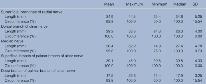
Positive dye staining of all nerves was observed in 12/12 limbs (Table 2). When targeting the RSbr nn., the dye was deposited too laterally in 1/12 limbs, resulting in patchy staining of the nerve. When targeting the UDbr n., the dye was located in the groove between the styloid process of the ulna and the ACb, and stained the nerve well in all cases. For the M n. and UPbrS n., the dye appeared to spread under the superficial digital flexor tendon in 2/12 limbs owing to the injection being made too deeply. This resulted in the injection being deposited dorsal to the M n. in one case, and therefore only the dorsal aspects of that nerve were stained well. The nerves were stained well in the other case. The
injection was deposited intramuscularly (into the medial edge of the ulnar head of the deep digital flexor immediately proximal to the continuing deep digital flexor tendon) when targeting the UPbrDp n. in 1/12 limbs owing to the injection being made too medially, and not directly adjacent the ACb. This resulted in patchy staining of the nerve in this case. The dye stained the nerve well in the other case. Overall, it was deter-mined that all nerves targeted were stained well and consistently with this developed four-point injection method (Figure 2).
Discussion
Preoperatively administered local anesthetics affect postoperative pain relief by blocking the transmission of nociceptive signals, thereby intercepting the nocice-ptive message.14 This should help avoid cellular wind up and subsequent central sensitization that appears to contribute to postoperative pain in clinical cases.15 A local nerve block together with systemic analgesia is recommended for cats undergoing onychectomy.16 However, neither the described techniques of nerve blockade nor irrigation of the wound with bupivacaine before skin closure have been shown to be associated Table 1 Measured and calculated parameters for the location of proposed injection sites for distal limb nerves
Mean Maximum Minimum Median SD CC
Superficial branches of radial nerve
(A) Distance from the lateral aspect of the limb (mm) 13.0 14.7 9.3 13.6 1.38 0.69* (B) Width of the limb at the level of injection point (mm) 15.7 17.8 12.8 16.0 1.47 0.65* (C) Distance proximally from the accessory carpal bone (mm) 17.8 20.9 14.1 18.2 1.62 0.71* (D) Distance from the accessory carpal bone to lateral
epicondyle of the humerus (cm) 9.6 11.1 8.4 9.7 0.91 0.91*
(E) Width% (the ratio of A/B expressed as %) 82.5 87.5 72.7 83.3 4.23 0.29 (F) Length% (the ratio of C/D expressed as %) 18.5 21.1 16.2 18.7 1.32 −0.34 Dorsal branch of ulnar nerve
(G) Distance from the accessory carpal bone (mm) 4.0 4.6 3.0 4.0 0.42 −0.12 (H) Width of the carpus at the level of accessory
carpal bone (mm) 17.4 20.2 14.5 17.2 1.63 0.76*
(I) Width% (the ratio of G/H expressed as %) 23.2 30.0 18.1 23.1 3.15 −0.55* Median nerve
(J) Distance from lateral aspect of the limb (mm) 11.5 14.2 9.8 11.2 1.14 0.04 (K) Width of the limb at the level of injection point (mm) 17.6 19.2 15.2 17.6 1.24 0.68* (L) Proximodistal distance from the accessory
carpal bone (mm) 11.5 13.5 10.0 11.5 0.90 0.55*
(M) Width% (the ratio of J/K expressed as %) 65.5 74.6 55.7 65.3 5.59 −0.54* Superficial and deep branch of palmar branch
of ulnar nerve
(N) Distance from the lateral aspect of the limb (mm) 6.8 9.5 5.5 6.4 1.19 −0.10 (O) Distance from the median nerve (mm) 4.8 5.1 4.3 4.8 0.27 0.39 (P) Width% (the ratio of N/K expressed as %) 38.4 50.8 29.7 36.3 5.89 −0.43 (Q) Distance from the accessory carpal bone (mm) 11.5 12.5 10.3 11.6 0.59 0.56*
with beneficial analgesic effects, using subjective or objective outcome measures.6,7 In our preliminary test-ing, NMB dye injection using the currently described four-point block technique resulted in incomplete nerve staining.11 The currently accepted injection tech-nique has not been described in depth,11 and this lack of a detailed description might be partially responsible for the inaccuracy of the injections performed. The most obvious deficit of the original technique is that the injectate is likely to be deposited more medially than the optimal injection point when targeting the M n., and this could result in intramuscular injection and poor blockade of the M n. In this report, we describe in detail both the anatomical location of nerves in relation to palpable landmarks and a proposed nerve block technique. In our dye injection study using our pro-posed injection technique, all branches of nerves responsible for innervating the feline thoracic limb digits were successfully stained with NMB. This detailed description and associated diagrams, we believe, will allow veterinarians to perform a more successful nerve blockade. However, whether this described technique results in a more successful nerve blockade needs to be clinically tested. We included cal-culations of ratios when developing the technique, assuming that the ratios we calculated would not be affected by body weight. However, several were, and so the described technique uses descriptions based on anatomical landmarks instead of calculated ratios. A shortcoming of this study was the lack of knowledge of the specific age of the feline cadavers. However, we know the cadavers were young animals that had almost completed their maturity or were young adults and it is most typical for onychectomy to be performed in young cats.17 We therefore believe our results are
generalizable to the target population of cats undergo-ing declawundergo-ing procedures. Furthermore, we also believe this nerve block technique is potentially useful for cats undergoing other distal thoracic limb surgeries such as complete digit amputation, biopsy and fracture repair.
Bupivacaine is known to cause cardiac and neurological side effects, and doses of bupivacaine recommended for perineural administration in dogs and cats are 1–2 mg/kg.18 Bupivacaine should not be administered intravascularly to cats.18 Although we did not detect any vascular trauma or intravascular injection, precautions should be taken to avoid this, including syringe aspiration to confirm an absence of intravascular needle position before injection is performed. From our observations during dissections, we believe that the highest risk for intravascular injection is into the median artery, which courses in association with the M n. When tar-geting the M n., keeping the needle as superficial as possible may help to avoid the median artery, because the vessel lies deep to the nerve. Our observations suggest that keeping the injection superficial will also minimize the chance of a poor block of the M n.
Limitations of this study include the use of cadavers and the use of NMB dye for injections. Because of the possible uptake of the local anesthetic solution by the lymph and blood vessels the distribution of injectate in a clinical patient may be different than that in cadavers.19 We used NMB as a substitute for local anesthetic solu-tion, but NMB distribution might not reflect the action of local anesthetics in vivo, where the action on the nerve fiber depends on several factors, including drug lipid solubility, tissue pH and molecular weight.20 Additionally, although we estimated the circumference of the nerve that was stained, this is not easily assessed accurately, as previously reported.13
Table 2 Degree of new methylene blue distribution on the surface (length and circumference stained) of each nerve
Mean Maximum Minimum Median SD
Superficial branches of radial nerve
Length (mm) 34.9 44.3 25.4 34.6 5.25
Circumference (%) 93.8 100.0 50.0 100.0 15.54
Dorsal branch of ulnar nerve
Length (mm) 29.2 38.8 24.8 28.3 4.00
Circumference (%) 100.0 100.0 100.0 100.0 0.00
Median nerve
Length (mm) 26.4 32.2 14.9 27.4 4.78
Circumference (%) 95.8 100.0 75.0 100.0 9.73
Superficial branch of palmar branch of ulnar nerve
Length (mm) 39.1 45.0 30.6 39.8 4.33
Circumference (%) 100.0 100.0 100.0 100.0 0.00
Deep branch of palmar branch of ulnar nerve
Length (mm) 17.5 22.6 11.4 17.9 3.25
Conclusions
All branches of nerves responsible for innervation of the feline thoracic limb digits were successfully stained with NMB using the described four-point injection technique. To assess the utility and effectiveness of this distal limb nerve block injection technique it should be evaluated in clinical patients.
Supplementary material Degree of new methylene blue
dis-tribution on the surface of each nerve using a four-point block.
Acknowledgements We are grateful to Alice Harvey for
the medical illustrations and to Wendy Savage for the photog-raphy. We also thank Dr Margaret Gruen for comments on the manuscript. The authors of this report do not support feline onychectomy; however, we do believe cats undergoing this procedure should be provided effective pain relief of an appro-priate duration.
Conflicts of interest The authors do not have any potential
conflicts of interest to declare.
Funding Internally funded by the Comparative Pain Research
Laboratory. This research received no specific grant from any funding agency in the public, commercial or not-for-profit sectors.
References
1 Landsberg GM. Feline scratching and destruction and the
effects of declawing. Vet Clin North Am Small Anim Pract
1991; 21: 265–279.
2 Patronek GJ. Assessment of claims of short- and long-term
complications associated with onychectomy in cats. J Am Vet Med Assoc 2001; 219: 932–937.
3 Swiderski J. Onychectomy and its alternatives in the feline
patient. Clin Tech Small Anim Pract 2002; 17: 158–61.
4 Lockhart LE, Motsinger-Reif AA, Simpson WM, et al.
Prev-alence of onychectomy in cats presented for veterinary care near Raleigh, NC and educational attitudes toward the procedure. Vet Anaesth Analg 2014; 41: 48–53.
5 Jankowski AJ, Brown DC, Duval J, et al. Comparison of
effects of elective tenectomy or onychectomy in cats. J Am Vet Med Assoc 1998; 213: 370–373.
6 Romans CW, Gordon WJ, Robinson DA, et al. Effect of
postoperative analgesic protocol on limb function fol-lowing onychectomy in cats. J Am Vet Med Assoc 2005; 227: 89–93.
7 Curcio K, Bidwell LA, Bohart GV, et al. Evaluation of signs
of postoperative pain and complications after forelimb onychectomy in cats receiving buprenorphine alone or with bupivacaine administered as a four-point regional nerve block. J Am Vet Med Assoc 2006; 228: 65–68.
8 Melzack R, Coderre TJ, Katz J, et al. Central
neuroplas-ticity and pathological pain. Ann N Y Acad Sci 2001; 933: 157–174.
9 Naja ZM, Raf M, El Rajab M, et al. Nerve stimulator-guided
paravertebral blockade combined with sevoflurane seda-tion versus general anesthesia with systemic analgesia for postherniorrhaphy pain relief in children: a prospective randomized trial. Anesthesiology 2005; 103: 600–605.
10 Siddiqui ZI, Cepeda MS, Denman W, et al. Continuous
lumbar plexus block provides improved analgesia with fewer side effects compared with systemic opioids after hip arthroplasty: a randomized controlled trial. Reg Anesth Pain Med 2007; 32: 393–398.
11 Ringwood PB and Smith JA. Anesthesia case of the month.
J Am Vet Med Assoc 2000; 217: 1633–1635.
12 Richman JM, Liu SS, Courpas G, et al. Does continuous
peripheral nerve block provide superior pain control to opioids? A meta-analysis. Anesth Analg 2006; 102: 248–257.
13 Trumpatori BJ, Carter JE, Hash J, et al. Evaluation of a
mid-humeral block of the radial, ulnar, musculocutaneous and median (RUMM block) nerves for analgesia of the distal aspect of the thoracic limb in dogs. Vet Surg 2010; 39: 785–796.
14 Buback JL, Boothe HW, Carroll GL, et al. Comparison of
three methods for relief of pain after ear canal ablation in dogs. Vet Surg 1996; 25: 380–385.
15 Lascelles BD, Cripps PJ, Jones A, et al. Post-operative central
hypersensitivity and pain: the pre-emptive value of pethidine for ovariohysterectomy. Pain 1997; 73: 461–471.
16 Steagall PV and Monteiro-Steagall BP. Multimodal
analge-sia for perioperative pain in three cats. J Feline Med Surg
2013; 15: 737–743.
17 Clark K, Bailey T, Rist P, et al. Comparison of 3 methods of
onychectomy. Can Vet J 2014; 55: 255–262.
18 Robertson SA and Taylor PM. Pain management in cats –
past, present and future. Part 2. Treatment of pain – clini-cal pharmacology. J Feline Med Surg 2004; 6: 321–333.
19 Kull K, Baer GA, Samarutel J, et al. Distribution of local
anesthetic solution in retromediastinal block. Prelimi-nary experimental results. Reg Anesth 1997; 22: 308–312.
20 Portela DA, Otero PE, Tarragona L, et al. Combined