Copyright © 2004, American Society for Microbiology. All Rights Reserved.
Detection and Typing of Human Papillomavirus by E6 Nested
Multiplex PCR
K. Sotlar,
1* D. Diemer,
1A. Dethleffs,
1Y. Hack,
1A. Stubner,
1N. Vollmer,
2S. Menton,
2M. Menton,
2K. Dietz,
3D. Wallwiener,
2R. Kandolf,
1and B. Bu¨ltmann
1 Institute of Pathology,1Department of Obstetrics and Gynecology,2and Department of Medical Biometry,3University of Tu¨bingen, Tu¨bingen, Germany
Received 21 August 2003/Returned for modification 9 October 2003/Accepted 25 March 2004
A nested multiplex PCR (NMPCR) assay that combines degenerate E6/E7 consensus primers and type-specific primers was evaluated for the detection and typing of human papillomavirus (HPV) genotypes 6/11, 16, 18, 31, 33, 35, 39, 42, 43, 44, 45, 51, 52, 56, 58, 59, 66, and 68 using HPV DNA-containing plasmids and cervical scrapes (nⴝ1,525). The performance of the NMPCR assay relative to that of conventional PCR with MY09-MY11 and GP5ⴙ-GP6ⴙprimers, and nested PCR with these two primer sets (MY/GP) was evaluated in 495 cervical scrapes with corresponding histologic and cytologic findings. HPV prevalence rates determined with the NMPCR assay were 34.7% (102 of 294) in the absence of cervical intraepithelial neoplasia (CIN 0), 94.2% (113 of 120) in the presence of mild or moderate dysplasia (CIN I/II), and 97.8% (44 of 45) in the presence of severe dysplasia (CIN III). The combination of all four HPV detection methods applied in the study was taken as “gold standard”: in all three morphological subgroups the NMPCR assay had significantly (P< 0.0001) higher sensitivities than the MY09-MY11 and GP5ⴙ-GP6ⴙassays and sensitivities comparable or equal to those of the MY/GP assay. All 18 HPV genotypes investigated were detected among the clinical samples. The ratio of high- to low-risk HPV genotypes increased from 4:1 (80 of 103) in CIN 0 to 19:1 (149 of 157) in CIN I to III. Multiple infections were detected in 47.9% (124 of 259) of the patients. In conclusion, the novel NMPCR method is a sensitive and useful tool for HPV DNA detection, especially when exact HPV genotyping and the identification of multiple HPV infections are required.
Human papillomomaviruses (HPVs) constitute a group of more than 100 different genotypes associated with benign and malignant neoplasms of skin and mucous membranes (5, 34). Approximately 40 different HPV genotypes have been de-tected in the anogenital mucosa (34). On the basis of their epidemiological association with the development of cervical carcinoma, a group of so-called high-risk HPV genotypes has been defined. These include HPV genotype 16 (HPV-16), HPV-18, -31, -33, -35, -39, -45, -51, -52, -56, -58, -59, -66, and -68 (3, 8, 17, 34). Other genotypes, such as HPV-6, -11, -42, -43, and -44, are classified as low-risk types (22).
Detection of high-risk HPV infections might identify women who are at increased risk of development or progression of a cervical lesion (7, 9, 26), and vice versa, negative tests have a very high negative predictive value for the development of a cervical lesion (4, 27). The diversity of the HPV spectrum and the high incidence of multiple infections make it necessary to establish reliable methods for the identification of the various HPV genotypes, not only for epidemiologic studies but also for patient management. Future applications of such HPV detec-tion methods may include the selecdetec-tion and monitoring of women in studies of antiviral treatment or type-specific vac-cines.
A number of PCR-based assays for the identification of the various HPV genotypes have been described. Type-specific PCR primer sets allow the identification of individual
geno-types (1, 32). However, they require the performance of mul-tiple parallel amplifications from each sample and have only been described for a limited number of HPV genotypes. Al-ternatively, PCR assays utilizing consensus or general primers, e.g., GP5⫹-GP6⫹, MY09-MY11, PGMY, and SPF10, allow the amplification of a broad spectrum of HPV genotypes in a single reaction (10, 12, 20, 23). The use of MY09-MY11 and either GP5-GP6 or GP5⫹-GP6⫹ primers in a nested PCR assay has been shown to increase the overall sensitivity com-pared to that of each primer pair alone (11, 15). General primer-mediated amplification products can be analyzed by various methods, e.g., direct sequencing, restriction fragment length polymorphism analysis, and hybridization with type-specific probes (5, 13, 14, 17, 29). Recently, two independent reverse hybridization assays were introduced, allowing the de-tection and typing of all known mucosal HPV genotypes, in-cluding multiple infections (13, 19). These two methods utilize highly conserved regions of the viral L1 major capsid gene as the target for their primers. Targeting the E6/E7 region, Sasa-gawa et al. recently described an LCR-E7 PCR test utilizing consensus primers for the detection and restriction fragment length polymorphism analysis for the genotyping of 34 differ-ent HPVs (29).
We report a novel PCR assay with the viral E6/E7 oncogenes as the primer target region. In this assay, consensus primers for first-round amplification of a broad spectrum of mucosal HPV genotypes, including all high-risk HPV genotypes, were com-bined with type-specific primers for nested PCR amplifications. In order to reduce the number of nested PCRs these primers were used in multiplex primer cocktails (Sotlar et al., 17th Int.
* Corresponding author. Mailing address: Institute of Pathology, University of Tu¨bingen, Liebermeisterstraße 8, 72076 Tu¨bingen, Ger-many. Phone: 49 7071 2982265. Fax: 49 7071 292258. E-mail: klsotlar @med.uni-tuebingen.de.
3176
on May 15, 2020 by guest
http://jcm.asm.org/
Papillomavirus Conf. 1999, abstr. Dia 31, p. 292). This strategy allows (i) HPV genotyping based on PCR product size, (ii) extension of the assay with multiplex primer cocktails for ad-ditional HPV genotypes, and (iii) direct detection of the viral oncogenes.
The sensitivity of this novel GP-E6/E7 nested multiplex PCR (NMPCR) assay was analyzed and compared with single and nested PCR assays using MY09-MY11 and GP5⫹-GP6⫹ prim-er sets. The clinical pprim-erformance of the new assay was tested by investigating the cervical scrapes of 1,699 women. Morpholog-ical and molecular biologMorpholog-ical findings were associated in 459 cases with corresponding cytologic and histologic diagnoses.
MATERIALS AND METHODS
Patients.A total of 1,699 women were referred to the Colposcopy Unit, Department of Obstetrics and Gynecology, University of Tu¨bingen, between January 2001 and June 2002 because of atypical Pap smears. Colposcopy was performed and cervical scrapes were obtained for a repeat Pap test and HPV detection in all cases. Cervical biopsy specimens were also taken for histopatho-logical investigation in cases in which the colposcopy findings were suspicious for cervical dysplasia (n⫽864). Total DNA extracted from cervical scrapes in the latter group was used to test the clinical performance of the NMPCR assay and to compare the method with the established assays using MY09-MY11 and GP5⫹-GP6⫹and a nested PCR assay with these two primer sets (MY/GP).
DNA preparation.The cervical scrapes obtained for HPV DNA detection were transferred into tubes containing 400 l of phosphate-buffered saline, snap-frozen in liquid nitrogen, and stored at⫺70°C until further processing. Extraction of total DNA was performed using the QIAmp DNA mini kit (QIA-GEN, Hilden, Germany) according to the manufacturer’s instructions. In brief, the tubes were thawed and rigorously vortexed for 1 min. The swab was then removed. Twenty microliters of QIAGEN protease stock solution and 400l of lysis buffer were added. The tubes were immediately vortexed for 15 s, centri-fuged, and incubated at 56°C for 1 h. Four hundred microliters of pure ethanol was added, and the mixture was transferred into spin columns. After centrifu-gation at 6,000 ⫻gfor 2 min, the spin columns were transferred into new collection tubes and 500l of washing buffer 1 was added to each column. After centrifugation at 20,000⫻gfor 3 min, the flowthrough was discarded and 500l of washing buffer 2 was added. The columns were again centrifuged at 20,000⫻
gfor 3 min and then transferred into new sterile 1.5-ml collection tubes, to which 150l of elution buffer was added. After 10 min of incubation at room temper-ature, the columns were centrifuged at 6,000⫻gfor 1 min and then discarded. The concentration of extracted DNA was determined by spectrophotometry at 260 nm. One hundred nanograms of total extracted DNA was used for PCR amplifications.
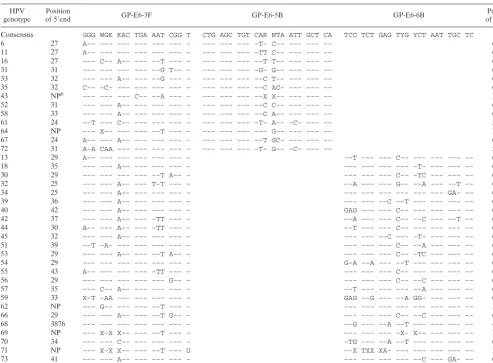
[image:2.603.44.538.80.443.2]HPV plasmid DNA.Newly designed PCR primers were tested for their sen-sitivity and specificity using HPV DNA-containing plasmids kindly provided by E.-M. de Villiers, Deutsches Krebsforschungszentrum, Heidelberg, Germany (genotypes 6, 11, 16, 18, 45, and 51); A. Lo¨rincz, Digene Corporation, Silver Spring, Md. (genotypes 31, 35, 43, 44, and 56); G. Orth, Institut Pasteur, Paris, France (genotypes 33 39, 42, 66, and 68); T. Matsukura, National Institute of Health, Tokyo, Japan (genotypes 58 and 59); and W. D. Lancaster, Wayne State TABLE 1. Sequences of GP-E6/E7 consensus primers and alignment with E6 and E7 regions of 38 HPV genotypesa
HPV
genotype of 5Position⬘end GP-E6-3F GP-E6-5B GP-E6-6B of 3Position⬘end
Consensus GGG WGK KAC TGA AAT CGG T CTG AGC TGT CAR NTA ATT GCT CA TCC TCT GAG TYG YCT AAT TGC TC
6 27 A–– ––– ––– ––– ––– ––– – ––– ––– ––– –T– C–– ––– ––– –– 607
11 27 A–– ––– ––– ––– ––– ––– – ––– ––– ––– –TT C–– ––– ––– –– 607
16 27 ––– C–– A–– ––– ––T ––– – ––– ––– ––– ––T T–– ––– ––– –– 636
31 31 ––– ––– ––– ––– ––G T–– – ––– ––– ––– –G– G–– ––– ––– –– 634
33 32 ––– ––– A–– ––– ––G ––– – ––– ––– ––– ––C T–– ––– ––– –– 647
35 32 C–– –C– ––– ––– ––– ––– – ––– ––– ––– ––C AC– ––– ––– –– 636
43 NPb ––– ––– ––– C–– ––A ––– – ––– ––– ––– ––X X–– ––– ––– –– NP
52 31 ––– ––– A–– ––– ––– ––– – ––– ––– ––– ––C C–– ––– ––– –– 627
58 33 ––– ––– A–– ––– ––– ––– – ––– ––– ––– ––C A–– ––– ––– –– 648
61 24 ––T ––– C–– ––– ––– ––– – ––– ––– ––– –T– A–– –C– ––– –– 592
64 NP ––– X–– ––– ––– ––T ––– – ––– ––– ––– ––– G–– ––– ––– –– NP
67 24 A–– ––– A–– ––– ––– ––– – ––– ––– ––– ––T GC– ––– ––– –– 637
72 31 A–A CAA ––– ––– ––– ––– – ––– ––– ––– –T– G–– –C– ––– –– 598
13 29 A–– ––– ––– ––– ––– ––– – ––T ––– ––– C–– ––– ––– ––– –– 607
18 35 ––– ––– A–– ––– ––– ––– – ––– ––– ––– ––– –T– ––– ––– –– 674
30 29 ––– ––– ––– ––– ––T A–– – ––– ––– ––– C–– –TC ––– ––– –– 644
32 25 ––– ––– A–– ––– T–T ––– – ––A ––– ––– G–– ––A ––– ––T –– 632
34 25 ––– ––– A–– ––– ––– ––– – ––– ––– ––– ––– ––– ––– GA– –– 629
39 36 ––– ––– A–– ––– ––– ––– – ––– ––– ––C ––T ––– ––– ––– –– 679
40 42 ––– ––– A–– ––– ––– ––– – GAG ––– ––– C–– ––– ––– ––– –– 602
42 37 ––– ––– A–– ––– –TT ––– – ––A ––– ––– C–– ––C ––– ––T –– 626
44 30 A–– ––– A–– ––– –TT ––– – ––T ––– ––– C–– ––– ––– ––– –– 611
45 32 ––– ––– A–– ––– ––– ––– – ––– ––– ––C ––– –T– ––– ––– –– 674
51 39 ––T –A– ––– ––– ––– ––– – ––– ––– ––– C–– ––A ––– ––– –– 638
53 29 ––– ––– A–– ––– ––T A–– – ––– ––– ––– C–– –TC ––– ––– –– 647
54 29 ––– ––– ––– ––– ––– ––– – G–A ––A ––– ––T ––– ––– ––– –– 611
55 43 A–– ––– ––– ––– –TT ––– – ––– ––– ––– C–– ––– ––– ––– –– 608
56 29 ––– ––– ––– ––– ––– G–– – ––– ––– ––– C–– ––C ––– ––– –– 650
57 35 ––– C–– A–– ––– ––– ––– – ––T ––– ––– ––– ––A ––– ––– –– 608
59 33 X–T –AA ––– ––– ––– ––– – GAG ––G ––– ––A GG– ––– ––– –– 659
62 NP ––– G–– ––– ––– ––T ––– – ––– ––– ––– ––– ––– ––– ––– –– NP
66 29 ––– ––– A–– ––– ––T G–– – ––– ––– ––– C–– ––C ––– ––– –– 650
68 3876 ––– ––– ––– ––– ––– ––– – ––G ––– ––A ––T ––– ––– ––– –– 4516
69 NP ––– X–X X–– ––– ––T ––– – ––– ––– ––– –X– X–– ––– ––– –– NP
70 34 ––– ––– C–– ––– ––– ––– – –TG ––– ––A ––T ––– ––– ––– –– 679
71 NP ––– X–X X–– ––– ––T ––– G ––X TXX XA– ––– ––– ––– ––– –– NP
73 41 ––– ––– A–– ––– ––– ––– – ––– ––– ––– ––– ––C ––– GA– –– 628
74 2787 ––– X–X X–– ––– ––T ––– – ––– ––– ––– ––– ––– ––– ––– –– 3451
aSingle-letter code: W, A/T; K, G/T; R, A/G; Y, C/T; N, A/C/G/T; X, unknown nucleotides. –, identity with consensus sequence. bNP, not published.
on May 15, 2020 by guest
http://jcm.asm.org/
University School of Medicine, Detroit, Mich. (genotype 52). Virtually all HPV DNA-containing plasmids have a total length of about 10,000 to 12,000 bp (10 to 12 kb). The viral copy number per unit mass was calculated by assuming that 1 bp weighs about 660 Da or 1.66⫻10⫺24g, giving a mass of 1.1⫻10⫺17to 1.3⫻
10⫺17g or approximately 10 ag per plasmid molecule. To determine the
sensi-tivities of the PCR assays investigated, 10-fold dilutions of all HPV DNA-containing plasmids were made. The dilution series (10 dilution steps) started with 10 ng (109viral target copies) and ended with 10 ag (1 viral target copy).
E6/E7 consensus PCR primers.The E6/E7 genomic sequences were obtained from GenBank. Alignment of these sequences identified relatively well-con-served regions from nucleotides 28 to 46 and from nucleotides 636 to 658 according to the HPV-16 sequence (GenBank accession number K02718). The appropriate E7 region for HPV-18-related genotypes was located from nucleo-tides 674 to 696 (GenBank accession number X05015). Based on these sequence alignments a single consensus forward primer (GP-E6-3F) and two consensus back primers (GP-E7-5B and GP-E7-6B) were synthesized. The sequences of these primers, designated GP-E6/E7, and their alignments with the correspond-ing regions in the E6 and E7 open readcorrespond-ing frames of 38 of the most prevalent HPV genotypes are listed in Table 1.
The assay was designed to allow specific detection of a broad spectrum of HPV genotypes, including all high-risk types (Fig. 1). To increase the sensitivity, a nested PCR-based format was chosen. First-round PCR with GP-E6/E7 consen-sus primers should facilitate initial amplification of the genomic DNA of all known mucosal HPV genotypes and provide enough material to be reamplified in numerous nested PCRs with type-specific primers.
Type-specific nested multiplex primers.Nested amplification of the GP-E6/E7 PCR products with type-specific primers was chosen to achieve exact typing of the HPV infections. These primers were again selected from sequence align-ments of the E6/E7 genes, now aimed to indicate sequence variations even between closely related genotypes. To reduce the number of nested PCRs nec-essary to discriminate among a broad spectrum of different HPV genotypes, nested primers were arranged in multiplex PCR primer cocktails. Primers for the identification of high-risk genotypes 16, 18, 31, 33, 35, 39, 45, 51, 52, 56, 58, 59, 66, and 68 and low-risk genotypes 6/11, 42, 43, and 44 were synthesized. The primers were used in four cocktails, each containing four to five different primer pairs (Fig. 1). The identification of each HPV type present was achieved by determining the size of the nested PCR amplification product by gel
electro-phoresis. Table 2 gives the sequences of the 18 type-specific primers used in the study. For HPV-6 and -11 only one specific primer pair was designed because of their high sequence homologies and their biological similarity.
PCR procedures.HPV detection with primers MY09-MY11 was performed as described by the authors elsewhere (13, 14, 23), except for one minor modifica-tion (indicated parenthetically below). In brief, PCRs were performed in a final volume of 50l. Each PCR mixture contained 50 mM KCl, 10 mM Tris-HCl (pH 8.5), 6 mM MgCl2, a 200M concentration of each deoxynucleoside
triphos-phate (dNTP), 5 U (instead of 7 to 10 U) of AmpliTaq Gold DNA polymerase (PE Applied Biosystems, Weiterstadt, Germany), 50 pmol of primers MY09 and MY11, 5 pmol of primer HMBB01 (5⬘-GCG ACC CAA TGC AAA TTG GT-3⬘), and 5 pmol of primers PC04 (5⬘-GAA GAG CCA AGG ACA GGT AC-3⬘) and GH20 (5⬘-CAA CTT CAT CCA CGT TCA CC-3⬘) for the simul-taneous amplification of a 248-bp product of the human beta-globin housekeep-ing gene (28). Amplifications were performed with the followhousekeep-ing cyclhousekeep-ing profile: AmpliTaq Gold activation was performed by incubation at 94 C for 10 min followed by 40 cycles of 1-min denaturation at 94°C, 1-min annealing at 55°C, and 1-min elongation at 72°C. The last cycle was followed by a final extension step of 7 min at 72°C (instead of 5 min).
PCRs with primers GP5⫹-GP6⫹were performed as described in the original publication with two minor modifications which are indicated parenthetically below (10). In brief, PCRs were carried out in a final volume of 50l containing 50 mM KCl, 10 mM Tris-HCl (pH 8.5), 3.5 mM MgCl2, a 200M concentration
of each dNTP, 5 U (instead of 1 U conventional AmpliTaq) of AmpliTaq Gold DNA polymerase (PE Applied Biosystems), and 50 pmol of primers GP5⫹
(5⬘-TTT GTT ACT GTG GTA GAT ACT AC-3⬘) and GP6⫹(5⬘-GAA AAA TAA ACT GTA AAT CAT ATT C-3⬘). The cycling conditions were as follows: a 10-min activation step for the AmpliTaq Gold polymerase was followed by 40 cycles of 1-min denaturation at 94°C, 2-min annealing at 40°C, and 1.5-min elongation at 72°C. The last cycle was followed by a final 7-min extension step at 72°C (instead of 4 min).
To increase the sensitivity of HPV detection, nested PCRs were performed using MY09-MY11 as outer and GP5⫹-GP6⫹as inner primers. Two microliters of the MY09-MY11 PCR product was used as template for the nested PCR ampli-fication with GP5⫹-GP6⫹primers. This nested PCR assay was designated MY/GP. The conditions for PCRs with E6 consensus primers were 94°C for 1 min, 40°C for 1 min, and 72°C for 2 min for a total of 40 amplification cycles. The first cycle was preceded by a 4-min denaturation step at 94°C. The last cycle was followed by an additional 10-min elongation step at 72°C. NMPCRs with type-specific primers were performed under the following conditions: 35 cycles of 94°C for 30 s, 56°C for 30 s, and 72°C for 45 s. The first cycle was preceded by a 4-min denaturation step and the last cycle was followed by a 4-min elongation step (Sotlar et al., 17th Int. Papillomavirus Conf. 1999).
All PCRs were performed in a final volume of 50l containing 50 mM KCl, 10 mM Tris-HCl pH 8.3, a 200M concentration of each dNTP, 1.5 mM MgCl2,
1 U of thermostable DNA polymerase (AmpliTaq DNA polymerase; Perkin-Elmer Cetus), and 15 pmol of each primer. Two microliters of the PCR product served as template for the nested PCRs. Tenmicroliters of the amplification products were analyzed by electrophoresis on 2% agarose gels and ethidium bromide staining. With clinical samples, 100 fg of HPV-16, -33, -35, and -68 DNA-containing plasmids were used as positive controls.
Sequencing of PCR products.PCR products were excised from 2% Tris-acetate-EDTA (TAE) agarose gels and purified with a gel extraction kit (QIA-GEN) according to the manufacturer’s instructions. PCR products were se-quenced by the dye-deoxy terminator method on a 377 ABI Prism Sequencer (PE Applied Biosystems) using 5 pmol of either forward or back primers.
Statistical analysis.The “gold standard” was taken to be represented by the combination of all four tests applied in the study, i.e., MY09-MY11, GP5⫹ -GP6⫹, MY/GP nested PCR, and NMPCR. HPV prevalences and the sensitivi-ties, together with the 95% confidence intervals (CI), of the four methods were estimated separately for subjects without dyplasia (cervical intraepithelial neo-plasia score of 0 [CIN 0]), subjects with mild dysneo-plasia (CIN I), subjects with moderate dysplasia (CIN II), and subjects with severe dysplasia (CIN III). The McNemar test was used to compare the sensitivities of the four methods. The kappa statistic was calculated to adjust for chance agreement between the four HPV detection methods. The calculations were performed by the statistical software package JMP version 5.0.1.2.
RESULTS
[image:3.603.46.283.69.338.2]E6/E7 consensus primers.The ability of the newly designed GP-E6/E7 consensus primers to amplify viral DNA of all the
FIG. 1. HPV detection and typing by GP-E6/E7 NMPCR. Diagram of PCR amplicon positions relative to the HPV-16 genome. Type-specific nested PCR primers were arranged in four cocktails.
on May 15, 2020 by guest
http://jcm.asm.org/
HPV genotypes addressed in this study—i.e., genotypes 6, 11, 16, 18, 31, 33, 35, 39, 42, 43, 44, 45, 51, 52, 56, 58, 59, 66, and 68—was tested on HPV DNA-containing plasmids of these genotypes (100 pg of plasmid DNA, 107viral copies). Success-ful amplification was achieved with all available plasmids, ex-cept the one containing HPV-51 DNA. For this genotype a patient sample known to contain HPV-51 DNA was used to demonstrate successful amplification of HPV-51 DNA with GP-E6/E7 primers. The length of the amplicons generated by amplification with GP-E6/E7 consensus primers ranged from 602 bp (HPV-6 and -11) to 666 bp (HPV-39) due to sequence variations of the viral genomic DNA (Fig. 2). According to the sequence alignments shown in Table 1, viral DNA of many other additional HPV genotypes should be amplifiable as well.
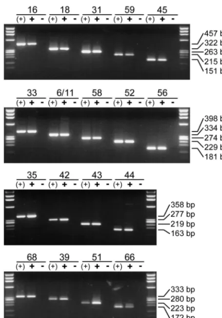
Specificity of nested PCR primers.The ability of all type-specific nested PCR primers to amplify genomic DNA from the corresponding HPV genotype was analyzed by utilizing 100 pg (107viral copies) of HPV DNA-containing plasmids. Am-plification of viral DNA from the relevant single plasmid [Fig. 3, lanes (⫹)] as well as from a plasmid cocktail containing DNA of all HPV genotypes addressed in the study (Fig. 3, lanesⴙ) generated sharp bands of the expected length for each single primer pair. Direct sequencing of amplification products
generated from the plasmid cocktails demonstrated the geno-type specificity of each single primer pair for the relevant HPV genotype.
HPV DNA-containing plasmids were again used to investi-gate the specificity of all the nested PCR primers used. This time, for each nested PCR primer pair to be analyzed, a plas-mid cocktail containing all (100 pg each; 107viral copies) but the relevant HPV DNA was produced. Amplification of these plasmid cocktails did not generate any PCR product for any type-specific primer pair used in the study (Fig. 3, lanes⫺).
[image:4.603.48.539.80.455.2]To facilitate the application of these type-specific nested
TABLE 2. Sequences of type-specific nested PCR primers used in this study
Primer cocktail HPV genotype Amplicon (bp) Sequence (5⬘-3⬘) Position (bp)
I 16 457 CAC AGT TAT GCA CAG AGC TGC 141–161
CAT ATA TTC ATG CAA TGT AGG TGT A 597–573
18 322 CAC TTC ACT GCA AGA CAT AGA 170–190
GTT GTG AAA TCG TCG TTT TTC A 491–470
31 263 GAA ATT GCA TGA ACT AAG CTC G 137–158
CAC ATA TAC CTT TGT TTG TCA A 399–378
59 215 CAA AGG GGA ACT GCA AGA AAG 159–179
TAT AAC AGC GTA TCA GCA GC 373–354
45 151 GTG GAA AAG TGC ATT ACA GG 82–101
ACC TCT GTG CGT TCC AAT GT 232–213
II 33 398 ACT ATA CAC AAC ATT GAA CTA 172–192
GTT TTT ACA CGT CAC AGT GCA 569–549
6/11 334 TGC AAG AAT GCA CTG ACC AC 201–220
TGC ATG TTG TCC AGC AGT GT 534–515
58 274 GTA AAG TGT GCT TAC GAT TGC 297–317
GTT GTT ACA GGT TAC ACT TGT 570–550
52 229 TAA GGC TGC AGT GTG TGC AG 178–197
CTA ATA GTT ATT TCA CTT AAT GGT 406–383
56 181 GTG TGC AGA GTA TGT TTA TTG 294–314
TTT CTG TCA CAA TGC AAT TGC 475–455
III 35 358 CAA CGA GGT AGA AGA AAG CAT C 157–178
CCG ACC TGT CCA CCG TCC ACC G 514–493
42 277 CCC AAA GTA GTG GTC CCA GTT A 85–106
GAT CTT TCG TAG TGT CGC AGT G 361–340
43 219 GCA TAA TGT CTG CAC GTA GCT G 102–123
CAT GAA ACT GTA GAC AGG CCA AG 320–298
44 163 TAA ACA GTT ATA TGT AGT GTA CCG 248–271
TAT CAG CAC GTC CAG AAT TGA C 410–389
IV 68 333 GCA GAA GGC AAC TAC AAC GG 4049–4068
GTT TAC TGG TCC AGC AGT GG 4381–4362
39 280 GAC GAC CAC TAC AGC AAA CC 213–232
TTA TGA AAT CTT CGT TTG CT 492–473
51 223 GAG TAT AGA CGT TAT AGC AGG 319–339
TTT CGT TAC GTT GTC GTG TAC G 541–520
66 172 TTC AGT GTA TGG GGC AAC AT 353–372
AAA CAT GAC CCG GTC CAT GC 520–501
FIG. 2. Amplification of HPV DNA-containing plasmids (100 pg of plasmid DNA; 107viral copies) with consensus primers GP-E6-3F and GP-E7-5B/6B. The PCR products generated with GP-E6/E7 primers from HPV-containing plasmids and from a clinical sample (HPV-51) range from 602 to 666 bp in size. HPV genotypes are indicated at the top. M, length marker⌽X 174 phage DNA, digested by HaeIII.
on May 15, 2020 by guest
http://jcm.asm.org/
PCR primers in a multiplex PCR assay, they were selected to amplify products of markedly different sizes. In total, four primer cocktails, each containing four to five type-specific primers, were used. The length of their amplification products ranged from 457 bp (HPV-16) down to 151 bp (HPV-45), thus allowing easy discrimination and identification of the underly-ing HPV type by simple gel electrophoresis (see Fig. 5, lanes on the right). This is demonstrated for NMPCR cocktails I to IV, containing primers specific for HPV-16, -18, -31, -59, and -45 (cocktail I); HPV-33, -6/11, -58, -52, and -56 (cocktail II); HPV-35, -42, -43, and -44 (cocktail III); and HPV-68, -39, -51, and -66 (cocktail IV). The amplification products varied in size by steps of about 50 to 60 bp (Fig. 1; also see Fig. 5).
Sensitivity of the E6/E7 NMPCR assay.To determine the sensitivity of the novel NMPCR, 10-fold dilutions of all plas-mids containing the genomic DNA of the HPV genotypes addressed in the study were tested. The dilution series started with 10 ng (109viral target copies) and ended with 10 ag (1 viral target copy; 10 dilution steps). The sensitivity of the assay was compared with that of a nested PCR assay using
MY09-MY11 and GP5⫹-GP6⫹ primers (MY/GP). The two assays showed equal sensitivity for HPV-16, -18, -33, -43, and -59 (100 to 1 fg; 104to 102viral copies). The MY/GP nested PCR assay was about 10 times more sensitive for HPV-6 and -45 (1 fg; 102 viral copies). For all other HPV genotypes the NMPCR assay was 10 to 100 times more sensitive (1 fg; 102 viral copies). Plasmid DNA of HPV-51 could not be amplified with the NMPCR assay. Equally, plasmid DNA of HPV-68 could not be amplified with the MY/GP assay. These failures are most prob-ably due to the fact that the relevant primer target regions were not present in their entirety in the plasmid amplified. The sensitivities indicated above were those that could reproducibly be achieved. The results for HPV-6, -35, -52, and -66 are shown in Fig. 4.
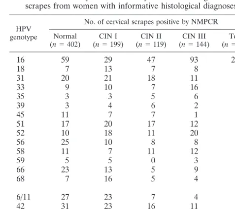
Evaluation of the novel E6/E7 NMPCR assay with clinical samples.The clinical performance of the novel NMPCR assay was tested by the investigation of 864 cervical scrapes from women for whom histologic diagnoses were available. No his-tologic signs of cervical dysplasia (CIN 0) were found in 402 women, 199 had mild dysplasia (CIN I), 119 had moderate dysplasia (CIN II), and 144 had severe dysplasia (CIN III). HPV infection was detected in 63.7% of the women (550 of 864), being due to a single genotype in 54.9% of them (302 of 550) and multiple genotypes in 45.1% of them (248 of 550). Two, three, four, and five different genotypes were detected in 27.8% (153 of 550), 12.0% (66 of 550), 4.4% (24 of 550), and 0.9% (5 of 550) of the HPV-infected women, respectively. All 18 different HPV genotypes investigated were specifically de-tected by NMPCR in multiple subjects. Table 3 shows the absolute frequencies of these 18 HPVs according to the histo-logic diagnosis. HPV-16 was detected in 41.5% of HPV-posi-tive subjects (228 of 550) and was by far the most widespread genotype. The highest relative frequency of HPV-16 was found in the presence of severe dysplasia, being almost 65% (93 of 144) in such subjects. By contrast, it was detected in only 15% (88 of 601) of women with mild (n⫽199) or no dysplasia (n⫽
402).
[image:5.603.49.277.67.393.2]DNA extracted from the cervical scrapes of these 864 women was also investigated by conventional PCR with MY09-MY11 primers, conventional PCR with GP5⫹-GP6⫹primers, and a nested PCR assay using MY09-MY11 as outer and GP5⫹-GP6⫹as inner primers (MY/GP). By these four meth-ods HPV DNA was detected in 49.7% (429 of 864; MY09-MY11), 49.9% (431 of 864; GP5⫹-GP6⫹), 62.6% (541 of 864; MY/GP), and 63.7% (550 of 864; NMPCR) of subjects. A comparison of PCR data with the morphological findings for each of the four methods demonstrated an increase in appar-ent prevalence of HPV infection with the grade of the cervical lesions. The two nested PCR assays (MY/GP and NMPCR) gave almost equal values for prevalence rates in the four mor-phological subgroups (CIN 0, CIN I, CIN II, and CIN III) and, as expected, much higher prevalence rates than detected by the two single-round PCR assays (MY09-MY11 and GP5⫹ -GP6⫹). The results are given in detail in Table 4. With regard to analytical sensitivity, the gold standard (as defined in Ma-terials and Methods) was represented by the combination of the four PCR assays. Table 4 gives the results of the sensitiv-ities (relative to the gold standard) of the four methods. The sensitivity of the NMPCR assay was significantly higher than that of the MY09-MY11 and GP5⫹-GP6⫹assays for all four
FIG. 3. Type-specific identification of HPV DNA. Eighteen primer pairs for type-specific HPV detection by nested PCR were arranged in four multiplex cocktails. The ability of each primer pair to detect HPV DNA is demonstrated by amplification of the corresponding HPV type from a single plasmid [lanes (⫹)] and from a cocktail containing all 18 HPV genotypes addressed in the study (lanes⫹). The specificity of each primer pair was demonstrated by the absence of unspecific am-plification products when the HPV plasmid or HPV DNA belonging to the primer tested was missing from the cocktail (lanes ⫺). In all experiments 100 pg of plasmid DNA (107viral copies) was amplified.
on May 15, 2020 by guest
http://jcm.asm.org/
groups (i.e., CIN 0, CIN I, CIN II, and CIN III;P⬍0.0002 for all comparisons). By contrast, no significant difference in sen-sitivity between MY/GP and NMPCR was observed for these groups. Since the sensitivities of each method differ signifi-cantly among the four morphological subgroups it is not ap-propriate to combine the estimates for these four groups. The values obtained for HPV prevalence and the sensitivities of the four methods, together with their 95% CI, for each of the four morphological subgroups are listed in Table 5. The extent of agreement between the four diagnostic tests and the corre-sponding kappa values are shown in Table 6. Of 69 samples with discordant results between the MY/GP and NMPCR as-says, 39 were positive by the NMPCR assay only and 30 were positive by the MY/GP assay only. Interestingly, only 10 of these women had moderate or severe dysplasia. Thus, the concordance of the two assays in patients with so-called high-grade dysplastic lesions is 96% (253 of 263), with a kappa value of 0.81 (standard error, 0.11). Direct sequencing of NMPCR-negative and MY/GP-positive samples revealed HPV-11, -74, and -84 (one sample); HPV-54, -67, and -70 (two samples); HPV-61 (three samples); HPV-53 and -73 (four samples); and HPV-62 (five samples). In five cases sequencing results were inconclusive because of multiple infections. Apart from these five cases, the NMPCR assay missed only 1 HPV (HPV-11)-positive sample targeted by its specific nested PCR primers. The 39 NMPCR-positive and MY/GP-negative samples com-prised 34 samples with single infections with HPV-6/11 (n⫽3), -16 (n⫽7), -18 (n⫽2), -33 (n⫽1), -35 (n⫽2), -39 (n⫽1), -42 (n⫽4), -43 (n⫽2), -44 (n⫽1), -51 (n⫽2), -52 (n⫽3), -56 (n⫽2), -58 (n ⫽1), -59 (n⫽ 1), and -66 (n⫽ 2). The remaining five samples showed evidence of double and triple infections involving HPV-6/11, -16, -31, -42, -56, -59, and -68. With NMPCR, multiple HPV infections were found in 40.0% (72 of 180) of the HPV-positive women without cervical dysplasia, 54.5% (67 of 123) of the women with CIN I lesions, 46.7% (50 of 107) of the women with CIN II lesions, and 42.1%
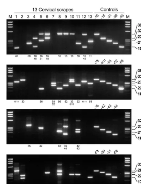
[image:6.603.128.457.70.265.2](59 of 140) of the women with CIN III lesions (Table 4). In these four groups, infections with at least one high-risk HPV genotype were detected by NMPCR assay in 83.3% (150 of 180; CIN 0), 88.6% (10 of 123; CIN I), 96.3% (103 of 107), and 99.3% (139 of 140; CIN III) of samples (Table 4). Figure 5 shows the results of HPV detection and typing by the novel NMPCR assay in 13 samples investigated in the study. Only samples with multiple HPV genotypes were chosen. Not only the specific detection of the different HPV genotypes (Fig. 5, right), but also the ability of the assay to detect infections with
FIG. 4. Sensitivity of MY09-MY11 and GP5⫹-GP6⫹nested PCR (MY/GP) and NMPCR. A 10-fold dilution series of HPV DNA-containing plasmids of genotypes 6, 35, 52, and 66 was amplified by both methods. Lanes 1 to 7 represent PCR products of 10 pg to 10 ag (106to 1 viral copy). MY09-MY11 and GP5⫹-GP6⫹nested PCR is 10 times more sensitive for HPV-6, while the NMPCR method is 10 times more sensitive for HPV-35 and HPV-66 and nearly 100 times more sensitive for HPV-52. M, length marker⌽X 174 phage DNA, digested by HaeIII.
TABLE 3. Absolute frequencies of the 18 different HPV genotypes addressed in the study and detected by NMPCR among 864 cervical
scrapes from women with informative histological diagnoses
HPV genotype
No. of cervical scrapes positive by NMPCR Normal
(n⫽402) (nCIN I⫽199) (nCIN II⫽119) (nCIN III⫽144) (nTotal⫽864)
16 59 29 47 93 228
18 7 13 7 8 35
31 20 21 18 11 70
33 9 10 7 16 42
35 3 3 5 6 17
39 3 4 6 2 15
45 11 7 7 1 26
51 17 20 17 12 66
52 10 18 11 20 59
56 25 10 8 8 51
58 11 7 11 12 41
59 5 5 0 3 13
66 23 13 5 9 50
68 7 16 5 4 32
6/11 27 23 7 4 61
42 31 23 16 11 81
43 8 7 5 4 24
44 7 4 3 3 17
on May 15, 2020 by guest
http://jcm.asm.org/
[image:6.603.301.540.511.724.2]multiple HPV genotypes within each multiplex primer cocktail is demonstrated (Fig. 5, lanes 2 and 4 to 12).
DISCUSSION
Today, the diagnosis of HPV infections is based on the detection of viral DNA. Highly conserved regions in different parts of the viral genome have enabled the development of general or consensus PCR primer sets, such as MY09-MY11, PGMY09-MY11, GP5⫹-GP6⫹, SPF10, and LCR-E7 (12, 16, 20, 29), which allow the detection of a broad spectrum of different HPV genotypes. However, differences in malignant potential mean that it is particularly important to accurately identify infections with the high-risk HPV genotypes. After amplification with general or consensus primers, additional techniques are necessary to identify the underlying HPV ge-notype. These include restriction enzyme digestion (6, 29), dot blot or Southern blot hybridization (25), direct sequencing (30), and analysis by enzyme immunoassays (17) or line probe assays (13).
We have developed a novel fully PCR-based approach for the sensitive and type-specific detection of HPV infections based on the amplification of the viral E6/E7 oncogenes (Sot-lar et al., 17th Int. Papillomavirus Conf. 1999). Degenerate PCR primers hybridizing to short consensus regions within the E6 and E7 oncogenes were demonstrated to amplify the DNA of all high-risk and some of the most prevalent low-risk HPV genotypes. In addition, sequence alignments with the corre-sponding E6/E7 regions of a broad spectrum of other (low-risk) mucosal HPV genotypes indicates that the E6/E7 consen-sus primers would also be able to amplify DNA of these types with comparable efficiency. A similar approach has recently been described by Sasagawa and colleagues (29). Their LCR-E7 primers are located in consensus regions close to those targeted by the GP-E6/E7 primers used in our study. To allow
exact genotyping, not only in single, but also in multiple HPV infections, a total of 18 pairs of type-specific internal nested PCR primers were used in multiplex cocktails. This approach reduced the number of nested PCRs necessary for the ampli-fication of the 18 different genotypes addressed in the study down to four. Gel electrophoresis and ethidium bromide stain-ing then allowed specific identification of the HPV type or types present in a sample by simple determination of the am-plicon length.
The sensitivity of the novel NMPCR assay was determined by amplification of HPV DNA-containing plasmid dilutions. The NMPCR assay was found to be about as sensitive as nested PCR with MY09-MY11 and GP5⫹-GP6⫹primers and much more sensitive than conventional PCR with either of these two primer sets.
The exact identification of each single HPV type may be necessary, for example, in the follow-up of HPV-infected women or in detailed epidemiological studies. Each nested PCR primer pair in our assay was successfully tested on two HPV multiplasmid cocktails. The cocktail with plasmids cor-responding to all 18 HPV genotypes investigated could specif-ically be amplified with each primer pair. By contrast, not a single (unspecific) amplification product resulted when the HPV plasmid belonging to the primer tested was absent from the cocktail.
[image:7.603.43.550.90.179.2]Because of limited concordance between the histologic and cytologic findings, which is a well-known phenomenon (18, 21) but was not the topic of this study, we confined the evaluation of the clinical value of the NMPCR assay to the investigation of cervical scrapes from women in whom cervical biopsies had also been performed and histologic diagnoses (considered the gold standard) were available (n⫽864). In these subjects the NMPCR assay had a significantly (P ⬍0.0002) higher sensi-tivity in both women with and those without cervical dysplasia compared to single PCRs with MY09-MY11 or GP5⫹-GP6⫹
TABLE 4. HPV detection rates, sensitivities of the four assays relative to the gold standard, and distribution of high- and low-risk infections in 864 cases with available histologic diagnoses
Morphology Total HPV-positiveNo. of results (%)a
No. of HPV-positive results
(% sensitivity) genotypes (%)No. of HPV Ratio of high-riskgenotypes to low-risk genotypes MY09-MY11 GP5⫹-GP6⫹ MY/GP NMPCR Multiple High risk
CIN 0 402 196 (48.8) 114 (58.2) 117 (59.7) 174 (88.8) 180 (91.8) 72 (40.0) 150 (83.3) 5:1 CIN I 199 133 (66.8) 100 (75.2) 95 (71.4) 121 (91.0) 123 (92.4) 67 (54.5) 109 (88.6) 8:1 CIN II 119 111 (93.3) 88 (79.3) 91 (82.0) 107 (96.4) 107 (96.4) 50 (46.7) 103 (96.3) 26:1 CIN III 144 141 (97.9) 126 (89.4) 127 (90.1) 139 (98.6) 140 (99.3) 59 (42.1) 139 (99.3) 139:1 Total 864 581 (67.2) 428 (73.7) 430 (74.0) 541 (93.1) 550 (94.7) 248 (45.1) 501 (91.1) 10:1 aPercent relative to gold standard (either MY/GP or NMPCR positive).
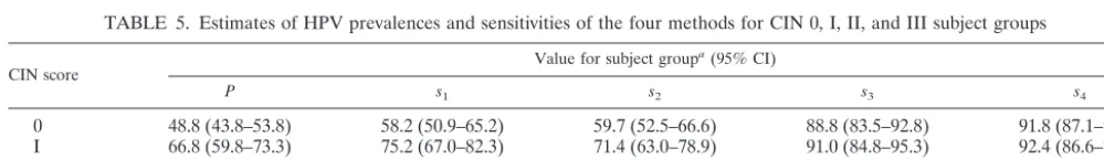
TABLE 5. Estimates of HPV prevalences and sensitivities of the four methods for CIN 0, I, II, and III subject groups
CIN score Value for subject group
a(95% CI)
P s1 s2 s3 s4
0 48.8 (43.8–53.8) 58.2 (50.9–65.2) 59.7 (52.5–66.6) 88.8 (83.5–92.8) 91.8 (87.1–95.3) I 66.8 (59.8–73.3) 75.2 (67.0–82.3) 71.4 (63.0–78.9) 91.0 (84.8–95.3) 92.4 (86.6–96.3) II 93.3 (87.2–97.1) 79.3 (70.5–86.4) 82.0 (73.6–88.6) 96.4 (91.0–99.0) 96.4 (91.0–99.0) III 97.9 (94.0–99.6) 89.4 (83.1–93.9) 90.1 (83.9–94.5) 98.6 (95.0–99.8) 99.3 (96.1–100) aAbbreviations:p, estimated HPV prevalence;s
1, sensitivity method 1 (MY09-MY11);s2, sensitivity method 2 (GP5⫹-GP6⫹);s3, sensitivity method 3 (MY/GP
nested PCR);s4, sensitivity method 4 (NMPCR).
on May 15, 2020 by guest
http://jcm.asm.org/
[image:7.603.41.541.636.708.2]primers. When the latter primers were applied in a nested PCR assay (MY/GP), the sensitivity was very similar to that of the NMPCR assay. In cases with high-grade cervical dysplasia (CIN II and III), the MY/GP and NMPCR assays showed sensitivities and a concordance rate of about 98% percent each. With regard to the clinical sensitivity of the MY09-MY11 and GP5⫹-GP6⫹assays, it has to be stated that the results obtained in this study may not reflect their performance in previously published studies in which hybridization steps were applied rather than, as in this study, simple gel analysis, which is known to be less sensitive. Nevertheless, the HPV preva-lence rates detected in the various disease categories with the NMPCR assay (by gel analysis) are close to the values obtained by the SPF10 line probe assay method (19) and are consider-ably higher than HPV detection rates reported in other studies (2, 16, 30).
All 18 HPV genotypes addressed in the present study were detected in multiple samples among the cervical scrapes inves-tigated. As in other studies, HPV-16 was by far the most prevalent genotype in all of the morphological subgroups. The highest relative frequency of HPV-16 was found in the pres-ence of severe dysplasia. The relative frequency of other HPV genotypes is more difficult to compare between studies because the primer sets used usually have differing sensitivities for the various HPV genotypes (24). The ratio of high- to low-risk HPV genotypes detected by the NMPCR assay increased as a function of morphological abnormality. Of women without morphological evidence of dysplasia, 83.3% were infected with high-risk HPV genotypes while 16.7% exhibited low-risk types only (ratio, 5 to 1). In women with cervical dysplasia the ratio increased to 18 to 1 (94.9% high-risk HPV genotypes). Al-though not reaching the compelling ratio of 32.2 to 1 (97.0% high-risk HPV genotypes) reported by Kleter et al. (20), these figures confirm the ability of the NMPCR assay to detect those HPV genotypes characteristically associated with cervical dys-plasia and carcinoma.
Despite the fact that besides all 14 known high-risk HPV genotypes, the NMPCR assay in its current form is able to
detect only five low-risk HPVs, the rate of multiple infections detected by the NMPCR assay was higher than that reported by other groups (12, 19, 33). However, as in other studies, the highest prevalence of multiple HPV genotypes was detected in women with mild dysplasia (54.5%).
[image:8.603.43.284.88.262.2]In conclusion, we report the development of a novel, fully PCR-based assay that combines the advantages of consensus and type-specific primers for the sensitive detection and spe-cific identification of 18 different HPV genotypes, including all high-risk genotypes. The spectrum of this assay may easily be extended by addition of new multiplex primer cocktails for supplementary HPV genotypes. By utilizing the E6 and E7 regions as primer binding sites, the presence of the viral on-cogenes is also documented. In addition, many primers em-ployed in this assay can also be used for the amplification of oncogene transcripts by reverse transcription-PCR, thus pos-sibly detecting an additional risk factor for the development or progression of a cervical lesion (31). With regard to sensitivity and performance with clinical samples, the novel NMPCR assay is a potentially useful tool for HPV DNA detection in
[image:8.603.306.539.309.613.2]FIG. 5. HPV detection and typing by GP-E6/E7 NMPCR. The results of 13 samples with multiple infections are shown (lanes 1 to 13). The individual lanes in each of the four gels represent amplification products from the same sample. The genotypes identified per primer cocktail are indicated below each gel. Not only the specific detection of the various HPV genotypes as determined by the amplification of control DNA (lanes 4 and 5 on the right), but also the ability of the assay to detect infections with multiple HPV genotypes within each multiplex primer cocktail is demonstrated (lanes 2 and 4 to 12). One hundred femtograms of HPV DNA-containing plasmids was amplified under the same conditions and served as positive controls. M, length marker⌽X 174 phage DNA, digested by HaeIII.
TABLE 6. Kappa coefficients for all four HPV detection methods for CIN 0, I, II, and III
Methods
compareda Morphol-ogy
No. of specimens with result by:
(SE) Both tests Only NMPCR
Positive Negative Positive Negative
1,4 CIN 0 108 216 72 6 0.59 (0.04) CIN I 93 69 30 7 0.63 (0.05) CIN II 86 10 21 2 0.37 (0.10) CIN III 126 4 14 0 0.33 (0.13)
2,4 CIN 0 113 218 67 4 0.63 (0.04) CIN I 90 71 33 5 0.62 (0.05) CIN II 90 11 17 1 0.48 (0.10) CIN III 127 4 13 0 0.35 (0.13)
3,4 CIN 0 158 206 22 16 0.81 (0.03) CIN I 111 66 12 10 0.77 (0.05) CIN II 103 8 4 4 0.63 (0.12) CIN III 138 3 2 1 0.66 (0.19) aSubscripts refer to method numbers as follows: 1, MY09-MY11; 2, GP5⫹ -GP6⫹; 3, MY/GP nested PCR; 4, NMPCR.
on May 15, 2020 by guest
http://jcm.asm.org/
epidemiologic and clinical follow-up studies, especially when exact HPV typing and the detection of multiple HPV infec-tions are required.
ACKNOWLEDGMENTS
This work was supported in part by a grant to K.S. and M.M. from the Deutsche Krebshilfe (70-2623-So I).
We thank Barbara Mankel, Sema Colak and Perikles Kosmidis for excellent technical assistance.
REFERENCES
1. Baay, M. F., W. G. Quint, J. Koudstaal, H. Hollema, J. M. Duk, M. P. Burger, E. Stolz, and P. Herbrink.1996. Comprehensive study of several general and type-specific primer pairs for detection of human papillomavirus DNA by PCR in paraffin-embedded cervical carcinomas. J. Clin. Microbiol. 34:745–747.
2. Bollen, L. J., A. H. Tjong, J. van der Velden, K. Brouwer, B. W. Mol, F. J. ten Kate, and J. ter Schegget.1997. Hum. papillomavirus deoxyribonucleic acid detection in mildly or moderately dysplastic smears: a possible method for selecting patients for colposcopy. Am. J. Obstet. Gynecol.177:548–553. 3. Bosch, F. X., M. M. Manos, N. Munoz, M. Sherman, A. M. Jansen, J. Peto,
M. H. Schiffman, V. Moreno, R. Kurman, K. V. Shah, et al.1995. Prevalence of human papillomavirus in cervical cancer: a worldwide perspective. J. Natl. Cancer Inst.87:796–802.
4. Burger, M. P., H. Hollema, W. J. Pieters, and W. G. Quint.1995. Predictive value of human papillomavirus type for histological diagnosis of women with cervical cytological abnormalities. BMJ310:94–95.
5. Chan, S. Y., H. Delius, A. L. Halpern, and H. U. Bernard.1995. Analysis of genomic sequences of 95 papillomavirus types: uniting typing, phylogeny, and taxonomy. J. Virol.69:3074–3083.
6. Contorni, M., and P. Leoncini.1993. Typing of human papillomavirus DNAs by restriction endonuclease mapping of the PCR products. J. Virol. Methods 41:29–36.
7. Cox, J. T., A. T. Lorincz, M. H. Schiffman, M. E. Sherman, A. Cullen, and R. J. Kurman.1995. Human papillomavirus testing by hybrid capture ap-pears to be useful in triaging women with a cytologic diagnosis of atypical squamous cells of undetermined significance. Am. J. Obstet. Gynecol.172: 946–954.
8. Cuzick, J., P. Sasieni, and A. Singer.1996. Risk factors for invasive cervix cancer in young women. Eur. J. Cancer32:836–841.
9. Cuzick, J., A. Szarewski, G. Terry, L. Ho, A. Hanby, P. Maddox, M. Ander-son, G. Kocjan, S. T. Steele, and J. Guillebaud.1995. Hum. papillomavirus testing in primary cervical screening. Lancet345:1533–1536.
10. de-Roda, H. A., J. M. Walboomers, B. A. van-den, C. J. Meijer, and P. J. Snijders.1995. The use of general primers GP5 and GP6 elongated at their 3⬘ends with adjacent highly conserved sequences improves human papillo-mavirus detection by PCR. J. Gen. Virol.76:1057–1062.
11. Evander, M., K. Edlund, E. Boden, A. Gustafsson, M. Jonsson, R. Karlsson, E. Rylander, and G. Wadell.1992. Comparison of a one-step and a two-step polymerase chain reaction with degenerate general primers in a population-based study of human papillomavirus infection in young Swedish women. J. Clin. Microbiol.30:987–992.
12. Gravitt, P. E., C. L. Peyton, T. Q. Alessi, C. M. Wheeler, F. Coutlee, A. Hildesheim, M. H. Schiffman, D. R. Scott, and R. J. Apple.2000. Improved amplification of genital human papillomaviruses. J. Clin. Microbiol.38:357– 361.
13. Gravitt, P. E., C. L. Peyton, R. J. Apple, and C. M. Wheeler.1998. Geno-typing of 27 human papillomavirus types by using L1 consensus PCR prod-ucts by a single-hybridization, reverse line blot detection method. J. Clin. Microbiol.36:3020–3027.
14. Hildesheim, A., M. H. Schiffman, P. E. Gravitt, A. G. Glass, C. E. Greer, T. Zhang, D. R. Scott, B. B. Rush, P. Lawler, M. E. Sherman, et al.1994. Persistence of type-specific human papillomavirus infection among cytolog-ically normal women. J. Infect. Dis.169:235–240.
15. Husnjak, K., M. Grce, L. Magdic, and K. Pavelic.2000. Comparison of five different polymerase chain reaction methods for detection of human papil-lomavirus in cervical cell specimens. J. Virol. Methods88:125–134. 16. Jacobs, M. V., A. M. de Roda Husman, A. J. van den Brule, P. J. Snijders,
C. J. Meijer, and J. M. Walboomers.1995. Group-specific differentiation between high- and low-risk human papillomavirus genotypes by general primer-mediated PCR and two cocktails of oligonucleotide probes. J. Clin. Microbiol.33:901–905.
17. Jacobs, M. V., P. J. Snijders, A. J. van den Brule, T. J. Helmerhorst, C. J. Meijer, and J. M. Walboomers.1997. A general primer GP5⫹/GP6(⫹ )-mediated PCR-enzyme immunoassay method for rapid detection of 14 high-risk and 6 low-high-risk human papillomavirus genotypes in cervical scrapings. J. Clin. Microbiol.35:791–795.
18. Jones, B. A., and D. A. Novis.1996. Cervical biopsy-cytology correlation. A College of American Pathologists Q-Probes study of 22 439 correlations in 348 laboratories. Arch. Pathol. Lab Med.120:523–531.
19. Kleter, B., L. J. van Doorn, L. Schrauwen, A. Molijn, S. Sastrowijoto, J. ter Schegget, J. Lindeman, B. ter Harmsel, M. Burger, and W. Quint.1999. Development and clinical evaluation of a highly sensitive PCR-reverse hy-bridization line probe assay for detection and identification of anogenital human papillomavirus. J. Clin. Microbiol.37:2508–2517.
20. Kleter, B., L. J. van Doorn, J. ter Schegget, L. Schrauwen, K. van Krimpen, M. Burger, B. ter Harmsel, and W. Quint.1998. Novel short-fragment PCR assay for highly sensitive broad-spectrum detection of anogenital human papillomaviruses. Am. J. Pathol.153:1731–1739.
21. Lonky, N. M., M. Sadeghi, G. W. Tsadik, and D. Petitti.1999. The clinical significance of the poor correlation of cervical dysplasia and cervical malig-nancy with referral cytologic results. Am. J. Obstet. Gynecol.181:560–566. 22. Lorincz, A. T., R. Reid, A. B. Jenson, M. D. Greenberg, W. Lancaster, and
R. J. Kurman.1992. Hum. papillomavirus infection of the cervix: relative risk associations of 15 common anogenital types. Obstet. Gynecol.79:328–337. 23. Manos, M. M., Y. Ting, D. K. Wright, A. J. Lewis, T. R. Broker, and S. M.
Wolinski.1989. Use of polymerase chain reaction amplification for the detection of genital human papillomaviruses. Cancer Cells7:209–213. 24. Qu, W., G. Jiang, Y. Cruz, C. J. Chang, G. Y. Ho, R. S. Klein, and R. D. Burk.
1997. PCR detection of human papillomavirus: comparison between MY09/ MY11 and GP5⫹/GP6⫹primer systems. J. Clin. Microbiol.35:1304–1310. 25. Resnick, R. M., M. T. Cornelissen, D. K. Wright, G. H. Eichinger, H. S. Fox,
J. ter Schegget, and M. M. Manos.1990. Detection and typing of human papillomavirus in archival cervical cancer specimens by DNA amplification with consensus primers. J. Natl. Cancer Inst.82:1477–1484.
26. Richart, R. M.1995. Screening. The next century. Cancer76:1919–1927. 27. Rozendaal, L., J. M. Walboomers, J. C. van der Linden, F. J. Voorhorst, P.
Kenemans, T. J. Helmerhorst, M. van Ballegooijen, and C. J. Meijer.1996. PCR-based high-risk HPV test in cervical cancer screening gives objective risk assessment of women with cytomorphologically normal cervical smears. Int. J. Cancer68:766–769.
28. Saiki, R. K., T. L. Bugawan, G. T. Horn, K. B. Mullis, and H. A. Erlich.1986. Analysis of enzymatically amplified beta-globin and HLA-DQ alpha DNA with allele-specific oligonucleotide probes. Nature324:163–166.
29. Sasagawa, T., Y. Minemoto, W. Basha, H. Yamazaki, M. Nakamura, H. Yoshimoto, J. Sakaike, and M. Inoue.2000. A new PCR-based assay ampli-fies the E6–E7 genes of most mucosal human papillomaviruses (HPV). Virus Res.67:127–139.
30. Smits, H. L., L. M. Tieben, A. H. Tjong, M. F. Jebbink, R. P. Minnaar, C. L. Jansen, and J. ter Schegget.1992. Detection and typing of human papillo-maviruses present in fixed and stained archival cervical smears by a consen-sus polymerase chain reaction and direct sequence analysis allow the iden-tification of a broad spectrum of human papillomavirus types. J. Gen. Virol. 73:3263–3268.
31. Sotlar, K., H. C. Selinka, M. Menton, R. Kandolf, and B. Bultmann.1998. Detection of human papillomavirus type 16 E6/E7 oncogene transcripts in dysplastic and nondysplastic cervical scrapes by nested RT-PCR. Gynecol. Oncol.69:114–121.
32. van den Brule, A. J., E. C. Claas, M. du Maine, W. J. Melchers, T. Helmer-horst, W. G. Quint, J. Lindeman, C. J. Meijer, and J. M. Walboomers.1989. Use of anticontamination primers in the polymerase chain reaction for the detection of human papilloma virus genotypes in cervical scrapes and biop-sies. J. Med. Virol.29:20–27.
33. van Doorn, L.-J., W. Quint, B. Kleter, A. Molijn, B. Colau, M.-T. Martin, I. Kravang, N. Torrez-Martinez, C. L. Peyton, and C. M. Wheeler.2002. Geno-typing of human papillomavirus in liquid cytology cervical specimens by the PGMY line blot assay and the SPF10line probe assay. J. Clin. Microbiol.
40:979–983.
34. zur-Hausen, H.1996. Papillomavirus infections—a major cause of human cancers. Biochim. Biophys. Acta1288:F55–F78.