Copyright © 2002, American Society for Microbiology. All Rights Reserved.
New Simple and Rapid Test for Culture Confirmation of
Mycobacterium tuberculosis
Complex: a Multicenter Study
Naoki Hasegawa,
1* Takao Miura,
2Koudou Ishii,
2Kazuhiro Yamaguchi,
1Thomas H. Lindner,
3Samuel Merritt,
4Janis D. Matthews,
4and Salman H. Siddiqi
5Cardiopulmonary Division, Department of Internal Medicine, Keio University, School of Medicine, Tokyo,1and Department of
Internal Medicine, Minami-Yokohama National Hospital, Yokohama,2Japan; Tuberculosis Laboratory, Missouri State Chest
Hospital, Mount Vernon, Missouri3; North Carolina State Laboratory of Public Health, Raleigh, North Carolina4;
and Becton Dickinson Diagnostic Systems, Sparks, Maryland5
Received 19 September 2001/Returned for modification 3 November 2001/Accepted 19 December 2001
Mycobacterial antigen MPB64 has been identified as aMycobacterium tuberculoisiscomplex-specific secretory protein since 1984. Recently, a simple culture confirmation test forM. tuberculosiscomplex has been developed by using lateral flow immunochromatographic assay (ICA) to detect MPB64 with anti-MPB64 monoclonal antibody. The current multicenter study evaluated the performance of an ICA slide test for MPB64 antigen in the clinical setting. Primary positive cultures from clinical specimens, as well as stock cultures, were tested. Approximately 100l of positive liquid culture medium or suspension made from colonies on solid medium was placed into the test well of the plastic slide devise, and the test was read after 15 min. No processing or instrumentation was required. A total of 304 mycobacterial isolates consisting ofM. tuberculosiscomplex (171 isolates) and mycobacteria other thanM. tuberculosis(MOTT) complex (133 isolates) belonging to 18 different species were tested. Growth in liquid media (Mycobacteria Growth Indicator Tube [MGIT] and Radiometric 12B), as well as in solid (Löwenstein-Jensen and Middlebrook 7H10 agar) media, was evaluated. Results were compared with those obtained with nucleic acid-based and/or high-pressure liquid chromatography identifi-cation. All MOTT were found to be negative on the ICA slide with no cross-reaction. AllM. tuberculosisandM. africanumcultures were found to be positive, whereas the results ofM. bovisandM. bovisBCG cultures were variable since some of the BCG strains are known to lack MPB64 antigen production. The results did not change with prolonged storage of cultures. This low-tech rapid test with high sensitivity and specificity could provide an alternative to currently available identification methods, particularly for recently introduced nonradiometric liquid culture systems such as MGIT.
Tuberculosis is a significant reemerging infectious disease in many parts of the world, which is of great concern. Prompt detection, isolation, identification and susceptibility testing of
Mycobacterium tuberculosisfrom clinical specimens is essential for appropriate management of patients with tuberculosis. The Centers for Disease Control and Prevention has recommended the use of liquid medium for primary culture and susceptibility testing to achieve better as well as faster results (17).
Even though liquid medium allows less time for the detec-tion of positive cultures, it is crucial for identifying isolated acid-fast cultures quickly as well as accurately. The
differenti-ation ofM. tuberculosisfrom mycobacteria other thanM.
tu-berculosis(MOTT) is most important from a clinical point of view, since tuberculosis is an infectious disease. Management and treatment of a patient with tuberculosis is different from that of patients infected with other mycobacteria. The radio-metric BACTEC 460 TB system (Becton Dickinson Diagnostic
Systems, Sparks, Md.) offers a NAP (p-nitro-␣-acetylamino-
-hydroxypropiophenone) test, which discriminates theM.
tuber-culosis complex from MOTT (16). The NAP test, however, requires ca. 4 to 6 days to report results. Recently,
nonradio-metric liquid culture media such as Mycobacteria Growth Indicator Tube (MGIT; Becton Dickinson) have been intro-duced and extensively evaluated (1, 3, 13). There is no rapid
and simple test available to differentiate the M. tuberculosis
complex from MOTT in these new media. Other technologies, such as the use of molecular probes, gas-liquid chromatogra-phy, and high-performance liquid chromatograchromatogra-phy, are avail-able, but these tests are technically complex, cumbersome, and expensive, especially the ones that are based on molecular analyses (5, 7). In countries with limited resources such tests are not practical. Therefore, a rapid and simple test is greatly needed, one that is as sensitive and as specific as the probe-based tests.
Several secreted mycobacterial proteins have been reported that showed some promise in diagnostic use (4, 9, 19). Among these secreted proteins, MPB64, which was first described as
MPT64, has been found in unheated culture media of M.
tuberculosis, M. bovis, and some but not all substrains of M. bovisBCG. This is a highly specific protein for theM. tuber-culosiscomplex and has shown potential for diagnostic use (4, 10, 14, 15). The MPB64 gene has been well characterized, and the antigen has been widely studied (12, 20). This immuno-genic protein elicits delayed-type hypersensitivity reactions in sensitized guinea pigs and lymphoproliferative responses in patients with tuberculosis (4, 8, 14). Studies with MPB64 an-tigen showed promise as a skin test anan-tigen to differentiate
* Corresponding author. Mailing address: Cardiopulmonary Divi-sion, Department of Medicine, Keio University School of Medicine, 35 Shinanomachi, Shinjuku-ku, Tokyo 160-8582, Japan. Phone: 81-3-3353-1211, ext. 62310. Fax: 81-3-3353-2502. E-mail: [email protected] .keio.ac.jp.
908
on May 15, 2020 by guest
http://jcm.asm.org/
patients with active disease from BCG-vaccinated people (10, 11).
Recently, MPB64 has been studied to differentiate the M.
tuberculosiscomplex from MOTT in isolated cultures (2, 18). Utilizing specificity of MPB64 Tomiyana et al. recently devel-oped a simple immunochromatographic assay (ICA) with anti-MPB64 monoclonal antibody (18). Abe et al. demonstrated that the MPB64-ICA on a slide format could be easily used for
rapid identification of theM. tuberculosiscomplex as a liquid
culture confirmation test by using the MGIT and MB-REDOX (2). In the present multicenter study, the clinical usefulness of MPB64-ICA test and its different parameters were evaluated by using fresh clinical isolates and stock cultures of different mycobacterial species grown in several liquid and solid media.
MATERIALS AND METHODS
The study was conducted at four different sites: the Clinical Microbiology Laboratory of the National Minami Yokohama Hospital, Yokohama City, Ka-nagawa Prefect, Japan (site 1); the Missouri State Tuberculosis Laboratory, Mount Vernon, Mo. (site 2); the North Carolina State Lab of Public Health, Raleigh, N.C. (site 3); and the Research and Development Department of Becton Dickinson, Sparks, Md. (site 4).
Clinical samples were mostly sputa but also included extrapulmonary speci-mens (i.e., 15 bronchial-washing, 8 pleural-effusion, 15 gastric-lavage, 3 urine, 8 stool, 3 pus, and 1 knee joint fluid specimens) and were processed according to the routineN-acetyl-L-cysteine-NaOH (NALC-NaOH) method for specimens suspected to have contaminating bacteria (6). The digested, decontaminated, and concentrated specimens were inoculated into BACTEC MGIT 960 culture tubes containing 7 ml of Middlebrook 7H9 broth base supplemented with en-richment and PANTA (Becton Dickinson). The tubes were placed in the BACTEC MGIT 960 instrument (Becton Dickinson), where they were incubated and monitored continuously for fluorescence. The time to detection of positive growth in the MGIT was defined as the interval between the day of specimen inoculation and the day of positive fluorescence detected by the BACTEC 960 instrument. At site 4, growth in radiometric BACTEC 12B medium and Middle-brook 7H10 medium was tested. At two sites (sites 2 and 4) Löwenstein-Jensen (LJ) medium was also evaluated. After the detection of a positive culture the presence of acid-fast bacilli (AFB) was confirmed by Ziehl-Neelsen staining.
As a reference identification method, all of the cultures tested in this study were identified using either with the AccuProbe culture confirmation kits (Gen-Probe, San Diego, Calif.), by the DNA-DNA hybridization method (Kyokuto Pharmaceutical Co., Ltd., Tokyo, Japan) (7) (site 1), by high-pressure liquid chromatography, or by biochemical testing. In some instances more than one method was used for the final identification.
In order to have additional isolates and a wider range of different mycobac-terial species, stock cultures were also tested. The frozen cultures were first inoculated into liquid and/or solid media. Once liquid culture was instrument positive or colonies on solid media were observed, the growth was tested by the ICA method.
Identification by ICA slide test.The ICA slide test kit for MPB64 (MPB64-ICA, CapiliaTB; TAUNS, Numazu, Japan) consists of a plastic slide with a sample pad, a reagent pad, a nitrocellulose membrane, and an absorbent pad. On the slide there is a sample well where the sample is applied and two test reaction zones where the results of the test and control are observed as pink to reddish purple bands. An⬃100-l sample is applied to the test well, and it flows through the nitrocellulose membrane laterally. The monoclonal antibody against MPB64, which is conjugated with colloidal gold and is immobilized at the membrane, is rehydrated by the liquid from the sample and reacts with the MPB64 antigen if present in the sample. The MPB64-antibody complex then flows further laterally and is captured by the test band, where anti-MPB64 antibody is present, pro-ducing a color band. A second anti-mouse immunoglobulin G antibody immo-bilized at the control band reacts with the free gold conjugate antibody when the sample continues to migrate through the control band area and gives another reddish purple color band. The test is read after 15 min. If the test band is positive it indicates the presence of theM. tuberculosiscomplex, but if there is no band then it indicates the presence of MOTT bacilli. The control band color indicates that all of the test reactions performed satisfactorily and should be positive in every testing. The test was performed according to the method
developed by Tomiyama et al. (18). All of the testing was carried out inside a biological safety cabinet in a properly equipped tuberculosis laboratory.
Testing growth from liquid media.All cultures, positive in liquid media, were applied to the ICA test slide directly without any manipulation. Growth in the MGIT was tested within 1 to 3 days of positive signal in the MGIT 960 instru-ment. At site 1, with 10 cultures, growth in MGIT was also monitored daily to determine the time (in days) that a culture turns positive by the ICA slide test compared to the time to detection by the instrument. At site 4, growth in radiometric BACTEC 12B medium was tested at different growth index (GI) values. The test results and the intensity of the color was recorded after 15 min for both the test and the control bands.
Testing growth from solid media.A few colonies from the surface of the medium were scraped by using a loop (1l) and suspended in 0.2 ml of Extrac-tion Buffer (TAUNS, Numazu, Japan). The suspension was vortexed to homog-enize it. Approximately 100l of the suspension was applied into the sample well of the ICA slide test. The presence or absence of red to pink bands both for control and the test was observed after 15 min. The intensity of the color was also recorded.
Stability of MPB64 in MGIT.The effect of age of MGIT and LJ cultures was studied at two sites (sites 1 and 4) to evaluate stability of MPB64 in the media stored at room temperature without any treatment. Cultures were tested at different intervals up to 1 year. As a control, MOTT cultures were also tested.
Evaluation of the effect of chemotherapy.At site 1, 25 fresh patients with pulmonary tuberculosis were followed up by culturing sputum specimen monthly with BACTEC MGIT 960 to evaluate effect of chemotherapy on the ICA test. During the follow-up, samples determined to be culture positive in the MGIT and the first time culture-negative sample in MGIT medium were tested by ICA slide test. The patients were monitored every 4 weeks for up to 12 weeks.
RESULTS
In all, 304 isolated cultures were tested at the four sites.
Among 171 cultures belonging to theM. tuberculosiscomplex,
26 were stock cultures, while 145 were fresh clinical isolates. Among 131 isolates belonging to 18 different species of MOTT bacilli, 43 were from stock cultures and 90 were fresh clinical
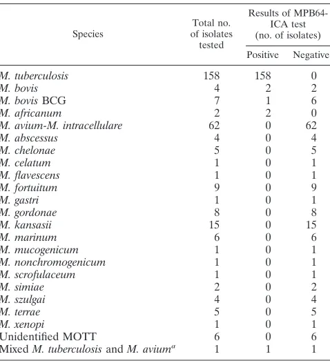
isolates. All M. tuberculosis and M. africanum isolates were
determined to be positive by ICA slide test, while 3 of 11M.
bovisorM. bovis BCG were determined to be positive. One
mixed culture ofM. tuberculosisandM. aviumwas also
deter-mined to be negative in the initial testing but positive on repeat testing after 3 days (Table 1).
All of these cultures were tested in MGIT medium, whereas
only selected cultures were tested in LJ (n ⫽ 63), 7H10
Middlebrook agar (n⫽9), and BACTEC 12B (n⫽25). The
ICA slide test was positive in the case ofM. tuberculosis
com-plex in all of the different media tested (Table 2). The positive band developed within ca. 5 to 10 min, but the intensity of the color was maximum at 15 min. The intensity of a positive test
color band varied from a 4⫹ (dark reddish purple) to 1⫹
(easily visible pink band). The control band was strongly pos-itive in all of the testing. There was no difference in the out-come of the results when 235 fresh primary isolated cultures were tested compared to the 69 stock cultures. Sequential evaluation of MGIT on a daily basis by ICA slide test demon-strated that the day of conversion from negative to positive by this test was concurrent or sometimes 1 to 2 days earlier than the day when the BACTEC MGIT 960 instrument detected
culture positive (Table 3). In the case of the M. tuberculosis
complex, there was no sample for which the ICA slide test was negative when the BACTEC MGIT 960 identified it as culture positive. In our earlier experiments the CFU count at the time
the MGIT medium flagged it as positive was ca. 2⫻105.
Radiometric BACTEC 12B medium was not positive at the
threshold of instrument positive (GI ⫽10 to 50). Even at a
on May 15, 2020 by guest
http://jcm.asm.org/
higher GI value of 500 the ICA test was not always positive.
The CFU counts at GI values of 50, 100, and 500 were ca. 4⫻
104, 1⫻105, and 5⫻105, respectively. Once a 12B vial withM.
tuberculosisgrowth achieved GI of ca. 500 and was incubated for one additional day it become positive for the ICA slide test.
At this GI, the CFU count is usually 106. Once the test was
positive in a BACTEC 12B vial, it gave a positive reaction even if the medium was diluted 1:2 to 1:4, whereas with MGIT medium a positive reaction was observed even at a 1:8 dilution. It was also observed that MPB64 antigen, once secreted in the medium, is stable, since the test remained positive even if performed 1 year after the detection of growth both in solid
and in liquid media. On the other handM. kansasiiand other
MOTT bacilli remained negative upon prolonged incubation and storage (Table 4).
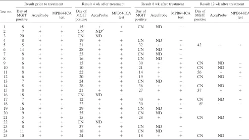
The data in Table 5 demonstrates that there is no effect of antituberculosis treatment on the test results when sequentially obtained sputum samples were tested up to 4 months after the initiation of chemotherapy. All culture-positive MGIT media
were also ICA slide test positive, except for one sample (Table
5). This patient, who was initially culture positive forM.
tuber-culosis and positive for the ICA slide test, became MGIT culture positive but ICA slide test negative. On further evalu-ation it was found that at this point of therapy the patient’s
sample did not yield M. tuberculosis growth, but actually
M. gordonaewas identified by the DNA-DNA hybridization method. During the chemotherapy none of the first-time cul-ture-negative samples in MGIT medium showed a positive ICA reaction, even though some of them were initially smear positive but remained culture negative.
DISCUSSION
We demonstrate here the usefulness of the ICA slide test for
routine laboratory testing for differentiation of theM.
tubercu-losiscomplex from MOTT bacilli. In addition, this test is ex-tremely simple and does not require any sample preparation or instrumentation. It is very rapid and can be performed using growth in both liquid and solid media.
Among the different media tested we concentrated on the BACTEC MGIT 960 system, as this is the most recent nonra-diometric method, the performance of which is comparable to
the radiometric BACTEC method (3, 13). All of theM.
tuber-culosis cultures once determined to be positive by the BACTEC MGIT 960 instrument were also determined to be positive by the ICA slide test, and the test results were avail-able within 15 min, which makes the ICA slide test more rapid and attractive than any other available technique.
[image:3.587.44.283.91.354.2]The production of MPB64 antigen in 12B medium is rather slow and could not always be detected even at a GI of 500. However, when the 12B vial is incubated one more day there is enough MPB64 antigen produced and the ICA slide test is positive. The delay in the production of MPB64 antigen in 12B medium could be due to the fact that this medium is not very rich and so mycobacteria may not be able to produce this antigen at an early stage of growth. On the solid media LJ and 7H10 the ICA slide test is positive once there is visible growth on the surface of medium. The positive color bands on the ICA slide were easily visible with a dark red-purple to a light pink color. There was no color reaction observed in the case of
TABLE 1. Results of different species of mycobacteria tested with the MPB64-ICA test
Species of isolatesTotal no. tested
Results of MPB64-ICA test (no. of isolates)
Positive Negative
M. tuberculosis 158 158 0
M. bovis 4 2 2
M. bovisBCG 7 1 6
M. africanum 2 2 0
M. avium-M. intracellulare 62 0 62
M. abscessus 4 0 4
M. chelonae 5 0 5
M. celatum 1 0 1
M. flavescens 1 0 1
M. fortuitum 9 0 9
M. gastri 1 0 1
M. gordonae 8 0 8
M. kansasii 15 0 15
M. marinum 6 0 6
M. mucogenicum 1 0 1
M. nonchromogenicum 1 0 1
M. scrofulaceum 1 0 1
M. simiae 2 0 2
M. szulgai 4 0 4
M. terrae 5 0 5
M. xenopi 1 0 1
Unidentified MOTT 6 0 6
MixedM. tuberculosisandM. aviuma 1 1 1
[image:3.587.303.542.93.218.2]aNegative in the initial testing but positive after 3 days.
TABLE 2. Growth on different culture media
Medium
No. of isolates tested (no. positive
for MPB64-ICA test)a Total no.
of tests
M. tuberculosiscomplex MOTT
LJ 30 (30) 33 (0) 63
7H10 9 (9) 0 9
BACTEC MGIT 960 171 (171) 133 (0) 304
BACTEC 12B 9 (9) 16 (0) 25
aAllM. tuberculosiscultures were positive, while all MOTT bacilli were
neg-ative, by the MPB64-ICA test.
TABLE 3. Daily evaluation of growth in BACTEC MGIT 960 and MPB64-ICA test
Case
no. AFB smearresulta
Day of positivitybby:
BACTEC 960 MPB64-ICA
1 Positive 10 10
2 Positive 12 11
3 Positive 6 6
4 Positive 9 8
5 Positive 12 11
6 Positive 13 13
7 Positive 4 3
8 Negative 18 17
9 Positive 8 7
10 Positive 12 12
aSmears were prepared with decontaminated and concentrated sputum
spec-imens by the NALC-NaOH method.
bThat is, the number of days required to detect first positivity by the BACTEC
MGIT 960 instrument or the MPB64-ICA test.
on May 15, 2020 by guest
http://jcm.asm.org/
[image:3.587.44.283.631.710.2]MOTT bacilli cultures. This test, in terms of its sensitivity and specificity, compares very well with the probe-based tests since
in our analyses allM. tuberculosiscultures and M. africanum
cultures were positive (100%). AmongM. bovisstrains, two of
four tested strains were determined to be negative by this test.
The isolation ofM. bovisis rare from human samples.
Occa-sionally, the BCG strain is isolated from patients who receive
BCG immunotherapy. All of the four M. bovis strains were
from stock cultures frozen for some time. Many laboratories
do not differentiateM. bovisfromM. bovisBCG. It is possible
that the two of the four strains that tested negative are actually BCG strains and were not further identified. Further study of
theseM. bovisisolates is needed in order to establish whether
these were indeed non-BCGM. bovisstrains. It has been
re-ported that some BCG strains, such as BCG Glaxo, do not
produce MPB64 antigen whereas others, such as BCG Japan, are good secretors of the antigen (2, 8). In our analyses, one of
sevenM. bovisBCG isolates was determined to be positive. We
were not able to obtain the history of these BCG strains. On the other hand, we did not observe any cross-reaction of this antigen with MOTT bacilli. This indicates excellent specificity of the test. Abe et al. in their earlier studies reported a very
high specificity for this test with only oneM. marinumstrain
(one of three) and oneM. flavescensstrain (one of one) giving
a very weak reaction (2). In our study six strains ofM. marinum
and one strain ofM. flavescens were tested with no positive
reaction. Besides confirming the high sensitivity and specificity of the test that Abe et al. had reported, our multicenter study evaluated some other parameters of the test.
Although we did not perform an in-depth study on the
sta-TABLE 4. Effect of age of culture on the MPB64-ICA test
Time after positive growth
MPB64-ICA test resulta(no. of cultures tested)
M. tuberculosis M. kansasii Other MOTT bacilli
(MGIT)
MGITb LJ MGIT LJ
5 days Pos (1) Pos (1) Neg (1) Neg (1) ND
10 days Pos (1) Pos (1) Neg (1) Neg (1) ND
15 days Pos (1) Pos (1) Neg (1) Neg (1) ND
1 mo Pos (1) Pos (1) Neg (1) Neg (1) ND
1 yr Pos (20) Pos (20) Neg (2) ND Neg (20)
[image:4.587.42.544.84.181.2]aPos, positive; Neg, negative; ND, no data. bTested in duplicate with or without PANTA.
TABLE 5. Sequential evaluation of sputum cultures prior to and after antituberculosis treatmenta
Case no.
Result prior to treatment Result 4 wk after treatment Result 8 wk after treatment Result 12 wk after treatment Day of
MGIT
positive AccuProbe MPB64-ICAtest Day of MGIT
positive AccuProbe MPB64-ICAtest Day of MGIT
positive AccuProbe MPB64-ICAtest Day of MGIT
positive AccuProbe MPB64-ICAtest
1 8 ⫹ ⫹ 15 ⫹ ⫹ CN ND ⫺
2 7 ⫹ ⫹ CNc NDd ⫺
3 20 ⫹ ⫹ CN ND ⫺
4 8 ⫹ ⫹ 19 ⫹ ⫹ CN ND ⫺
5 5 ⫹ ⫹ 21 ⫹ ⫹ 32 ⫹ ⫹ 42 ⫹ ⫹
6 14 ⫹ ⫹ 28 ⫹ ⫹ CN ND ⫺
7 8 ⫹ ⫹ 23 ⫹ ⫹ CN ND ⫺
8 5 ⫹ ⫹ 16 ⫹ ⫹ CN ND ⫺
9 6 ⫹ ⫹ 15 ⫹ ⫹ 30 ⫹ ⫹ CN ND ⫺
10 5 ⫹ ⫹ 10 ⫹ ⫹ 21 ⫹ ⫹ CN ND ⫺
11 8 ⫹ ⫹ 22 ⫹ ⫹ 14 ⫹ ⫹ 56 ⫹ ⫹
12 6 ⫹ ⫹ 20 ⫹ ⫹ 19 ⫹ ⫹ CN ND ⫺
13 7 ⫹ ⫹ 24 ⫹ ⫹ CN ND ⫺
14 5 ⫹ ⫹ 28 ⫹ ⫹ 36 ⫹ ⫹ CN ND ⫺
15 8 ⫹ ⫹ 21 ⫹ ⫹ 27 ⫹ ⫹ 37 ⫹ ⫹
16 18 ⫹ ⫹ CN ND ⫺
17 7 ⫹ ⫹ 12 ⫹ ⫹ 40 ⫹ ⫹ CN ND ⫺
18 8 ⫹ ⫹ 22 ⫹ ⫹ 30 ⫹ ⫹ 8b ⫺ ⫺
19 16 ⫹ ⫹ 29 ⫹ ⫹ CN ND ⫺
20 9 ⫹ ⫹ 35 ⫹ ⫹ CN ND ⫺
21 5 ⫹ ⫹ 15 ⫹ ⫹ 28 ⫹ ⫹ CN ND ⫺
22 6 ⫹ ⫹ CN ND ⫺
23 8 ⫹ ⫹ 37 ⫹ ⫹ CN ND ⫺
24 11 ⫹ ⫹ 18 ⫹ ⫹ CN ND ⫺
25 10 ⫹ ⫹ 24 ⫹ ⫹ 18 ⫹ ⫹ CN ND ⫺
a“Day of MGIT positive” means the number of days required to detect first positivity by the BACTEC MGIT 960 instrument. bOnlyM. gordonaewas isolated.
cCN, culture negative. dND, no data.
on May 15, 2020 by guest
http://jcm.asm.org/
[image:4.587.46.539.417.695.2]bility of MPB64 antigen in the medium, we did look at the effect of age of growth in a culture medium on the perfor-mance of the test. It is quite clear from the findings that the antigen is stable in both liquid and solid media. On the other hand, MOTT bacilli did not produce detectable antigen with an extended incubation or when growth in a medium was left for a long period. We also observed that a positive reaction band on the slide was very stable and could be stored for months without any loss of color intensity.
We also evaluated the effect of chemotherapy on this test.
The results indicate that as long as the culture is positive forM.
tuberculosisfrom the sputum of a patient on chemotherapy, the test would be positive, indicating that the antituberculosis treatment has no effect on the production of this antigen. Once sputum becomes culture negative, the test also becomes neg-ative even if the smear made from the specimen shows AFB, indicating that tubercle bacilli, if present in the specimen, are not viable.
In our experience there have been some instances in which a tuberculosis patient, after beoming culture negative can
be-come culture positive again, though at this time not for M.
tuberculosis but for MOTT bacilli (usually M. avium or M. gordonae). This could represent environmental contamination, colonization, or secondary infection. In such situations the ICA slide test offers a very simple and quick way to monitor the
presence or persistence ofM. tuberculosisin follow-up
speci-men cultures of a patient receiving antituberculosis treatspeci-ment. Molecular methods can rapidly identify mycobacteria within a few hours with great accuracy, but these methods, compared to the ICA slide test, are complex and cumbersome, require instrumentation, and are labor-intensive. We found that the ICA slide test was extremely simple, requiring very little tech time, and that the results are ready within 15 min.
One of the disadvantages of a culture system with liquid medium is the high potential for contamination or for the
coexistence ofM. tuberculosisand MOTT bacilli, which may go
undetected or may lead to erroneous reporting of results. Even though the ICA slide test could be an excellent identification tool and could serve as an alternative to molecular-analysis-based approaches, the positive result of this test does not
necessarily rule out the coexistence of M. tuberculosis and
MOTT bacilli. We experienced one case in the present study supporting this concern. In this case a PCR test with sputum
sample for primary cultures detectedM. tuberculosis;
there-fore, PCR forM. avium-M. intracellularewas not performed.
Initially, BACTEC MGIT 960 gave a positive result and the
ICA slide test was negative, indicating the absence ofM.
tu-berculosis. Since this MGIT tube turned positive within a short time, the test was repeated after 3 days of further incubation,
and the ICA test was then clearly positive. BothM. tuberculosis
andM. aviumwere present in the culture, and the additional
incubation allowedM. tuberculosisto produce sufficient
anti-gen detectable by this test. This is an inherent problem of molecular tests as well. However, in a probe test, such as
GenProbe, there is a provision of testing forM. aviumand a
few other species if there is any suspicion of having MOTT in the culture. Development of a similar kind of lateral flow
immunochromatography test for M. avium and a few
com-monly isolated mycobacteria would be desirable.
In conclusion, this immunochromatography test with mono-clonal antibody against MPB64 is a low-tech and rapid method
for the differentiation ofM. tuberculosisfrom other
mycobac-teria. Considering its accuracy as well as its simplicity, the method should offer significant savings of both labor and time and could be a good alternative to probe-based tests, especially in countries with limited resources.
REFERENCES
1.Abe, C., S. Hosojima, Y. Fukasawa, Y. Kazumi, M. Takahashi, K. Hirano, and T. Mori.1992. Comparison of MB-Check, BACTEC, and egg-based media for recovery of mycobacteria. J. Clin. Microbiol.30:878–881. 2.Abe, C., K. Hirano, and T. Tomiyama.1999. Simple and rapid identification
ofMycobacterium tuberculosiscomplex by immunochromatographic assay using anti-MPB64 monoclonal antibodies. J. Clin. Microbiol.37:3693–3697. 3.Badak, F. Z., D. L. Kiska, S. Setterquist, C. Hartley, M. A. O’Connell, and R. L. Hopfer.1996. Comparison of Mycobacteria Growth Indicator Tube with BACTEC 460 for detection and recovery of mycobacteria from clinical specimens. J. Clin. Microbiol.34:2236–2239.
4.Harboe, M., S. Nagai, M. E. Patarroyo, M. L. Torres, C. Ramirez, and N. Cruz.1986. Properties of proteins MPB64, MPB70 and MPB80 of Mycobac-terium bovisBCG. Infect. Immun.52:293–302.
5.Ichiyama, S., Y. Iinuma, S. Yamori, Y. Hasegawa, K. Shimokata, and N. Nakashima.1997. Mycobacterium Growth Indicator Tube testing in con-junction with the AccuProbe or the AMPLICOR-PCR assay for detecting and identifying mycobacteria from sputum samples. J. Clin. Microbiol.35:
2022–2025.
6.Kubica, G. P., W. E. Dye, M. L. Cohn, and G. Middlebrook.1963. Sputum digestion and decontamination with N-acetyl-L-cystein-sodium hydroxide for culture of mycobacteria. Am. Rev. Respir. Dis.87:775–779.
7.Kusunoki, S., T. Ezaki, M. Tamesada, Y. Hatanaka, K. Asano, Y. Hashi-moto, and E. Yabuuchi.1991. Application of colorimetric microdilution plate hybridization for rapid genetic identification of 22Mycobacterium spe-cies. J. Clin. Microbiol.29:1596–1603.
8.Li, H., J. C. Ulstrup, T. O. Jonassen, K. Melby, S. Nagai, and M. Harboe.
1993. Evidence for absence of the MPB64 gene in some strains of Mycobac-terium bovisBCG. Infect. Immun.61:1730–1734.
9.Nagai, S., H. G. Wiker, M. Harboe, and M. Kinomoto.1991. Isolation and partial characterization of major protein antigens in the culture fluid of
Mycobacterium tuberculosis. Infect. Immun.59:372–382.
10.Nakamura, R. M., M. A. Velmonte, K. Kawajiri, C. F. Ang, R. A. Frias, M. T. Mendoza, J. C. Montoya, I. Honda, S. Haga, and I. Toida.1998. MPB 64 mycobacterial antigen: a new skin test reagent through patch method for rapid diagnosis of active tuberculosis. Int. J. Tuberc. Lung Dis.2:541–546. 11.Nakamura, R. M., L. Einck, M. A. Velmonte, K. Kawajiri, C. F. Ang, C. E.
Delasllagas, and C. A. Nancy.2001. Detection of active tuberculosis by an MPB-64 transdermal patch: a field study. Scand. J. Infect. Dis.33:405–407. 12.Oettinger, T., and A. B. Andersen.1994. Cloning and B-cell-epitope mapping of MPT64 from mycobacterium tuberculosis H37Rv. Infect. Immun.62:
2058–2064.
13.Pfyffer, G. E., H. M. Welscher, P. Kissling, C. Cieslak, M. J. Casal, J. Gutierrez, and S. Rusch-Gerdes. 1997. Comparison of the Mycobacteria Growth Indicator Tube (MGIT) with radiometric and solid culture for re-covery of acid-fast bacilli. J. Clin. Microbiol.35:364–368.
14.Roche, P. W., J. A. Triccas, D. T. Avery, T. Fifis, H. Billman-Jacobe, and W. J. Britton.1994. Differential T cell responses to mycobacteria-secreted proteins distinguish vaccination with Bacille Calmette-Guerin from infection withMycobacterium tuberculosis. J. Infect. Dis.170:1326–1330.
15.Roche, P. W., N. Winter, J. A. Triccas, C. G. Feng, and W. J. Britton.1996. Expression of Mycobacterium tuberculosisMPT64 in recombinant Myco. smegmatis: purification, immunogenicity, and application to skin tests for tuberculosis. Clin. Exp. Immunol.103:226–232.
16.Siddiqi, S. H., C. C. Hwangbo, V. Silcox, R. C. Good, D. E. Snider, Jr., and G. Middlebrook.1984. Rapid radiometric method to detect and differentiate
Mycobacterium tuberculosis/M. bovisfrom other mycobacteria species. Am. Rev. Respir. Dis.130:634–640.
17.Tenover, F. C., J. T. Crawford, R. E. Huebner, L. J. Geiter, C. R. Horsburgh, Jr., and R. C. Good.1993. The resurgence of tuberculosis: is your laboratory ready? J. Clin. Microbiol.31:767–770.
18.Tomiyama, T., K. Mastuo, and C. Abe.1997. Rapid identification of Myco-bacterium tuberculosis by an immunochromatography using anti-MPB64 monoclonal antibodies. Int. J. Tuberc. Lung Dis.1(Suppl. 1):S59. 19.Wiker, H. G., S. Nagai, M. Harboe, and L. Ljungqvist.1992. A family of
cross-reacting proteins secreted bymycobacterium tuberculosis. Scand. J. Im-munol.36:307–319.
20.Yamaguchi, R., K. Matsuo, A. Yamazaki, C. Abe, S. Nagai, K. Terasaka, and T. Yamada.1989. Cloning and characterization of the gene for immunogenic protein MPB64 ofMycobacterium bovisBCG. Infect. Immun.57:283–288.