Chemistry Dissertations Department of Chemistry
12-14-2017
Understanding Function of Carbohydrates by
Synthesis of Structurally-defined Glycopeptides/
Glycoproteins and Glycans
Zhigang Wu
Follow this and additional works at:https://scholarworks.gsu.edu/chemistry_diss
This Dissertation is brought to you for free and open access by the Department of Chemistry at ScholarWorks @ Georgia State University. It has been accepted for inclusion in Chemistry Dissertations by an authorized administrator of ScholarWorks @ Georgia State University. For more information, please [email protected].
Recommended Citation
Wu, Zhigang, "Understanding Function of Carbohydrates by Synthesis of Structurally-defined Glycopeptides/Glycoproteins and Glycans." Dissertation, Georgia State University, 2017.
STRUCTURALLY-DEFINED GLYCOPEPTIDES/GLYCOPROTEINS AND GLYCANS
by
ZHIGANG WU
Under the Direction of Peng George Wang, PhD
ABSTRACT
Carbohydrates are crucial biomacromolecules found in organisms across all domains of
life and play pivotal roles in biological processes. To better understand the biological function of
glycans, structurally-defined glycopeptides/glycoproteins and glycans are highly demanded. In
this dissertation, the study has been focusing on acquiring homogenous
glycopeptides/glycoproteins and glycans which are good candidates for decoding their function.
Chapter 1 provides a brief introduction of function of glycosylation for proteins,
significance of glycosylation for therapeutic protein and artificial glycosylation approaches;
glycan synthetic approaches and glycan microarray and an overview of synthesis of
facile and efficient method for site-directed glycosylation of peptide/protein. The method
contains two sequential steps: generation of a GlcNAc-O-peptide/protein, and subsequent
ligation of a eukaryotic N-glycan to the GlcNAc moiety. (2) Peptides can be GlcNAcylated at
one or two natural N-glycosylation sites via two-step enzymatic reactions catalyzed by an
evolved N-glycosyltransferase (ApNGT Q469A) and a glucosamine N-acetyltransferase (GlmA),
respectively. Then, the resulting GlcNAc-peptides were further modified by an endo-β-N
-acetylglucosaminidase M mutant (EndoM N175Q) to generate glycopeptides carrying
complex-type N-glycans.
In Chapter 3, two focused glycan libraries were synthesized which were N-glycans
containing tandem epitopes and a new class of asymmetric N-glycans. (1) A focused library of
36 biantennary asparagine (Asn)-linked glycans with some presenting tandem glycan epitopes
was synthesized and glycan microarray study showed that the internal epitopes and/or
modifications of terminal epitopes influence the binding of glycans. (2) A class of asymmetric N
-glycans was synthesized by coupling glycan-oxazoline and N-glycans. Branch-biased binding
and spacial inhibition caused by bulky group on the other branch were observed in glycan
protein interactions involving lectins and these glycans.
In Chapter 4, a microbial glycosyltrasferases Hp1,3FTα66 were readily expressed in
Escherichia coli (E. coli) was biochemically characterized and applied as a diverse catalyst in the
facile synthesis of fucosylated human milk oligosaccharides. In addition, multi-gram scale of
fucosylated human milk oligosaccharides, fucopentaose (LNFP) III and
lacto-N-neofucopentaose (LNnFP) V were synthesized by three major combined engineered E. coli
STRUCTURALLY-DEFINED GLYCOPEPTIDES/GLYCOPROTEINS AND GLYCANS
by
ZHIGANG WU
A Dissertation Submitted in Partial Fulfillment of the Requirements for the Degree of
Doctor of Philosophy
in the College of Arts and Sciences
Georgia State University
Copyright by Zhigang Wu
STRUCTURALLY-DEFINED GLYCOPEPTIDES/GLYCOPROTEINS AND GLYCANS
by
ZHIGANG WU
Committee Chair: Peng George Wang
Committee: Binghe Wang
Jenny Yang
Jun Yin
Electronic Version Approved:
Office of Graduate Studies
College of Arts and Sciences
Georgia State University
DEDICATION
ACKNOWLEDGEMENTS
All the study in this dissertation are carried out under the direction of Prof. Peng George
Wang. First and foremost, I would like to sincerely thank my advisor Dr. Peng George Wang
who is always full of enthusiasm, energy and sparkling ideas during the dissertation. Under the
support, guidance and pressure from him throughout all these years, I am more mature and
trained as an independent thinker in science.
Secondly, I want to thank my committee, Dr. Binghe Wang, Dr. Jenny Yang and Dr. Jun
Yin for their help and insightful discussion during my annual report.
I especially want to thank my collaborators, Dr. Xi Chen, Dr. George Pierce, Dr. David
Smith, Dr. Lianwen Zhang, Dr. Jiansong Cheng, Dr. Jian Zhang and Dr. Xiu-feng Wan for
discussion and guide during my research. In addition, I would like to thank Dr. Yanyi Chen for
CD study, Dr. Lauren Ashley Byrd-Leotis for HA virus preparation, Dr. Jian Zhang and Yi
Lasanajak for microarray study, Kelly Cannon and Maurice de la Croix for fermentation study.
I also want to thank all my labmates, especially Lei, Yunpeng, Cheng, Jingyao, Hailiang,
Madhusudhan, He, Mohui, Christopher, Zhongying, Yuxi, Xuan, Angie, Jack, Ding and all
others for their help in my research. I am so happy and fortunate to be able to work with such a
wonderful large group of people.
Finally, I am especially grateful to my wife Wanyi Guan and my son Hongyi Wu for their
TABLE OF CONTENTS
ACKNOWLEDGEMENTS ... v
LIST OF TABLES ... vi
LIST OF FIGURES ... vii
LIST OF SCHEMES ... ix
LIST OF ABBREVIATIONS ... x
1 INTRODUCTION... 1
1.1 Function of glycosylation for proteins and artificial glycosylation approaches ... 1
1.1.1 Introduction of protein glycosylation ... 1
1.1.2 Glycosylation is significant for therapeutic proteins ... 2
1.1.3 Chemical and chemoenzymatic synthesis of glycoproteins ... 6
1.2 Glycan function, structure-defined glycan synthesis approaches and glycan array ... 11
1.2.1 Function of glycans ... 11
1.2.2 Structural diversity and classification of glycans ... 12
1.2.3 Preparation of structurally defined glycans ... 14
1.2.4 Glycan microarray ... 17
1.3 Synthesis of oligosaccharides in large scale ... 20
1.3.1 Significance of oligosaccharide synthesis in large scale ... 20
1.3.2 Challenges for current large-scale oligosaccharide production ... 21
1.4 Outline of the work in this dissertation ... 26
1.5 References ... 26
2 CHEMOENZYMATIC OR ENZYMATIC SYNTHESIS OF GLYCOPEPTIDES/PROTEINS ... 35
2.1 Site-directed glycosylation of peptide/protein with homogeneous O-linked eukaryotic N-glycans ... 35
2.1.1 Introduction ... 35
2.1.2 Experimental Methods ... 36
2.1.3 Results and Discussion ... 40
2.2 A novel enzymatic method for synthesis of glycopeptides carrying natural eukaryotic N-glycans ... 68
2.2.1 Introduction ... 68
2.2.3 Results and Discussion ... 73
2.3 References ... 86
3 GLYCAN SYNTHESIS AND DECODE BIOLOGICAL FUNCTION OF GLYCANS ... 90
3.1 Identification of the binding roles of terminal and internal glycan epitopes using enzymatically synthesized N-glycans containing tandem epitopes. ... 90
3.1.1 Introduction ... 90
3.1.2 Experimental Methods ... 94
3.1.3 Results and Discussion ... 103
3.2 Decoding glycan protein interactions by a new class of asymmetric N-glycans. 156 3.2.1 Introduction ... 156
3.2.2 Experimental Methods ... 159
3.2.3 Results and Discussion ... 176
3.3 References ... 193
4 LARGE-SCALE SYNTHESIS OF OLIGOSACCHARIDES ... 198
4.1 Biochemical characterization of Helicobacter pyloriα1–3/4FT and synthesis of fucosylated human milk oligosaccharides ... 198
4.1.1 Introduction ... 198
4.1.2 Experimental Methods ... 199
4.1.3 Results and Discussion ... 206
4.2 Fucosylated human milk oligosaccharide (HMOs) production in large scale by combined engineered E.coli. ... 212
4.2.1 Introduction ... 212
4.2.2 Experimental Methods ... 214
4.2.3 Results and Discussion ... 218
4.3 References ... 229
5 PUBLICATIONS ... 231
LIST OF TABLES
Table 1. 1 Four types of glycoprotein. ... 2
Table 1. 2 Different types of Endoglycoosidases. ... 10
Table 2. 1 N-Glycan modification of selected peptides. ... 41
Table 2. 2 OGA activity for GlcNAc-O-peptides. ... 45
Table 2. 3 Specificity of GlmA toward glycosylated peptides. ... 74
Table 2. 4 Modification of N-glycoprotein derived peptides and mGLP-1. ... 76
Table 3. 1 Conversion percentage of reactions catalyzed by NmCSS and CjCstII. ... 109
Table 3. 2 Acceptor substrate specificity of PmST1. ... 110
Table 4. 1 Kinetic parameters of Hp3/4FT. ... 208
Table 4. 2 ECAM medium (1 L) ... 215
Table 4. 3 Batch medium (10 L) ... 215
Table 4. 4 Feed medium (5 L) ... 216
Table 4. 5 Trace metal solution (1 L) ... 216
Table 4. 6 The strains used in this strategy. ... 221
LIST OF FIGURES
Figure 1. 1 Four types of glycosylated proteins. ... 2
Figure 1. 2 Aranesp has a 3-fold increase in serum half-life compared to rHuEPO. ... 5
Figure 1. 3 Mechanism of native chemical ligation. ... 8
Figure 1. 4 Glycosylation remodeling of glycoproteins. ... 9
Figure 1. 5 Interactions between glycans and receptors mediate biological processes. ... 12
Figure 1. 6 Mechanism of glycan-amine bound to slides. ... 18
Figure 1. 7 Glycans can be specific bound by proteins, pathogens and cells. ... 19
Figure 1. 8 OPME strategy for oligosaccharides synthesis. ... 22
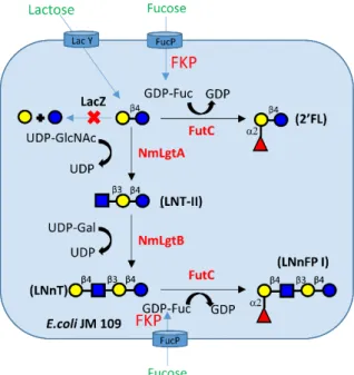
Figure 1. 9 Biosynthetic pathway of LNnFP I from lactose in engineered E. coli cells. ... 24
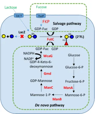
Figure 1. 10 Whole cell biosynthesis of 2’-FL by de novo pathway ... 25
Figure 2. 1 Site-directed glycosylation of peptide/protein with uniform eukaryotic N-glycan. ... 36
Figure 2. 2 HPLC chromatograph and ESI-MS spectra of glycan-O-Ser-01. ... 41
Figure 2. 3 Glycosylation of GLP-1 and circular dichroism study. ... 43
Figure 2. 4 HPLC and MALDI-TOF MS profiles of Ser-02-OGA. ... 44
Figure 2. 5 Glycosylation of bovine α-crystallin-A (Crys-A) and identification by immunoblot and in-gel digestion plus mass spectrometry. ... 46
Figure 2. 6 ESI-MS/MS characterization of glycosylated Crys-A. ... 47
Figure 2. 7 Enzymatic synthesis of glycopeptides carrying natural eukaryotic N-glycans. . 69
Figure 2. 8 Enzymatic production of GlcNAc-linked peptide. ... 73
Figure 2. 9 Transglycosylation of GlcN-peptide. ... 76
Figure 3. 1 Biantennary N-glycans synthesized in this study... 93
Figure 3. 2 Binding profile of plant lectins with defined binding specificities. ... 112
Figure 3. 3 Binding profile of antibodies. ... 115
Figure 3. 4 Binding profile of influenza A viruses. ... 116
Figure 3. 5 Glycan microarray analyses [binding profiles with two viral hemoagglutinin (HA)]... 178
Figure 3. 6 Glycan microarray analyses (binding profiles with five plant lectins). ... 179
Figure 4. 1 pH profile of His6-Hp3FT∆66. ... 207
Figure 4. 2 Metal cation effects on Hp3FT∆66. ... 207
Figure 4. 3 LNFP III (4) characterization by MALDI-TOF tandem mass spectrometry and 2D NMR. ... 210
Figure 4. 4 Specificity of Hp3FT∆66 was identified by tandem MS. ... 211
Figure 4. 5 LNT-II production by direct fermentation. ... 220
LIST OF SCHEMES
Scheme 3. 1 Preparation of disialylated N-glycan BA-01, the core N-glycan (BA-02) and
one-pot enzymatic synthetic schemes for producing N-glycans with only terminal
glycan epitopes. ... 104
Scheme 3. 2 One-pot synthetic scheme for N-glycans with tandem sugar epitopes without
sialic acids (BA-24 to BA-36). ... 107
Scheme 3. 3 One-pot synthetic scheme for N-glycans with sugar epitopes with sialic acids
(BA-09 to BA-16, BA-19 to BA-22). ... 108
Scheme 3. 4 Groups A and B glycans were synthesized by EndoM N175Q with α1-3Man
branch glycan oxazoline as donor. ... 158
Scheme 3. 5 Groups C and D glycans were synthesized by EndoM N175Q with α1-6Man
branch glycan oxazoline as donor ... 159
Scheme 4. 1 Fucosylated HMOs were synthesized by Hp3FT∆66 according its acceptor
specificity. ... 208
Scheme 4. 2 Fucosylated HMOs were synthesized by Hp3FT∆66 according its acceptor
specificity. ... 213
Scheme 4. 3 LNFP III and LNnFP V synthesis in large scale by combined engineered E.coli
LIST OF ABBREVIATIONS
Amp ampicillin
ATCC American Type Culture Collection
ATP adenosine 5'-triphosphate
bp base pair
BSA bovine serum albumin
CD circular dichroism
cm centimeter
DF-paraLNnH difuco-para-Lacto-N-neohexaose
DTT dithiothreitol Fuc fucose g gram Gal galactose Glc glucose GlcN glucosamine
GlcNAc N-acetylglucosamine
GT glycosyltransferase
GTP guanosine 5'-triphosphate
GDP-Fuc guanosine 5'-diphosphate-fucose
h hour
HRMS high-resolution mass spectrometry
IPTG isopropyl-β-D-thiogalactopyranoside
kDa kilo Dalton
L liter
LB Luria-Bertani (medium)
LPS lipopolysaccharide
LNT-II lacto-N-triose II
LNFP III lacto-N-fucopentaose III
LNnT lacto-N-neotetraose
LNnFP V Lacto-N-neofucopentaose V M molar
m/z mass to charge ratio
MALDI matrix-assisted laser desorption-ionization
Man mannose mg milligram min minute mL milliliter mM millimolar
MS mass spectrometry
NMR nuclear magnetic resonance
oC degree centigrade
OD optical density
PAGE polyacrylamide gel electrophoresis
PBS phosphate-buffered saline
PBST phosphate-buffered saline with Tween
PCR polymerase chain reaction
PEP phosphoenolpyruvate
PPA yeast inorganic pyrophosphatase
PPi pyrophosphate
rpm round per minute
s second
SDS sodium dodecyl sulfate
Sia sialic acid
TF-paraLNnH trifuco-para-lacto-N-neohexaose
TLC thin-layer chromatography
UDP uridine diphosphate
UDP-Glc uridine 5'-diphosphate-glucose
UDP-GlcNAc uridine 5'-diphosphate-N-acetylglucosamine
UDP-Gal uridine 5'-diphosphate-galactose
μg microgram
1 INTRODUCTION
1.1 Function of glycosylation for proteins and artificial glycosylation approaches
1.1.1 Introduction of protein glycosylation
Glycosylation is a ubiquitous post-translational modification of protein in most
eukaryotes and some prokaryotes. Glycan structures play important roles in biological and
physiological function of proteins, such as contributions in protein folding, quality control and
involvement in biological recognition events [1, 2]. It is reported that the full diversity of
mammalian glycans (estimated to be ≥7,000 structures), which are assembled from only ten
monosaccharides: fucose (Fuc), galactose (Gal), glucose (Glc), N‑acetylgalactosamine
(GalNAc), N-acetylglucosamine (GlcNAc), glucuronic acid (GlcA), iduronic acid (IdoA),
mannose (Man), sialic acid (Sia) and xylose (Xyl), are generated by around 700 proteins [3, 4].
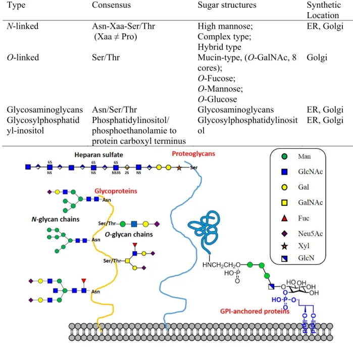
Four types of glycosylated proteins are known as glycoproteins: glycans are linked to
proteins through (1) amide linkages to asparagine (Asn) side chains which is N-glycosylation, (2)
glycosidic linkages to Ser/Thr which is O-glycosylation, (3) glycosaminoglycans, and (4)
glycosylphosphatidylinositol (GPI)-anchored proteins [5-7] (Table 1.1 and Figure 1.1).
Structures of glycans attached to proteins are highly complex because of numerous possibilities
for branching and anomeric linkage, which lead to greater structural diversity than linear nucleic
acid or polypeptide[3] (Figure 1.1). Complexity and diversity of glycans is due to potential
competition among multiple enzymes for the same glycan substrates in intricate biosynthetic
pathways [5, 8]. The complexity of glycan is relevant to the stability and recognition of protein.
Sialic acids on glycans form highly hydrophilic structures, which can increase the solubility of
the proteins by shielding hydrophobic residues and extend the half-life of proteins which are very
antibody with addition of bisecting GlcNAc or lacking core fucose residue exhibit strong
antibody-dependent cell-mediated cytotoxicity (ADCC) at lower concentrations with much
higher efficacy, which means that core fucosylation significantly influence ADCC [11-13].
Table 1. 1 Four types of glycoprotein.
Type Consensus Sugar structures Synthetic
Location
N-linked Asn-Xaa-Ser/Thr
(Xaa ≠ Pro)
High mannose; Complex type; Hybrid type
ER, Golgi
O-linked Ser/Thr Mucin-type, (O-GalNAc, 8
cores);
O-Fucose;
O-Mannose;
O-Glucose
Golgi
Glycosaminoglycans Asn/Ser/Thr Glycosaminoglycans ER, Golgi
Glycosylphosphatid
yl-inositol Phosphatidylinositol/ phosphoethanolamie to protein carboxyl terminus
Glycosylphosphatidylinosit
ol ER, Golgi
Figure 1. 1 Four types of glycosylated proteins.
For the past decades, protein based drugs have developed fast because of their great
economic and clinical importance. Protein therapeutics have many advantages in therapeutically
favorable properties, such as higher target specificities, lower side effects and better
pharmacological potencies, however, their development is hindered by intrinsic limitations in
their physicochemical and pharmacological properties [14-16]. The strategic manipulation of the
protein’s surface glycosylation is a promising method now [7, 17]. Also, glycosylation plays a
crucial role in the safety of protein therapeutics. Therapeutic proteins being modified by N
-glycan have demonstrated increased efficacy in patients and is considered as a safe way during
process development [18, 19].
Glycans plays crucial roles from four aspects in therapeutic glycoproteins: (1) physical
properties; (2) biological activity; (3) serum half-life and clearance; (4) immunogenicity.
(1) Physical properties
Glycosylation is important for protein to maintain molecular integrity, increase solubility
and reduce the possibility of proteolysis. For example, fibronectin can be easily hydrolyzed if
glycans are removed[20]; thermal stability of glycosylated interferon beta (IFN-beta 1a, Avonex)
is higher than unglycosylated form (IFN-beta 1b, Betaseron) which is expressed by Escherichia
coli (E. coli) [21]. Only 11% biological activity of fully deglycosylated recombinant human
erythropoietin (rHuEPO) can be kept after heat treatment, however, the activity of glycosylated
rHuEPO remains the same. Non-glycosylated rHuEPO can be easily denatured to be aggregates
and precipitates by heat, pH and guanidine-HCl, while glycosylated rHuEPO can be reversibly
folded and remains soluble [22-24]. In addition, glycans can protect rHuEPO from oxygen free
radical damage [25].
Glycosylated form (IFN-beta 1a, Avonex) had a 10-fold increased anti-viral activity as
compared to the E. coli-expressed product (IFN-beta 1b, Betaseron) [21]. It was reported that
research of glycoengineered EPO analogs demonstrated that a direct and positive relationship
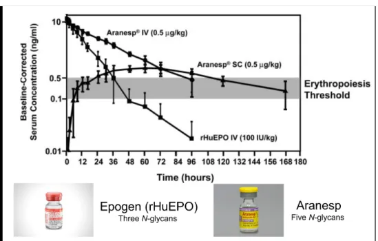
between number of glycans and in vivo activity. Although Aranesp (glycoengineered rHuEPO)
with five N-glycans exhibits similar conformation and stability compared to Epogen (rHuEPO)
with three N-glycan chains, to keep similar erythropoietic response, 3-fold more Epogen than
Aranesp was required at three times per week dosing in mice [26, 27], which increased to 13-fold
at weekly intervals.
Mpl ligand is hormone (glycoprotein) involved in maturation, megakaryocyte growth,
and platelet production. Glycosylated analogs of the Mpl ligand, N39 (four additional N-linked
glycans) and N40 (a total of six N-linked glycans) can increase duration and amount of platelet in
mice compared to native rHuMpI ligand [28].
(3) Serum half-life and clearance
Aranesp has an approximately 3-fold longer terminal half-life than Epogen and more than
2-fold greater area under the curve (AUC) resulting in a 2.5-fold lower clearance [7] (Figure
Figure 1. 2 Aranesp has a 3-fold increase in serum half-life compared to rHuEPO.
Aranesp administered intravenously (IV) or subcutaneously (SC), and rHuEPO administered IV in dialysis patients are shown. Serum half-lives were: Aranesp IV, 25.3±2.2 h;
Aranesp SC, 48.8±5.2 h; and rHuEPO IV, 8.5±2.4 h. Modified figure[7]. Follicle stimulating hormone (FSH) is used clinically for the treatment of anovulatory
conditions and of infertility. FSH1208 which had extended N-linked glycosylation sequence had
an increased half-life (3- to 4-fold) and AUC (4- to 6-fold) compared to native FSH [29].
Glycosylated interleukin 3 (IL-3) can be bound by the extracellular matrix (ECM), which
leads to glycosylated IL-3 slowly released into the circulation and increase 30%–40% activity to
stimulate histidine carboxylase activity in bone marrow compared with non-glycosylated IL-3
because of a 2-fold increase in serum half-life [30].
But it was reported that not all glycans can increase the half-life of therapeutic proteins.
The following important aspects are the relationship between glycan types and the circulatory
behavior of glycoproteins. (a) By specific receptor-based mechanisms, improperly glycosylated
proteins can be quickly removed from circulation; (b) Sialic acid terminated glycoproteins
(negative charge) have longer half-life compared with non-glycosylated proteins or partially
Some specific glycan patterns on the surface of therapeutic proteins can be targeted to certain
tissue or organs [31].
(4) Immunogenicity
Protein therapeutics can lose efficacy through neutralization of activity. It was reported
that immunoreactivity of antibodies to rHuEPO increased in pure red cell aplasia (PRCA) patient
serums if N-glycans were removed from rHuEPO, which may be explained by that glycans can
block the binding between antibody and rHuEPO by shielding the protein molecule from
immune system [32]. In addition, carbohydrate can increase the solubility and stability of
proteins by shielding hydrophobic residues, which may reduce the possibility of protein
aggregation and lead to decreased immunogenicity of proteins[33]. In summary, glycosylation of
protein therapeutics is a practical approach to reduce the possibility of immunogenicity.
Although glycosylation is well accepted for increasing the stability and efficacy of
therapeutic protein, technique still challenge the artificial homogeneous glycosylation of
therapeutic proteins because of complex structure of glycans. Chemical and enzymatic glycan
synthesis or remodeling strategies are currently pursued in academic and industrial area, which
may be alternative approaches for acquiring homogenous target glycoproteins which can be used
for long-term therapeutics and decoding fundamental effects and mechanism of protein
physicochemical and pharmacological properties.
1.1.3 Chemical and chemoenzymatic synthesis of glycoproteins
From the point of view of chemical synthesis, the synthesis of glycoproteins is one of
most challenging technique because of the very large size (normally over 15 kDa), complex
structure of glycans, the heterogeneity of glycosylation. Small to middle size N- and O
strategy[34, 35]. However, synthesis of larger glycopeptides or glycoproteins by the strategy is
difficult because of low coupling efficiency, poor solubility of protected glycopeptides and
strong acid hydrolysis (TFA and HF treatment) for deprotection of final product[36]. The solve
the above problems, three major strategies have been developed, which include (1) native
chemical ligation (NCL), (2) direct enzymatic glycosylation, and (3) chemoenzymatic
transformation for synthesis of homogenous larger glycopeptides and glycoproteins.
(1) Native chemical ligation
In reaction of native chemical ligation, the thiol group in N-terminal Cys from peptide b
can attack the thioester in C-terminal from peptide a to form a new larger peptide (Figure 1.3).
Bertozzi group firstly reported that they synthesize an glycoprotein with 82 amino acid and two
O-GalNAc residues [37]. Unverzagt group reported synthesis of a glycopeptide thioester using
alkanesulfonamide “safety-catch” linker for NCL strategy [38]. Then, Kajihara group firstly
synthesize a full size glycoproteins, monocyte chemotactic protein-3 (MCP-3) with 76 amino
acid and a complex type N-glycans and acquire the native glycoproteins by folding approach
[39]. However, Cys residues for ligation in NCL still limit the synthesis of glycoproteins because
many targets are lack of Cys residues. Therefore, different strategies have been developed to
mimic the cysteine ligation. A series of thiolated amino acids as potential residues for Lys, Phe,
Pro, Ala, Gln, Leu, Val and Thr have been tried and then the thiol group can be selectively
removed, which significantly expanded application of NCL for synthesis of glycoprotein [40,
41]. Recent research on synthsis of glycoprotein hormone α- and β-subunits [42, 43], human
interferon-β [44] and human EPO [45, 46] demonstrated that NCL is powerful strategy for
Figure 1. 3 Mechanism of native chemical ligation.
(2) Direct enzymatic glycosylation
Naturally, N-glycoprotein can be synthesized by the oligosaccharyl transferases (OST)
which can transfer glycans from a dolichol-phosphate glycolipid to Asn in a consensus
Asn-Xaa-Ser/Thr (Xaa ≠ Pro) sequon of the nascent protein. OST can transfer glycans to proteins to form
glycoproteins in vitro [47-49], however, OST is hard to handle for in vitro protein glycosylation
because it is a nine-subunit enzyme and is membrane-bound. In contrast, PglB, is a single
subunit transferase that was discovered for protein N-glycosylation in Campylobacter jejuni,
which can be used as a tool for glycoprotein glycosylation [50-54]. PglB is capable to transfer
relaxed polysaccharide substrate to a protein to acquire polysaccharide-protein conjugates for
application of bacterial glycoconjugate vaccines [55, 56]. But it needs strict sequence
(Asp/Glu-Xaa-Asn-Xaa-Ser/Thr) in protein as a substrate [51] and cannot efficiently use mammalian N
-glycan as donor [53], which limit its application in protein glycosylation.
N-glycosyltransferase (NGT) from a pathogenic bacterium was reported to transfer a
glucose residue to Asn-Xaa-Ser/Thr (Xaa ≠ Pro) consensus sequence in a polypeptide from
UDP-Glc, rather than glycolipids as a donor [57]. Wang group reported that glycosylated
endoglycosidase-catalyzed transglycosylation, however, the product was not a natural complex
type N-glycan because NGT cannot transfer GlcNAc residue to protein due to strict substrate
specificity [58].
Alternatively, direct enzymatic glycosylation of proteins in vitro maybe a very promising
and attractive method to synthesize homogenous glycoproteins if the enzymes can be engineered
to have board substrate specificity by protein engineering and directed evolution.
(3) Glycosylation remodeling of glycoproteins
Glycosylation remodeling of heterogeneous glycoproteins is a promising approach for
synthesis of homogenous glycoproteins in vitro. The approach involved two key steps: (a)
GlcNAc-glycoproteins preparation by enzymatic hydrolysis to remove N-glycan from
glycoproteins or chemical synthesis; (b) Protein transglycosylation by endoglycosidases mutant
[59] (Figure 1.4).
Endoglycosidases is a class of enzymes that release oligosaccharides from glycoproteins
to produce GlcNAc- or GlcNAc-(Fuc)-proteins. After site-directed mutagenesis (SDM), the
enzymes lose or decrease activity of hydrolysis and largely increase activity of
transglycosylation. After mutation, several endoglycosidases from bacteria and fungus, including
EndoA [59], EndoH [60], EndoM [59], EndoD [61], EndoS [62, 63], EndoS2 [64] and EndoF3
[65] were applied for synthesis of homogenous glycoproteins (Table 1.2).
Table 1. 2 Different types of Endoglycoosidases.
Enzyme Name
Source Mutant Protein substrate Glycan
substrate EndoA Arthrobacter
protophormiae
EndoA N171A GlcNAc-Peptide;
GlcNAc-protein
High mannose
EndoH Streptomyces
plicatus No GlcNAc-peptide; GlcNAc-protein High mannose; Hybrid type EndoM Mucor hiemalis EndoM N175A
and N175Q GlcNAc-Peptide; GlcNAc-protein. High mannose; Complex type; Hybrid type EndoD Streptococcus
pneumoniae EndoD N322Q and N322A GlcNAc-antibody (high activity) GlcNAc-(Fuc)-antibody (low activity). Man3GlcNAc (small glycan)
EndoF3 Elizabethkingia meningoseptica endoglycosidase F3
EndoF3 D165A and D165Q
Only
GlcNAc-(Fuc)-antibody Only bi- and triantennary glycan EndoS S. pyogenes EndoS D233A
and EndoS D233Q
Only
GlcNAc-(Fuc)-antibody Only biantennary
complex
type N-glycan
EndoS2 S, pyogenes
(serotype M49) EndoS2 D184M and D184Q GlcNAc-antibody and GlcNAc-(Fuc)-antibody. High mannose; Complex type; Hybrid type
Throughout the strategy, HIV-1 glycopeptide antigens [66-68], glycan remodeling of
homogeneous glycoproteins harboring natural or modified N-glycans [67-74], glycoengineering
developed. A novelty of the strategy is relatively easy for synthesizing homogenous and larger
glycoproteins, antibodies, e.g. which can be applied for deciphering biological functions.
1.2 Glycan function, structure-defined glycan synthesis approaches and glycan array
1.2.1 Function of glycans
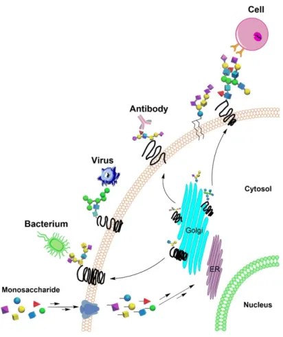
Glycans commonly existed on cell surfaces. In addition to their effect on protein
folding and properties, they also mediate numerous biological processes, including but not
limited to signaling, fertilization, embryogenesis, cell proliferation and differentiation[79]
(Figure 1.5). Plentiful data have shown that glycosylation is related to pathogen recognition,
inflammation, innate immune response, autoimmune diseases and development of cancer
[80, 81]. For instance, glycan-binding protein CD22 on the surface of activated B
lymphocytes can recognize terminal Neu5Ac-α-2,6-Gal sequence in Asn-linked glycans
[82]. Another example is that sialyl Lewis x tetrasaccharide can mediate cell adhesion,
which has been used for drug development to treat common diseases such as rheumatoid
arthritis and heart diseases [83]. However, more glycan functions are yet to be discovered,
Figure 1. 5 Interactions between glycans and receptors mediate biological processes.
1.2.2 Structural diversity and classification of glycans
(1) Structural diversity of glycans
Glycans are monosaccharide chains linked by glycosidic linkages. Since (1) each
monosaccharide has several hydroxyl groups that can form glycosidic linkage with other
monosaccharides, (2) the formed glycosidic linkages can be in α or β configuration, (3) the
glycans can be linear or branched, (4) there are over ten common monosaccharides, when
glycans are formed by certain number of monosaccharides, the sequence of sugar residues,
the position where glycosidic bonds form, the configuration of anomeric carbons in the
formed glycosidic bond, and the types of monosaccharides can all vary, and will result in
encode and carry numerous information, and deliver the information by interacting with
specific molecules.
(2) Classification of glycans
In nature, glycans can exist as unbonded compounds, e.g. human milk
oligosaccharides (HMOs) and hyaluronan, or in glycoconjugates by covalent bonding to
protein or lipid. Among these glycans, the ones that exist in glycoconjugates (glycoproteins
and glycolipids) are the glycans function mostly in intercellular interactions. In this work,
we focus on the glycans in glycoproteins. Per the linkage between glycan and
protein/peptide, glycans in glycoproteins can be sub-grouped into N-glycans and O-glycan.
N-glycans are linked to protein/peptide by linkages between the anomeric carbon of
N-acetylglucosamine (GlcNAc) at its reducing end and the amino group on the side chain
of Asn residue. They exist widely in immunoglobulin and other therapeutic proteins. The
structures of N-glycans are relatively clearly examined. Almost all N-glycans contain a
common pentasaccharide core structure, composing of three mannose (Man) residues and
two GlcNAc residues. They are further divided into high-mannose type, complex type and
hybrid type, depending on the detailed structures of the appendages on the N-glycan core
[84].
O-glycan are connected to protein/peptide through the glycosidic bond between the
anomeric carbon of the reducing sugar and the hydroxyl group in the side chain of Ser, Thr,
4-hydroxylproline (Hyp) or 5-hydroxylysine (Hyl). O-glycans are also structurally
diversified. For example, the O-glycans rich in mucin are linked to the hydroxyl group in
amino acid through α-glycosidic bond from N-acetylgalactosamine (GalNAc) [85]. Eight
can be further extended with galactose (Gal), GlcNAc, fucose (Fuc) and/or sialic acid (Sia)
[84]. Many antigenic epitopes and cancer-related glycan epitopes exist in the extension
structure of O-GalNAc glycans [86]. Another type of O-glycans, O-Man glycans, forms
linkages between the Man residue at the reducing end and protein. The aberrant structure
will lead to congenital muscular dystrophies [87, 88] and cancer [89-91]. In addition, many
proteins are modified by O-Fuc, O-glucose (Glc) and O-xylose (Xyl) glycans through α
-Fuc, β-Glc and β-Xyl linkages to Ser/Thr residue For instance, the epidermal growth factor
(EGF)-like repeats of Notch protein are O-glucosylated [92], while the EGF-like repeats in
Notch receptor are O-fucosylated, and the O-fucosylation is related to protein folding [93].
1.2.3 Preparation of structurally defined glycans
Although the critical functions of protein glycosylation have attracted researchers’
attention, it is still challenging to fully interpret the molecular mechanisms under their
functions. The lack of structurally defined complex glycans as standard has hindered the
analysis of accurate structures of glycans in complicated mixtures [94, 95], and hampered
the specificity and characterization study of glycan-binding proteins (GBPs) existed in
nature [96-98]. Therefore, structurally defined homogeneous glycans are a prerequisite and
are highly demanded for structural and functional study of glycans. Currently, isolation and
synthesis are two main approaches for glycan preparation.
(1) Isolation of glycans
Glycans isolated from natural sources include that human milk oligosaccharides,
glycans from glycolipids and glycoproteins by chemical hydrolysis or endoglycosidases,
proteoglycans (GAGs) and fragments obtained by chemical or enzymatic degradation, and
glycans from glycoproteins/glycopeptides, natural sources that are rich in glycans (highly
glycosylated proteins/peptides) are required, but usually only limited amount of glycan can
be obtained by this method [99]. In addition, except for a few glycoproteins which contain
glycans with relatively homogeneous structures such as Man9GlcNAc2 N-glycan from
soybean agglutinin [100] and complex type N-glycan from sialylglycopeptide (SGP) from
egg yolk [101], glycans isolated from most glycoproteins are mixtures and further
separation of glycans with similar structures is very challenging [99]. Therefore, isolation
from natural sources is not a reliable strategy to afford substantial structurally defined
glycans.
(2) Synthesis of glycans
Preparation of glycan library by chemical, chemoenzymatic or enzymatic synthesis
has been widely applied to perform biological and structural studies [97, 98, 102-104]. In
chemical synthesis, a series of protecting groups with different reactivities have been
developed to selectively protect the hydroxyl groups located at different positions in the
sugar ring but have similar properties. Then, the protecting group at the target site is
removed to expose the hydroxyl group for glycosidic linkage formation. Also, protecting
groups with different properties are used to create specific spatial hindrance near the
anomeric carbon and force the formation of glycosidic linkage with specific configuration
(α or β) [105]. Furthermore, to simplify the synthetic approach and omit purification
process, one-pot solution-based synthesis and solid-phase synthesis have been developed.
In the former strategy, glycosyl donors with different reactivities are mixed into one reactor.
The synthesis of complex glycans is achieved by controlling the reaction condition to
Two hexosaccharyl tumor-associated carbohydrate antigens (TACAs) Globo-H and GM1
have been accomplished by this strategy [107-109]. In the latter strategy, glycosyl acceptors
are immobilized to the surface of solid-phase, and are well exposed to solutions containing
glycosyl donors to react. Based on this strategy, Seeberger group developed automated
glycan synthesis, and generated histo-blood antigen Lewis x and Lewis y, TACAs Gb-3
and Globo-H and so on [110, 111].
Despite its major progress, chemical synthesis of complex glycans is still
time-consuming, especially in the cases when the target glycans are very complicated [97].
Therefore, chemoenzymatic strategy with higher efficiency has been developed, in which
a glycan precursor is synthesized chemically, and then is extended by a set of
glycosyltransferases (GTs) to provide more complex glycans [103, 104]. The GTs used
here are a group of enzymes that can transfer saccharide (usually monosaccharide) from
glycosyl donor to glycosyl acceptor and form a glycosidic linkage between them[103].
Most GTs require sugar nucleotides as their donor, while a minority of GTs can use sugar
phosphate, glycolipid or glycan itself as sugar donor. The reactions catalyzed by most GTs
have strict stereo-selectivity and regio-selectivity with only a few exceptions [103]. In
addition, some GTs can also recognize and utilize analogs of their natural sugar donors[103]
and provide glycans containing bioorthoganol groups which can assist the study of
biological functions of glycans.
Many research groups have been focusing on synthesizing branching glycans
chemically or chemoenzymatically [99, 112-116]. Majority of these work dealt with
symmetric glycans, i.e. the glycan structures of each branch are the same, the best synthetic
glycan structures on different appendages are different, chemical synthesis is employed. To
achieve this, protecting groups with different reactivities are used at the branching point,
and suitable conditions are used to sequentially deprotect each protecting group followed
by extension of the branch to generate the final asymmetric glycans [99], which can better
mimic natural branching glycans that usually carry different appendages at each branching
point [2].
However, both chemical and chemoenzymatic synthesis is dependent on expertise
related to chemical synthesis. If no chemists are available, enzymatic synthesis is the only
option. With glycopeptide isolated from egg yolk as precursor, Kajihara et al prepared a
series of asymmetric bi-antennary complex-type N-glycans by using glycosidases and GTs
[117, 118]. The glycosidases used can partially hydrolyze some sugar residues from natural
glycans, and GTs can selectively extend the glycans. Nevertheless, structures of glycans
generated solely by enzymatic synthesis are usually limited.
1.2.4 Glycan microarray
Glycans on the surface of living cells is diverse structures and mediate biological
function by glycan binding proteins (GBPs) [119-124]. Recently, glycan microarray provides
high-throughput approaches for decoding the biological function of glycans in vitro and is
applied as a tool to screen many sugar-biomolecular interations. Glycan microarray was firstly
reported in 2002 and various approaches for glycan immobilization have been developed by
several research group [125-131]. So far, all glycans that have been applied for glycan
microarray were synthetic glycans or/and glycans isolated from natural sources.
When structure-defined glycans are obtained, an appropriate approach for functional
categorized into two methods: (1) non-covalent immobilization: nitrocellulose and oxidized
polystyrene coated glass slides normally designed for immobilization, and (2) covalent
immobilization: the most covalent methods for glycan microarray have employed robust amine
and thiol chemistries, which provide more specific attachment for the structure-defined glycan
probes [132]. Amine chemistry is the most widely applied for glycan microarray now (Figure
1.6).
Figure 1. 6 Mechanism of glycan-amine bound to slides.
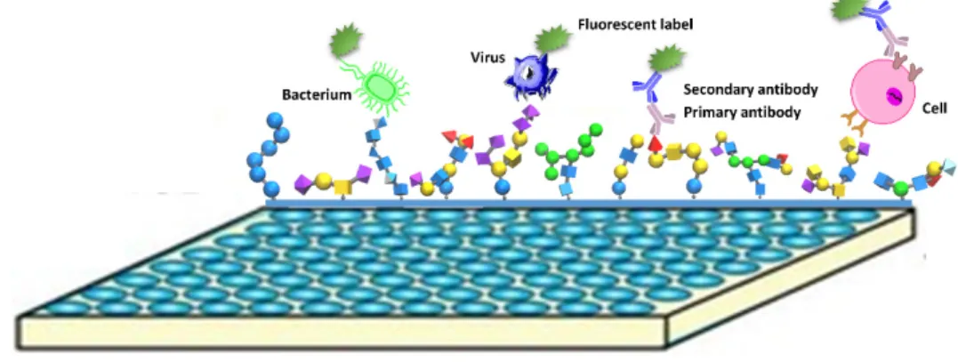
Glycan microarray can be successfully applied to analyze the binding specificity between
glycans and proteins (e.g. lectins and antibodies), pathogens, as well as cells (Figure 1.7). Plant
lectins can recognize specific epitopes in glycans. However, some plant lectins have specific
requirement for binding research. For example, conconavalin A (ConA), a plant lectin with
different concentration showed similar binding affinity to mannose when concentration of
glycans in the slide was high. But it can only bind the high affinity glycans when concentration
of glycan was low [133]. Glycan microarray were applied to study infectious disease because
For instance, by glycan microarray analysis, it was reported that seasonal H1N1 virus only bound
α2–6 sialyl sequences, however, H1N1 pandemic virus can bind α2–6 and α2–3 sialyl sequences
[136]. The clinical application of glycan microarrays has also been developed. For example, a
glycan microarray containing 37 glycans was applied for identification of changes of glycan
structure that can initiate humoral immune response in classical Hodgkin's lymphoma (cHL)
patients [137].
Figure 1. 7 Glycans can be specific bound by proteins, pathogens and cells.
So far, a major issue for microarray is how to design a library to unveil biological
functions of glycans. Due to complexity of glycans, the availability of structure-defined glycans
is very limited, which is an obstacle for glycan microarray application. Focused libraries which
are composed of a small group of glycans are relatively easy to prepare and can address a
specific issue, however, it has limitation for broad application in biological research. Glycans
libraries with large size of structure-defined glycans are still hard to acquire. Therefore, a glycan
microarray which can cover most of the natural glycan structural space of the glycome are highly
1.3 Synthesis of oligosaccharides in large scale
1.3.1 Significance of oligosaccharide synthesis in large scale
Some significant functional complex oligosaccharides are required in large scale in
industry, such as Globo-H and HMOs. The presence of Globo-H oligosaccharide in glycolipids
is strongly associated with the aggressiveness of several types of tumors [138]. As a result, the
Globo-H oligosaccharide has been used in a glycoconjugate vaccine, which is under Phase II
clinical trials for its immunotherapy activity in patients with metastatic breast cancer. Human
milk oligosaccharides (HMOs), which are unconjugated and unique to human breast milk, are
another group of structurally diverse glycans. HMOs function as prebiotics that facilitates the
establishment of commensal bacteria in the human gut [139]. Studies have shown that
inflammatory bowel diseases or their chronic manifestation might be prevented in breast-fed
infants, due to interactions between HMOs and mucosal leukocytes [140]. Abbott Nutrition
launched New Similac® which is cow milk-based infant formula supplemented with the simple
HMO 2’-fucosyllactose (2’-FL), and clinical results demonstrated that infants fed by formula
containing 2’-FL showed lower plasma and ex vivo inflammatory cytokine profiles, comparable
to a breast-fed reference group [141]. Oligosaccharides are therefore a promising area of study
for biological evaluation and roles in human disease. However, the availability of large amounts
of structurally complex, pure oligosaccharides at low cost has lagged behind the demand of
research and applications. There is an urgent need to develop efficient large-scale production
strategies to meet the demand for structurally defined oligosaccharides in pharmaceutical and
1.3.2 Challenges for current large-scale oligosaccharide production
Generally, three strategies are employed for large-scale oligosaccharide production: (1)
chemical synthesis, (2) enzymatic synthesis, and (3) whole-cell synthesis/living cell factory.
Each strategy has distinct challenges.
(1) Chemical synthesis.
Chemical synthesis of oligosaccharides from tetrasaccharides to octasaccharides have
been reported [142]. But the difficulty of chemical synthesis is associated with the intrinsic
structural complexity of oligosaccharides, and has long hindered their production. For simple
trisaccharides, such as 2’-FL (Fucα1–2Galβ1–4Glc) and 6’-SL (Siaα2–6Galβ1–4Glc), total
chemical synthesis has been developed in large scale. However, for large-scale synthesis of more
complex oligosaccharides, lengthy synthetic steps and relatively low overall yield impair the
practicality of pure chemical synthesis. In addition, chemical synthesis of oligosaccharide in
large scale is still challenging due to toxic reagents which are not suitable for applications in
pharmaceutical and nutritional food area.
(2) Enzymatic synthesis.
Bacterial glycosyltransferases (GTs) have been used as suitable tool for oligosaccharide
synthesis beucause of their relaxed substrate specificity. The high regioselectivity and
stereoselectivity of enzyme-catalyzed reactions have made one-pot multi-enzyme (OPME)
reactions an attractive method for oligosaccharide synthesis. OPME simultaneously combines
several enzymes in a pathway for sugar nucleotides synthesis and transfer, and synthesizes
oligosaccharides with relatively high yield and low cost. Using OPME, Wong and coworkers
combined sugar nucleotide regeneration enzymes and glycosyltransferases (GTs) to achieve
reduced by replacing expensive sugar nucleotides sugar with nucleotide regenerating enzymes,
however, preparation of multitude of enzymes required for the addition of a single sugar to the
antigen made the whole process very time- and labor- intensive. Chen and coworkers employed a
similar strategy, including sugar nucleotide generating enzymes instead of regenerating enzymes
[143], but a large number of enzymes were still required and likely to hinder scaled-up
production of oligosaccharides (Figure 1.8).
Figure 1. 8 OPME strategy for oligosaccharides synthesis.
(3) Whole-cell synthesis/living cell factory.
In a third strategy to bypass protein purification, whole cells containing the necessary
cell factories” during cell growth. Using whole-cell catalysts by freeze-and-thaw strategy,
Kyowa Hakko Kogyo Co., has established a 10-100 gram-scale oligosaccharide production
platform by combining low-cost starting materials and metabolically engineered bacteria to
specialize in the production of nucleotides, sugar nucleotides, or oligosaccharides [144-146].
Similarly, Wang and coworkers achieved multi-gram scale production of lactose-based
trisaccharides using “superbugs,” which contained recombinant enzymes responsible for sugar
nucleotide regeneration or generation in one E. coli strain [147, 148]. Neverthless, the Kyowa
Hakkko and Wang whole-cell strategies were limited to oligosaccharides with relatively simple
structures.
To synthesize oligosaccharides by whole-cell strategy, sugar donor and acceptor need to
be prepared. In E. coli, uridine-5’-diphosphate N-acetylglucosamine (UDP-GlcNAc) and
uridine-5’-diphosphate galactose (UDP-Gal), the donor substrates that are required by
glycosyltransferases (GTs) for oligosaccharide synthesis, can be produced in the growing cells as
required precursors of lipopolysaccharide (LPS) and peptidoglycan polysaccharide for cell wall
construction. Therefore, the intracellular UDP-GlcNAc and UDP-Gal can be “borrowed” by the
recombinant GTs for extending oligosaccharide in E. coli. For acceptor, lactose is an inexpensive
candidate for oligosaccharide biosynthesis in E. coli [149]. LacY is a transporter which can
transfer lactose from the medium into cytosol [150]. The activity of LacZ, a β-galactosidase that
can hydrolyze lactose, should be diminished while the lactose uptake must be retained or even
enhanced [151, 152]; therefore, LacZ-, LacY+E. coli is chosen as preferred strains for using
lactose as an acceptor to synthesize oligosaccharide in E. coli. Based on this technology, many
groups attempted to produce oligosaccharide by direct fermentation. Samain et al attempted to
substrate with a single genetically engineered E. coli strain containing all GTs needed for
oligosaccharide synthesis (Figure 1.9). By Samain’s strategy, lacto-N-triose (LNT) II (6 g/L),
GD3 (0.83 g/L) and lacto-N-neofucopentaose (LNnFP)-I (1.5 g/L) can be produced[153-155].
Figure 1. 9 Biosynthetic pathway of LNnFP I from lactose in engineered E. coli cells.
2'-FL, a trisaccharide, can be synthesized in engineered E. coli by two pathways: (1) de
novo pathway and (2) salvage pathway, which are reported by different research groups
[155-160]. (Figure 1.10). The de novo pathway is the natural metabolic pathway in E. coli. In this
pathway, five recombinant enzymes (Man A. ManB, ManC, Gmd, WcaG/Fcl) were utilized to
synthesize GDP-Fuc from fructose-6-phosphate. Under catalysis of recombinant α1–2
fucosyltransferase in E. coli, 2’-FL can be produced in large scale in vivo. By this strategy,
Albermann group produced 2’-FL with concentration of 20.28±0.83 g/L [157]. Salvage pathway
is naturally present in some Bacteroides strains and mammalian cells. In this pathway, the key
Bacteroides fragilis which can synthesize GDP-fucose from L-Fucose. By this strategy, Seo
group synthesized 2’-FL with concentration of 23.1 g/L [159].
Figure 1. 10 Biosynthesis of 2’-FL by de novo pathway and salvage pathway in engineered E.
coli.
Unfortunately, this “living cell factory” strategy had two major restrictions for production
of oligosaccharides larger than trisaccharides: (a) Larger oligosaccharide substrates could not be
transported into living E. coli for additional single-step extensions; (b) Several intermediates
were formed along the biosynthetic pathway when multiple GTs were coexpressed for mono- or
disaccharide acceptor extension, leading to difficulty in purifying the intended product. For
example, LNT-II, lacto-N-neotetraose (LNnT) and 2'-FL were produced as intermediates when
Based on the challenges described above, a practical, cost-effective strategy for
large-scale synthesis of structurally complex oligosaccharides remains a high-priority for driving
advances in the glycoscience field.
1.4 Outline of the work in this dissertation
The work in this dissertation mainly focuses on investigating the significance of glycans
by synthesis of structurally-defined glycopeptiedes/glycoproteins and glycans. More specifically,
in Chapter 2, two techniques were developed for synthesis of glycopeptide/glycoprotein. (1) O
-linked-N-glycan-peptide/protein were synthesized and GLP-1 with O-linked N-glycan kept
unchanged secondary structure after glycosylation, suggesting the potential application of this
approach for peptide/protein drug production. (2) By three steps of enzymatic catalysis,
glycopeptides carrying complex-type N-glycans can be efficiently synthesized. In Chapter 3, two
focused glycans libraries were synthesized which were N-glycans containing tandem epitopes
and a new class of asymmetric N-glycans. By glycan microarray study, N-glycans containing
tandem epitopes library showed when interpreting the binding between glycans and
GBPs/viruses, not only the structure of the terminal glycan epitopes, but also the internal
epitopes and/or modifications of terminal epitopes need to be taken into account; a new class of
asymmetric N-glycans library showed that branch-biased binding and spacial inhibition caused
by bulky group on the other branch when glycans were bound to GBPs. In Chapter 4, a microbial
glycosyltrasferase Hpα1–3/4FT was characterized and applied in the facile synthesis of
fucosylated human milk oligosaccharides (HMOs). In addition, multi-gram scale of fucosylated
HMOs were synthesized by combined engineered E. coli strains.
1.5 References
1. Haltiwanger, R.S. and J.B. Lowe, Role of glycosylation in development. Annu Rev Biochem, 2004.
2. Moremen, K.W., M. Tiemeyer, and A.V. Nairn, Vertebrate protein glycosylation: diversity,
synthesis and function. Nat Rev Mol Cell Biol, 2012. 13(7): p. 448‐62.
3. Cummings, R.D., The repertoire of glycan determinants in the human glycome. Mol Biosyst, 2009. 5(10): p. 1087‐104.
4. Nairn, A.V., et al., Regulation of glycan structures in animal tissues: transcript profiling of glycan‐
related genes. J Biol Chem, 2008. 283(25): p. 17298‐313.
5. Spiro, R.G., Protein glycosylation: nature, distribution, enzymatic formation, and disease
implications of glycopeptide bonds. Glycobiology, 2002. 12(4): p. 43R‐56R.
6. Hoseki, J., R. Ushioda, and K. Nagata, Mechanism and components of endoplasmic reticulum‐
associated degradation. J Biochem, 2010. 147(1): p. 19‐25.
7. Sinclair, A.M. and S. Elliott, Glycoengineering: the effect of glycosylation on the properties of
therapeutic proteins. J Pharm Sci, 2005. 94(8): p. 1626‐35.
8. Schachter, H., The joys of HexNAc. The synthesis and function of N‐ and O‐glycan branches. Glycoconj J, 2000. 17(7‐9): p. 465‐83.
9. Goldwasser, E., C.K. Kung, and J. Eliason, On the mechanism of erythropoietin‐induced
differentiation. 13. The role of sialic acid in erythropoietin action. J Biol Chem, 1974. 249(13): p.
4202‐6.
10. Fukuda, M.N., et al., Survival of recombinant erythropoietin in the circulation: the role of
carbohydrates. Blood, 1989. 73(1): p. 84‐9.
11. Davies, J., et al., Expression of GnTIII in a recombinant anti‐CD20 CHO production cell line:
Expression of antibodies with altered glycoforms leads to an increase in ADCC through higher
affinity for FC gamma RIII. Biotechnol Bioeng, 2001. 74(4): p. 288‐94.
12. Shinkawa, T., et al., The absence of fucose but not the presence of galactose or bisecting N‐
acetylglucosamine of human IgG1 complex‐type oligosaccharides shows the critical role of
enhancing antibody‐dependent cellular cytotoxicity. J Biol Chem, 2003. 278(5): p. 3466‐73.
13. Yamane‐Ohnuki, N. and M. Satoh, Production of therapeutic antibodies with controlled
fucosylation. MAbs, 2009. 1(3): p. 230‐6.
14. Frokjaer, S. and D.E. Otzen, Protein drug stability: a formulation challenge. Nat Rev Drug Discov, 2005. 4(4): p. 298‐306.
15. Manning, M.C., K. Patel, and R.T. Borchardt, Stability of protein pharmaceuticals. Pharm Res, 1989. 6(11): p. 903‐18.
16. Jefferis, R., Glycosylation as a strategy to improve antibody‐based therapeutics. Nat Rev Drug Discov, 2009. 8(3): p. 226‐34.
17. Sola, R.J. and K. Griebenow, Effects of glycosylation on the stability of protein pharmaceuticals. J Pharm Sci, 2009. 98(4): p. 1223‐45.
18. Chung, C.H., et al., Cetuximab‐induced anaphylaxis and IgE specific for galactose‐alpha‐1,3‐
galactose. N Engl J Med, 2008. 358(11): p. 1109‐17.
19. Sethuraman, N. and T.A. Stadheim, Challenges in therapeutic glycoprotein production. Curr Opin Biotechnol, 2006. 17(4): p. 341‐6.
20. Bernard, B.A., K.M. Yamada, and K. Olden, Carbohydrates selectively protect a specific domain of
fibronectin against proteases. J Biol Chem, 1982. 257(14): p. 8549‐54.
21. Runkel, L., et al., Structural and functional differences between glycosylated and non‐
glycosylated forms of human interferon‐beta (IFN‐beta). Pharm Res, 1998. 15(4): p. 641‐9.
22. Tsuda, E., et al., The role of carbohydrate in recombinant human erythropoietin. Eur J Biochem, 1990. 188(2): p. 405‐11.
24. Endo, Y., et al., Heat‐induced aggregation of recombinant erythropoietin in the intact and
deglycosylated states as monitored by gel permeation chromatography combined with a low‐
angle laser light scattering technique. J Biochem, 1992. 112(5): p. 700‐6.
25. Uchida, E., et al., Effect of active oxygen radicals on protein and carbohydrate moieties of
recombinant human erythropoietin. Free Radic Res, 1997. 27(3): p. 311‐23.
26. Elliott, S., et al., Control of rHuEPO biological activity: the role of carbohydrate. Exp Hematol, 2004. 32(12): p. 1146‐55.
27. Egrie, J.C., et al., Darbepoetin alfa has a longer circulating half‐life and greater in vivo potency
than recombinant human erythropoietin. Exp Hematol, 2003. 31(4): p. 290‐9.
28. Elliott, S., et al., Enhancement of therapeutic protein in vivo activities through glycoengineering. Nat Biotechnol, 2003. 21(4): p. 414‐21.
29. Perlman, S., et al., Glycosylation of an N‐terminal extension prolongs the half‐life and increases
the in vivo activity of follicle stimulating hormone. J Clin Endocrinol Metab, 2003. 88(7): p. 3227‐
35.
30. Ziltener, H.J., et al., Carbohydrate does not modulate the in vivo effects of injected interleukin‐3. Exp Hematol, 1994. 22(11): p. 1070‐5.
31. Byrne, B., G.G. Donohoe, and R. O'Kennedy, Sialic acids: carbohydrate moieties that influence
the biological and physical properties of biopharmaceutical proteins and living cells. Drug Discov
Today, 2007. 12(7‐8): p. 319‐26.
32. Casadevall, N., et al., Pure red‐cell aplasia and antierythropoietin antibodies in patients treated
with recombinant erythropoietin. N Engl J Med, 2002. 346(7): p. 469‐75.
33. Moore, W.V. and P. Leppert, Role of aggregated human growth hormone (hGH) in development
of antibodies to hGH. J Clin Endocrinol Metab, 1980. 51(4): p. 691‐7.
34. Seitz, O., Glycopeptide synthesis and the effects of glycosylation on protein structure and
activity. Chembiochem, 2000. 1(4): p. 214‐46.
35. Hojo, H. and Y. Nakahara, Recent progress in the field of glycopeptide synthesis. Biopolymers, 2007. 88(2): p. 308‐24.
36. Wang, L.X. and M.N. Amin, Chemical and chemoenzymatic synthesis of glycoproteins for
deciphering functions. Chem Biol, 2014. 21(1): p. 51‐66.
37. Roberts, A.G., et al., Fully Synthetic Granulocyte Colony‐Stimulating Factor Enabled by Isonitrile‐
Mediated Coupling of Large, Side‐Chain‐Unprotected Peptides. J Am Chem Soc, 2015. 137(40): p.
13167‐75.
38. Mezzato, S., M. Schaffrath, and C. Unverzagt, An orthogonal double‐linker resin facilitates the
efficient solid‐phase synthesis of complex‐type N‐glycopeptide thioesters suitable for native
chemical ligation. Angew Chem Int Ed Engl, 2005. 44(11): p. 1650‐4.
39. Yamamoto, N., et al., Chemical synthesis of a glycoprotein having an intact human complex‐type
sialyloligosaccharide under the Boc and Fmoc synthetic strategies. J Am Chem Soc, 2008. 130(2):
p. 501‐10.
40. Payne, R.J. and C.H. Wong, Advances in chemical ligation strategies for the synthesis of
glycopeptides and glycoproteins. Chem Commun (Camb), 2010. 46(1): p. 21‐43.
41. Unverzagt, C. and Y. Kajihara, Chemical assembly of N‐glycoproteins: a refined toolbox to address
a ubiquitous posttranslational modification. Chem Soc Rev, 2013. 42(10): p. 4408‐20.
42. Aussedat, B., et al., Total synthesis of the alpha‐subunit of human glycoprotein hormones:
toward fully synthetic homogeneous human follicle‐stimulating hormone. J Am Chem Soc, 2012.
134(7): p. 3532‐41.
43. Nagorny, P., et al., Probing the frontiers of glycoprotein synthesis: the fully elaborated beta‐
subunit of the human follicle‐stimulating hormone. Angew Chem Int Ed Engl, 2012. 51(4): p. 975‐