comment
reviews
reports
deposited research
interactions
information
refereed research
Research
Quod erat demonstrandum
? The mystery of experimental validation
of apparently erroneous computational analyses of protein
sequences
Lakshminarayan M Iyer*, L Aravind*, Peer Bork
†
, Kay Hofmann
‡
, Arcady R
Mushegian
§
, Igor B Zhulin
¶
and Eugene V Koonin*
Addresses: *National Center for Biotechnology Information, National Library of Medicine, National Institutes of Health, Bethesda, MD 20894, USA. †EMBL, Biocomputing, Meyerhofstrasse 1, 69117 Heidelberg, Germany. ‡MEMOREC Stoffel GmbH, Köln D-50829,
Germany. §Stowers Institute for Medical Research, 1000 E 50th Street, Kansas City, MO 64410, USA. ¶School of Biology, Georgia Institute of
Technology, Atlanta, GA 30332, USA.
*Correspondence: Eugene V Koonin. E-mail: [email protected]
Abstract
Background: Computational predictions are critical for directing the experimental study of
protein functions. Therefore it is paradoxical when an apparently erroneous computational prediction seems to be supported by experiment.
Results: We analyzed six cases where application of novel or conventional computational
methods for protein sequence and structure analysis led to non-trivial predictions that were subsequently supported by direct experiments. We show that, on all six occasions, the original prediction was unjustified, and in at least three cases, an alternative, well-supported computational prediction, incompatible with the original one, could be derived. The most unusual cases involved the identification of an archaeal cysteinyl-tRNA synthetase, a dihydropteroate synthase and a thymidylate synthase, for which experimental verifications of apparently erroneous computational predictions were reported. Using sequence-profile analysis, multiple alignment and secondary-structure prediction, we have identified the unique archaeal ‘cysteinyl-tRNA synthetase’ as a homolog of extracellular polygalactosaminidases, and the ‘dihydropteroate synthase’ as a member of the --lactamase-like superfamily of metal-dependent hydrolases.
Conclusions:In each of the analyzed cases, the original computational predictions could be refuted and, in some instances, alternative strongly supported predictions were obtained. The nature of the experimental evidence that appears to support these predictions remains an open question. Some of these experiments might signify discovery of extremely unusual forms of the respective enzymes, whereas the results of others could be due to artifacts.
Published: 13 November 2001
GenomeBiology2001, 2(12):research0051.1–0051.11
The electronic version of this article is the complete one and can be found online at http://genomebiology.com/2001/2/12/research/0051 © 2001 Iyer et al., licensee BioMed Central Ltd
(Print ISSN 1465-6906; Online ISSN 1465-6914)
Received: 3 July 2001 Revised: 7 September 2001 Accepted: 4 October 2001
Background
The availability of a large number of protein sequences, including complete protein sets encoded in diverse genomes, and the rapidly growing database of protein structures have
structural similarities between proteins. In particular, methods that extract information from multiple alignments to construct various types of sequence profiles and use the resulting sequence profiles for iterative database searching, such as PSI-BLAST and Hidden-Markov-Model (HMM)-based approaches, have substantially improved the detection of subtle similarities between proteins that previously were amenable only to direct structural comparison [3,4]. The sensitivity and accuracy of these methods have been exten-sively tested and statistical approaches for validating the observed similarities are available [5-11].
Despite these achievements, detection and interpretation of relationships between homologous proteins that have limited sequence similarity remains a major challenge. Such studies typically require a case-by-case approach that is guided by a detailed understanding of protein sequence-structure pat-terns and is rooted in the biology of the proteins analyzed. Prediction of structures and function(s) of uncharacterized proteins is one of the principal outcomes of these analyses, and experimental verification of such predictions tends to increase confidence in the validity of sequence-structure comparative approaches. The negative feedback from experi-ments that failed to confirm a computational prediction is potentially even more important, because it could result in revision and refinement of the computational methods. When examining cases of reported prediction followed by experimental validation, however, we encountered several paradoxical situations. In each of these, a prediction that has been reportedly confirmed by experiment was incompatible with results obtained with several standard computational procedures. More importantly, alternative predictions, sup-ported by statistically significant sequence and/or structural similarity, were made in some of these cases. Here we present several such mysteries, describe the refutation of the original predictions and the new predictions, wherever feasi-ble, and discuss the discrepancy between the computational and experimental results. The choice of the cases was not systematic; rather, those chosen were notable because they relied on novel computational techniques, exploited particu-larly subtle sequence or structural motifs, and dealt with crucial biological problems.
Results
MJ1477: a predicted archaeal cysteinyl-tRNA synthetase
Aminoacyl-tRNA synthetases (aaRSs) specific for 17 of the 20 amino acids are universally present in cellular life forms. The three exceptions are GlnRS, AsnRS and CysRS. GlnRS and AsnRS are missing in many bacteria and archaea because glutamine and asparagine are incorporated into proteins through transamidation of glutamate and aspartate, respectively. CysRS is missing in two archaeal methanogens whose genomes have been sequenced - Methanobacterium
thermoautotrophicumand Methanococcus jannaschii[12]. No alternative mechanism for cysteine incorporation into proteins is known; hence the absence of CysRS in these organisms was an enigma.
Two solutions to this puzzle, both unusual, have recently been proposed and experimentally validated. One involves non-orthologous gene displacement, a situation in which the same essential function is carried out by distantly related or even unrelated proteins in different organisms [13,14]. It has been shown that M. jannaschiiProRS, a class II synthetase that is unrelated to the class I CysRS, substituted for the missing CysRS activity [15-17]. The other solution involved a new candidate for the role of CysRS, the MJ1477 protein from M. jannaschii. This protein and its orthologs (direct evolutionary counterparts related by vertical descent from a common ancestor) from the bacteria Thermotoga maritima and Deinococcus radiodurans were identified as ‘distant orthologs’ of the Bacillus subtilisCysRS by using a computa-tional method specifically designed to detect distantly related orthologs [18]. The method is based on application of discriminant analysis to alignment scores, in order to sepa-rate the scores for pairs of functionally identical proteins from different genomes from the scores for proteins with dif-ferent functions. This prediction was then validated experi-mentally by showing that MJ1477 had CysRS activity in vitro and that an ortholog of MJ1477 from D. radiodurans, DR0705, complemented a CysRS deficient, temperature-sensitive, lethal E. coli mutant strain [18]. An important corollary of these surprising findings is a rapid divergence of the MJ1477 family from CysRS, such that all the catalytic and otherwise functionally important residues characteristic of this enzyme, and also present in other class I aaRSs, have changed. Furthermore, MJ1477 and its orthologs do not have the accessory domains found in all known CysRS, namely the DALR domain (named after a distinct amino-acid signature), which is shared by aaRSs of several specifici-ties, and another domain specific to CysRS [19].
comment
reviews
reports
deposited research
interactions
information
refereed research
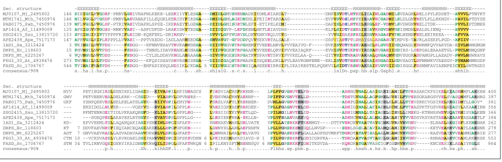
residue; p, polar residue), in which the conserved aspartate is likely to directly participate in catalysis (Figure 1). The hybrid-fold-recognition method, which combines sequence-profile analysis with alignment-based secondary-structure prediction [22] and the 3D-PSSM method [23] both sug-gested a likely ,-amylase-like triosephosphate isomerase (TIM) barrel structure for this protein family. Thus, although the identification of MJ1477 as a secreted poly-galactosaminidase or a related polysaccharide hydrolase with a different specificity awaits experimental verification, it shows all the signs of a correct computational prediction: statistically significant similarity between the analyzed protein and an experimentally characterized enzyme; con-servation of distinct motifs implicated in catalysis; potential presence of a structural fold compatible with the experimen-tally demonstrated enzymatic activity; and confident predic-tion of the extracellular localizapredic-tion that is, again, compatible with a polysaccharide hydrolase activity involved in environmental carbohydrate utilization or capsular metabolism. None of this evidence is offered by the analysis that led to the CysRS prediction for MJ1477.
Therefore we are forced to conclude that MJ1477 and its homologs are not related to CysRS and there is nothing in the computational analysis of these proteins that would point to an aaRS activity. In contrast, we predict these pro-teins to be extracellular polygalactosaminidases or similar polysaccharide hydrolases. The polysaccharide hydrolase and aaRS functions seem to be essentially incompatible. First, a secreted enzyme is unlikely to function as an aaRS
whose site of action is, by definition, intracellular. Second, even if an entirely new class of aaRSs is postulated, the reac-tion catalyzed by this new aaRS does not resemble polysac-charide hydrolysis or its reversal. Aminoacyl-tRNA synthetases catalyze a succession of reactions, which involve: hydrolysis of the ,--phosphate bond in ATP; condensation of AMP with the cognate amino acid, resulting in the forma-tion of an aminoacyl-adenylate; displacement of the AMP moiety of the aminoacyl-adenylate with the cognate tRNA, producing aminoacyl-tRNA. Even if the two condensation reactions, in very general terms, could be considered a rever-sal of the polysaccharide hydrolysis reaction, there is no indication that polysaccharide hydrolases could bind and hydrolyze ATP, and the multiple alignment of the MJ1477 family did not include any conserved signatures typical of potential phosphate-binding loops (Figure 1). Neither does this family contain any recognizable RNA-binding domains. Finally, M. thermoautotrophicum does not encode any homologs of MJ1477, ruling out the possibility that this family encompasses CysRS of both archaeal methanogens. Taken together, these observations appear to effectively refute the prediction of a CysRS activity, thus pitting compu-tational results against experimental data.
MJ0301: a predicted dihydropteroate synthase
[image:3.609.55.556.87.257.2]Dihydropteroate synthase (DHPS) catalyzes the condensation of p-aminobenzoic acid with 7,8-dihydro-6-hydroxy-methylpterin pyrophosphate to give 7,8-dihydropteroate, an intermediate in folate metabolism. The protein from Staphy-lococcus, a Gram-positive bacterium, has been crystallized
Figure 1
Multiple alignment of the polygalactosaminidase family that includes MJ1477, the alleged archaeal CysRS. Proteins are denoted by their gene name, followed by their species abbreviations and GenBank identifier (GI) numbers. The coloring reflects the 100% consensus. The consensus abbreviations and coloring scheme used in this and subsequent figures are as follows. Hydrophobic residues (h; LIYFMWACV) and aliphatic (l; LIAV) residues are shaded yellow. Colored magenta are alcohol (o; ST), charged (c; KERDH), basic (+; KRH), acidic (- ; DE), and polar (p; STEDRKHNQ) residues. Small (s; SAGDNPVT) residues are colored green and big (b; LIFMWYERKQ) residues are shaded gray. The hydrophobic residues of the signal peptide are highlighted in yellow. In the Secondary Structure line, H indicates a helix and E indicates extended conformation (b strand). Aqa, Aquifex aeolicus; Dr, Deinococcus radiodurans; Mj, Methanococcus jannaschii; Pa, Pseudomonas aeruginosa; Ps,Pseudomonasspecies; Scoe, Streptomyces coelicolor; Strgi, Streptomyces griseus; Tm, Thermotoga maritima.
PHD Sec Structure ---EEEE---EEEEEE---EEEEEEE---HHHHHH---MJ1477_Mj_11387333 13 KSHILGIIICIILIVGFFISFDSTFLDNPKMMSKSKN---NIRNAENLTNISKNSNNLKFLWAYQLQNA 5 ANSNFTLIVIDYSKDGTENGKYSEEEIEKLKKAGKIPIAYISI GEAEDYRFYWDNEWLKNPPKWLG TM1410_Tm_11387367 1 MSHLKNILF-IIIVSLFFISSCSTVMSTEGW---FMPFDNWLYQLQNA 5 SSSGFEIAVIDYSKDGSESGEYSPEEIKIMVDAGVVPVAYVNI GQAEDYRFYWKESWYTNTPEWLG DR0705_Dr_11387356 1 MKPLLCSLLLLSACAGPSEPQASGAGPAQTPPPVTVPMTPPSKK---PALSAVQHWGVQLTGY 8 HTSPFELVVVDPF---DDDGTPWPAAEVRAAAQGRWLIAYLSM GAAESYRSYWQKGWKVGAPAWLL aq_1993_Aqa_7517523 1 MVTLFNLLIFFAFSCDGGGGTDK---KAPQITWYIQLKGK VDTTKNVELYDI----DLFDNS-VQVINELKAKGKTVICYFSA GTWEEWRPDANE----FPKEAIG DR0706_Dr_7472861 1 ---MSAPTEPPAPQLAVYYGPATAPA 2 TLAAFPRVVVQA----PLYTPE---QLAALRGGSTHVLAYLSV GEDHPLG-DWEC--RPGSAAYHQ PA3064_Pa_11349783 14 LPSRRNILRPIECPLAWLAGLALALCAGTAAGAAGG---PSSVAFWYAERPPL 1 ELSQFDWVVLEA----AHLKPA---DVGYLKEQGSTPFAYLSV GEFDGDAAAIADSGLARGKS--A DR0164_Dr_7473475 1 MRLPLTVLLPLSAVLLASCGGTPSPVAATVFPATPAAQPTRTLAAQATPPLRLP---PAGKLAWDWQIGAA 4 VALPAGVSLLDL----DGFETS-AAKVADLKAQGVYTVCYLNV GSYESYRPDAAQ----YPDSLKI SCF11.17_Scoe_6137039 1 MSRTQVHTPRKPVLAGIGATAVLVTAAALVPGSTPAEAATYSP---PPAHAGFDYQIGGA YTPPAGVEVVSR----DHTASP---APGLYNICYVNA 5 GAEGDWDDDLLL----RDANGDV PGAm_Stgri_1731855 1 MATGAGLLLSNAFASGAAPEVVP---PEADVAFDYQIGGA YTPPDGVGAVSR----DRGDEP---AEGLYNVCYVNA 8 DRWEKDDPDLLL----RDGDGEL PGAm_Ps_286165 1 MNKKTVRNLSGVAALALCASILQACGDGSSDPLSAKAFAAPPPAAAAPARAAAHWT---PTVADTWQWQLKGK LNTSYNVAIYDI----DLFDTD-PATIAALKQAGRKVVCYFSA GSSENWRPDFSK----FKASDQG SC5F7.23c_Scoe_7480659 1 MRRPVLPTALLLLLLAGCTSAPGGDGGVGGVGGEGGEGGEGGDGGVGATAGHWR---PTPGTAWQWQLSGR LDTSVDVPVYDI----DGFDHD-EATVAGLHDDGRKVICYVST GAWEDFRPDADA----FPKKVLG consensus/100% ...h...h...h...s...hsYhs....s...h...
PHD Sec Structure ---EEE--HHHHHH---HHHHHHHHHHHHH---HHHHH---HHHHHHHHHHHHHHHHHHHH---EEEE---HHHHHHH---HHH----HHHHHHHHH---HHHHHHHHHH---EEEEEEE---MJ1477_Mj_11387333 DENPEWEGCYAVKYWHPEWKK IIFSYLDKIIQQGFCGVYLDKVDEFEYWA 4 DEDFTAKEMIKFIVEISNYCRNKTNNSFIIIPQNGERLLEYDK 15 LFYDGVEQKTEEEINE 1 IKLLDKVKDSGKFVLVVDYVDDG 301 TM1410_Tm_11387367 EEDPAWPGNYFVKYWYNEWKE IVFSYLDRVIDQGFKGIYLDRIDSFEYWA 5 SRRSAARKMINFVLEIAEYVRER-KPDMLIIPQNGENILDFDD 14 LFYLKTIPLEENETKS 1 LEYLIRLNRKGKFILSVDYVDDG 266 DR0705_Dr_11387356 NEDPDWPGNFDVAYWDPAWQA IALAQLDRVIAQGFDGVYMDLIDAYQRHD NRPGARAEMVAWVCKIAAHARAQ-NPQFVIIPQNAAELIRDPG 13 YVYAANRPTEAARQRE 1 LASYRLWQQAGKPVFTIEYANQP 276 aq_1993_Aqa_7517523 KPYEGWEGEYFLDVRNEKVRE LMVKRLKLAKQKGCDGVDPDNLDIYLYDT GFNLTKEDLKDYAVFLSREAKKI---GLKIGLKNNGVLVEELL NYFDFSVVEECHKFKE 4 LSPYRLWQARF* 215 DR0706_Dr_7472861 QPNAAWPS-VVVDAAHPLWHS TLLTRAEQAL-EHTDGLLLDTLESAD--- ---PAATLALLRHLRAV--FPQAALWANRGFTLWPAL 17 STHHTPYALHDVAGLA 2 AAWLTEVRRSGLAVHALDYADRP 216 PA3064_Pa_11349783 VRNQAWNS-QVMDLAAPSWRA HLLKRAAELRKQGYAGLFLDTLDSFQLQA 2 RREGQRR---ALASFLA-QLHRQ--EPGLKLFFNRGFEVLPEL 16 DAAAGQYREVPQDDRD 2 KGHLDALRAQGMPIVAIDYLPPE 265 DR0164_Dr_7473475 QTDPNWPDESFVDIRDVFREG 5 ILDRRLALCAAKGFDAVEPDNLQNDQNVT SGVISRQDQLDFNGWLADRAHAH---GLAILQKNGPDYVLQAD 7 DLFDGVLNESCQRYKE CGPLTEYVRRGKLALNVEYRQAD 270 SCF11.17_Scoe_6137039 VYDTDW-GEAFLDIRTAGKRE 4 QVGTWIDGCADKGFQAVEPDNYDSYTR-A GDLLDAADAQGLIKLLAERAHAD---GLAIGQKNTVELAPNRK 1 NGLDFAVAEECGEWDE CGDYTDA--FGDRVIVIEYTAKG 241 PGAm_Stgri_1731855 VNDEAW-GEALLDTSTADRRS 4 IVGGWIDGCAKAGFQAVEPDNLDSYER-S KGLLTRAHNAASAKLLADRAHAA---GLAIGQKNTTDLLGQRD 1 IGFDFAVAEECGRYDE CADYADA--YGDRVFVVEYTDGD 224 PGAm_Ps_286165 NKLDDWEGERWLDIRSSNVRD IMTARLDRAVAKGCDGVEPDNVDGYANDT GFPLQDTDQYAFNVFIANEAHKR---NLAVGLKNDVDQLVALE PSFDFAVNEECNEQKE CDGYTVFTSKNKPVLNAEYAGKY 256 SC5F7.23c_Scoe_7480659 KGN-GWEGERWLDIRATDVLE 1 LMAERLDMCRDKGFDAVEPDNMDGYKNDT GFPLTGDDQLRYNRLIAKLAHDR---GMAVGLKNDLDQIPDLV DDFDFAVNEQCAQYGE CADNRPFVDADKAVFHVEYELPT 254 consensus/95% ...W.s...h...h...h..h...sl..D.hp...l...h+...N....h...h...
and shown to adopt a TIM-barrel structure [24]. Although it has been indicated that no DHPS could be detected in archaeal genomes [25], orthologs of bacterial DHPS are readily identifiable in all archaea; this enzyme is missing only in animals and in several intracellular bacterial pathogens, such as Rickettsia prowazekii, spirochetes and mycoplasmas (COG0294 in the database of Clusters of Orthologous Groups of proteins (COGs)) [26]. Most archaea have a distinct version of DHPS that shows relatively low sequence similarity to the bacterial orthologs and contains an additional unchar-acterized carboxy-terminal domain. This previously unde-tected domain is also present in some other enzymes of pterin biosynthesis, such as tetrahydromethanopterin-S -methyl-transferase from Streptomyces (L.M.I., L.A. and E.V.K., unpublished observation). Some archaeal species, including Thermoplasmaand Halobacterium, have the bacterial-type DHPS, which was probably acquired by horizontal gene transfer and displaced the original archaeal version. Despite the relatively low sequence similarity to bacterial DHPS, all archaeal orthologs have the conserved catalytic residues iden-tified in DHPS (Figure 2) and are confidently predicted, by the hybrid-fold-recognition method, to assume the same fold as DHPS from Pneumocystis carinii and Staphylococcus aureuswhose crystal structures have been determined. An analysis using ORF, a program developed to recognize folds by comparing predicted secondary structures of pro-teins ([27]; we are unaware of a published detailed descrip-tion of this method), identified MJ0301 as a homolog of DHPS, although, given the low sequence similarity, a conver-gent origin of the relationship between MJ0301 and DHPS was deemed likely (there seems to be a terminological confu-sion involved here, but we are quoting the results of the origi-nal computatioorigi-nal aorigi-nalysis of this protein as they have been
presented). It was acknowledged that MJ0107 (a member of COG0294) could be identified as a possible homolog of DHPS by sequence-based methods, and this protein was assayed for dihydropteroate synthase activity, but none was detected [25]. In contrast, DHPS activity (albeit relatively low) was shown in vitrofor the partially purified MJ0301 protein [25]. However, MJ0301 has been shown to belong to the
metallo---lactamase superfamily of enzymes and, in the evolutionary classification of metallo---lactamases, belongs to an archaea-specific family (Figure 2; COG1237) [28]. Metallo-- -lactamases encompass a wide range of metal-dependent hydrolytic and oxidoreductase activities with a variety of sub-strates and are particularly abundant in archaea where some of them are involved in RNA processing [28]. None of these enzymes catalyzes a reaction resembling the condensation reaction catalyzed by DHPS. The characteristic motifs of metallo---lactamases, which mostly include metal-binding histidines, are highly conserved in MJ0301 and its orthologs (Figure 3). In contrast, most of the MJ0301 residues described as equivalent to the functionally important residues of Escherichia colidihydropteroate synthase are not conserved, even among the archaeal orthologs of this protein. Finally, the --lactamase fold consists of two subdomains of the -4-,---,topology whose -sheets are sandwiched against each other; in structural terms, these domains are completely different from the TIM-barrel, with which the ORF program matched the MJ0301 structural prediction. Taken together, these observations are sufficient to reject the proposed rela-tionship between MJ0301 and dihydropteroate synthases.
MJ0757: a predicted thymidylate synthase
[image:4.609.60.557.499.660.2]Thymidylate synthase is a central enzyme of pyrimidine metabolism that catalyzes the formation of deoxythymidine monophosphate (dTMP) from deoxyuridine monophosphate
Figure 2
Multiple alignment of predicted archaeal dihydropteroate synthases. The scheme for displaying multiple alignments is as described in the legend to Figure 1. The consensus secondary structure was derived from the crystal structures of the
Staphylococcus aureus, Mycobacterium tuberculosis and Escherichia coliDHPS (Protein Data Bank ID: 1AD1, EYE, 1AJ0). Residues are colored at 90% consensus. Af, Archaeoglobus fulgidus; Ape, Aeropyrum pernix; At, Arabidopsis thaliana; Ec,Escherichia coli; Mj,Methanococcus jannaschii; Mt, Mycobacterium tuberculosis; Mth, Methanobacterium thermoautotrophicum; Sa, Staphylococcus aureus; Sc, Saccharomyces cerevisiae; Pab, Pyrococcus abyssi.
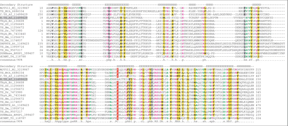
(dUMP) by transfer of a methyl group to its pyrimidine ring. This reaction is catalyzed by at least two unrelated enzymes. The canonical thymidylate synthase (TS), such as the E. coli ThyA, is a protein with a distinct ,/--fold that transfers a methyl group to dUMP from 5,10-methylenetetrahydrofolate [29]. This classic TS is readily identifiable in many (but not all) bacteria, eukaryotes and three archaeal species, Archaeoglobus fulgidus, M. jannaschii, and M. thermoau-totrophicum (COG0207). The archaeal members of the TS family share with their bacterial orthologs all the conserved residues involved in catalysis (Figure 4).
An alternative TS or its subunit is predicted to be encoded by a gene from Dictyostelium that rescues a slime mold mutant auxotrophic for thymidylate [30]. This protein is not homol-ogous to the canonical TS, but its orthologs in bacteria and archaea show an almost perfect complementary phyletic dis-tribution (COG1351).
In a screen for the TS in M. jannaschii, the ORF method picked the MJ0757 protein as the most likely homolog of the canonical TS family [27]. In the validation experiment, MJ0757 overexpressed in E. coli was shown to possess TS activity [25]. Sequence searches show that MJ0757 belongs to a small family of euryarchaea-specific proteins of uncharacter-ized function (COG1810). Of the 17 residues reported to be conserved between MJ0757 and the TS family, only seven were conserved thoughout the MJ0757 family (Figure 5). Moreover, a comparison of the secondary structure elements
derived from the reported three-dimensional model of MJ0757 [27] and those derived from a prediction generated using a multiple alignment query with the structure-predic-tion program PHD (such predicstructure-predic-tions typically exceed 70% accuracy), showed an overlap of just two of the 16 or so sec-ondary structural elements (Figure 5). Conversely, several sequence motifs that are characteristic of the MJ0757 family did not overlap with the conserved regions in the MJ0757-TS alignment (Figure 5). Furthermore, some, but not all, members of the MJ0757 family contain an amino-terminal insertion of a small, metal-chelating module (Figure 5), which was used to improve the alignment with the E. coli TS [25], although this region was variable even within the MJ0757 family itself. On the basis of these observations, a relationship between MJ0757 and the canonical TS has to be rejected. The actual fold and function of MJ0757 and its homologs cannot be predicted at present. However, these proteins have several features that suggest that they might be metal-dependent enzymes potentially involved in redox reactions. These sug-gestive features include the fusion with a ferredoxin domain seen in the M. thermoautotrophicummember MTH601, the insertion of the metal-binding module in certain members, including MJ0757 (see above), and the presence of three cys-teines that are conserved throughout this family.
Cmpp16: a plant ‘paralog’ of plant viral movement proteins
Viral movement proteins (MPs) are encoded by diverse, unrelated families of plant viruses, such as positive-strand
comment
reviews
reports
deposited research
interactions
information
[image:5.609.59.555.88.287.2]refereed research
Figure 3
Multiple alignment of the archaea-specific family of predicted metallo---lactamase superfamily hydrolases that includes the alleged archaeal dihydropteroate synthase, MJ0301. The scheme for displaying multiple alignments is as described in the legend to Figure 1. A consensus secondary structure was derived from the crystal structure metallo---lactamases from
Stenotrophomonas maltophilia(1SML) and Bacteroides fragilis(1A7T). Residues are colored at 90% consensus. Bfr,Bacteroides fragilis; Bsp, Bacillusspecies 170; Mj, M. jannaschii; Mth, M. thermoautotrophicum; Pab, P. abyssi; Ph, P. horikoshii; Stma,
S. maltophilia; Tm, Thermotoga maritima.
Sec. Structure EEE EEEEEEEEEE----EEEEEEEEEEEE---EEEEE---HHHHHHHHHH---EEEEEE---HHHHHHH---HHH--HHHHHH
MJ0301_Mj_2495901 14 YTLAEDYAGYNSPF----WSQHGLSFLIEVESNGIKKRILFDTATY---AEPILFNMKLLNINPKSIDMIILSHNHFDHTGGLFGIMKEIN-KEIPIFAHP--NIFKVS FATEP-EFMLAGTL---MTH1101_Mth_7482082 4 TCLVEDRARPPL---RAEHGLSLHIEGDV---NILFDTGQS---G-LFMENAELLGVDLGDVDLAILSHGHYDHGGGLTHFM-EIN-ENTEVLTGE--GAFRGR WAVEDGAERFIG---PH0874_Ph_7518869 6 YTLADDYAGYNSPF----WAQHGVSFLVEVEGK----KILFDTASY---AEPILFNMKLLNLNPRDIDMIVLSHNHFDHTGGLLGILREIN-RKVPIFAHP--EIFKVS FALEP-EFMYAG---PAB0518_Pab_7517873 9 TIVFENHAGYRKGL----IGYHGFSALVESNGY----KVLVDTGTD---GKVLLNNMHELGIDANDIDVLFITHGHYDHTGGLKEFLKERS-EPIEVYAHP--GIFDER IALKP-RKRDIG---PH1382_Ph_7519119 11 TIVFENHAGYRKGL----IGYHGFSALVEANGY----RVLVDTGTD---GKVLLNNMSELGISPDDIDALFITHGHYDHTGGLRELLSART-EALDIYAHP--GIFKRR LALKP-KKREIG---MJ0448_Mj_2495991 3 KILVDNTA-YKKF---FAQHGFSALIEINNK----RILFDAGQN---SITLRENLRLFN-EKEGFDYIVLSHGHYDHCDGLKYVIENDL-INGKVIAHK--DAFLDK YA----GNRYIG---TM1679_Tm_7462229 4 HVLCDDSSQNGF---ESEHGFSVLVD---SVLFDTGKS---D-VFLKNARKLGIDLP--KDVLISHGHYDHAGGLLYLSGKR---VWLRK--EALDQK YS----GERYAG---PH1079_Ph_7518967 5 IVLNDNTPSRGL---INDWGWSILIEGRE---RFLFDADTN---PLVLAHNSKVLNVNLRNLDFAVLSHWHYDHYGGFEYIA-ELN-PGIKFYAPPQGLALAMR WGFQPIAINF---PAB1761_Pab_7518230 5 IILNDNVPSKGL---MNDWGWSVLVEGRK---RFLFDADTN---PLVLAHNSKALNVNLRNLDFAVLSHWHYDHYGGFQYIA-ELN-PGIKFYAPIQGLAMAMR WGFQPIAINS---BLA2_Bsp_115023 49 SQLNKNVWVHTELGYFN-GEAVPSNGLVLNTSKG---LVLVDSSWDNK-LTKELIEMVEKK-FQKRVTDVIITHAHADRIGGITALKERGI-KAHSTALTA--ELAKKS GYEEPL---1a7t_Bfr_3318914 14 TQLSDKVYTYVSLAEIEGWGMVPSNGMIVINNHQ---AALLDTPIND--AQTEMLVNWVTDSLHAKVTTFIPNHWHGDCIGGLGYLQRKGV-QSYANQMTI--DLAKEK GLPVPE---1SMLA_Stma_6137470 24 LQIADHDWQIGT---EDLTALLVQTPDG---AVLLDGGMPQMASHLLDNMKARGVT-PRDLRLILLSHAHADHAGPVAELKRRTGAKVAANAESA--VLLARGGSDDLHFGDGITYPPANAD consensus/90% ..l.-p...s.s.hl..p...hLhDss...s..b...s...hp.hhloH.HhD+.GGh..h..p...h...hh..p...
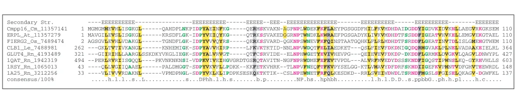
RNA, negative-strand RNA, single-stranded DNA and double-stranded DNA viruses, and are essential for cell-to-cell movement of all these viruses [31,32]. To isolate poten-tial host homologs of the red clover necrotic mosaic virus (RCNMV) MP, antibodies to this protein were used to screen phloem extracts of Cucurbita maxima, resulting in the detection of a protein designated Cmpp16. This protein was identified as a ‘paralog’ (generally, this term refers to homol-ogous genes related by duplication within the same genome) of the viral MPs on the basis of sequence similarity detected using the Megalign program [33]. Subsequently, Cmpp16 was shown to bind RNA, which is a common property of viral MPs, and to induce an increase of the size-exclusion limit of plasmodesmata, also a mechanism associated with the MPs [33].
However, computational analysis of the Cmpp16 sequence reveals a picture that is incompatible with a homologous relationship with MPs. Cmpp16 consists mostly of a C2 domain that is readily detected by PSI-BLAST or by profile-searching engines such as the CD-search [34]. The Cmpp16 sequence contains all critical residues of the C2 domain (Figure 6). C2 domains bind a variety of substrates, such as Ca2+, phospholipids, inositol polyphosphates and other pro-teins, but apparently not RNA [35]. There is no detectable similarity between C2 domains and the MPs, and conserved motifs in the published alignment of Cmpp16 and the
RCNMV MP do not correspond to those in C2 domains; moreover, many of the residues described as conserved in Cmpp16 and MP are not conserved within the viral move-ment protein family itself. Thus, we conclude that viral MPs and Cmpp16, a C2-domain protein, are not homologs. Sub-sequently, a similar methodology has been employed to detect a relationship between Cmpp36 (a cytochrome B5 reductase), Cmpp16 and the RCNMV movement protein [36]. As in the above case of Cmpp16, this relationship of a cytochrome B5 reductase with the viral movement proteins appears to be spurious (data not shown).
Human activating transcription factor-2 (ATF-2): a predicted histone acetyltransferase
[image:6.609.60.556.88.305.2]Histone acetyltransferases (HAT) are key regulators of eukaryotic transcription. GCN5-like HATs, which modulate chromatin-associated transcription, belong to a vast super-family of amino-group acetyl- and myristoyl-transferases with extremely diverse functions [37]. ATF-2 is a basic leucine zipper (b-ZIP) family transcription factor that binds to cyclic AMP-response elements (CRE) and activates transcription [38]. Vertebrate ATF-2 also has an amino-terminal zinc finger, which is involved in transcription activation [39]. Non-vertebrate orthologs of ATF-2, in Drosophila, Caenorhabditis elegans and yeasts, lack the zinc finger. In experiments designed to isolate ATF-2-associated HAT, ATF-2 alone was shown to be sufficient for the acetyltransferase activity.
Figure 4
Multiple alignment of predicted archaeal thymidylate synthases (TS). The scheme for displaying multiple alignments is as described in the legend to Figure 1. Residues are colored at 90% consensus. A consensus secondary structure was derived using known TS structures from R. norvegicus, E. coliand bacteriophage T4 deoxycytidylate hydroxymethyltransferase (1B5D). The Archaeoglobus fulgidusTS has a duplication of the TS domain and the amino-terminal domain (N.TS_Af; shaded gray) is predicted to be inactive. Af, Archaeoglobus fulgidus; At, Arabidopsis thaliana; BPSP1; bacteriophage SP1; Bs, B. subtilis;
Dm,Drosophila melanogaster; Dr, D. radiodurans; Ec, E. coli; Mj, M. jannaschii; Mt, M. tuberculosis; Mth, M. thermoautotrophicum; Nm, Neisseria meningitidis; Rn, R. norvegicus; T2, bacteriophage T2; Xf, Xylella fastidiosa.
Secondary Structure -HHHHHHHHHHHH--EEEEE---EEEEEEEEEEEEEE---HHHHHHHHHHH---HHHH---HHHH---HHHHH---MJ0511_Mj_3219867 10 ASAFNELIPKILKDGEVVETEFE---ERTKEIRNTIIEITNPKLKK---VPEK-YPL--GE---KAVEEYTKNLLYG-S---KNVFSYDYHQRL FEYPYA---TS_Mth_6686334 11 ADGWKKLVEKIMHDGREIRDERG---SLTREVMNTVVTIKKPLGKSDDFYHIRRGSLLNIKVPEG-YFWS-GE---KLEK-YSEQFLSG-D---RKGFVYTYGNRL RA---C.TS_Af_11499629 250 SSAWHSALETIYTNGKKKRTEWGDIF---EGQKE-SLFVHRLFLEVEKPEE---NKLHDK-APF--TE---KYGIEYAHDYIMH-AAKLDGEVRRS--ILKEGEEYTYAERA RYCDRD---N.TS_Af_11499629 18 SLADFFSICLAMLKRFSMADKTFF---HGKLFNLLRKWVLMILTKTPEEA---KEMLVS-KVIG-GE---TCRSRF-GDYRLSKPTM---VVVEEPTSFGFEFDY DVCGEKYS---ThyA_Ec_136608 1 MKQYLELMQKVLDEGTQKNDRTG---TGTLSIFGHQMRFNLQDGFPLVTTKRCH---LRSIIHELLWFLQ-GDTNIAYLHENNVTIWDEWA---DENGDLGPVYGKQW RAWPTPD---TS_Xf_11256665 1 MKQYLELLNDVLVHGIQKPDRTG---TGTRSVFGWQMRFDLSQGFPLVTTKKLH---LRSIIHELLWFLR-GETNIAYLKKHQVHIWDEWA---DATGELGPVYGKQW RRWAGAD---TS_Nm_11256672 1 MKAYLDLMRHVLDNGTDKSDRTG---TGTRSVFGYQMRFDLGKGFPLLTTKKLH---LRSIIHELLWFLK-GDTNIKYLKDNNVSIWDEWA---DENGDLGPVYGYQW RNWPAPD---TS_Dr_7473980 124 VKQYLDFLRHIRDHGTDKMDRTG---TGTRSVFGYQMRFDLSEGFPLVTTKRVH---LKSIIYELLWFLR-GDSNVRWLQEHGVTIWDEWA---REGGELGPVYGVQW RSWPDYG---ThyA_Mt_7433440 1 MTPYEDLLRFVLETGTPKSDRTG---TGTRSLFGQQMRYDLSAGFPLLTTKKVH---FKSVAYELLWFLR-GDSNIGWLHEHGVTIWDEWA---SDTGELGPIYGVQW RSWPAPS---TS_Nm_11256672 1 MKAYLDLMRHVLDNGTDKSDRTG---TGTRSVFGYQMRFDLGKGFPLLTTKKLH---LRSIIHELLWFLK-GDTNIKYLKDNNVSIWDEWA---DENGDLGPVYGYQW RNWPAPD---TS_Bs_1174837 5 DKQYNSIIKDIINNGISDEEFDVRTKWDSDGTPAHTLSVISKQMRFDNSE-VPILTTKKVA---WKTAIKELLWIWQLKSNDVNDLNMMGVHIWDQWK---QEDGTIGHAYGFQL GKKNRSLN---DHFRTS_At_1169423 235 EYLYLNLVKEIISNGNLKDDRTG---TGTLSKFGCQMKFNLRRNFPLLTTKRVF---WRGVVEELLWFIS-GSTNAKVLQEKGIRIWDGNASRAYLDGIGLTEREEGDLGPVYGFQWRHFGAKYTDMHADYT TS_Dm_13959716 36 EMHYLDLLRHIIANGEQRMDRTE---VGTLSVFGSQMRFDMRNSFPLLTTKRVF---FRAVAEELLWFVA-GKTDAKLLQAKNVHIWDGNSSREFLDKMGFTGRAVGDLGPVYGFQWRHFGAQYGTCDDDYS TS_Rn_9507217 24 ELQYLRQVEHIMRCGFKKEDRTG---TGTLSVFGMQARYSLRDEFPLLTTKRVF---WKGVLEELLWFIK-GSTNAKELSSKGVRIWDANGSRDFLDSLGFSARQEGDLGPVYGFQWRHFGADYKDMDSDYS DUHMase_BPSP1_399407 10 TQLYMDILSTVIKEGDVLAPRG---KRIKEIRPVMIEFKNPIRRTTFLKGRNIN--PFFQVAESLWILA-GRSDVGFLLDYNKNMGQ-FS---DDGVFFNAPYGERLRFWNRSDANNFIYNP dCHMT_T4_118788 8 VEEIRLHLGLALKEKDFVVDKTG---VKTIEIIGASFVA---DEPFIFGALN---DEYIQRELEWYKS-KSLFVKDIPGETPKIWQQVA---SSKGEINSNYGWAI WS---consensus/90% ...h...h..hb.p...sc...pbp.b...h.h...h.-..h.h...p...ph...bs.sY..ph...
Examining the region of ATF-2 that showed HAT activity, the authors found some sequence similarity and at least one motif resembling the acetyltransferase superfamily and con-cluded that ATF-2 contained a GCN5-like acetyltransferase domain [40]. Subsequent site-directed mutagenesis sup-ported the importance of the resup-ported acetyltransferase motifs for the HAT activity of ATF-2.
However, profile-based sequence searches and attempts at fold recognition failed to detect any relationship between ATF-2 and the acetyltransferase superfamily. The region des-ignated as having HAT activity and containing the acetyl-transferase domain shows poor conservation between orthologs and closely related paralogs of the ATF-2 family, especially in the sequence identified as the most prominent A motif of the acetyltransferase family (Figure 7). Further-more, complexity analysis using the SEG program, with the
parameters adjusted for decomposition of a protein into glob-ular and non-globglob-ular regions [41], predicted that the entire region of the ATF-2 protein between the amino-terminal zinc finger and the carboxy-terminal helical b-ZIP was unstruc-tured. This is consistent with the structural prediction derived using the PHD program that indicated no regular sec-ondary structure in this region. Thus, the relationship between ATF-2 and the GCN5-like acetyltransferase super-family seems to be invalid, leaving the structural basis for the reported acetyltransferase activity of ATF-2 an open issue.
Predicted PAS domain in the phytochrome-interacting transcription factor PIF3
PAS domains are sensory modules in various signal trans-duction proteins from all major lineages of cellular life [42]. PAS domains are typically implicated in sensing oxygen, redox potential, light and small ligands [43]. In addition,
comment
reviews
reports
deposited research
interactions
information
[image:7.609.57.556.88.252.2]refereed research
Figure 5
Multiple alignment of the uncharacterized archaeal protein family that includes the alleged archaeal thymidylate synthase, MJ0757. The scheme for displaying multiple alignments is as described in the legend to Figure 1. Residues are colored at 100% consensus. In addition, metal-chelating residues in an inserted module shared by orthologs of MJ0757 are shaded blue. The asterisks denote residues in MJ0757 that were predicted to be conserved between MJ0757 and TS. Also shown are predicted secondary structures for the MJ0757 family that were obtained by using the PHD program, and the TS-like secondary structure predicted for MJ0757 in [25]. Af, A. fulgidus; Mj, M. jannaschii; Mth, M. thermoautotrophicum.
Proposed con residues * --- * * * * * * *
MJ0757_Mj_2496119 17 MRAVFIYHKNNQRMEKFYKNLLNEPDFCR---ICD--DCYNCR-GNWTFKNNVKNIVIEEVY---EEFVDNPYDYLPE--LPEGDICIA-QLHEDLLYELPLLLKE-KGYKALIVPSETPHDLSLALRRDLKRV AF2108_Af_11499691 6 LKLIVFQHGFF--GERFVANLMNYPNSCPSYGACGIDGCTQCKEGLYNFSKN---IIAVFSMPDPTTM-PDFIENAEDFLPKV-IPEADIAVAINLHPDVLAVMPEKLKG-K-VKALIVPVEEPRWCSPGLAKQIREK AF1307_Af_11498905 1 MELGIIYSGEF--GKRFVSNLA-YPYLCPTFGACGINGCDYCKR--YDFSSS---IVYAKELAEPQEI-GLYVEEPESYLEP---FECDIAVAINVHPDILVSLPEI----GEFKALIVPACNQNWCLPGLRKQLAEK MJ0575_Mj_2496052 2 AKILVVTDGAY--GYRIKGTINSFGKKNK---FIGIYKINKPDDLIVDDIEFPDELLEK--IKEADILLLYTQHPDNTYYLCYEARRLNKDIAIIVA---TWSGEGEKK AF1687_Af_11499277 1 MKLGVVTRK----GKRQDDIRMFSQFFEVKV---YEIPEEL---PELIDEPAEILRLPDDFDVDMIVSFAAHPDINLELIKQAAE-RGIGLVIISGGAKGGAYKQLKEEGEK -MTH601_Mth_7482230 2 MRIIMVTAGNY--GARVVNTMAVHGLAPQ---IVAVFDYTGEG---GDFLDDPSSLLPAR-TPDADLTVAAGLGGDLNLVAAEIAAE-SGSGGIIVESHAPGQLPDGLRSEIDSL MTH1356_Mth_7430198 2 IKVAIVTDGPY--GERAYENIAREF---ETMFIELEAPSGIFADEVDIPADKLKA--IRSADIVITYILHPDLTLELVDEIHG--DVDWIIIG---AWRGDGFRN MJ0838_Mj_2496132 1 MKVAILTDGVY--GDRAYNTIKSKFPCD---FITVKYYGD---FDEITISENTIEK--LKDYDLFITYTLNPDLTYELVRKIKELNNKAFVLVG---AWKGEGFKK consensus/100% hch.hh...+....b...h...h...lp.s.p.l...p.Dh.l...D..h.h...lll...h..p..p. PHD Sec. Structure -EEEEEE---HHHHHHHHHH---EEEEEE---HHH---HHHHHH---EEEEEE----HHHHHHHHHHHH---EEEEEE---HHHHHH Pred. TS like Structure --HHHHHH-EEE---EEE---EEE---HHHHHHHH---EE---EEE
HHHHHHHHHH---Proposed con residues * * * * * * * * *
MJ0757_Mj_2496119 CSNYNIEFENPKPFCSLEKKEGN---EYINKFIDYFKIGKPELEIEVENGLIKDVKVKISAPCGETYYIAKRLKGKAIDDLKE---EIANAHHNYPCLASMEMDKELGDTILHKAGYIAFEVVEKALKK 260 AF2108_Af_11499691 CDELGLEFAAPKPFCNLRPSEEH---PTINRMIEEMRIGYPEFEIELLEDGKAYVRISRSQPCGCAYYIGVKLRGFDFSEVKEGKMRELWNVVAEAHHSYPCTASMERDNEYNETLLHVGGYIARHAVNRALGY 265 AF1307_Af_11498905 CGELGIEFASPKPFCSLTGNSG---WISRFIDEFKIGRPEFRVERGGDAIKKVEVIKSDPCGSAYFVAKRMTGYIIESKEDFWK---EIHQHQCAYPCMASMDRDVELKEAPFHLAGYIMVYQFSTAAGV 246 MJ0575_Mj_2496052 ELKKF-DAICPEEMCLLDENEVGSLIDK---YPKLKEFLEEF--GTPKVKVYVKDNKVIDVEVLRTSICGSTLFMAKLMKGMEVNDIEEFAK---KSAMLIQRYPCVAGKIKIF-RGDCRKQKALNIHKEAVLEGIIF 228 AF1687_Af_11499277 -RGVRVVWEE---ICCATPKVED---ERTREFFEHF--GAPEVEVEVENGKVKDVRVKRSAFCGATYYVAEKIKGLSVEEAPT---KAGYYTQIYPCLAPRGHE---GGIHKAARAHKRAVEKAIEK 213 MTH601_Mth_7482230 VD----CAIFPTPFCSLEPV-GN---PHIDAFASRF--GKPEVEME-GNERVVSVKVKRSAPCGSTFYVAENIRGVPLDEVDV---AAGEKFHNYPCLASMEVDPRFGDTHLHVAGYLTREAFKRAAGV 221 MTH1356_Mth_7430198 QLLSYGNLTAPENMCDLEEN-GN---PSFDEFVSRF--GRPLVEVDLEGDTVKEIRVLRSSPCGATLFVAEELTGEDAQDLPL---KAGLKIQHYPCRAPKMRLFSDDECKKEMAARMHSEAFERAIGV 214 MJ0838_Mj_2496132 QIESFGNAFCPYLMCDIDEDELKDYKDYLDNYPHLKEFLKYF--GKPKVKLYIKNNKIEKIDVLREAPCGSTSETLKEFVGREFNDKTLI---DIGLRVQHF-CRAGKIRLFVEKEGKKTKAGKILVSGIQ-VIQI 222 consensus/100% ...h...hC.hp...p.hhp.h..G.P.hch...lcl..p..CG.s.h....h.G..hpp...p.aPC.As...s..p.s..h....h..sh.. PHD Sec. Structure ---HHHHHHHHH---EEEEEEE----EEEEEEE----HHHHHHHHHH---HHHHHHH---HHHHHH---HHHHHHHHHHHHHHHH---Pred. TS like Structure EEEE---EEEEEE-
EE---EEE---HHHHHHHHHHHHHHHHHH---EEE---HHHHHHHH---EEEEE---EEEEE---Figure 6
Multiple alignment of a selection of C2 domains including the alleged ‘paralog’ of plant virus movement proteins, Cmpp16. The scheme for displaying multiple alignments is as described in the legend to Figure 1. Residues are colored at 100% consensus. A consensus secondary structure was derived from known structures of the C2 domains in phospholipase C-/1 (1QAT), synaptotagmin (1RSY), and protein kinase C (1A25). At: A. thaliana, Cm: Cucurbita maxima, Le: Lycopersicon esculentum, Os: Oryza sativa, Rn: R. norvegicus.
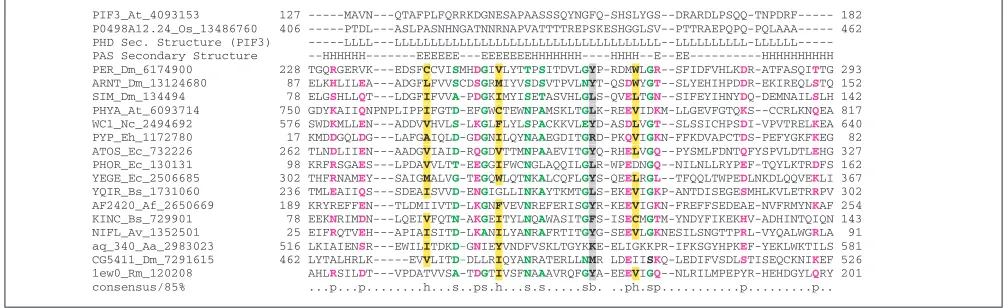
[image:7.609.52.557.357.445.2]PAS domains are sites for protein-protein interactions and are responsible for the formation of homo- and hetero-dimers in several signal transduction pathways that involve transcriptional activation. A PAS domain has been reported in the transcription factor PIF3 from Arabidopsis, which interacts with a phytochrome photoreceptor and transduces light signals to photoresponsive plant genes [44]. It has been hypothesized that the purported PAS domain of PIF3 directly interacts with the PAS domains of the phytochrome [44]. This hypothesis was later tested experimentally and evidence was presented that the PAS domain of PIF3 indeed was a major contributor to the interaction between the two proteins [45].
PIF3 belongs to a plant-specific family of basic helix-loop-helix (bHLH)-domain- containing proteins that, in addition to the bHLH domain, have an uncharacterized conserved domain at the amino terminus present in single or duplicate copies (L.M.I., I.Z., L.A. and E.V.K., unpublished observa-tions). The PIF3 family currently consists of about eight par-alogous proteins in Arabidopsisand an ortholog from rice. The region predicted to be a PAS domain is poorly conserved in the rice ortholog of PIF3 and the paralogs from Arabidop-sis. An alignment with the rice ortholog indicated that the proposed PAS domain was a rapidly diverging, composition-ally biased sequence (Figure 8). Complexity analysis using the SEG program showed that the reported PAS domain mapped to a region that was predicted to be entirely non-globular. All attempts to objectively detect a PAS domain in PIF3 using sensitive profile methods based on PSI-BLAST-derived scoring matrices or Hidden Markov Models (HMM) failed. Additionally, secondary-structure prediction for the proposed PAS region using PHD indicated that this region is largely unstructured. These observations appear to be suffi-cient to reject the presence of a PAS domain in PIF3 although the region thought to be a PAS domain could indeed be involved in the interaction with phytochrome.
Discussion and conclusions
In the six cases described above, we provide evidence for rejecting the homologous relationships and functional pre-dictions inferred for the proteins in question by using
computational methods. The number of examples in this category could be increased, and some have already been considered in the literature, for example the spurious discov-ery of a ‘functional PDZ domain’ in the molecular chaperone ClpA ([46], see refutation in [47]) or the finding of an ATPase domain and death effector domains in the apoptosis-associated protein FLASH ([48], see refutation in [49]). The common and most striking aspect of all these cases is that the predictions based on apparently erroneous computa-tional analysis were supported by experiments. What are the solutions to this clash between computational and experi-mental evidence?
We envisage three main possibilities. The first, experiment-centered view would hold that experimental evidence always has the upper hand and that, even if the alternative compu-tational solutions that we describe here seem more plausible than the original predictions, the latter are correct insofar as they are supported by experiment. Epistemologically, this argument is not sound because hypotheses (computational predictions in this case) cannot be proved by the success of the experiments they prompt. They can only be falsified by experiments producing results incompatible with the predic-tions [50]. Simply put, the experiments could have worked for a wrong reason. For example, this seems particularly likely in the case of the site-directed mutagenesis of the tran-scription factor ATF-2 discussed above. The mutagenized residues probably are indeed important for the function of this protein, but not because they are part of a GCN5-like acetyltransferase domain, which this protein does not contain. Similar logic applies to the case of the predicted, but apparently nonexistent, PAS domain in the transcription factor PIF3. More important, however, computational pre-dictions are falsifiable within the realm of computational analysis itself. Falsification is offered by alternative, unequivocally supported predictions that are incompatible with the original ones. In four of the six cases described (CysRS, DHPS, TS and MP), such evidence was obtained by computational methods.
[image:8.609.61.548.89.154.2]The second possibility is that, although the computational predictions described here are correct, whereas the original ones are wrong, the experimental evidence is also solid. In
Figure 7
Multiple alignment of the region of the ATF-2 transcription factor and its homologs identified as a GCN5-like
acetyltransferase domain. The scheme for displaying multiple alignments is as described in the legend to Figure 1. Residues are colored at 100% consensus. Ce: Caenorhabditis elegans,Hs: Homo sapiens, Sp: Schizosaccharomyces pombe.
Predicted HAT motif A <--->
Region masked by SEG <--->
PHD Sec. Str. LLLLLLLLLLLLLLLLLLLLLLLLLLLLLLLLLLLLLLLLLLLLLLLLLLLLLLLLLLLLLLLLLLLLLLLLLLLLHHHHHHHHHHHHHHHHHHHHHHHHHHHHHHHHHHHHHHHHHHHHHHHHHHHHHHHHHHHHHHH ATF-2_Hs_87016 288 PVTNGDT---VKGHGSGLVRTQSEESRPQSLQQPATSTTETPASPAH---TTPQTQSTSGRRRRAANEDPDEKRRKFLERNRAAASRCRQKRKVWVQSLEKKAEDLSSLNGQLQSEVTLLRNEVAQLKQLLLAHKD-CPV 420 ATF-7_Hs_5802980 270 SINGGCGM---VVGTASTMVTARPEQSQILIQHPDAPSPAQPQVSPAQ---PTPSTGGRRRRTVDEDPDERRQRFLERNRAAASRCRQKRKLWVSSLEKKAEELTSQNIQLSNEVTLLRNEVAQLKQLLLAHKD-CPV 400 ATF-1_Sp_1236269 408 PTANSMP---VKLENGTDYSTSQEPSSNANNQSSPTSSINGKASSE---SANGTSYSKGSSRRNSKNETDEEKRKSFLERNRQAALKCRQRKKQWLSNLQAKVEFYGNENEILSAQVSALREEIVSLKTLLIAHKD-CPV 540 C07G2.2a_Ce_6580209 255 PYFNDDAMMLMERSNMSSSGSDQDQSADMSNAGSTASTSTGNPVGRPQ---NGTPGRGRGRGRST-TADMQPDERRNTILERNKAAAVRYRKRKKEEHDDMMGRVQAMEAEKNQLLTQNQVLRRELERVTALLTERESRCVC 392 CRE-BPa_Hs_13630551 297 QPHHQQNHP----HHHSHSHLHAHPAHHQTSPHPPLHTGNQAQVSPATQQMQPTQTIQPPQPTGGRRRRVVDEDPDERRRKFLERNRAAATRCRQKRKVWVMSLEKKAEELTQTNMQLQNEVSMLKNEVAQLKQLLLTHKD-CPI 436 consensus/100% ...p..s...s.s..p.p...p...sss....s.s...sppp.s.sbps-E+RpphLERN+.AA.+hRb++K....shb.+sb.h...p.bL.sbsphL+pEl.plp.LL..+cs.Csh
each of the described cases, this would elevate the biochemi-cal activities identified through these experiments to the status of major, unexpected discoveries, because the chem-istry underlying them would have to be extremely unusual. In particular, if the identification of the M. jannaschii cys-teinyl-tRNA synthetase is indeed correct, this enzyme would have to be a derivative of a specific family of polysaccharide hydrolases containing a signal peptide but no recognizable ATP-binding or RNA-binding domains.
The third explanation is that the original computational pre-dictions triggered over-interpretation of the experimental results that, in reality, might have been obtained as a result of nonspecific activities, contamination or other artifacts. In this regard, it is important to realize that not only computa-tional predictions, but biological experiments also, are intrinsically error-prone and open to conflicting interpreta-tions. The probabilistic nature of computational analyses is well realized (and at times, perhaps, overrated) by most researchers, probably because explicit calculation of proba-bility or likelihood is at the core of most widely used com-puter methods for sequence and structure analyses. In this regard, it is prudent to note that the alternative computa-tional predictions presented here should be considered to be ‘more likely’ than the original ones, rather than to contradict the latter in an absolute sense. As we attempted to show above, however, the difference in the likelihood of two mutu-ally incompatible predictions can be overwhelming, with one supported by multiple lines of evidence as opposed to the other. In contrast to computational studies, experimental ones are often, consciously or unconsciously, treated as demonstration of ‘final truth’. In reality, however, proba-bilistic inference is inherent in practically any interpretation of experimental results when questions are asked such as
“How likely is it that the protein under study has a particular biochemical activity in vivo?” or “How central is this activity for the in vivofunction of the protein under study, given the results of a surrogate in vitroassay?” Thus, certain experi-mental designs may not be appropriate to ascertain the actual in vivobiochemistry of a protein. Furthermore, even if the particular activities detected under these conditions are genuine, the likelihood of these being relevant in vivo needs to be additionally assessed. Accordingly, when strong computational predictions seem not to be borne out by experiment, the conditions and design of the experiments deserve special scrutiny: they might have given a negative result for a wrong reason. A case in point is the MJ0107 protein, the apparent archaeal ortholog of DHPS, which failed to show dihydropteroate synthase activity [25]. We strongly believe that this issue needs to be revisited. All this considered, the results of independent application of compu-tational and experimental techniques tend to be comple-mentary, and useful in adding or reducing confidence in the biological conclusions of a particular study.
Finally, it should be emphasized that these cautionary notes on application of computational methods in protein function prediction in no way suggest that new computational approaches that depart sharply from more established ones are doomed to failure. Indeed, the most popular advanced search methods based on sequence profiles - PSI-BLAST and Hidden Markov Model (HMM) search - are rather recent innovations [11,51,52]. Furthermore, methods based on a different principle, such as protein sequence-structure threading, have a recent history of success despite uncertain-ties in their statistical foundations [22,53-56]. It does seem, however, that when a structurally and functionally plausible prediction is produced, with a high confidence, by a well
comment
reviews
reports
deposited research
interactions
information
[image:9.609.52.556.89.243.2]refereed research
Figure 8
A comparison of the multiple alignments of PIF3, its rice ortholog, and PAS domain proteins. The scheme for displaying multiple alignments is as described in the legend to Figure 1. Residues are colored at 90% consensus. A consensus secondary structure was derived from those available for FixL (1EW0) and photoactive yellow protein (3PYP). Aa, A. aeolicus;
Af,A. fulgidus; At, A. thaliana; Av, Azotobacter vinelandii; Bs, B. subtilis; Dm, D. melanogaster; Ec, E. coli; Eh, Ectothiorhodospira halophila; Nc: Neurospora crassa; Os, O. sativa, Rm: Rhizobium meliloti.
tested, statistically sound computational method, an incom-patible prediction yielded by a new method without a clear statistical foundation is most likely to be incorrect.
Materials and methods
The non-redundant protein-sequence database at the National Center for Biotechnology Information (NCBI) was searched using the gapped version of the BLAST program [9]. Sequence-profile searches were carried out using the PSI-BLAST program, with the cut-off for inclusion of sequences into the profile set at E = 0.01 [3,9], and the HMMer program package [57]. Multiple alignments of amino-acid sequences were generated using the T_Coffee program [58]. Protein secondary-structure predictions were generated using the PHD program [59,60], with multiple alignments of individual protein families used as queries. Sequence-structure threading was carried out using the combined-fold-prediction algorithm [22] or the 3D-PSSM algorithm based on the use of a three-dimensional position-specific scoring matrix [23]. Signal peptides in protein sequences were predicted using the SignalP program [61]. The COG database [62,63] was used as a source of informa-tion on orthologous relainforma-tionships between proteins.
References
1. Bork P, Dandekar T, Diaz-Lazcoz Y, Eisenhaber F, Huynen M, Yuan Y: Predicting function: from genes to genomes and back. J Mol Biol 1998, 283:707-725.
2. Koonin EV, Aravind L, Kondrashov AS: The impact of compara-tive genomics on our understanding of evolution. Cell 2000,
101:573-576.
3. Aravind L, Koonin EV: Gleaning non-trivial structural, func-tional and evolutionary information about proteins by itera-tive database searches. J Mol Biol 1999, 287:1023-1040. 4. Murzin AG: Progress in protein structure prediction. Nat Struct
Biol 2001, 8:110-112.
5. Karlin S, Bucher P, Brendel V, Altschul SF: Statistical methods and insights for protein and DNA sequences. Annu Rev Biophys Biophys Chem 1991, 20:175-203.
6. Karlin S, Brendel V: Chance and statistical significance in protein and DNA sequence analysis.Science 1992, 257:39-49. 7. Karlin S, Altschul SF: Applications and statistics for multiple
high-scoring segments in molecular sequences. Proc Natl Acad Sci USA 1993,90:5873-5877.
8. Karlin S: Statistical studies of biomolecular sequences: score-based methods.Phil Trans R Soc Lond B 1994, 344:391-402. 9. Altschul SF, Madden TL, Schaffer AA, Zhang J, Zhang Z, Miller W,
Lipman DJ: Gapped BLAST and PSI-BLAST: a new genera-tion of protein database search programs.Nucleic Acids Res 1997, 25:3389-3402.
10. Altschul SF, Bundschuh R, Olsen R, Hwa T: The estimation of sta-tistical parameters for local alignment score distributions.
Nucleic Acids Res 2001, 29:351-361.
11. Durbin R, Eddy S, Krogh A, Mitchison G: Biological Sequence Analysis: Probabilistic Models of Proteins and Nucleic Acids.Cambridge, MA: Cam-bridge University Press; 1998.
12. Ibba M, Soll D: The renaissance of aminoacyl-tRNA synthesis.
EMBO Rep 2001, 2:382-387.
13. Koonin EV, Mushegian AR, Bork P: Non-orthologous gene dis-placement. Trends Genet 1996, 12:334-336.
14. Galperin MY, Walker DR, Koonin EV: Analogous enzymes: inde-pendent inventions in enzyme evolution.Genome Res 1998,
8:779-790.
15. Stathopoulos C, Li T, Longman R, Vothknecht UC, Becker HD, Ibba M, Soll D: One polypeptide with two aminoacyl-tRNA syn-thetase activities. Science 2000, 287:479-482.
16. Lipman RS, Sowers KR, Hou YM: Synthesis of cysteinyl-tRNA(Cys) by a genome that lacks the normal cysteine-tRNA synthetase. Biochemistry 2000, 39:7792-7798.
17. Stathopoulos C, Jacquin-Becker C, Becker HD, Li T, Ambrogelly A, Longman R, Soll D: Methanococcus jannaschii prolyl-cysteinyl-tRNA synthetase possesses overlapping amino acid binding sites. Biochemistry 2001, 40:46-52.
18. Fabrega C, Farrow MA, Mukhopadhyay B, de Crecy-Lagard V, Ortiz AR, Schimmel P: An aminoacyl tRNA synthetase whose sequence fits into neither of the two known classes. Nature 2001, 411:110-114.
19. Wolf YI, Aravind L, Grishin NV, Koonin EV: Evolution of aminoa-cyl-tRNA synthetases - analysis of unique domain architec-tures and phylogenetic trees reveals a complex history of horizontal gene transfer events. Genome Res 1999, 9:689-710. 20. Nielsen H, Brunak S, von Heijne G: Machine learning
approaches for the prediction of signal peptides and other protein sorting signals. Protein Eng 1999, 12:3-9.
21. Tamura J-I, Kaname H, Kadowaki K, Igarashi Y, Kodama T: Molecu-lar cloning and sequence analysis of the gene encoding an endo ,,-1,4 polygalactosaminidase of Pseudomonassp. 881. J Ferment Bioeng 1995, 80:305-310.
22. Fischer D: Hybrid fold recognition: combining sequence derived properties with evolutionary information. Pac Symp Biocomput 2000:119-130.
23. Kelley LA, MacCallum RM, Sternberg MJ: Enhanced genome annotation using structural profiles in the program 3D-PSSM.J Mol Biol 2000, 299:499-520.
24. Hampele IC, D’Arcy A, Dale GE, Kostrewa D, Nielsen J, Oefner C, Page MG, Schonfeld HJ, Stuber D, Then RL:Structure and func-tion of the dihydropteroate synthase from Staphylococcus aureus. J Mol Biol 1997, 268:21-30.
25. Xu H, Aurora R, Rose GD, White RH: Identifying two ancient enzymes in Archaea using predicted secondary structure alignment. Nat Struct Biol 1999, 6:750-754.
26. COG: Phylogenetic classification of proteins encoded in complete genomes
[http://www.ncbi.nlm.nih.gov/COG/]
27. Aurora R, Rose GD: Seeking an ancient enzyme in Methanococcus jannaschii using ORF, a program based on predicted secondary structure comparisons.Proc Natl Acad Sci USA 1998, 95:2818-2823.
28. Aravind L: An evolutionary classification of the metallo--- -lactamase fold proteins. In Silico Biology 1998, 1:8; available at [http://www.bioinfo.de/isb/1998/01/0008/].
29. Matthews DA, Appelt K, Oatley SJ: Crystal structure of
Escherichia coli thymidylate synthase with FdUMP and
10-propargyl-5,8-dideazafolate. Adv Enzyme Regul 1989, 29:47-60. 30. Dynes JL, Firtel RA: Molecular complementation of a genetic
marker in Dictyosteliumusing a genomic DNA library. Proc Natl Acad Sci USA 1989, 86:7966-7970.
31. Mushegian AR, Koonin EV: Cell-to-cell movement of plant viruses. Insights from amino acid sequence comparisons of movement proteins and from analogies with cellular trans-port systems. Arch Virol 1993, 133:239-257.
32. Melcher U: The ‘30K’ superfamily of viral movement pro-teins. J Gen Virol 2000, 81:257-266.
33. Xoconostle-Cazares B, Xiang Y, Ruiz-Medrano R, Wang HL, Monzer J, Yoo BC, McFarland KC, Franceschi VR, Lucas WJ: Plant paralog to viral movement protein that potentiates transport of mRNA into the phloem.Science 1999, 283:94-98.
34. CD-search[http://www.ncbi.nlm.nih.gov/Structure/cdd/wrpsb.cgi] 35. Ponting CP, Parker PJ: Extending the C2 domain family: C2s in
PKCs delta, epsilon, eta, theta, phospholipases, GAPs, and perforin.Protein Sci 1996, 5:162-166.
36. Xoconostle-Cazares B, Ruiz-Medrano R, Lucas WJ: Proteolytic processing of CmPP36, a protein from the cytochrome b(5) reductase family, is required for entry into the phloem translocation pathway.Plant J 2000, 24:735-747.
37. Neuwald AF, Landsman D: GCN5-related histone N-acetyl-transferases belong to a diverse superfamily that includes the yeast SPT10 protein.Trends Biochem Sci 1997, 22:154-155. 38. Hai TW, Liu F, Coukos WJ, Green MR: Transcription factor ATF
39. Nagadoi A, Nakazawa K, Uda H, Okuno K, Maekawa T, Ishii S, Nishimura Y: Solution structure of the transactivation domain of ATF-2 comprising a zinc finger-like subdomain and a flexible subdomain.J Mol Biol 1999, 287:593-607. 40. Kawasaki H, Schiltz L, Chiu R, Itakura K, Taira K, Nakatani Y,
Yokoyama KK: ATF-2 has intrinsic histone acetyltransferase activity which is modulated by phosphorylation. Nature 2000,
405:195-200.
41. Wootton JC: Non-globular domains in protein sequences: automated segmentation using complexity measures.
Comput Chem 1994, 18:269-285.
42. Taylor BL, Zhulin IB: PAS domains: internal sensors of oxygen, redox potential, and light. Microbiol Mol Biol Rev 1999, 63:479-506. 43. Anantharaman V, Koonin EV, Aravind L: Regulatory potential, phyletic distribution and evolution of ancient, intracellular small-molecule-binding domains. J Mol Biol 2001, 307:1271-1292. 44. Ni M, Tepperman JM, Quail PH: PIF3, a phytochrome-interact-ing factor necessary for normal photoinduced signal trans-duction, is a novel basic helix-loop-helix protein.Cell 1998,
95:657-667.
45. Zhu Y, Tepperman JM, Fairchild CD, Quail PH: Phytochrome B binds with greater apparent affinity than phytochrome A to the basic helix-loop-helix factor PIF3 in a reaction requiring the PAS domain of PIF3.Proc Natl Acad Sci USA 2000, 97: 13419-13424.
46. Levchenko I, Smith CK, Walsh NP, Sauer RT, Baker TA: PDZ-like domains mediate binding specificity in the Clp/Hsp100 family of chaperones and protease regulatory subunits.Cell 1997, 91:939-947.
47. Neuwald AF, Aravind L, Spouge JL, Koonin EV: AAA+: A class of chaperone-like ATPases associated with the assembly, operation, and disassembly of protein complexes. Genome Res 1999, 9:27-43.
48. Imai Y, Kimura T, Murakami A, Yajima N, Sakamaki K, Yonehara S:
The CED-4-homologous protein FLASH is involved in Fas-mediated activation of caspase-8 during apoptosis. Nature 1999, 398:777-785.
49. Koonin EV, Aravind L, Hofmann K, Tschopp J, Dixit VM: Apoptosis. Searching for FLASH domains.Nature 1999, 401:662-663. 50. Popper K: The Logic of Scientific Discovery. New York/London:
Rout-ledge; 1999.
51. Altschul SF, Koonin EV: PSI-BLAST - a tool for making discov-eries in sequence databases. Trends Biochem Sci 1998, 23:444-447. 52. Eddy SR: Profile hidden Markov models. Bioinformatics 1998,
14:755-763.
53. Bryant SH, Altschul SF: Statistics of sequence-structure thread-ing. Curr Opin Struct Biol 1995, 5:236-244.
54. Jones DT: GenTHREADER: an efficient and reliable protein fold recognition method for genomic sequences. J Mol Biol 1999, 287:797-815.
55. Panchenko A, Marchler-Bauer A, Bryant SH: Threading with explicit models for evolutionary conservation of structure and sequence.Proteins 1999, 37:133-140.
56. Panchenko AR, Marchler-Bauer A, Bryant SH: Combination of threading potentials and sequence profiles improves fold recognition.J Mol Biol 2000, 296:1319-1331.
57. Eddy SR: Hidden Markov models. Curr Opin Struct Biol 1996,
6:361-365.
58. Notredame C, Higgins DG, Heringa J: T-Coffee: A novel method for fast and accurate multiple sequence alignment.J Mol Biol 2000, 302:205-217.
59. Rost B, Schneider R, Sander C: Protein fold recognition by pre-diction-based threading. J Mol Biol 1997, 270:471-480.
60. Rost B, Sander C, Schneider R: PHD - an automatic mail server for protein secondary structure prediction. Comput Appl Biosci 1994, 10:53-60.
61. Nielsen H, Engelbrecht J, Brunak S, von Heijne G: A neural network method for identification of prokaryotic and eukaryotic signal peptides and prediction of their cleavage sites. Int J Neural Syst 1997, 8:581-599.
62. Tatusov RL, Koonin EV, Lipman DJ:A genomic perspective on protein families. Science 1997, 278:631-617.
63. Tatusov RL, Natale DA, Garkavtsev IV, Tatusova TA, Shankavaram UT, Rao BS, Kiryutin B, Galperin MY, Fedorova ND, Koonin EV:
The COG database: new developments in phylogenetic clas-sification of proteins from complete genomes.Nucleic Acids Res 2001, 29:22-28.
comment
reviews
reports
deposited research
interactions
information