Journal of Chemical and Pharmaceutical Research, 2015, 7(10):593-599
Research Article
CODEN(USA) : JCPRC5
ISSN : 0975-7384
Characterization of microstructural changes on electroacupuncture induced
rat skeletal muscle by low-field NMR and microscope image
Qingwen Ni
1*, Cesar Rivera¹, Samuel De Los Reyes², Kijeong Kim
3, Junyoung Hong
4and Sukho Lee
51Texas A&M International University, Laredo, TX, USA 2Texas A&M University, College Station, TX, USA 3University of Ulsan, Republic of Korea, South Korea
4University of Houston, Houston, TX, USA 5
Texas A&M University-San Antonio, San Antonio, TX, USA
_____________________________________________________________________________________________
ABSTRACT
An electroacupuncuture method has been applied on rat skeletal muscle for determining whether this treatment can attenuate or prevent muscle degradation during the disuse. It was tested on a hind limb suspension (HS) disuse rat model, and by using a non-destructive Nuclear Magnetic Resonance (NMR) relaxation technique, and microscope image analysis technique, as well the muscle mass and density measurements to characterize the muscle microstructural changes. Our study suggests that electroacupubcuture technique is an effective method in preventing muscle degradation in a disuse rat model.
Keywords: acupuncture, NMR, Microscope image, Skeletal muscle.
_____________________________________________________________________________________________
INTRODUCTION
Osteopenia is a disease characterized by long term loss of bone tissue, particularly in the weight-supporting skeleton [1,2]. Meanwhile, sarcopenia, the loss of muscle mass and strength that occurs with aging, is believed to play a major role in the pathogenesis of frailty and functional impairment during aging process as well as muscle atrophy with these who experienced various injuries and disuse conditions. Therefore, finding an effective intervention for attenuating and/or preventing osteopenia and muscle atrophy become major interests in those individuals.
acupuncture has been extensively investigated [11]. EA increases blood flow in and oxygenation of skeletal muscles [12,13] and evokes somatosensory responses of the brain, spinal cord and muscles in humans [14], and these factors are thought to contribute to its ameliorating effect on muscle fatigue.
Hind limb suspension (HS) model has been accepted by the scientific community as the rodent model of choice for simulating weightless condition, and widely used for induction of muscle atrophy in animal model [15]. Acupuncture treatments have been used to restore muscle strength from the injury. However, little is known of effect of acupuncture treatment on the skeletal muscle function, particularly in animal model. To find an effective countermeasure on the muscle degradation, the characterization design for the muscle samples will be performed on 3 rat groups: 1) Control (CON), 2) Hind limb Suspension (HS), and 3) HS + Electroacupuncture (HS+EA/HSA).
Here we hypothesize that acupuncture in place of exercise training is an alternative non-pharmacological intervention that can help to prevent muscle atrophy. To elucidate the effects of acupuncture on skeletal muscle atrophy caused by hind limb suspension (HS), we characterized the samples with/without acupuncture performed on HS rats. In this research study it was to apply a non-destructive nuclear magnetic resonance (NMR) technique and micro-scope image to characterize skeletal muscle development associated with water distribution and cell structural changes, and these measures could subsequently be used to predict muscle quality. In this study, the NMR relaxation technique was used to assess disuse, normal, and physical treated rat skeletal muscle quality in vitro. Specifically, we tested the changes in an alteration of the NMR spin-spin (T2) relaxation time signal due to the structural changes within the different treatments. The obtained NMR signal was further processed to produce a T2 relaxation distribution spectrum [18, 20] related to water distribution and cell size changes, as well as observed by microscope image, and their derived parameters were used as descriptors of muscle microstructural changes related to the different treatments. Meanwhile, by using low-field nuclear magnetic resonance and combined with muscle soleus weighing and micro image methods to characterize the skeletal muscle changes among control, disuse (hind limb suspension (HS)), and HS plus electroacupunture (HSA) in vitro. The purpose of this study was to determinate that whether the electrocupunture treatment can attenuate or prevent muscle degradation during the disuse.
EXPERIMENTAL SECTION
In vivo animal test: disuse hind limb suspension (HS) rat model
The hind limb suspension of rats is a model of stimulated microgravity for inducing muscle atrophy (decrease in muscle mass and bone density). In HS experiment, the tail of rat was attached by a triangular metal clip, thus rat’s hind limb was elevated off the ground. The animals were able to move freely with their fore limbs in the cage as shown in Figure 1.
Figure1. HS consists of rat tail suspension from the cage ceiling with free access to food and water
Sample corrections
______________________________________________________________________________
Electroacupuncture
In electroacupuncture experiment, the sparse-wave was used, with 2-10 Hz frequency, 4-6 Voltage for 15 mins in each treatment, which is good for treating muscle atrophy. It was applied to HS rat by needing GB34 and ST36 [16] as shown in Figure 2 (a and b). The electroacupuncture treatments were 3 times per week for 3 weeks (21 days). The specific instrument for electroacupuncture treatment on rat is shown below (Figure 2c).
Relationship between NMR data and cell size
It is found that the T2 relaxation time as measured by NMR is non-invasive and non-destructive technique for bone
quality evaluation [17-21]. These techniques also can be applied to effectively determine skeletal muscle quality under various testing conditions for the animals (e.g. HS, HS+EA/HSA, and normal only). It is known that the total intensity of magnetization from the NMR free induction decay (FID) signal is due to the water (protons) present inside the muscle (mobile water), and the water (protons) that has undergone hydration with the muscle (bound water). In addition, the total amplitude of T2 relaxation envelopes, measured by the NMR spin echo train (CPMG)
[22,23], is a representation of the liquid phase inside the muscle.
In low-field NMR, at the fast diffusion limit (diffusion effect is negligible), the relaxation rate 1/T2 is proportional to
the surface-to-volume (S/V) ratio of the pore [24]:
1/T2 = ρ(S/V)pore (1)
where ρ is the surface relaxivity – a measure of the pore surface’s ability to enhance the relaxation rate – which falls within a reasonably narrow band. For compact bone material, it ranges roughly from micron to tens of microns per second [20].
In the CPMG [21,22] NMR sequence (900 - t - 1800 - echo - delay) for spin-spin relaxation measurement, for a fluid contained in a single pore size, the echo following the 1800 rotation of the magnetization vector is given by
M(t) = Moexp(-t/T2) (2)
where Mo is the magnetization of the nuclei at equilibrium and M(t) is the observed magnetization at a variable
delay time t, between the 90o and 180o measurement pulses. For muscle the observed nuclear magnetization (NMR signal) depends on the T2 (i.e., pore size) of all cells’ water. As shown in Equation (1), the NMR relaxation time is
M(ti) =
∑
=
m
j 1
f(T2,j)exp(-ti/T2,j) (3)
Where f(T2,j) is proportional to the number of spins, which relax with a time constant, T2,j. M(ti) is the NMR
magnetization decay from protons (water) within muscle in different sizes of cells with the longer relaxation time corresponding to larger cell size similar as porous bone [17, 20]. Equation (3) can be inverted into a T2 relaxation
time distribution. Thus, instead of estimating a single relaxation time from a magnetization decay, it is necessary to estimate a spectrum or distribution of relaxation times M(T2i). Since T2 depends linearly on pore size, the T2
distribution corresponds to pore-size distribution, with the longer relaxation times being from larger cell size [20].
NMR Experimental Studies
An BRUKER built NMR system are set up at a proton frequency of 20 MHz for these measurements. 1H spin-spin (T2) relaxation profiles can be obtained by using NMR CPMG {900 [- τ - 1800 – τ (echo)]n– TR} spin echo method
with a 6.2 µs wide RF-90o pulse, τof 500 - 1000 µs, and TR (sequences repetition rate) of 15 s. The obtained data from NMR measurement, after inversion T2 relaxation process are combined and compared with results obtained
from the microscope image measurements.
Microscope image
Transverse sections (10µm) were taken from in the middle of the soleus muscle by a cryostat microtome at – 20 degree C. Myofiber of soleus muscle was visualized by Mayer’s Hematoxylin& Eosin counter staining (Hu-manson, 1972). Then image was captured by cooled CCD camera (Carl Zeiss, Thronwood, NY) connected to NIKON microscope using 20 X, N.A. 0.16 (NIKON OPTIPHOT) and analyzed cross-sectional area using image J.
Muscle density determination
The volume of the muscle is determined by Archimedes’ principle (water displaced method). The calibrated muscle is VB = (Wair – Wwater)/dwater, where VB is the volume of the muscle, Wair is the weight of muscle in the air, Wwate is
the weight of muscle in the water, dwater is the density of water. Therefore, the density of muscle can be deter-mined
by Wair/VB.
RESULTS AND DISCUSSION
[image:4.595.174.429.501.644.2]Our result shows our test example of the CPMG inversion T2 spin-spin relaxation spectra from HS (▲) and HSA (•). The shorter relaxation time (▲) corresponds to HS muscle (Figure 3).
Figure 3. CPMG inversion T2 spin-spin relaxation spectra between HS (▲) and HSA (•). The shorter relaxation time (▲) corresponds to muscle degradation
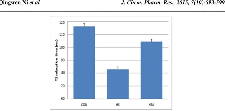
The average T2 relaxation times changes among control (CON), hind limb suspension (HS) and hind limb
______________________________________________________________________________
[image:5.595.174.421.321.477.2]Figure 4. The average T2 relaxation times among CON, HS, and HSA groups obtained from NMR CPMG measurement
Figure 5. The average weight of Soleus mass changes among CON, HS, and HSA groups
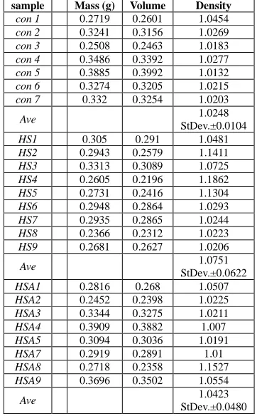
[image:5.595.92.506.503.649.2]Table 1. The average muscle densities among CON, HA, and HAS samples
sample Mass (g) Volume Density
con 1 0.2719 0.2601 1.0454
con 2 0.3241 0.3156 1.0269
con 3 0.2508 0.2463 1.0183
con 4 0.3486 0.3392 1.0277
con 5 0.3885 0.3992 1.0132
con 6 0.3274 0.3205 1.0215
con 7 0.332 0.3254 1.0203
Ave 1.0248
StDev.±0.0104
HS1 0.305 0.291 1.0481
HS2 0.2943 0.2579 1.1411
HS3 0.3313 0.3089 1.0725
HS4 0.2605 0.2196 1.1862
HS5 0.2731 0.2416 1.1304
HS6 0.2948 0.2864 1.0293
HS7 0.2935 0.2865 1.0244
HS8 0.2366 0.2312 1.0223
HS9 0.2681 0.2627 1.0206
Ave 1.0751
StDev.±0.0622
HSA1 0.2816 0.268 1.0507
HSA2 0.2452 0.2398 1.0225
HSA3 0.3344 0.3275 1.0211
HSA4 0.3909 0.3882 1.007
HSA5 0.3094 0.3036 1.0191
HSA7 0.2919 0.2891 1.01
HSA8 0.2718 0.2358 1.1527
HSA9 0.3696 0.3502 1.0554
Ave 1.0423
StDev.±0.0480
The significantly difference between HS and HSA is clearly observed. The soleus muscle mass is significantly higher (11%), the skeletal muscle cell sizes are 15-20% larger, and the T2 relaxation time is longer (20%) on HSA
than on HS, respectively. Since the muscle size is decreased after HS, however, it is observed that the average muscle density is slightly increased. As shown in Table 1 the average muscle densities are HSA 1.71% higher than CON; and HS 4.91% higher than CON. It is clearly to find that the electroacupunture has the function to improve or prevent muscle degradation during the disuse.
CONCLUSION
The results from our electroacupuncture tested on the dis-use hind limb suspension (HS) rat model are positive. It is suggested this technique can be effective in attenuating muscle atrophy induced by HS rat model. In addition, the success test is quite significant, especially, this non-pharmacological intervention can be applied on human. Since NMR measurement is non-destructive and non-invasive, and it is also suggested to use MRI technique to find the recovery function by continuing to apply electroacupuncture after HAS test on rat model.
Acknowledgements
This work was supported by Texas A&M International University internal research project.
REFERENCES
[1]Riggs, B. L. and L. J. Melton, III. N.Engl.J.Med. 327:620-627,8-27-1992. [2]Riggs, B. L. and L. J. Melton, III. Bone. 17:505S-511S,1995.
[3]D.J. Glass, Trends Mol. Med. 9 (8) (2003) 344–350.
[4]C.M. Ferrara, A.P. Goldberg, H.K. Ortmeyer, A.S. Ryan, J. Gerontol. A Biol. Sci. Med. Sci. 61 (5) (2006) 480– 487.
______________________________________________________________________________
[6]NIH, J Am Med Assoc. 280 (1998) 1518–1524.
[7]NIH Consensus Conference, Acupuncture, JAMA 280 (17) (1998) 1518–1524. [8]J.G. Lin, S.H. Yang, Am. J. Chin. Med. 27 (3–4) (1999) 299–305.
[9]M.L. Bullock, A.M. Pheley, S.K. Lenz, P.D. Culli-ton, J. Altern. Com-plement. Med. 5 (3) (1999) 253– 260. [10]A. Chandola, Y. Young, J. McAlister, J.S. Axford, J. R. Soc. Med. 92 (1) (1999) 13–16.
[11]V. Napadow, A. Ahn, J. Longhurst, L. Lao, E. Stener-Victorin, R. Harris, et al., J. Altern. Complement.Med. 14 (7) (2008) 861–869.
[12]E. Noguchi, H. Ohsawa, S. Kobayashi, M. Shi-mura, S. Uchida, Y. Sato, J. Auton. Nerv. Syst. 75 (2–3) (1999) 78–86.
[13]M. Ohkubo, T. Hamaoka, M. Niwayama, N. Murase, T. Osada, R. Kime, et al., Dyn. Med. 8 (2009) 2.
[14]E. Ikezono, K. Ohama, K. Nagayama, T. Sawa, T. Yoshida, Am. J. Chin. Med. (Gard City NY) 4 (1) (1976) 53– 59.
[15]Morey-Holton, E. R. and R. K. Globus. Bone. 22:83S-88S,1998.
[16]Rong-Tsung Lin, Chung-YuhTzeng, Yu-Chen Lee, Wai-Jane Ho, Juei-Tang Cheng, Jaung-Geng Lin and Shih-Liang Chang. BMC Complemen-tary and Alternative Medicine 2009, 9:26.
[17]Wang, X. and Ni, Q. 2003. J. Orthop. Res. 21(2): 312-319.
[18]Ni, Q., King, J.D. and Wang, X. 2004. Measurement Science and Technology, 15, 58 – 66. [19]Ni, Q. and Nicolella, D.P. 2005. Measurement Science and Technology, 16, 659-668.
[20]Ni, Q., Nyman, J., Wang, X., De Los Santos, A. and Nicolella, D. 2007 Measurement Science and Technology, 18, 715-723.
[21]Fantazzini, P., Brown, R.J.S. and Borgia, G.C., 2003. Magnetic Resonance Imaging 21, 227-234. [22]Carr, H.Y., and Purcell, E. M., 1954 Phys. Rev. 904, No.3 630.