129 C5aR = C5a receptor; CIA = collagen-induced arthritis; CII = collagen type II; Cn= complement component n; CR = complement receptor; FcγR = Fcγreceptor; FcRn = intracellular Fc receptor; GPI = glucose-6-phosphate isomerase; IC = immune complex; IL = interleukin; IVIG = intravenous therapy with high doses of normal IgG; RA = rheumatoid arthritis; RF = rheumatoid factor; SF = synovial fluid; TNF = tumor necrosis factor.
Abstract
Autoantibodies in sera from patients with autoimmune diseases have long been known and have become diagnostic tools. Analysis of their functional role again became popular with the availability of mice mutant for several genes of the complement and Fcγreceptor (FcγR) systems. Evidence from different inflammatory models suggests that both systems are interconnected in a hierarchical way. The complement system mediators such as complement component 5a (C5a) might be crucial in the communication between the complement system and FcγR-expressing cells. The split complement protein C5a is known to inactivate cells by its G-protein-coupled receptor and to be involved in the transcriptional regulation of FcγRs, thereby contributing to the complex regulation of autoimmune disease.
Introduction
Rheumatoid arthritis (RA) is a severe chronic disease characterized by the inflammation of synovial tissue in joints, which causes pain and dysfunction and ultimately leads to the destruction of joints. The pathogenesis of RA is not yet fully understood [1,2]. A general pathogenic hallmark of RA is the infiltration of T cells, B cells, macrophages, granulocytes and particular neutrophils into the synovial lining and fluid and the periarticular spaces. These infiltrating cells produce abundant cytokines, dominated mainly by the inflammatory type of cytokines such as tumor necrosis factor (TNF-α) and IL-1, which further activate effector cells such as macrophages and synoviocytes, finally leading to the damage of joint tissue. The occurrence of elevated levels of rheumatoid factors (RFs), which are autoantibodies against the Fc portion of IgG molecules, are a diagnostic marker for RA, even though they are not specific for the disease. RF is present not only in patients with RA but also in patients with other autoimmune diseases or healthy donors. The role of RF in the pathogenesis of RA, or even whether it has one, is still not clear [3].
More recently, autoantibodies against citrullinated proteins have been shown to be specifically present in patients with RA [4]. Studies involving animal models have shed more light on the role of autoantibodies in the pathogenesis of the disease [5,6]. There has been increasing evidence of the importance of autoantibodies and innate immunity cellular factors (Fc receptors and components of the complement system) in the pathophysiology of immunological diseases. In collagen-induced arthritis (CIA) [7], a model in which arthritis is induced in certain susceptible mouse strains by injecting collagen type II (CII) in complete Freund’s adjuvant, the antibodies directed to CII epitopes exposed on the cartilage surface in the joints have a crucial role in pathogenesis [8,9].
In a more recent mouse model of arthritis known as the K/B × N model [5,10,11], arthritis occurs spontaneously and autoantibodies reactive to the ubiquitously expressed protein glucose-6-phosphate isomerase (GPI) are produced. These IgG autoantibodies bind to GPI present on the cartilage surface in the joints and trigger a destructive arthritis. An acute transient form of synovitis can also be produced by the passive transfer of anti-GPI antibodies alone, supporting the view that antibodies alone can trigger the disease [12]. Studies conducted in this model supported the concept that whereas T and B cells are important for the initiation of RA, the pathogenicity is brought about mostly by autoantibodies and innate immune mediators. This review discusses the emerging concepts of a combined role of complement components and Fc receptors in RA pathogenesis.
Role of complement and complement
receptors
The complement system is a major innate defense system against various pathogenic agents, including bacteria and
Review
The role of the complement and the Fc
γγ
R system in the
pathogenesis of arthritis
Samuel Solomon
1, Daniela Kassahn
1and Harald Illges
1,2,31Immunology, Department of Biology, Faculty of Sciences, University of Konstanz, Konstanz, Germany 2Biotechnology Institute Thurgau, Tägerwilen, Switzerland
3University of Applied Sciences, Department of Natural Sciences, Immunology and Cell Biology, Rheinbach, Germany
Corresponding author: Harald Illges, [email protected]
Published: 16 May 2005 Arthritis Research & Therapy2005, 7:129-135 (DOI 10.1186/ar1761) This article is online at http://arthritis-research.com/content/7/4/129
130
viruses. Using different mechanisms (through both cell-bound and soluble proteins) it is able to discriminate self from non-self and its major role is in the induction and progression of inflammation against microbial pathogens. The complement system contributes to immune complex (IC) clearance by complement receptor 1 (CR1)-dependent and CR3-dependent phagocytosis, cell lysis by the terminal membrane attack complex, and mobilization of inflammatory immune cells through proteolytic products of soluble complement proteins known as the anaphylatoxins C3a, C4a and C5a. These proteins also modulate the inflammatory process by the complement receptors CR1/CD35, CR2/CD21, CR3/ CD11b-CD18, CR4/CD11c-CD18 and C5aR, expressed on leukocytes [13-17]. However, aberrant activation can lead to tissue damage and disease. In human disease, the comple-ment system has been shown to have a role in the patho-genesis of various immune-mediated disorders, including systemic lupus erythematosus, vasculitis, glomerulonephritis and RA [18]. Interestingly, deficiencies of early complement proteins of the classical pathway lead to autoimmunity, both in human and in mouse. Decreased levels of native comple-ment components and increased levels of complecomple-ment metabolites in plasma and synovial fluid (SF) of patients with RA have indirectly implicated a role of complement in the pathogenesis of RA.
A significant role for complement in the pathogenesis of RA has also been demonstrated by a variety of molecular and pathological studies. The total hemolytic complement, C3, and C4 are drastically reduced in SF relative to the total protein in patients with RA. Measurement of the activated proinflammatory complement products that are generated after C5 cleavage, namely C5a and C5b-9, showed significantly elevated levels in RA joints. Furthermore, positive correlations have been shown between SF complement activation (for example levels of plasma C3dg, a C3 activation metabolite) and local as well as general disease activity in RA, and between C2 and C3 expression in RA synovium and inflammation [19,20]. Furthermore, studies have shown high levels of C5a in SF [21] and correlation of the levels of C5a with the number of neutrophils present in the SF of patients with RA [22].
More direct evidence for the role of the complement system in RA came from experimental work with different animal models. Initial indications that activation of the complement cascade might have a role in RA was shown by the observation that rats depleted of complement C3 by using cobra venom factor are resistant to CIA [13]. Similar observations were seen with SWR mice that, in spite of expressing the I-Aq susceptibility gene, are resistant to CIA
because of a genetic deficiency in producing C5 [15].
Recent uses of knockout mice have clearly shown that complement activation is an integral component in the pathogenesis of CIA. Genetic deletion of C5, C3 or factor B
in DBA/1 mice resulted in each case in mice being highly resistant to CIA induction [23,24] despite the presence of high titers of anti-CII antibodies. The anaphylatoxin C3a is a product of complement network activation via all three initiating pathways. Subsequently the most potent anaphylatoxin, C5a, is generated from C5 during activation of the complement system by the C5 convertase. C5a not only induces cellular chemotaxis but also a wide array of effects, including the increase of vascular permeability and cellular degranulation. C5a exerts its bioactivity by binding to a G-protein-coupled receptor, called C5a receptor (C5aR). Expression of C5aR was also noted in synovial mast cells in RA joints [25], and mast cells are essential in arthritis induced by anti-GPI sera [26]. Both C5 deficiency and the systemic administration of anti-C5 antibodies ameliorated CIA, whereas both cellular and antibody responses to immunization with collagen II were normal [23,27].
Similarly, a gene therapy approach using retrovirally transduced soluble complement receptor 1 (CD35), an inhibitor of the classical and alternative pathway of complement activation in DBA/1 mice, has also been shown to have a beneficial effect, reducing inflammation and the development of CIA in these mice [16]. These results suggest that both the classical and alternative pathways of complement activation may be involved in CIA.
More recently, studies showing the importance of autoantibodies and both the complement and the FcR system came from the K/B × N mice model, in which arthritis arises spontaneously by crossing a C57BL/6 KRN TCR transgenic mouse line with the nonobese diabetic (NOD) mouse strain. The arthritogenic activity of K/B × N serum resides solely in the glucose-6-phosphate isomerase-reactive IgG fraction, allowing the passive transfer of antibodies in naive mice to induce arthritis [11,12,17,28]. A thorough study of the genetic influences on the end-stage effector phase of the arthritis in K/B × N mice revealed a prominent role of the C5 locus on chromosome 2 [29]. Arthritis was seen to progress in the progeny of crosses between the K/B × N mice and C1q or the C4 knockout mice similar to that in wild-type mice, showing that C1q and the classical complement activation pathway is not an effector in disease progression in this model. However, progeny mice of crosses between K/B × N and knockout mice deficient for either complement factor B, C3, C5 or C5aR were found to be completely resistant to arthritis.
131
C6-deficient mice also developed disease, suggesting that the late complement activation step (complement lysis by the membrane attack complex) is not involved in disease activity, but rather that the C5a interaction with C5aR expressed on many cell types such as neutrophils, macrophages and mast cells might be involved. C5a functions as a chemoattractant and induces acute inflammation by activating neutrophils and mast cells [29,30]. C5-targeting therapy prevented not only synovitis but also bone and cartilage degradation in the arthritic mouse model. Considering these data, C5a/C5aR may be an important modulator not only of inflammation in synovitis but also of cartilage destruction in arthritis.
How the alternative pathway is activated is still a matter of speculation: one method would be the formation and stabilization of surface-bound C3b-IgG fragments on the acellular cartilage surface, leading to formation of the C3 and C5 convertase. Because there are no complement regulatory proteins – which usually inhibit complement activation on eukaryotic cells – present on the cartilage surface and because the bound IgGs are able to bind C3b and prevent the binding of the complement regulatory plasma proteins factor I and factor H, formation of the C3 convertase is possible. The chemotactic effects of C5a–C5aR ligation then attracts neutrophils to sites of inflammation to produce properdin, which, upon binding to C3b–IgG complexes, enhances the association with factor B, leading to stabilization of the alternative pathway C3 convertase and amplification of the alternative pathway C3 consumption. However, properdin may bind preferentially to microbial surfaces and not to self surfaces [31]. These results are surprising because in human RA the involvement of the complement network in the development of arthritis has generally been assumed to reflect the classical pathway of activation involving the recognition of ICs by C1. One exception is juvenile arthritis, for which reports have provided evidence for the role of the alternative pathway [20].
The complement receptor CR1 binds the C3b, iC3b and C4b fragments, CR2 binds the C3d and iC3b fragments, and CR3 binds the iC3b fragments. These interactions have been implicated in the immune adherence of opsonized particles, phagocytosis, IC clearance and signal transduction. However, it could be shown that not even a combined deficiency of CR1 and CR2 had a detectable effect of conferring resistance or susceptibility on K/B × N serum-induced RA [17,30].
Role of Fc
γγ
receptors
Fc receptors act as a link between humoral and cellular responses, coupling antibody specificity with effector cell function. Engagement of Fc receptors triggers inflammatory, cytolytic, allergic or phagocytic activities [32]. An important role for FcγRs in RA pathogenesis is supported by the linkage of FcγR polymorphisms with RA [33,34] and the striking effects of FcγR deficiency on disease incidence and severity
in several animal models of inflammatory arthritis [35-37]. In mice, three subtypes of cell-surface membrane-bound IgG receptors (FcγRI, FcγRIIB and FcγRIII) have been identified. FcγRI and FcγRIII are regarded as stimulatory FcγRs because the receptors transmit stimulatory signals through an immunoreceptor tyrosine-based activation motif on self aggregation. In mice the high-affinity FcγRI is expressed on monocytes, macrophages and dendritic cells, whereas the low-affinity FcγRIII has a broader expression and is seen on neutrophils, macrophages and dendritic cells.
In contrast, FcγRII is regarded as an inhibitory receptor of immunoglobulin-induced B cell activation; it contains an immunoreceptor tyrosine-based inhibition motif and inhibits the activation of FcγRI and FcγRIII on aggregation with them. FcγRII is expressed on B cells, dendritic cells, neutrophils, macrophages, NK cells and mast cells in mice. These FcRs have been shown to have central roles in disease models such as allergy, nephritis and arthritis [32]. In the Arthus reaction mice model, the crucial role of the FcγR is well established. Mice lacking the stimulatory FcγRIII show an impaired Arthus reaction [38], whereas the FcγRII−/− mice
showed an enhanced Arthus reaction owing to the absence of its modulatory role in inhibiting FcγR activation [39].
Similarly, in RA several lines of evidence have implicated FcRs, in particular IgG-binding receptors (FcγRs), in disease pathogenesis: FcγRIII was detected on synovial intima in normal and arthritic human joints and on invading macrophages [40]. A FcγRIII gene polymorphism has been correlated with human RA susceptibility [33]. A dominant role for FcγRIII in the induction of both TNF-αand IL-1 production by human macrophages in RA after receptor ligation by small ICs has been shown recently. Mice lacking the common γ chain of the FcγRs (and thereby FcγRI and FcγRIII) were not susceptible to arthritis induction after an injection of collagen or adjuvant [35]; in addition, a lack of the inhibitory receptor FcγRIIB was found to exacerbate CIA in susceptible mouse strains [35].
132
FcR action determined in animal models of RA were also found in other inflammatory disease models such as the Arthus reaction [38] and IC peritonitis [44], suggesting a more general mechanism of these receptors in inflammatory diseases.
Maintaining autoantibody titers through
FcRn-mediated recycling
One of the most striking characteristics of the K/B × N model is the extremely high titer of specific autoantibodies throughout the life of the diseased mice. Titers in the order of 10 mg/ml specific anti-GPI IgG1 can be measured even in 2-year-old sick K/B × N mice (the spontaneous disease starts at about the third week after birth and is chronic). The particular receptor responsible for maintaining IgG titers in blood is the intracellular Fc receptor (FcRn) that is abundantly expressed in endothelial cells. The FcRn binds pinocytosed IgG only in the acidic environment of the endosome and releases intact IgG when its transport vesicle is redirected to the neutral pH of the cell surface. IgG not bound to the FcRn is transferred to lysosomes for degradation [45]. Accelerated catabolism of IgG is found when the FcRn in states of hypergammaglobulinemia is saturated. This IgG-depleting mechanism plausibly explains the beneficial results of intravenous therapy with high doses of normal IgG (IVIG) in autoimmune diseases mediated by pathogenic IgG. The synthesis of IgG is driven by immunogenic stimulation and is not affected by the rate of catabolism [46]. Since the first application of IVIG pooled from the plasma of healthy donors in the treatment of idiopathic thrombocytopenic purpura in children [47], many patients with a variety of autoimmune disorders have benefited from this therapy [48].
Numerous mechanisms have been proposed to explain the beneficial action of high doses of normal IgG in antibody-mediated disorders. Recently Akilesh and colleagues [49] crossed FcRn deficient mice with the genetically determined K/B × N arthritis model. This resulted in partial or complete protection from arthritis. The same was found for the anti-GPI sera transfer model. In both cases disease severity was correlated with the concentration of anti-GPI antibody titers in blood. Moreover, transferring large amounts of sera from sick mice could override the protective effect of FcRn deficiency, suggesting a dependence of disease induction on antibody concentration. Interestingly, IVIG treatment of mice resulted in protection from arthritis induced by K/B × N sera in wild-type animals but not in FcRn-deficient animals [49,50]. This suggests that saturation of the FcRn is the mechanism indirectly shortening the half-life of the pathogenic IgG by decreasing the concentration below pathological thresholds. In contrast, the FcRn is responsible for maintaining high auto-antibody titers by prolonging their half-life. It is speculative, but plausible, that different glycosylation patterns as found in autoimmune conditions [51] may enhance or reduce FcRn-dependent recycling, because the FcRn seems to have some specificity for alterations of antibodies [52].
Convergence of complement and Fc receptor
activation in RA pathogenesis
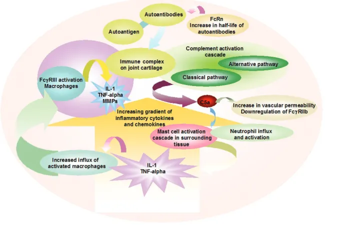
It has been widely accepted that ICs initiate inflammatory responses in IC diseases such as arthritis or the Arthus reaction either by activation of the complement system or by the direct engagement and activation of FcγR-bearing inflammatory cells. Complement and FcγRs act in concert in many inflammatory responses, with complement both attracting and activating FcγR-bearing cells at sites of inflammation. Antibodies, particularly as constituents of antibody–antigen ICs, have a central role in triggering inflammation in several autoimmune diseases (Fig. 1).
Although the concept of IC-triggered inflammation through the activation of the complement cascade is known, studies in FcR-deficient mutant mice have promoted an opposing view that ICs induce inflammation predominantly through FcR engagement, with complement proteins subserving primarily immunoregulatory functions. Studies on relative contributions of either complement or FcγRs in the inflammatory response, especially in Arthus reaction mice models, have resulted in a better understanding of the role of the complement system and FcγRs, although the relative contributions and the hierarchy of these two pathways in the manifestation of disease are still unclear. In FcγR−/−mice, in which the surface
expression of FcγRI and FcγRIII is downregulated, the inflammatory response in the reverse Arthus reaction in skin, peritoneum and lung is impaired, arguing for the predominance of FcγR-expressing effector cells for disease manifestation. This view is opposed by studies showing that C5aR−/−mice are completely protected from lung injury in the
reverse Arthus reaction, although this protection was not complete for injury to skin and peritoneum [53].
Another elegant study in an IC-induced inflammation model argued for a codominant role of inflammatory pathways mediated by FcγRI/III and C5aR [54]. In support of this view it was shown that the C5a anaphylatoxin, acting through the C5aR, is a major regulator of the transcription of activating and inhibitory FcγRs in IC-induced lung disease. C5a increased the expression of activating FcγRIII and decreased the expression of the inhibitory FcγRII on alveolar macrophages, which led to a lower threshold for cellular activation [55]. This elegant set of experiments supports the hypothesis that the FcγR activation is downstream of complement activation in the inflammatory cascade. This is of particular interest because it shows clearly how an immune response initially triggered by the complement system can regulate the cellular response through the regulation of FcRs much like the instructive role of the innate immune system for the adaptive response [56].
133
results in complement activation. This activation depends on the structural changes imposed on the antibody molecule after binding to antigen and, at least in case of the classical pathway of complement, on the distance or density of antigen-bound antibodies. Once the complement system has been activated, anaphylatoxic molecules, most importantly C5a, trigger cellular migration to sites of inflammation and changes in FcR expression. At that time the inhibitory receptor FcγRII is downregulated and these cells subsequently lack the ability to downregulate FcγRI and FcγRIII and the ability to clear and endocytose ICs efficiently [55]. Both complement and FcγRs are co-expressed on key cellular players of inflammation such as mast cells and macrophages.
Recently it was shown that autoantibodies develop long before the onset of disease in systemic lupus erythomatosus and anti-phospholipid syndrome [57,58]. Disease may develop through epitope spreading and changes in the concentration of the autoantibodies. In particular the latter may also depend on FcRn-mediated recycling. ICs containing
[image:5.612.145.490.113.338.2]antigen, antibody and proteolytically split complement products can bind to FcRs, complement receptors and antigen receptors at the same time and, for B cells, on the very same cell. Both FcRs and complement receptors are involved in the binding of ICs and phagocytosis; they deliver signals leading to the activation and differentiation of cells. Differences in the expression of these receptors on cells such as mast cells and macrophages as well as in the tissue localization determine the reaction to ICs, but are far from understood. Moreover, both the FcR and complement system, as part of the innate immune system, are involved in early immune defense and subsequently activate and instruct the adaptive immune system; at the same time they are involved in immune regulation when the adaptive response is ongoing. Taken together, coordinated activation and signaling through the complement system and the FcR system in a hierarchical manner leads to inflammation. Although the results obtained from several disease models fit more or less well, the diverse activities of complement and FcRs on the different types of cells, even if one focuses only on antibody-mediated diseases, remains to be analyzed in detail, as is true Figure 1
Schematic representation of a local autoantibody-induced inflammatory network in an arthritic joint. Autoantibodies and autoantigen form immune complexes (ICs) in the vascular system and are captured on cartilage surfaces. The intracellular Fc receptor (FcRn) may sustain certain
134
of the first step in antibody-dependent activation of the alternative pathway of the complement system.
Competing interests
The author(s) declare that they have no competing interests.
Authors’ contributions
All authors contributed equally to this paper.
Acknowledgements
This work was supported by the Thurgauische Stiftung für Wis-senschaft und Forschung, the Hans-Hench-Stiftung and the Bunde-samt für Bildung und Wissenschaft (BBW), Bern, Switzerland through grants QLG1-CT-2001-01536 and QLG1-CT-2001-01407 to HI.
References
1. Feldmann M, Brennan FM, Maini RN: Rheumatoid arthritis.Cell 1996, 85:307-310.
2. St Clair EW: Therapy of rheumatoid arthritis: new develop-ments and trends.Curr Rheumatol Rep1999, 1:149-156. 3. Steiner G, Smolen J: Autoantibodies in rheumatoid arthritis and
their clinical significance.Arthritis Res2002, 4 (Suppl 2):S1-S5. 4. van Venrooij WJ, Pruijn GJM: Citrullination: a small change for a protein with great consequences for rheumatoid arthritis. Arthritis Res2000, 2:249-251.
5. Kouskoff V, Korganow AS, Duchatelle V, Degott C, Benoist C, Mathis D: Organ-specific disease provoked by systemic autoimmunity.Cell1996, 87:811-822.
6. Holmdahl: Rheumatoid arthritis viewed using a headache par-adigm.Arthritis Res2000, 2:169-171.
7. Myers LK, Rosloniec EF, Cremer MA, Kang AH: Collagen-induced arthritis, an animal model of autoimmunity.Life Sci 1997, 61:1861-1878.
8. Holmdahl R, Jansson L, Larsson A, Jonsson R: Arthritis in DBA/1 mice induced with passively transferred type II collagen immune serum. Immunohistopathology and serum levels of anti- type II collagen auto-antibodies.Scand J Immunol1990, 31:147-157.
9. Burkhardt H, Koller T, Engstrom A, Nandakumar KS, Turnay J, Kraetsch HG, Kalden JR, Holmdahl R: Epitope-specific recogni-tion of type II collagen by rheumatoid arthritis antibodies is shared with recognition by antibodies that are arthritogenic in collagen-induced arthritis in the mouse.Arthritis Rheum2002, 46:2339-2348.
10. Kyburz D, Corr M: The KRN mouse model of inflammatory arthritis.Springer Semin Immunopathol2003, 25:79-90. 11. Korganow AS, Ji H, Mangialaio S, Duchatelle V, Pelanda R, Martin
T, Degott C, Kikutani H, Rajewsky K, Pasquali JL, et al.: From sys-temic T cell self-reactivity to organ-specific autoimmune disease via immunoglobulins.Immunity1999, 10:451-461. 12. Matsumoto I, Maccioni M, Lee DM, Maurice M, Simmons B,
Brenner M, Mathis D, Benoist C: How antibodies to a ubiqui-tous cytoplasmic enzyme may provoke joint-specific autoim-mune disease.Nat Immunol2002, 3:360-365.
13. Morgan K, Clague RB, Shaw MJ, Firth SA, Twose TM, Holt PJ: Native type II collagen-induced arthritis in the rat: the effect of complement depletion by cobra venom factor.Arthritis Rheum 1981, 24:1356-1362.
14. Banerjee S, Anderson GD, Luthra HS, David CS: Influence of complement C5 and V beta T cell receptor mutations on sus-ceptibility to collagen-induced arthritis in mice. J Immunol 1989, 142:2237-2243.
15. Barrington R, Zhang M, Fischer M, Carroll MC: The role of com-plement in inflammation and adaptive immunity.Immunol Rev 2001, 180:5-15.
16. Dreja H, Annenkov A, Chernajovsky Y: Soluble complement receptor 1 (CD35) delivered by retrovirally infected syngeneic cells or by naked DNA injection prevents the progression of collagen-induced arthritis.Arthritis Rheum2000, 43:1698-1709. 17. Solomon S, Kolb C, Mohanty S, Jeisy-Walder E, Preyer R, Schollhorn V, Illges H: Transmission of antibody-induced arthritis is indepen-dent of complement component 4 (C4) and the complement receptors 1 and 2 (CD21/35).Eur J Immunol2002, 32:644-651.
18. Tsokos GC, Fleming SD: Autoimmunity, complement activa-tion, tissue injury and reciprocal effects.Curr Dir Autoimmun 2004, 7:149-164.
19. Jarvis JN, Pousak T, Krenz M, Iobidze M, Taylor H: Complement activation and immune complexes in juvenile rheumatoid arthritis.J Rheumatol1993, 20:114-117.
20. Aggarwal A, Bhardwaj A, Alam S, Misra R: Evidence for activa-tion of the alternate complement pathway in patients with juvenile rheumatoid arthritis.Rheumatology (Oxford)2000, 39: 189-192.
21. Hogasen K, Mollnes TE, Harboe M, Gotze O, Hammer HB, Oppermann M: Terminal complement pathway activation and low lysis inhibitors in rheumatoid arthritis synovial fluid. J Rheumatol1995, 22:24-28.
22. Gerard C, Gerard NP: C5A anaphylatoxin and its seven trans-membrane-segment receptor. Annu Rev Immunol 1994, 12: 775-808.
23. Wang Y, Kristan J, Hao L, Lenkoski CS, Shen Y, Matis LA: A role for complement in antibody-mediated inflammation: C5-defi-cient DBA/1 mice are resistant to collagen-induced arthritis.J Immunol2000, 164:4340-4347.
24. Hietala MA, Nandakumar KS, Persson L, Fahlen S, Holmdahl R, Pekna M: Complement activation by both classical and alter-native pathways is critical for the effector phase of arthritis. Eur J Immunol2004, 34:1208-1216.
25. Kiener HP, Baghestanian M, Dominkus M, Walchshofer S, Ghan-nadan M, Willheim M, Sillaber C, Graninger WB, Smolen JS, Valent P: Expression of the C5a receptor (CD88) on synovial mast cells in patients with rheumatoid arthritis. Arthritis Rheum1998, 41:233-245.
26. Lee DM, Friend DS, Gurish MF, Benoist C, Mathis D, Brenner MB: Mast cells: a cellular link between autoantibodies and inflammatory arthritis.Science2002, 297:1689-1692. 27. Wang Y, Rollins SA, Madri JA, Matis LA: Anti-C5 monoclonal
antibody therapy prevents collagen-induced arthritis and ameliorates established disease. Proc Natl Acad Sci U S A 1995, 92:8955-8959.
28. Matsumoto I, Staub A, Benoist C, Mathis D: Arthritis provoked by linked T and B cell recognition of a glycolytic enzyme. Science1999, 286:1732-1735.
29. Ji H, Gauguier D, Ohmura K, Gonzalez A, Duchatelle V, Danoy P, Garchon HJ, Degott C, Lathrop M, Benoist C, et al.: Genetic influences on the end-stage effector phase of arthritis.J Exp Med2001, 194:321-330.
30. Ji H, Ohmura K, Mahmood U, Lee DM, Hofhuis FM, Boackle SA, Takahashi K, Holers VM, Walport M, Gerard C, et al.: Arthritis critically dependent on innate immune system players. Immu-nity2002, 16:157-168.
31. Schwaeble WJ, Reid KB: Does properdin crosslink the cellular and the humoral immune response?Immunol Today1999, 20: 17-21.
32. Ravetch JV, Bolland S: IgG Fc receptors.Annu Rev Immunol 2001, 19:275-290.
33. Nieto A, Caliz R, Pascual M, Mataran L, Garcia S, Martin J: Involvement of Fcγγreceptor IIIA genotypes in susceptibility to rheumatoid arthritis.Arthritis Rheum2000, 43:735-739. 34. Johansson AC, Sundler M, Kjellen P, Johannesson M, Cook A,
Lindqvist AK, Nakken B, Bolstad AI, Jonsson R, Alarcon-Riquelme M, et al.: Genetic control of collagen-induced arthritis in a cross with NOD and C57BL/10 mice is dependent on gene regions encoding complement factor 5 and FcγγRIIb and is not associated with loci controlling diabetes.Eur J Immunol2001, 31:1847-1856.
35. Kleinau S, Martinsson P, Heyman B: Induction and suppression of collagen-induced arthritis is dependent on distinct Fcγγ receptors.J Exp Med2000, 191:1611-1616.
36. Corr M, Crain B: The role of FcγγR signaling in the K/B x N serum transfer model of arthritis.J Immunol2002, 169: 6604-6609.
37. Van den Berg WB: Lessons from animal models of arthritis. Curr Rheumatol Rep2002, 4:232-239.
38. Hazenbos WL, Gessner JE, Hofhuis FM, Kuipers H, Meyer D, Heijnen IA, Schmidt RE, Sandor M, Capel PJ, Daeron M, et al.: Impaired IgG-dependent anaphylaxis and Arthus reaction in Fc γγRIII (CD16) deficient mice.Immunity1996, 5:181-188. 39. Clynes R, Maizes JS, Guinamard R, Ono M, Takai T, Ravetch JV:
135 by the coordinate expression of activation and inhibitory Fc
receptors.J Exp Med1999, 189:179-185.
40. Edwards SW, Watson F, Gasmi L, Moulding DA, Quayle JA: Acti-vation of human neutrophils by soluble immune complexes: role of Fc γγRII and Fc gamma RIIIb in stimulation of the respi-ratory burst and elevation of intracellular Ca2+.Ann N Y Acad
Sci1997, 832:341-357.
41. Yuasa T, Kubo S, Yoshino T, Ujike A, Matsumura K, Ono M, Ravetch JV, Takai T: Deletion of Fcγγreceptor IIB renders H-2b
mice susceptible to collagen-induced arthritis. J Exp Med 1999, 189:187-194.
42. Nandakumar KS, Andren M, Martinsson P, Bajtner E, Hellstrom S, Holmdahl R, Kleinau S: Induction of arthritis by single mono-clonal IgG anti-collagen type II antibodies and enhancement of arthritis in mice lacking inhibitory FcγγRIIB.Eur J Immunol 2003, 33:2269-2277.
43. Kyburz D, Carson DA, Corr M: The role of CD40 ligand and tumor necrosis factor alpha signaling in the transgenic K/BxN mouse model of rheumatoid arthritis.Arthritis Rheum 2000, 43:2571-2577.
44. Heller T, Gessner JE, Schmidt RE, Klos A, Bautsch W, Kohl J: Cutting edge: Fc receptor type I for IgG on macrophages and complement mediate the inflammatory response in immune complex peritonitis.J Immunol1999, 162:5657-5661.
45. Ghetie V, Ward ES: FcRn: the MHC class I-related receptor that is more than an IgG transporter.Immunol Today1997, 18: 592-598.
46. Junghans RP: IgG biosynthesis: no ‘immunoregulatory feed-back’.Blood1997, 90:3815-3818.
47. Imbach P, Barandun S, d’Apuzzo V, Baumgartner C, Hirt A, Morell A, Rossi E, Schoni M, Vest M, Wagner HP: High-dose intra-venous gammaglobulin for idiopathic thrombocytopenic purpura in childhood.Lancet1981, i:1228-1231.
48. Geha R, Rosen F: Intravenous immunoglobulin therapy. In Therapeutic Immunology.Edited by Auston K, Burakoff S, Rosen FS, Strom T. Cambridge, MA: Blackwell Science; 1996:280-296. 49. Akilesh S, Petkova S, Sproule TJ, Shaffer DJ, Christianson GJ, Roopenian D: The MHC class I-like Fc receptor promotes humorally mediated autoimmune disease.J Clin Invest2004, 113:1328-1333.
50. Bruhns P, Samuelsson A, Pollard JW, Ravetch JV: Colony-stimu-lating factor-1-dependent macrophages are responsible for IVIG protection in antibody-induced autoimmune disease. Immunity2003, 18:573-581.
51. Parekh RB, Dwek RA, Sutton BJ, Fernandes DL, Leung A, Stan-worth D, Rademacher TW, Mizuochi T, Taniguchi T, Matsuta K, et al.: Association of rheumatoid arthritis and primary osteoarthritis with changes in the glycosylation pattern of total serum IgG.Nature1985, 316:452-457.
52. Popov S, Hubbard JG, Kim J, Ober B, Ghetie V, Ward ES: The stoichiometry and affinity of the interaction of murine Fc frag-ments with the MHC class I-related receptor, FcRn. Mol Immunol1996, 33:521-530.
53. Höpken U: Impaired inflammatory responses in the reverse Arthus reaction through genetic deletion of the C5a receptor. J Exp Med1997, 186:749–756.
54. Baumann U, Kohl J, Tschernig T, Schwerter-Strumpf K, Verbeek JS, Schmidt RE, Gessner JE: A codominant role of Fc γγRI/III and C5aR in the reverse Arthus reaction.J Immunol2000, 164: 1065-1070.
55. Shushakova N, Skokowa J, Schulman J, Baumann U, Zwirner J, Schmidt RE, Gessner JE: C5a anaphylatoxin is a major regula-tor of activating versus inhibiregula-tory FcγγRs in immune complex-induced lung disease.J Clin Invest2002, 110:1823-1830. 56. Fearon DT: Seeking wisdom in innate immunity.Nature1997,
388:323-324.
57. Arbuckle MR, McClain MT, Rubertone MV, Scofield RH, Dennis GJ, James JA, Harley JB: Development of autoantibodies before the clinical onset of systemic lupus erythematosus.N Engl J Med2003, 349:1526-1533.