ScholarWorks @ Georgia State University
ScholarWorks @ Georgia State University
Chemistry Dissertations Department of Chemistry
3-5-2010
Roles of Serine 101, Histidine 310 and Valine 464 in the Reaction
Roles of Serine 101, Histidine 310 and Valine 464 in the Reaction
Catalyzed by Choline Oxidase from Arthrobacter Globiformis
Catalyzed by Choline Oxidase from Arthrobacter Globiformis
Steffan Finnegan
Georgia State University
Follow this and additional works at: https://scholarworks.gsu.edu/chemistry_diss
Part of the Chemistry Commons
Recommended Citation Recommended Citation
Finnegan, Steffan, "Roles of Serine 101, Histidine 310 and Valine 464 in the Reaction Catalyzed by Choline Oxidase from Arthrobacter Globiformis." Dissertation, Georgia State University, 2010.
https://scholarworks.gsu.edu/chemistry_diss/37
This Dissertation is brought to you for free and open access by the Department of Chemistry at ScholarWorks @ Georgia State University. It has been accepted for inclusion in Chemistry Dissertations by an authorized
CATALYZED BY CHOLINE OXIDASE FROM ARTHROBACTER GLOBIFORMIS
by
STEFFAN FINNEGAN
Under the Direction of Giovanni Gadda
ABSTRACT
The enzymatic oxidation of choline to glycine betaine is of interest because organisms accumulate glycine betaine intracellularly in response to stress conditions, as such it is of potential interest for the genetic engineering of crops that do not naturally possess efficient pathways for the synthesis of glycine betaine, and for the potential development of drugs that target the glycine betaine biosynthetic pathway in human pathogens. To date, one of the best characterized enzymes belonging to this pathway is the flavin-dependent choline oxidase from Arthrobacter globiformis. In this enzyme, choline oxidation proceeds through two reductive half-reactions and two oxidative half-half-reactions. In each of the reductive half-half-reactions the FAD cofactor is reduced to the anionic hydroquinone form (2 e- reduced) which is followed by an oxidative half-reaction where the reduced FAD cofactor is reoxidized by molecular oxygen with formation and release of hydrogen peroxide.
as well as X-ray crystallography.
A comparison of the kinetic data obtained for the variant enzymes to previous data obtained for wild-type choline oxidase are consistent with the valine residue at position 464 being important for the oxidative half-reaction as well as the positioning of the catalytic groups in the active site of the enzyme. The kinetic data obtained for the serine at position 101 shows that serine 101 is important for both the reductive and oxidative half-reactions. Finally, the kinetic data for histidine at position 310 suggest that this residue is essential for both the reductive and oxidative half-reactions.
CATALYZED BY CHOLINE OXIDASE FROM ARTHROBACTER GLOBIFORMIS
by
STEFFAN FINNEGAN
A Dissertation Submitted in Partial Fulfillment of the Requirements for the Degree of Doctor of Philosophy
in the College of Arts and Sciences Georgia State University
Copyright by Steffan Finnegan
CATALYZED BY CHOLINE OXIDASE FROM ARTHROBACTER GLOBIFORMIS
by
STEFFAN FINNEGAN
Committee Chair: Dr. Giovanni Gadda
Committee: Dr. Markus W. Germann Dr. Aimin Liu
Electronic Version Approved by:
TABLE OF CONTENTS
LIST OF TABLES ix
LIST OF FIGURES xi
LIST OF SCHEMES xv
CHAPTER I 1
INTRODUCTION 1
1.1 Flavin dependent enzymes ... 1
1.2 Selected structural features of flavoenzymes with different oxygen reactivity ... 7
1.3 Oxygen reactivity... 12
1.3.1. General oxygen chemistry ... 12
1.3.2. Enzymes and oxygen ... 15
1.4. Choline Oxidase from Arthrobacter globiformis ... 28
1.5. Specific Goals ... 32
1.6. References ... 35
CHAPTER II 57 CRYSTALLOGRAPHIC, SPECTROSCOPIC, AND COMPUTATIONAL ANALYSIS OF A FLAVIN C4A-OXYGEN ADDUCT IN CHOLINE OXIDASE. 57 2.1. Abstract ... 57
2.2. Introduction ... 57
2.3. Materials and methods... 61
2.4. Results and discussion ... 65
2.5. Acknowledgment ... 79
CHAPTER III 87
SUBSTITUTION OF AN ACTIVE SITE VALINE UNCOVERS A KINETICALLY
SLOW EQUILIBRIUM BETWEEN COMPETENT AND INCOMPETENT FORMS OF
CHOLINE OXIDASE 87
3.1. Abbreviations ... 87
3.2. Abstract ... 87
3.3. Introduction ... 88
3.4. Experimental procedures ... 94
3.5. Results ... 97
3.6. Discussion ... 111
3.7. Appendix ... 117
3.8. Acknowledgements ... 121
3.9. References ... 121
CHAPTER IV 130 ROLE OF VAL464 IN THE FLAVIN OXIDATION REACTION CATALYZED BY CHOLINE OXIDASE 130 4.1. Abbreviations ... 130
4.1. Abstract ... 130
4.2. Introduction ... 131
4.3. Experimental procedures ... 135
4.4. Results ... 140
4.5. Discussion ... 151
4.7. References ... 157
CHAPTER V 163 ON THE IMPORTANCE OF SER101 FOR OVERALL TURNOVER IN CHOLINE OXIDASE 163 5.1. Abstract ... 163
5.2. Introduction ... 163
5.3. Experimental Procedures ... 165
5.4. Results ... 168
5.5. Discussion ... 174
5.6. Acknowledgement ... 176
5.7. References ... 176
CHAPTER VI 179 ON THE ROLE OF THE ACTIVE SITE RESIDUE HIS310 OF CHOLINE OXIDASE 179 6.1. Abstract ... 179
6.2. Introduction ... 180
6.3. Experimental Procedures ... 182
6.4. Results ... 185
6.5. Discussion ... 194
6.6. References ... 198
7.2. Introduction ... 201
7.3. Experimental Procedures ... 205
7.4. Results ... 208
7.5. Discussion ... 213
7.6. References ... 217
LIST OF TABLES
Table 1.1. Overall Fold of Selected Flavoenzymes 8
Table 1.2. Structural Features of Selected Flavoenzymes Important for Oxygen Reactivity 12
Table 1.3. Kinetic Parameters Influenced by Substitution of Various Residues in Choline
Oxidase 30
Table 2.1. Selected Bond Distances and Angles in the X-ray Structure and DFT Optimized
Models 63
Table 3.1. Reductive Half-reaction of the Val464Thr, Val464Ala and Wild-type Enzymes with
Choline as Substrate 104
Table 3.2. Effect of Deuterated Substrate and Solvent on the Reductive Half-reaction of the
Val464Ala, Val464Thr and Wild-type Enzymes 106
Table 3.3. Substrate and Solvent Deuterium Kinetic Isotope Effects on the Reductive Half-reaction of the Val464Ala, Val464Thr and Wild type Enzymes 106
Table 3.4. Effect of Glucose and PEG-6000 on the Reductive Half-reaction of the Val464Thr
Enzyme with Choline as Substrate 108
Table 3.5. Comparison of the pKa Values Sssociated with the Ionization of the Enzyme-bound Oxidized Form of 8α-N(3)-histidyl-FAD in the Val464Ala, Val464Thr, and His466Ala Enzymes
with Wild-type Choline Oxidase 111
Table 4.1. Crystallographic Data Collection and Refinement Statistics 140 Table 4.2.app(kcat/Koxygen) for the Val464Ala Enzyme at Fixed Saturating Concentrations of
3,3-dimethyl-butan-1-ol as Substrate 150
Table 5.2. Comparison of the Kinetic Parameters of Ser101Ala and Wild-type Choline Oxidase
at pH 10.0 173
Table 6.1. Flavin Properties of the His310Ala, His310Asp, and His310Asn Variant Enzymes 187
Table 6.2. Anaerobic Reduction of His310Ala and His310Asn with Choline 192
LIST OF FIGURES
Figure 1.1. Structures of Riboflavin (X=H), FMN (X = PO32-), and FAD (X = ADP) 1
Figure 1.2. Sample of Biological Functions that Involve Flavoenzymes 2
Figure 1.3. Redox and Ionization States of Flavins 4
Figure 1.4. Spectra of Glucose Oxidase in the Oxidized, Semiquinone (Anionic and Neutral),
and Fully Reduced States 4
Figure 1.5. Flavin Dependent Enzymes Displaying a Hydrophobic Residue Close to a Positive Charge that is Required for Oxygen Activation Close to the FAD-C(4a)-N5 Region 10
Figure 1.6. Flavin Dependent Enzymes Displaying a Hydrophobic Residue Close to a Positive
Charge Close to the FAD-N5 Atom 10
Figure 1.7. Standard Reduction Potentials for O2 13
Figure 1.8. Flavin Dependent Oxygen Reduction Pathways 15
Figure 1.9. Reaction Types Catalyzed by Flavin-dependent Monooxygenases 24
Figure 1.10. The Two Proposed Copper Oxygen Coordination Modes 26
Figure 1.11. The Coordination of the Side-chain of His310 in the Crystal Structure of Wild-type
Choline Oxidase 34
Figure 2.1. The Single Crystal Optical Absorption Spectroscopy Facility Installed at X26-C60
Figure 2.2. Spectroscopic Changes Observed in Single Crystals of Choline Oxidase upon
X-ray Irradiation at 100 K 67
Figure 2.3. Optical Absorption Spectrum of 0.04 mM Choline Oxidase at Room Temperature 68
Figure 2.4. Comparison of the X-ray Structures with the DFT Optimized Modes for the FAD
Figure 2.5. The Active Site Environment of Choline Oxidase Illustrating Potential Hydrogen Bonding Interactions that Stabilize the C4a-OH or C4a-OO(H) Adducts 73
Figure 2.6. Overlay of Several DFT Optimized C4a Adducts and the Crystal structure of Choline
Oxidase 75
Figure 2.7. The Proposed Reaction Scheme for the C4a Adduct Formation in Single Crystals of
Choline Oxidase at 100 K 76
Figure 2.8. Proposed Reaction Mechanism for Choline Oxidase 78
Figure 3.1. Close-up View of the Active Site of the Wild-type Form of Choline Oxidase 93
Figure 3.2. Anaerobic Reduction of the Val464Thr and Val464Ala Enzymes with Choline 100
Figure 3.3. Example of an Anaerobic Reduction Trace of the Val464Ala Enzyme with 75 mM
Choline in 50 mM Sodium Pyrophosphate, pH 6.0 and 25 °C 101
Figure 3.4. The Rates of Anaerobic Flavin Reduction Measured in a Stopped-flow
Spectrophotometer as a Function of the Mole Fraction of Deuterium Oxide 109
Figure 3.5. pH Dependence of the UV-visible Absorbance Spectra of Val464 Variant
Enzymes 110
Figure 3.6. Close-up View of the Active Site of the Wild-type Form of Choline Oxidase Showing the Hydrogen Bonding Interactions Involving the N(1) Atom of His99 116
Figure 4.1. Crystal Structure of Val464Ala Mutant 141
Figure 4.2. Dependence of the Initial Rates of Reaction with Choline as Substrate for the
Val464Thr Enzyme as a Function of [Oxygen] 145
Figure 4.3. Effect of pH on the kcat/Koxygen Values with Choline as Substrate for the Val464Ala
Figure 4.4. Time-resolved, Flavin Oxidation of the Val464Ala, Val464Thr and Wild-type Enzymes with Oxygen in 50 mM Sodium Pyrophosphate, pH 10.0 and 25 °C 147
Figure 4.5. Enzyme Monitored Turnovers with Choline and Oxygen as Substrates for the
Val464Ala, Val464Thr, and Wild-type Enzymes 148
Figure 4.6.kcat/Koxygen Values for the Wild-type Enzyme as well as Variant Forms of Choline Oxidase with Mutations in the Active Site at pH 10.0 and 25°C 151
Figure 5.1. Comparison of Crystal Structures of the Ser101Ala and Wild-type Enzymes of
Choline Oxidase 170
Figure 5.2. Double Reciprocal Plots of CHO-Ser101Ala Catalyzed Oxidation of Choline and
Betaine Aldehyde 172
Figure 5.3. Reductive Half-reaction of the Ser101Ala Enzyme with Betaine Aldehyde 174
Figure 6.1. Line Drawing Showing the Interaction of His466 with the N(1)−C(2)═O Locus of FAD and the Intermediate Alkoxide Species in the Transition State for the Oxidation of Choline
Catalyzed by Choline Oxidase 182
Figure 6.2. Crystal Structure of Choline Oxidase.X-ray Crystallographic Structure of the Active
Site of Choline Oxidase Determined at 1.86 Å Resolution 182
Figure 6.3. Spectral Properties of the His310 Variant Enzymes 186
Figure 6.4. Circular Dichroism and Fluorescence of the His310Ala, His310Asp and His310Asn
Variant Enzymes as well as the Wild-type Enzyme 188
Figure 6.5. Time Resolved, Anaerobic Reduction of the His310Ala, His310Asp and His310Asn Variant Enzymes with Choline in 20 mM Tris-Cl, pH 7.0 and at 15 °C 189
Figure 6.6. Anaerobic Spectra of the Oxidized and Reduced Form of the His310Ala Variant
Figure 6.7. Anaerobic Reduction Traces of the His310Ala Variant Enzyme with Choline, pH
8.0, at 15 C 191
Figure 6.8. Anaerobic Flavin Reduction of His310Ala and His310Asn 193
Figure 7.1. Close-up View of the Active Site of the Val464Ala Variant Form of Choline Oxidase Showing the Hydrogen Bonding Interactions Involving the N(1) Atom of His99 204
Figure 7.2. pH Dependence of the kred, Kd , k1, k3 and Kd* Values with Choline and 1,2-[2H4
]-Choline as Substrate for ]-Choline Oxidase. 210
LIST OF SCHEMES
Scheme 1.1. Generalized Flavin Catalytic Cycle 3
Scheme 1.2. Reaction Mechanism of Indoleamine 2,3-dioxygenase 17
Scheme 1.3. Generalized Reaction Catalyzed by Cytochrome P-450 18
Scheme 1.4. Simplified Catalytic Mechanism for Cytochrome P-450 19
Scheme 1.5. Monooxygenation of Monophenols Catalyzed by Tyrosinase 21
Scheme 1.6. Dehydrogenation of o-diphenols Catalyzed by Tyrosinase 21
Scheme 1.7. Catalytic Cycle of Tyrosinase Reaction with o-diphenols 22
Scheme 1.8. The Overall Reaction Catalyzed by Galactose Oxidase 25
Scheme 1.9. Minimal Kinetic Mechanism of Choline Oxidase 29
Scheme 3.1. The Flavin-mediated, Four-electron Oxidation of Choline to Glycine Betaine
Catalyzed by Choline Oxidase 89
Scheme 3.2. Hydride Transfer Mechanism for the Oxidation of Choline to Betaine Aldehyde
Catalyzed by Choline Oxidase 91
Scheme 3.3. Proposed Kinetic Mechanism for Choline Oxidation Catalyzed by the Val464Thr
and Val464Ala Enzymes. 102
Scheme A1. Pre-steady State Mechanism of Val464 Variant Enzymes 117
Scheme A2. Simplified Mechanism for Derivation of Pre-steady State Equations for Val464
Variant Enzymes 118
Scheme 4.1. Activation of Oxygen by a Positively Charged Group in Glucose Oxidase,
Monomeric Sarcosine Oxidase, and Choline Oxidase 132
Scheme 4.3. Minimal Kinetic Mechanism for Reductive Half-reaction of the Val464Ala and
Val464Thr Enzymes 134
Scheme 6.1. Reaction Catalyzed by Choline Oxidase 181
Scheme 6.2. Proposed Reaction Mechanism for the Reaction Catalyzed by Choline Oxidase . 197
Scheme 7.1.The Two-step Reaction for the Oxidation of Choline to Glycine Betaine Catalyzed
by Choline Oxidase 202
Scheme 7.2. Proposed Kinetic Mechanism for Choline Oxidation Catalyzed by the Val464Thr
and Val464Ala Enzymes 203
Scheme 7.3. The Thermodynamic Cycle Linking Ionization and Equillibria for the Competent
CHAPTER I
INTRODUCTION
1.1 Flavin dependent enzymes
Flavin dependent enzymes or flavoenzymes are enzymes that utilize the chemically versatile compound 7,8-dimethyl-10-alkylisoalloxazine or simply flavin (Figure 1.1) as a noncovalently or covalently bound cofactor for catalysis. The predominant form of flavin found in nature is flavin adenine dinucleotide (FAD), and to a lesser degree flavin mononucleotide (FMN).
N N
N N H3C
O
O
H HC
H2C
O 1 2 3 10 9 8 7 6 5 4a 4 10a Isoalloxazine Nucleus Ribityl Chain
H3C
9a 5a P O O -O P O O
-O CH2
O HO OH N N NH2 N N X OH C C H CH H OH OH OH
Figure 1.1. Structures ofRiboflavin (X=H), FMN (X = PO32-), and FAD (X = ADP).
Although synthesis of riboflavin, the precursor of all biological relevant flavins, only occurs in some lower organisms and plants, an estimated 1-3% of all prokaryotic and eukaryotic genes are thought to encode for flavin binding proteins. In humans, riboflavin is an essential nutrient
R Riboflavin
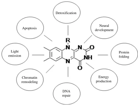
[image:20.612.108.542.266.577.2]required for FMN and FAD synthesis. FMN and FAD synthesis begin with flavokinase phosphorylating riboflavin to generate FMN, which then can be adenylated to form FAD by FAD synthetase (1-3). The abundance of flavin binding proteins in nature is likely due to the wide array of biochemical reactions that can be catalyzed by flavin dependent enzymes (Figure 1.2).
Figure 1.2. Sample of Biological Functions that Involve Flavoenzymes (4-10). (Modified from ref. (11))
The chemical versatility of flavoenzymes arises from the flavin cofactor having the ability to couple one- and two-electron transfer processes between substrates and various electron carriers as well as functioning as electrophile and nucleophile (12). Flavoenzymes generally achieve
Detoxification
Neural development
Protein folding
Energy production
DNA repair Chromatin
remodeling Light
emission
[image:21.612.94.555.206.558.2]catalysis through two half-reactions, a reductive half-reaction in which the flavin cofactor is either one- or two-electron reduced followed by an oxidative half-reaction in which the flavin is re-oxidized (Scheme 1.1).
Scheme 1.1. Generalized Flavin Catalytic Cycle. SA and SB are Substrate A and B Respectively.
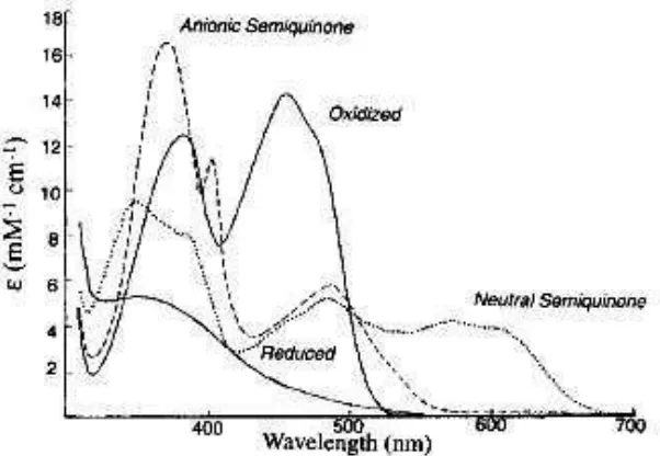
Flavin can exist in three different redox states, the oxidized, the semiquinoid (1 e- reduced), and the fully reduced (2 e- reduced) species (Figure 1.3). Additionally, the semiquinoid and the fully reduced states exist in both a neutral and an anionic form. Each of these 5 flavin states is accompanied with unique spectral properties (Figure 1.4) (13). These large spectral differences between the various flavin oxidation states make it possible to monitor events occurring during catalysis using the flavin itself as a spectral probe.
Figure 1.3. Redox and Ionization States of Flavins. (Modified from ref. (16))
[image:23.612.225.409.71.357.2] [image:23.612.182.483.441.649.2]The spectroscopic properties of flavins have allowed for a detailed kinetic characterization of many flavoenzymes, making them one of the best studied classes of enzymes. In addition, these kinetic studies have often been supplemented with structural studies. In recent years, structural studies of flavoenzymes have gained momentum as seen by the increase of entries in the Protein Databank that now contains ~1400 structures of proteins containing flavin as compared to ~200 in the year 2000.
Generally, flavoenzymes can be grouped into a relatively small number of subclasses, where members within the same subclass share several common properties, including the type of reactions that they catalyze. The most common grouping of flavoenzymes is based on the reactivity of their reduced form with dioxygen (17). Using these criteria there are four well defined groups of flavoenzymes.
1. Oxidases – react rapidly with oxygen to yield H2O2 and oxidized enzyme.
2. Monooxygenases – react rapidly with oxygen to yield H2O, oxidized enzyme and a substrate with an inserted single oxygen atom.
3. Electron transferases – react slowly with oxygen to yield an oxygen radical paired with the neutral flavoenzyme semiquinoneradical.
4. Dehydrogenases – react poorly with oxygen to yield O2-..
Oxidases. The flavin dependent oxidases react with oxygen in their reduced form by transferring a hydride equivalent to reform the oxidized flavin cofactor and hydrogen peroxide. A common feature of all the flavin dependent oxidases is that they stabilize the red anionic one-electron reduced semiquinone (18-22) as well as having the ability to form a stable flavin N(5)-sulfite adduct (19-20). These features both imply the presence of a positive charge proximal to the negative charge formed at the N(1)-C(2)=O locus of the flavin isoalloxazine ring as a result of flavin reduction. Indeed, the emergence of X-ray crystallographic structures of flavin dependent oxidases has shown the presence of a positively charged residue close to N(1)-C(2)=O region of the flavin cofactor, e.g. His466 in choline oxidase and Lys230 in glycolate oxidase ( 23-25). Amongst the most intensively studied flavin dependent oxidases are: choline oxidase (24, 26-38), cholesterol oxidase (39-61), glycolate oxidase (23-25), sarcosine oxidase, D-amino acid oxidase (62-69) and glucose oxidase (70-75).
Monooxygenases. Contrary to the oxidases, the flavin dependent monooxygenases activate oxygen through a detectable flavin hydro-peroxide intermediate. Upon formation the flavin hydroperoxide intermediate transfers an oxygen atom to the substrate with the formation of a C(4a)-hydroxyflavin, which returns to its oxidized state upon dehydration. However, even in the absence of the substrate, the flavin hydroperoxide converts slowly to H2O2 and oxidized flavin (76).
adducts. Amongst the best characterized flavoprotein electron transferases are flavodoxin ( 23-25), NADPH-cytochrome P-450 reductase (77), and ferredoxin-NADP+ reductase (78-83).
Dehydrogenases. The flavin-dependent dehydrogenases react poorly, if at all, with molecular oxygen. They utilize a specific electron acceptor such as NADP+ to oxidize the organic substrate. Amongst the best studied flavin-dependent dehydrogenases are the acyl-CoA dehydrogenases. These enzymes oxidize the organic substrate by transferring a hydride equivalent to the oxidized flavin, which subsequently transfers these electrons to the final electron acceptor, NAD(P)+ (84).
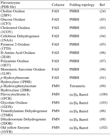
1.2 Selected structural features of flavoenzymes with different oxygen reactivity
Flavoenzymes are known to have a variety of folding topologies such as the (α/β)8-barrel and
Table 1.1. Overall Fold of Selected Flavoenzymes Flavoenzyme
(PDB file) Cofactor Folding topology Ref
Choline Oxidase (2JBV)
FAD PHBH (24)
Glucose Oxidase (1CF3)
FAD PHBH (85)
Cholesterol Oxidase (1COY)
FAD PHBH (93)
Cellobiose Dehydrogenase (1NAA)
FAD PHBH (94)
Pyranose 2-Oxidase (1TT0)
FAD PHBH (95)
D-Amino Acid Oxidase (2E48)
FAD PHBH (96)
Polyamine Oxidase (1B37)
FAD PHBH (97)
Monomeric Sarcosine Oxidase (1L9F)
FAD PHBH (98)
p-Hydroxybenzoate Hydroxylase (1PHH)
FAD PHBH (91)
p-Hydroxyphenylacetate Hydroxylase (2JBR)
FMN Tetrameric (99)
Flavocytochrome b2 (1FCB)
FMN (/β)8 Barrel (100) Glycolate Oxidase
(1GOX)
FMN (/β)8 Barrel (101) Trimethylamine Dehydrogenase
(2TMD)
FMN (/β)8 Barrel (86) Dihydroorotate Dehydrogenase
(2DOR)
FMN (/β)8 Barrel (88) Old yellow Enzyme
(1OYB)
FMN (/β)8 Barrel (102)
hydrophobic residues define a hydrophobic cavity in front of the C(4a) atom and in phenylacetone monooxygenase (104) where 5 residues create a similar cavity on the re side of the flavin. Other monooxygenases with similar cavities in front of the C(4a)-N5 locus of the flavin are p-hydroxybenzoate hydroxylase (91), 3-hydroxybenzoate hydroxylase (105) and tryptophan 7-halogenase (106).
is increased almost an order of magnitude upon binding of the positively charged product or product analog (112-113).
His466
FAD-C(4a)
Val464 3.2
3.8 5.5
choline oxidase glucose oxidase
His516
FAD-C(4a)
Val560 4.7
4.3 6.3
monomeric sarcosine oxidase
Lys265
FAD-C(4a)
Phe256 3.7
7.2 5.9
choline
Figure 1.5. Flavin Dependent Enzymes Displaying a Hydrophobic Residue Close to a Positive Charge that is Required for Oxygen Activation Close to the FAD-C(4a)-N5 Region.
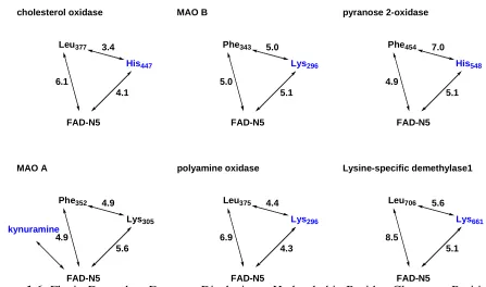
When expanding the structural analysis to encompass flavoenzymes that have not been as comprehensively characterized, several flavin-dependent oxidases are found with a similar motif consisting of a non-polar region close to a positive charge and the C(4a) atom of the flavin as exemplified by the flavoenzymes shown in Figure 1.6 (see Chapter IV for survey details) (24, 85, 107-108).
3.4 4.1 6.1 cholesterol oxidase 5.0 5.1 5.0 pyranose 2-oxidase 7.0 5.1 4.9 kynuramine His447 Leu377 Lys296 Phe343 His548 Phe454 4.9 5.6 4.9 polyamine oxidase 4.4 4.3 6.9 Lysine-specific demethylase1 5.6 5.1 8.5 Lys305 Phe352 Lys296 Leu375 Lys661 Leu706 FAD-N5 MAO B FAD-N5 FAD-N5 FAD-N5 MAO A FAD-N5 FAD-N5
[image:29.612.86.533.424.688.2]A structural and kinetic comparison of glycolate oxidase and flavocytochrome b2 revealed that flavoenzymes with structurally homologous and highly conserved active sites can have vastly different reactivities with oxygen. The major differences between these two flavoenzymes are the presence of a leucine (Leu230) residue and the backbone nitrogen of Ala198 close to the C(4a) atom of the flavin cofactor in flavocytochrome b2 whereas in glycolate oxidase it is a tryptophan (Trp108) and the methyl side-chain of alanine79 that is close to the C(4a)-atom (87, 100). Thus, the polarity and orientation of the sidechain of this region appears to be the predominant difference between these two flavoenzymes.
Consistent with the importance of the cavity in front of the C(4a)-N5 locus of the FAD isoalloxazine ring for oxygen reactivity are findings from recent studies on the enzymes belonging to the L-2-hydroxy acid oxidase family, which are FMN dependent enzymes. The members of the L-2-hydroxy acid oxidase family that display poor oxygen reactivity have been shown to have a constrained environment around the N5 position of the FMN cofactor (117). To further elaborate on the importance of the residues lining the cavity in front of the C(4a)-N5 locus of the flavin it is key to look at a study of L-galactono-γ-lactone dehydrogenase. The study showed that upon mutation of the alanine residue at position 113, which is positioned near the C(4a) locus of the isoalloxazine ring, to a glycine residue oxygen reactivity was increased 400-fold and effectively changing a dehydrogenase to a catalytically competent oxidase (118). In L-galactono-γ-lactone dehydrogenase, a bigger cavity is sufficient to allow for better oxygen reactivity likely due to improved oxygen accessibility.
Conversely, in the oxidases no such highly defined cavity is observed, most likely due to the oxidases having less stringent requirements for optimum reaction of the reduced flavin with molecular oxygen. These structurally different oxidases do seem to have some commonly recurring structural features, such as a non-polar region or a positive charge close to the C(4a)-atom of the flavin cofactor. A summary of common structural features of the surveyed flavoenzymes are shown in Table 1.2.
Table 1.2. Structural Features of Selected Flavoenzymes Important for Oxygen Reactivity Flavoenzyme Positive charge Effect of positive
charge on kcat/Koxygen
Non-polar residue
Ref
Choline Oxidase Substrate ~80 fold increase Val464 (24, 108) Glucose Oxidase His516 ~100 fold increase Val560 (85, 107)
Cholesterol Oxidase His447 Leu377 (93)
Pyranose 2-Oxidase His548 Phe454 (95)
D-Amino Acid Oxidase Product analog Ile230 (96)
Polyamine Oxidase Lys296 Leu375 (97)
Monomeric Sarcosine Oxidase
Lys265 ~8000 fold increase Phe256 (49, 98)
Glycolate Oxidase Lys236 Trp110 (119)
Monoamine oxidase A Product/Lys305 ~10 fold increase Phe352 (86) Monoamine oxidase B Product/Lys296 ~10 fold increase Phe343 (116)
Old yellow Enzyme His196 Phe250 (102)
1.3 Oxygen reactivity
1.3.1. General oxygen chemistry
in its triplet state has a magnetic moment and as such liquid oxygen is paramagnetic. Molecular oxygen in its singlet ground spin state, there are no unpaired electrons and as such the total spin is zero. Singlet oxygen is many times more reactive than triplet oxygen and readily reacts with other singlet molecules, which comprise the majority of all compounds, and is therefore rapidly depleted. Contrary to this is molecular oxygen in its triplet ground state, for which chemical reactions between reactants in their triplet and singlet spin states are forbidden by Wigner's spin selection rule (120). The conservation of spin allows triplet oxygen to readily react with molecules in a doublet state, such as radicals, to form a new radical.
Thus, the triplet multiplicity is the actual reason why most reactions of oxygen with organic substances, although being exergonic, do not readily proceed at room temperature but only upon heating or in the presence of catalysts that activate O2 via a series of one-electron transfer reactions (121). Figure 1.7 shows the reduction potentials of O2 at pH 7.0. These reduction potentials indicate that the limiting step in O2 reduction is the first single electron transfer (122). Furthermore the negative standard reduction potential for the one electron transfer to O2 makes it a non-spontaneous process with electron donors of higher standard potentials.
O H OH O H O H O
O V V V . 2.31V 2
2 38 . 0 2 2 89 . 0 . 2 33 . 0
2 2
Figure 1.7. Standard Reduction Potentials for O2 (123).
does not yield a single principle by which this is achieved. However, broad generalizations can be made and some general principles can be deduced.
hydrogen peroxide and oxidized flavin is not as clear. It can either go directly to the end product or through an intermediate as in the monooxygenases. So far, only two flavin dependent oxidases have experimental evidence that this reaction proceeds through an intermediate. Both pyranose-2-oxidase and choline oxidase have shown the presence of a C(4a)-oxygen-adduct (24, 126-127).
O
2-N
H
N
NH
N
O
O
R
O
2N
H
N
NH
N
O
O
R
O
(H)O
N
H
N
NH
N
O
O
R
N
N
NH
N
O
O
R
-+ HO
2-EFlH + O
2-monooxygenase reactions
Figure 1.8. Flavin Dependent Oxygen Reduction Pathways. (modified from (103))
1.3.2. Enzymes and oxygen
Generally, all the known enzymes that have molecular oxygen as one of their substrates can be divided in to two main categories:
1. Oxygenases. 2. Oxidases.
oxygen functions as an electron acceptor and is reduced to a superoxide radical, hydrogen peroxide, or water. A common feature of all enzymes activating molecular oxygen for reaction is that they are conjugated proteins with a metal or flavin prosthetic group. Oxidases as well as oxygenases have either or both types of prosthetic groups.
1.3.1.1.Metal-containing oxygenases
Iron is by far the most frequently used metal cofactor in oxygenases (124, 128), either as part of a heme group as in tryptophan 2,3-dioxygenase and indolamine 2,3-dioxygenase (124). Alternatively, iron may be in a non-heme form as seen in protocatechuate 3,4-dioxygenase ( 129-130) and lipoxygenase (124, 131-132).
1.3.1.1.1. Heme-dependent oxygenases
For the cofactor in heme-dependent proteins to interact directly with molecular oxygen the iron must be in its ferrous state and possess an available sixth coordination site.
There are four known types of interactions of heme-proteins and molecular oxygen: 1. Transport (hemoglobin) (133)
2. Reduction (cytochrome oxidase) (134) 3. Monooxygenation (cytochrome P-450) (135) 4. Dioxygenation (tryptophan oxygenase) (136)
electron acceptor catalyzing the electron flow from oxygen as it gets reduced to water. Contrary to these types of heme-containing proteins, are the heme-dependent monooxygenases and dioxygenases which activate oxygen and catalyze its insertion into organic substrates.
In the current hypothesis for how iron-dependent oxygenases, in which the iron cofactor is directly involved, activate molecular oxygen, the iron has to be reduced to the Fe2+ state prior to oxygen binding and involves an electron transfer from Fe2+ to the bound oxygen to form a oxyferrous intermediate, which is or will lead to the activated oxygen species. This type of mechanism has been established for most iron-dependent dioxygenases such as indoleamine 2,3-dioxygenase (138), however, not all oxygenases proceed through this type of mechanism. Protocatechuate 3,4-dioxygenase is an example of an iron-dependent oxygenase that must proceed through a mechanism that does not directly involve an electron-transfer from iron to oxygen as evidenced by the absence of a valency change in the iron cofactor (139).
As an example of the mechanism of oxygen activation that directly involves the iron cofactor, a closer look at indoleamine 2,3-dioxygenase is prudent, as it is one of the best characterized heme-dependent enzymes. Indoleamine 2,3-dioxygenase catalyzes the oxidative ring cleavage of indoleamine derivatives via an ordered sequential mechanism with the indoleamine derivative binding before oxygen in accordance to the mechanism shown in Scheme 1.2 (140-141).
(Fe2+)E + Substrate substrate-E(Fe2+)
substrate-E(Fe3+)O2-.
O2 Product
In vitro, indoleamine 2,3-dioxygenase requires methylene blue and a system to generate
2
O for maximum activity (140). As seen in Scheme 1.2, the oxygenated intermediate contains a
(Fe3+O2)-complex, and the resting enzyme has been shown to have its iron cofactor in the Fe3+
state. As a consequence, the intermediate complex can be formed from the resting enzyme and
2
O directly but not with molecular oxygen directly. Experimentally it has been shown that the
product is released from the Fe2+ enzyme, which can then undergo a second turnover with O2
(142-143). Upon binding of O2, an electron is transferred from Fe2+ to form the 2 3
O
Fe reaction
intermediate bound in the active site of the enzyme.
Cytochrome P-450 is an example of an iron-dependent monooxygenase. The term cytochrome P-450 (P450) refers to a group of heme-proteins that have a sulfur atom ligated to the iron (144). Cytochrome P-450 activates molecular oxygen to catalyze the monooxygenation of unactivated hydrocarbons (R-H). A generalized reaction scheme for the reaction catalyzed by cytochrome P-450 is shown in Scheme 1.3 (145).
R-H + O2 + NAD(P)H R-OH + H2O + NAD(P)+
Scheme 1.3. Generalized Reaction Catalyzed by Cytochrome P-450.
e
-O2
e
-(Fe3+(H2O))E + R-H E(Fe3+)-R-H
E(Fe2+)-R-H
R-OH + H2O
O2-E(Fe2+)-R-H
(1) (2)
(4) (3)
H2O
Scheme 1.4. Simplified Catalytic Mechanism for Cytochrome P-450 (124, 144).
The resting state of P450 has a hexacoordinated low spin heme (species (1) in Scheme 1.4). The sixth axial ligand is a weakly bound H2O molecule. Upon substrate binding, the water molecule is displaced to yield a pentacoordinated high spin heme (species (2) in Scheme 1.4) (145). The pentacoordinated ferric P450 is then reduced by its redox partner to give Fe2+ (species (3) in Scheme 1.4) to which oxygen binds to generate an oxyferrous intermediate (species (4) in
Scheme 1.4). A second electron is then transferred from the redox partner to the oxyferrous intermediate to form a peroxo (Fe-O-O2-) intermediate that is subsequently protonated to give a hydroperoxo (Fe-O-O-H-) intermediate. This is followed by heterolytic cleavage of oxygen to
form the oxyferryl (Fe=O+.) intermediate, which is the activated form of oxygen able to react
with the hydrocarbon to form the hydroxylated hydrocarbon and water (145).
1.3.1.1.2. Iron non-heme oxygenases
throughout the catalytic cycle. Molecular oxygen has not been found to complex with Fe3+, and as such the most likely role of the iron cofactor is to activate the substrate rather than oxygen (129). A proposed mechanism for the reaction catalyzed by protocatechuate 3,4-dioxygenase involves an initial keto-enol tautomerization of the substrate resulting from the interaction of one of the hydroxyl groups with the Fe3+. The activated keto form reacts with molecular oxygen to
form O2 that can then continue to form the enzyme-product complex. Resonance-Raman
spectroscopy show no signs of a charge-transfer complex, consistent with there being no
interactions between Fe3+ and O2 (146).
1.3.1.1.3. Copper oxygenases
OH
+ O2
O
O
+ OH
-Scheme 1.5. Monooxygenation of Monophenols Catalyzed by Tyrosinase.
OH
+ O2
O
O
+ 2 H2O OH
2 2
Scheme 1.6. Dehydrogenation of o-diphenols Catalyzed by Tyrosinase.
O2
(Cu2+)2E + o-diphenol E(Cu2+)2o-diphenol
E(Cu+)2o-quinone
E(Cu+)2O2
o-quinone + 2H+
E(Cu2+)2O2 + o-diphenol E(Cu2+)2O2 + o-diphenol
o-quinone + 2OH
-Scheme 1.7. Catalytic Cycle of Tyrosinase Reaction with o-diphenols.
It is assumed, that in the copper-dependent oxygenases oxygen activation proceeds via an electron transfer from reduced Cu2+ to oxygen to form a peroxo intermediate, which is the activated form of oxygen (150, 153).
1.3.1.2.Flavin-containing oxygenases
monoooxygenase, where the flavin is reduced as lactate is oxidized to pyruvate (156). Regardless of how the flavin is reduced, the first step in the reaction with oxygen is a single electron transfer from the reduced flavin to oxygen to form a caged radical pair consisting of superoxide and a flavin radical as shown in Figure 1.8. For most flavin-dependent monooxygenases, this is followed by the formation of a covalent adduct between oxygen and the C(4a) atom of the flavin radical. This leads to the formation of a C(4a)-hydroperoxyflavin intermediate, which is the reactive intermediate containing activated oxygen. In the flavin-dependent monooxygenases this highly unstable and reactive intermediate is stabilized long enough for it to transfer oxygen to the organic substrate rather than decaying to hydrogen peroxide and oxidized flavin as in the flavin-dependent oxidases thought to proceed through the formation of a C(4a) oxygen adduct (157). Depending on the protonation state of the peroxyflavin intermediate, either an electrophilic or a nucleophilic attack on the substrate will result in the transfer of a single atom of molecular oxygen to the substrate, while the other oxygen atom is reduced to water.
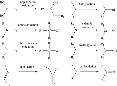
R1
R2
hydroxylation R1
R2
OH R1
R2
halogenation R1
R2
Br
R1
N
R2
amine oxidation R1
N+ R2 O -R1 Se R2 selenide oxidation R1 Se R2 O
R3 R3
R1 P R2 phosphite ester oxidation R1 P+ R2 O
-R3 R3
HO B HO organoboron oxidation OH B O
R1 HO
R1
R1
S
R2
sulfoxidation R1
S
R2
O R1
epoxidation R1
R2 R2
O
Figure 1.9. Reaction Types Catalyzed by Flavin-dependent Monooxygenases. (modified from (154)).
The flavin-dependent monooxygenases have been divided into several subclasses based on sequence and structural homology. Each subclass seems to only catalyze a limited range of oxygenation reactions suggesting that the types of reactions catalyzed is at least partially dependent on the overall folding topology (154).
[image:43.612.76.537.71.409.2]anionic semiquinone species. While the substrate radical is still bound in the active site of the enzyme molecular oxygen reacts with the one electron reduced flavin to yield an enzyme-associated superoxide species. The substrate radical and the superoxide is proposed to collapse in the active site of the enzyme and form an α-peroxynitrate.
1.3.1.3.Metal-containing oxidases
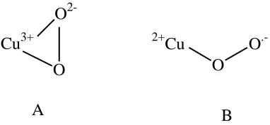
In contrast to the metal-containing oxygenases, the oxidases primarily use copper rather than iron as a cofactor. The copper-dependent oxidases generally use a free radical mechanism to activate oxygen for reaction. Galactose oxidase is one of the best characterized copper-dependent oxidases. It is a mononuclear copper-containing oxidase, belonging to the tyrosyl radical family (159). The overall reaction catalyzed by galactose oxidase shown in Scheme 1.8 is the oxidation of primary alcohols to the corresponding aldehydes (160), which in turn is oxidized to the corresponding carboxylates (161).
RCH2OH + O2 RCHO + H2O2
Scheme 1.8. The Overall Reaction Catalyzed by Galactose Oxidase.
Cu3+ O
2-O
A
2+
Cu O
.-O
[image:45.612.211.402.72.161.2]B
Figure 1.Error! Reference source not found.. The Two Proposed Copper Oxygen Coordination Modes (A = Side-on, B = End-on)
The side-on coordination mode is what would be expected if there is strong metal-ligand covalent interactions and it will result in the formation of a Cu3+-peroxide intermediate (164). Whereas the end-on coordination mode results in an intermediate with a Cu2+-superoxide adduct (164). In a sterically congested active site, the end-on oxygen coordination mode is the most likely favored, due to the side-on mode requiring both metal coordination sites to be available in order to form. The active site in galactose oxidase has been shown to be highly coordinated and sterically restricted with His496, His581 and Tyr272 occupying three of four equatorial
1.3.1.4.Flavin-containing oxidases
The chemical basis for flavin-dependent O2 reduction is not well understood. In summary, it is believed that the reaction proceeds by successive single electron transfers, the first of which is a single electron being transferred from the reduced flavin to O2 to form a caged radical pair consisting of a semi-reduced flavin and a superoxide anion. From here the oxygen can be further reduced through several different pathways as summarized in Figure 1.8.
Free flavin by itself can reduce oxygen with a second order rate constant in the 102 M-1s-1 range, whereas the flavin-dependent enzymes display second order rate constants spanning from 101 M-1s-1 range (flavocytochrome b2 (17)) to 106 M-1s-1 range (glucose oxidase (166)), indicative of the importance of the protein moiety in modulating the reactivity of the flavin cofactor with oxygen. However no clearly identifiable structural features seem to be recurring in the enzymes with high rate constants. For some of the fastest reacting flavin-dependent oxidases a preorganized electrostatic environment dominated by a positive charge close to the C(4a)-N5 locus of the flavin cofactor has been shown to be crucial for the fast reaction with oxygen. In the case of glucose oxidase it is a protonated histidine that provides the positive charge (85, 110). In cholesterol oxidase type I and II the need for a positive charge has also been demonstrated ( 167-168). In choline oxidase the positive charge in the active site required for efficient reaction with oxygen is provided by the positive charged head-group of choline and not by a residue in the active site (31, 109). Other examples of ligand binding enhancing oxygen reactivity are D-amino acid oxidase (112) and monoamine oxidase (113). In these two enzymes it is product binding that increases oxygen reactivity and in both cases the product or product analog is positively charged.
from these factors have not been well established. In some cases they have a positive effect on oxygen reactivity, whereas in others they have a detrimental effect (103).
Overall, it does not seem that there are general structural features that will always result in enhancement of oxygen reactivity in flavin-dependent oxidases, but rather several subtle factors with a combined effect that results in an enhanced oxygen reactivity being the main reason. Additionally it seems that there are multiple combinations possible to achieve enhanced oxygen reactivity, thereby making the identification of general principles elusive and problematic.
1.4. Choline Oxidase from Arthrobacter globiformis
For a recent comprehensive review on choline oxidase see “Hydride transfer made easy in the reaction of alcohol oxidation catalyzed by flavin-dependent oxidases” by G. Gadda published in Biochemistry 47, 13745-13753 (169). Choline oxidase from Arthrobacter globiformis is a homodimer with each subunit having a covalently attached FAD to His99 (24). This enzyme catalyzes the oxidation of choline to glycine betaine with betaine aldehyde as intermediate (170). The reaction proceeds through two reductive half-reactions and two oxidative half-reactions as shown in Scheme 1.9.
CH
Kd
kred, CH
k5
k9
k11
O2, H2O GB
O2 H2O2
H2O2 E-FADox
E-FADox-CH
E-FADred-BA
E-FADox-BA E-FADred-GB
E-FADox-GB
k4
k 8
Scheme 1.9. Minimal Kinetic Mechanism of Choline Oxidase. (E-FADox = Oxidized Enzyme Bound Flavin, -FADred = Reduced Enzyme Bound Flavin CH = Choline, BA = Betaine Aldehyde and GB = Glycine Betaine)
In the reductive half-reactions the FAD cofactor is reduced to the anionic hydroquinone form (2 e- reduced) by choline and the aldehyde intermediate respectively. Each reductive half reaction is followed by an oxidative half-reaction where the reduced FAD cofactor is reoxidized by molecular oxygen with formation of hydrogen peroxide (170-171).
The first reductive half-reaction is initiated by a kinetically fast abstraction of the hydroxyl proton of choline, which results in the formation of a transient alkoxide intermediate (37). This is followed by a rate-limiting hydride ion transfer to N(5) atom of the flavin cofactor from the α-carbon of the alkoxide resulting in the oxidation of choline to betaine aldehyde and reduction of the flavin (37). Prior to the second reductive half-reaction, betaine aldehyde is hydrated to form gem-diol choline in the active site (35). The subsequent oxidation of the reduced flavin occurs while the active site is still occupied with the gem-diol choline intermediate (38, 108-109). In the second reductive half-reaction, the gem-diol choline is oxidized to the product, glycine betaine. In both oxidative half-reactions the oxidized flavin cofactor is reformed upon a transfer of a hydride equivalent from the reduced flavin to molecular oxygen (32). Each of these half-reactions has specific requirements, that choline oxidase must balance in order to achieve maximum overall rate of catalysis. In the wild-type choline oxidase this yields the following kinetic parameters in the pH independent region, kcat = 60 s-1, kcat/Km = 237,000 M-1 s-1and kcat/Koxygen = 86,000 M-1 s-1 (37).
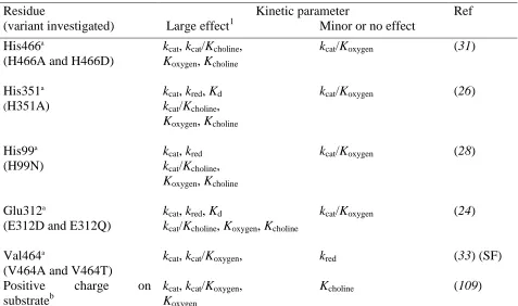
parameters affected by substitution of His99, Glu312, His351, Val464 and His466 are summarized in Table 1.3.
Even though the positively charged His466 does not affect oxygen activation, it is important for catalysis. The positive charge has been shown to be involved in the stabilization of a negatively charged reduced flavin as well as of the transient alkoxide species (30-31). Additionally, His466 is likely involved in choline activation as seen by the 60- and 1000-fold lowered kcat and kcat/Kcholine, respectively, in the pH independent region upon replacing the histidine with an alanine as compared to the wild-type enzyme (31).
Table 1.3. Kinetic Parameters Influenced by Substitution of Various Residues in Choline Oxidase.
Residue Kinetic parameter Ref
(variant investigated) Large effect1 Minor or no effect His466a
(H466A and H466D)
kcat, kcat/Kcholine, Koxygen, Kcholine
kcat/Koxygen (31)
His351a
(H351A)
kcat, kred, Kd kcat/Kcholine, Koxygen, Kcholine
kcat/Koxygen (26)
His99a
(H99N)
kcat, kred kcat/Kcholine, Koxygen, Kcholine
kcat/Koxygen (28)
Glu312a
(E312D and E312Q)
kcat, kred, Kd
kcat/Kcholine, Koxygen, Kcholine
kcat/Koxygen (24)
Val464a
(V464A and V464T)
kcat, kcat/Koxygen, kred (33) (SF) Positive charge on
substrateb
kcat, kcat/Koxygen, Koxygen
Kcholine (109)
a
The affected kinetic parameters were determined by investigating the variant enzymes where the residue of interest was replaced. b using 3,3-dimethyl-butanol instead of choline.
1 A large effect on a kinetic parameter is defined here as it being more than 3-fold different as compared to the
[image:49.612.69.546.346.628.2]His351 is also likely involved in choline activation as seen by the 60- and 350-fold lowered kcat and kcat/Kcholine, respectively, in the pH independent region upon replacing the histidine with an alanine as compared to the wild-type enzyme (26). Furthermore, His351 has a profound effect on the substrate binding affinity as well as the rate of flavin reduction in the first reductive half-reaction with choline as the reductant as seen by the Kd value increasing 10 times and the kred value decreasing 75 times (26).
Glu312 is involved in the preorganization of the enzyme-substrate complex as well as the initial binding and correct positioning of choline through interactions between its negatively charged side-chain and the positively charged trimethylammonium headgroup of choline (24).
Other than merely being the site of covalent attachment of the flavin cofactor to the protein moiety the FAD-histidyl covalent linkage between His99 and FAD is important for the optimal positioning of the flavin cofactor in the enzyme-alkoxide complex that is required for the environmentally assisted tunneling of the hydride ion in the oxidation of choline (28).
In summary, residues Glu312, His99, His351 and His466 do not contribute to oxygen activation, as indicated by site-directed mutagenesis studies where each of these four residues located in the active site of the enzyme were individually replaced. These studies revealed that the bimolecular rate constants for the reaction of the reduced flavin cofactor with oxygen (kcat/Koxygen) were not significantly altered in the mutant enzymes from the value of ~105 M-1s-1 of the wild-type (24, 26, 28, 30-32).
enzyme-bound ligand (108-109). Further evidence of the importance of this is seen in the pH studies of the His99Asn, His351Ala, His466Ala and wild-type forms of choline oxidase, which showed that the kcat/Koxygen values that are independent of pH, consistent with the fact that the trimethylammonium moiety of the choline cannot ionize (26, 28, 31-32, 108).
Further studies on choline oxidase from Arthrobacter globiformis are pertinent as they can help elucidate one of the puzzling unanswered questions in flavin chemistry, which is the chemical basis for the diverse oxygen reactivity in flavin-dependent enzymes.
1.5. Specific Goals
Flavin-dependent enzymes are in general amongst the most chemically versatile naturally occurring enzymes, as exemplified by the diverse reactivity of the reduced flavin with molecular oxygen. Depending upon the ability to react with oxygen and the product of oxygen reduction, three general classes of flavin-dependent enzymes have been distinguished (103, 155). Dehydrogenases have very poor, or no reactivity with oxygen, and utilize other electron acceptors for catalytic turnover. Monooxygenases and oxidases show high reactivity with oxygen, typically with second-order rate constants ≥105 M-1s-1 (155). Free reduced flavin in aqueous solution react with molecular oxygen with a bimolecular rate constant of 250 M-1s-1 (155). It is the interactions between the flavin cofactor and the protein moiety that modulate the reactivity of the flavin. (12, 103, 173).
elucidation of the roles of these three residues on both the reductive and the oxidative half-reactions of choline oxidase.
Ser101 is located ~4 Å away from the N(5) atom of FAD cofactor and within hydrogen bonding distance to DMSO, a co-crystallized ligand found in the active site in the crystal structure of wild-type choline oxidase (24). The location of Ser101 allows me to propose that this residue affects the flavin microenvironment while being able to actively participate in the oxidation of choline. To elucidate the effect exerted by Ser101 on oxygen reactivity, kinetic characterization of a variant enzyme where the serine residue has been replaced with an alanine was carried out (Hongling, Y. submitted for publication, 2010). In order to attribute any determined kinetic differences between the variant enzyme and the wild-type enzyme to the removal of the hydroxyl group at position 101 rather than to structural differences, the crystal structure of the variant enzyme was also investigated.
proton-transfer network was perturbed through site directed mutagenesis of His310 and subsequently the variant enzymes were subjected to kinetic analysis and characterization in order to establish the presence of a proton-transfer network and its effect on the reaction catalyzed by choline oxidase.
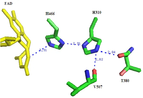
Figure 1.11. The Coordination of the Side-chain of His310 in the Crystal Structure of Wild-type Choline Oxidase (2JBV).
micro-environment of these two enzymes may be the cause for the rate constant for the reactivity of oxygen to drop from 8.5 x 10⁴ M-1s-1 in glycolate oxidase to 2 M-1s-1 in flavocytochrome b2 (103). Through X-ray crystallography, site-directed mutagenesis of position Val464 in choline oxidase, steady state and rapid kinetics approaches the role of the hydrophobic residue Val464 in the active site of choline oxidase was investigated.
Finally, the reaction of choline oxidation catalyzed by choline oxidase is very well characterized, making it ideally suited to investigate the chemical basis of how flavin-dependent enzymes react with molecular oxygen. As such, mechanistic data on Ser101, His310 and Val464 will provide important information about the roles of these active site residues in the reaction catalayzed by choline oxidase that may be relevance for a large number of flavin-dependent enzymes.
1.6.References
1. Fischer, M., and Bacher, A. (2005) Biosynthesis of flavocoenzymes, Nat Prod Rep 22, 324-350.
2. Schrecker, A. W., and Kornberg, A. (1950) Reversible enzymatic synthesis of flavin-adenine dinucleotide, J Biol Chem182, 795-803.
3. Santos, M. A., Jimenez, A., and Revuelta, J. L. (2000) Molecular characterization of FMN1, the structural gene for the monofunctional flavokinase of Saccharomyces cerevisiae, J Biol Chem275, 28618-28624.
5. Forneris, F., Binda, C., Vanoni, M. A., Mattevi, A., and Battaglioli, E. (2005) Histone demethylation catalysed by LSD1 is a flavin-dependent oxidative process, FEBS Lett 579, 2203-2207.
6. White, H. B., 3rd, and Merrill, A. H., Jr. (1988) Riboflavin-binding proteins, Annu Rev Nutr8, 279-299.
7. Susin, S. A., Lorenzo, H. K., Zamzami, N., Marzo, I., Snow, B. E., Brothers, G. M., Mangion, J., Jacotot, E., Costantini, P., Loeffler, M., Larochette, N., Goodlett, D. R., Aebersold, R., Siderovski, D. P., Penninger, J. M., and Kroemer, G. (1999) Molecular characterization of mitochondrial apoptosis-inducing factor, Nature397, 441-446.
8. Murty, C. V., and Adiga, P. R. (1982) Pregnancy suppression by active immunization against gestation-specific riboflavin carrier protein, Science216, 191-193.
9. Jorns, M. S., Wang, B., and Jordan, S. P. (1987) DNA repair catalyzed by Escherichia coli DNA photolyase containing only reduced flavin: elimination of the enzyme's second chromophore by reduction with sodium borohydride, Biochemistry26, 6810-6816.
10. Dagley, S. (1987) Lessons from biodegradation, Annu Rev Microbiol41, 1-23.
11. Joosten, V., and van Berkel, W. J. (2007) Flavoenzymes, Curr Opin Chem Biol11, 195-202.
12. Massey, V. (2000) The chemical and biological versatility of riboflavin, Biochem Soc Trans28, 283-296.
14. Draper, R. D., and Ingraham, L. L. (1968) A potentiometric study of the flavin semiquinone equilibrium, Arch Biochem Biophys125, 802-808.
15. Ehrenberg, A., Muller, F., and Hemmerich, P. (1967) Basicity, visible spectra, and electron spin resonance of flavosemiquinone anions, Eur J Biochem2, 286-293.
16. Ghisla, S., and Massey, V. (1986) New flavins for old: artificial flavins as active site probes of flavoproteins, Biochem J239, 1-12.
17. Massey, V. (2002) The reactivity of oxygen with flavoproteins, International Congress Series 1233, 3-11.
18. Massey, V., Muller, F., Feldberg, R., Schuman, M., Sullivan, P. A., Howell, L. G., Mayhew, S. G., Matthews, R. G., and Foust, G. P. (1969) The reactivity of flavoproteins with sulfite. Possible relevance to the problem of oxygen reactivity, J Biol Chem 244, 3999-4006.
19. Macheroux, P., Kieweg, V., Massey, V., Soderlind, E., Stenberg, K., and Lindqvist, Y. (1993) Role of tyrosine 129 in the active site of spinach glycolate oxidase, Eur J Biochem 213, 1047-1054.
20. Gadda, G., Wels, G., Pollegioni, L., Zucchelli, S., Ambrosius, D., Pilone, M. S., and Ghisla, S. (1997) Characterization of cholesterol oxidase from Streptomyces hygroscopicus and Brevibacterium sterolicum, Eur J Biochem250, 369-376.
22. Fitzpatrick, P. F., and Massey, V. (1983) The reaction of 8-mercaptoflavins and flavoproteins with sulfite. Evidence for the role of an active site arginine in D-amino acid oxidase, J Biol Chem258, 9700-9705.
23. Vrielink, A., Lloyd, L. F., and Blow, D. M. (1991) Crystal structure of cholesterol oxidase from Brevibacterium sterolicum refined at 1.8 A resolution, J Mol Biol 219, 533-554.
24. Quaye, O., Lountos, G. T., Fan, F., Orville, A. M., and Gadda, G. (2008) Role of Glu312 in binding and positioning of the substrate for the hydride transfer reaction in choline oxidase, Biochemistry47, 243-256.
25. Lindqvist, Y. (1989) Refined structure of spinach glycolate oxidase at 2 A resolution, J Mol Biol209, 151-166.
26. Rungsrisuriyachai, K., and Gadda, G. (2008) On the role of histidine 351 in the reaction of alcohol oxidation catalyzed by choline oxidase, Biochemistry47, 6762-6769.
27. Quaye, O., and Gadda, G. (2009) Effect of a conservative mutation of an active site residue involved in substrate binding on the hydride tunneling reaction catalyzed by choline oxidase, Arch Biochem Biophys489, 10-14.
28. Quaye, O., Cowins, S., and Gadda, G. (2009) Contribution of flavin covalent linkage with histidine 99 to the reaction catalyzed by choline oxidase, J Biol Chem 284, 16990-16997.
29. Hoang, J. V., and Gadda, G. (2007) Trapping choline oxidase in a nonfunctional conformation by freezing at low pH, Proteins66, 611-620.
31. Ghanem, M., and Gadda, G. (2005) On the catalytic role of the conserved active site residue His466 of choline oxidase, Biochemistry44, 893-904.
32. Ghanem, M., Fan, F., Francis, K., and Gadda, G. (2003) Spectroscopic and kinetic properties of recombinant choline oxidase from Arthrobacter globiformis, Biochemistry 42, 15179-15188.
33. Finnegan, S., and Gadda, G. (2008) Substitution of an active site valine uncovers a kinetically slow equilibrium between competent and incompetent forms of choline oxidase, Biochemistry47, 13850-13861.
34. Fan, F., Ghanem, M., and Gadda, G. (2004) Cloning, sequence analysis, and purification of choline oxidase from Arthrobacter globiformis: a bacterial enzyme involved in osmotic stress tolerance, Arch Biochem Biophys421, 149-158.
35. Fan, F., Germann, M. W., and Gadda, G. (2006) Mechanistic studies of choline oxidase with betaine aldehyde and its isosteric analogue 3,3-dimethylbutyraldehyde, Biochemistry 45, 1979-1986.
36. Fan, F., and Gadda, G. (2007) An internal equilibrium preorganizes the enzyme-substrate complex for hydride tunneling in choline oxidase, Biochemistry46, 6402-6408.
37. Fan, F., and Gadda, G. (2005) On the catalytic mechanism of choline oxidase, J Am Chem Soc127, 2067-2074.
39. Lyubimov, A. Y., Chen, L., Sampson, N. S., and Vrielink, A. (2009) A hydrogen-bonding network is important for oxidation and isomerization in the reaction catalyzed by cholesterol oxidase, Acta Crystallogr D Biol Crystallogr65, 1222-1231.
40. Fujishiro, K., Ohta, T., Hasegawa, M., Yamaguchi, K., Mizukami, T., Uwajima, T., and Ota, T. (1990) Isolation and identification of the gene of cholesterol oxidase from Brevibacterium sterolicum ATCC 21387, a widely used enzyme in clinical analysis, Biochem Biophys Res Commun172, 721-727.
41. Cheetham, P. S., Dunnill, P., and Lilly, M. D. (1982) The characterization and interconversion of three forms of cholesterol oxidase extracted from Nocardia rhodochrous, Biochem J201, 515-521.
42. Doukyu, N., and Aono, R. (1999) Two moles of O2 consumption and one mole of H2O2 formation during cholesterol peroxidation with cholesterol oxidase from Pseudomonas sp. strain ST-200, Biochem J341 ( Pt 3), 621-627.
43. Lim, L., Molla, G., Guinn, N., Ghisla, S., Pollegioni, L., and Vrielink, A. (2006) Structural and kinetic analyses of the H121A mutant of cholesterol oxidase, Biochem J 400, 13-22.
44. Smith, A. G., and Brooks, C. J. (1975) Studies of the substrate specificity of cholesterol oxidase from Nocardia erythropolis in the oxidation of 3-hydroxy steroids, Biochem Soc Trans3, 675-677.
46. Sampson, N. S., Kass, I. J., and Ghoshroy, K. B. (1998) Assessment of the role of an omega loop of cholesterol oxidase: a truncated loop mutant has altered substrate specificity, Biochemistry37, 5770-5778.
47. Buckland, B. C., Lilly, M. D., and Dunnill, P. (1976) The kinetics of cholesterol oxidase synthesis by Nocardia rhodocrous, Biotechnol Bioeng18, 601-621.
48. Piubelli, L., Pedotti, M., Molla, G., Feindler-Boeckh, S., Ghisla, S., Pilone, M. S., and Pollegioni, L. (2008) On the oxygen reactivity of flavoprotein oxidases: an oxygen access tunnel and gate in brevibacterium sterolicum cholesterol oxidase, J Biol Chem 283, 24738-24747.
49. Zhao, G., Bruckner, R. C., and Jorns, M. S. (2008) Identification of the oxygen activation site in monomeric sarcosine oxidase: role of Lys265 in catalysis, Biochemistry47, 9124-9135.
50. Hassan-Abdallah, A., Zhao, G., Chen, Z. W., Mathews, F. S., and Schuman Jorns, M. (2008) Arginine 49 is a bifunctional residue important in catalysis and biosynthesis of monomeric sarcosine oxidase: a context-sensitive model for the electrostatic impact of arginine to lysine mutations, Biochemistry47, 2913-2922.
51. Hassan-Abdallah, A., Zhao, G., and Jorns, M. S. (2008) Covalent flavinylation of monomeric sarcosine oxidase: identification of a residue essential for holoenzyme biosynthesis, Biochemistry47, 1136-1143.