Neuroscience Institute Dissertations Neuroscience Institute
Fall 11-21-2011
Dissociated Functional Pathways for Appetitive and
Dissociated Functional Pathways for Appetitive and
Consummatory Reproductive Behaviors in Male Syrian Hamsters
Consummatory Reproductive Behaviors in Male Syrian Hamsters
(Mesocricetus auratus)
(Mesocricetus auratus)
Laura E. Been
Georgia State University
Follow this and additional works at: https://scholarworks.gsu.edu/neurosci_diss
Part of the Neuroscience and Neurobiology Commons
Recommended Citation Recommended Citation
Been, Laura E., "Dissociated Functional Pathways for Appetitive and Consummatory Reproductive Behaviors in Male Syrian Hamsters (Mesocricetus auratus)." Dissertation, Georgia State University, 2011. https://scholarworks.gsu.edu/neurosci_diss/4
DISSOCIATED FUNCTIONAL PATHWAYS FOR APPETITIVE AND
CONSUMMATO-RY REPRODUCTIVE BEHAVIORS IN MALE SYRIAN HAMSTERS (MESOCRICETUS
AURATUS)
by
LAURA BEEN
Under the Direction of Aras Petrulis, Ph.D.
ABSTRACT
In many species, including Syrian hamsters, male reproductive behavior depends
on the perception of odor cues from conspecifics in the environment. Volatile odor cues
are processed primarily by the main olfactory system, whereas non-volatile cues are
processed primarily by the accessory olfactory system. Together, these two
chemosen-sory systems mediate appetitive reproductive behaviors, such as attraction to female
odors, and consummatory reproductive behaviors, such as copulation, in male Syrian
hamsters. Main and accessory olfactory information are first integrated in the medial
behav-iors. MA is densely interconnected with other ventral forebrain nuclei that receive
chemosensory information and are sensitive to steroid hormones. Specifically, several
lines of evidence suggest that MA may generate behavioral responses to socio-sexual
odors via functional connections with the posterior bed nucleus of the stria terminalis
(BNST) and medial preoptic area (MPOA). It is unknown, however, how these three
nu-clei act as functional circuit to adaptively regulate appetitive and consummatory
repro-ductive behaviors. Therefore, the overarching goal of this dissertation was to determine
how BNST and MPOA function, both uniquely and as a circuit with MA, to generate
at-traction to female odors and copulatory behaviors in male Syrian hamsters. We found
that BNST is required for attraction to female odors, but not for copulation, in
sexually-naïve males. In contrast, MPOA is required for both attraction to female odors and for
copulation in sexually-naïve males. Surprisingly, prior sexual experience mitigated the
requirement of BNST and MPOA for these behaviors. Next, we found that MA
preferen-tially transmits female odor information to BNST and to MPOA, whereas BNST relays
female and male odor information equivalently to MPOA. Finally, we found that the
func-tional connections between MA and BNST are required for attraction to female odors
but not for copulation, whereas the functional connections between MA and MPOA are
required for copulation but not for attraction to female odors. Ultimately, these data may
uncover a fundamental mechanism by which this ventral forebrain circuit regulates
ap-petitive and consummatory reproductive behaviors across many species and modalities.
DISSOCIATED FUNCTIONAL PATHWAYS FOR APPETITIVE AND
CONSUMMATO-RY REPRODUCTIVE BEHAVIORS IN MALE SYRIAN HAMSTERS (MESOCRICETUS
AURATUS)
by
LAURA BEEN
A Dissertation Submitted in Partial Fulfillment of the Requirements for the Degree of
Doctor of Philosophy
in the College of Arts and Sciences
Georgia State University
Copyright by Laura Been
DISSOCIATED FUNCTIONAL PATHWAYS FOR APPETITIVE AND
CONSUMMATO-RY REPRODUCTIVE BEHAVIORS IN MALE SYRIAN HAMSTERS (MESOCRICETUS
AURATUS)
by
LAURA BEEN
Committee Chair: Aras Petrulis, Ph.D.
Committee: Anne Murphy, Ph.D.
Timothy Bartness, Ph.D.
Larry Young, Ph.D.
Electronic Version Approved:
Office of Graduate Studies
College of Arts and Sciences
Georgia State University
ACKNOWLEDGEMENTS
Thank you to my dissertation committee for their thoughtful and constructive
guidance. Aras Petrulis, working with you has made me a better thinker, a better writer,
and a better person. I consider you not only a mentor, but also a friend. Anne Murphy, I
am continually flattered and encouraged by your confidence in me. Thank you for
treat-ing me like one of your own and investtreat-ing so much of yourself in my development. Tim
Bartness, if I am any good at any of this, it is due in large part to your training. You are
my litmus test for quality. Larry Young, your talent for distilling a problem down to its’
core elements is something I aspire to. I have so much respect for you and your work. I
sincerely appreciate each of your time and training—I could not have done this without
your help.
I am so grateful to my academic family for their friendship and support. Pam
Ma-ras, Luis Martinez, and Marc Badura, you are truly like siblings to me. I feel very lucky to
have worked in such a fun, challenging, and supportive environment for the past six and
half years. Luis and Marc, thank you for making me laugh every day. I hope it doesn’t
take too long to rehabilitate me back into a polite and respectful co-worker. Pam, I did
not realize how much better you made graduate school (and life in general) until you
left; working every day never felt like work when you were around. Many thanks also to
my academic stepsister, Kelli Duncan, for her support, encouragement, and advice.
During my time at GSU, I had the pleasure of mentoring many wonderful
under-graduates in the lab. Thanks in particular to Dennis VanLoozen, Yennhi Luu, Ashleigh
Hover, Shelease Johnson, Nina King, and Alix Pijeaux for their hard work and
Thanks so much to the great faculty in the Neuroscience Institute and the Center
for Behavioral Neuroscience. In particular, I’d like to thank Elliott Albers, Laura Carruth,
Andrew Clancy, Bradley Cooke, Chuck Derby, Don Edwards, Kyle Frantz, Matthew
Grober, Kim Huhman, Paul Katz, Sarah Pallas, Marise Parent,Vincent Rehder, and
Walt Wilcynski for serving as teachers, committee members, and mentors over the past
six and half years. I feel very fortunate to have been trained in such a diverse,
collabo-rative, and vibrant academic community.
I am also thrilled to have made so many great friends while in graduate school.
Thanks in particular to Pam Maras, Kelli Duncan, Amy Ross, Luis Martinez, Marc
Badu-ra, Josh Lillvis, Carrie Lippy, Stacie Lin-Taylor, and Victoria Smith for all of the fun
times.
Finally, I cannot thank my family enough for their unwavering support. Thank you
to my parents, Claudia and Ken Been, for instilling in me their value of education and
giving me the confidence to pursue my goals. The biggest thank you of all goes to my
husband: Andrew Golaszewski, your support over the past six and half years has meant
everything to me. Thank you for still wanting to marry me even though graduate school
TABLE OF CONTENTS
ACKNOWLEDGEMENTS ………. iv
LIST OF TABLES………. x
LIST OF FIGURES………. xi
LIST OF ABBREVIATIONS…..………... xiii
CHAPTER 1: Introduction……… 1
OVERVIEW………..… 2
CHEMICAL COMMUNICATION IN RODENTS……….. 2
APPETITIVE AND CONSUMMATORY REPRODUCTIVE BEHAVIORS……….. 4
SYRIAN HAMSTERS AS A MODEL SPECIES……….. 6
MA AND ODOR-GUIDED REPRODUCTIVE BEHAVIORS………. 7
BNST AND ODOR-GUIDED REPRODUCTIVE BEHAVIORS……… 8
MPOA AND ODOR-GUIDED REPRODUCTIVE BEHAVIORS………... 9
GOALS OF DISSERTATION……….. 10
CHAPTER 2: The role of the posterior bed nucleus of the stria terminalis in appet-itive and consummatory reproductive behaviors depends on odor volatility and sexual experience in male Syrian hamsters……… 13
ABSTRACT……… 14
INTRODUCTION…..……….……… 15
MATERIALS AND METHODS……… 17
DISCUSSION……….……… 35
ACKNOWLEDGEMENTS……….………... 42
CHAPTER 2 FIGURES……….………... 43
CHAPTER 2 TABLES………..….……… 48
CHAPTER 3: The role of medial preoptic area in appetitive and consummatory re-productive behaviors depends on odor volatility and sexual experience in male Syrian hamsters……….. 51
ABSTRACT……… 52
INTRODUCTION…..……….……… 53
MATERIALS AND METHODS……… 55
RESULTS………... 66
DISCUSSION……….……… 73
ACKNOWLEDGEMENTS……….………... 79
CHAPTER 3 FIGURES……….………... 80
CHAPTER 3 TABLES………..….……… 87
CHAPTER 4: Chemosensory and hormone signals are relayed directly between the medial amygdala, posterior bed nucleus of the stria terminalis, and medial preoptic area in male Syrian hamsters………..…………... 90
ABSTRACT……… 91
INTRODUCTION…..……….……… 92
RESULTS………. 101
DISCUSSION……….………. 110
ACKNOWLEDGEMENTS……….………. 118
CHAPTER 4 FIGURES……….………. 119
CHAPTER 4 TABLES………..….………. 128
CHAPTER 5: Dissociated functional pathways for appetitive and consummatory reproductive behaviors in male Syrian hamsters………..….………. 132
ABSTRACT……….. 133
INTRODUCTION…..……….………. 133
MATERIALS AND METHODS……….. 136
RESULTS………. 140
DISCUSSION……….………. 144
ACKNOWLEDGEMENTS……….………. 148
CHAPTER 5 FIGURES……….………. 149
CHAPTER 5 TABLES………..….………. 153
CHAPTER 6: General Discussion………. 154
SUMMARY………... 155
FUNCTIONAL NEUROANATOMY OF MA, BNST, AND MPOA……… 156
EXPERIENCE-DEPENDENT PLASTICIY AND REPRODUCTIVE
BEHAV-IORS..……… 170
VENTRAL FOREBRAIN REGULATION OF OTHER SOCIAL BEHAV-IORS………….………..…..… 173
CONSERVATION OF NEURAL SUBSTRATES OF MALE REPRODUCTIVE BEHAVIOR………... 177
CONCULSIONS………... 182
CHAPTER 6 FIGURES………... 183
REFERENCES………... 188
APPENDICES………... 218
LIST OF TABLES
Table 2.1: Summary of odor preference measures from males with incomplete lesion
damage to BNST ... 49
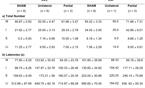
Table 2.2: Total number, duration, and derived measures of mating events ... 50
Table 2.3: Summary of copulatory behavior measures from males with incomplete
le-sion damage to BNST ... 51
Table 3.1: Summary of Odor Preference measures from males excluded from MPOA-X
lesion group ... 81
Table 3.2: Derived measures of mating events for EXP males ... 82
Table 3.3: Total number of mating events in males excluded from MPOA-X lesion group
... 83
Table 4.1: Densities of CTB+, Fos+, and Fos/CTB+ cells in cells that project to the
me-dial preoptic area (MPOA) ... 129
Table 4.2: Densities of CTB+, Fos+, and Fos/CTB+ cells that project to the posterior
bed nucleus of the stria terminalis (BNST) ... 130
Table 4.3: Densities of CTB+, ERG-1+, and CTB/ERG-1+ cells following female odor
exposure in cells the project to A) the medial preoptic area (MPOA) and B) the posterior
bed nucleus of the stria terminalis (BNST) ... 131
Table 4.4: Densities of CTB+, AR+, and CTB/AR+ cells in cells that project to A) the
medial preoptic area (MPOA) and B) the posterior bed nucleus of the stria terminalis
(BNST) ... 132
LIST OF FIGURES
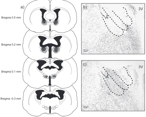
Figure 2.1: Lesion Reconstruction ... 44
Figure 2.2: Investigation Times for Non-Contact Preference Test ... 45
Figure 2.3: Investigation Times for Contact Preference Test ... 46
Figure 2.4: Investigation Times for Odor Discriminiation Test ... 47
Figure 2.5: Latencies to Mating Events ... 48
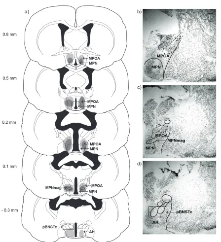
Figure 3.1: Lesion Reconstruction ... 81
Figure 3.2: Investigation Times for Non-Contact Preference Test ... 82
Figure 3.3: Investigation Times for Contact Preference Test ... 83
Figure 3.4: Investigation Times for Odor Discrimination Test ... 84
Figure 3.5: Total number of Mating Events in NVE and EXP males ... 85
Figure 3.6: Total Durations of Mating Events in NVE and EXP males ... 86
Figure 3.7: Latencies to Mating Events in EXP males ... 87
Figure 4.1: Counting domains for analysis of cholera toxin B (CTB), immediate early genes (Fos, EGR-1) and androgen receptor (AR) ... 120
Figure 4.2: Verification of cholera toxin B (CTB) injections ... 121
Figure 4.3: Co-localization of cholera toxin B (CTB) and immediate early genes ... 122
Figure 4.4: Percentages of Fos/CTB double-labeled cells in males with medial preoptic area (MPOA) injections ... 123
Figure 4.5: Percentages of Fos/CTB double-labeled cells in males with posterior bed nucleus of the stria terminalis (BNST) injections ... 124
Figure 4.7: Percentages of AR/CTB double-labeled cells in males with medial preoptic
area (MPOA) injections ... 126
Figure 4.8: Percentages of AR/CTB double-labeled cells in males with posterior bed nucleus of the stria terminalis (BNST) injections ... 127
Figure 4.9: Summary of chemosensory and steroid-sensitive projections to medial pre-optic area (MPOA) and posterior bed nucleus of the stria terminalis ... 128
Figure 5.1: Lesion Reconstructions ... 150
Figure 5.2: Non-Contact Odor Preference ... 151
Figure 5.3: Contact Odor Preference ... 152
Figure 5.4: Copulatory Behavior ... 153
Figure 6.1: Subnuclei in MA, BNST, and MPOA process chemosensory and hormonal information ... 183
Figure 6.2: MA, BNST, and MPOA Connectivity with Other Brain Areas ... 184
Figure 6.3: Possible Neurochemical Influences on Appetitive and Consummatory Re-productive Behaviors ... 186
LIST OF ABBREVIATIONS
ACo, anterior cortical nucleus of the amygdala
AHA, amygdalohippocampal area
AGI, anogenital investigation
AH, anterior hypothalamus
AOB, accessory olfactory bulbs
AOS, accessory olfactory system
A-P, anterior-posterior
AR, androgen receptors
AVP, vasopressin
BLA, basolateral amygdala
BNST, posterior bed nucleus of the stria terminalis
BNST-X, lesion of the posterior bed nucleus of the stria terminalis
BNSTpi, posterointermediate bed nucleus of the stria terminalis
BNSTpl, posterolateral bed nucleus of the stria terminalis
BNSTpm, posteromedial bed nucleus of the stria terminalis
BNSTpc, posterior caudal bed nucleus of the stria terminalis
CeA, central nucleus of the amygdala
CTB, cholera toxin B
CTF, central tegmental field
CONTRA, two unilateral lesions in contralateral hemispheres
D-V, dorsal-ventral
E, ejaculations
ER, estrogen receptors
EXP, sexually-experienced
HBI, head and body investigation
I, intromissions
I-E, intromissions to ejaculation
IEG, immediate early gene
INAH, interstitial nucleus of the hypothalamus
IPSI, two unilateral lesions in ipsilateral hemispheres
LI, long intromissions
M, mounts
MA, medial amygdala
MA-BNST-X, disconnect lesion of the medial amygdala and posterior bed nucleus of the
stria terminalis
MA-MPOA-X, disconnect lesion of the medial amygdala and medial preoptic area
ME, mount efficiency
MeA, anterior medial amygdala
MeP, posterior medial amygdala
M-L, medial-lateral
MOB, main olfactory bulbs
MOS, main olfactory system
MPNmag, magnocellular medial preoptic nucleus
MPOA, medial preoptic area
MPOA-X, lesion of the medial preoptic area
NAc, nucleus accumbens
NeuN, Neuronal Nuclei protein
nPGi, nucleus paragigantocellularis
NVE, sexually-naïve
OT, oxytocin
PAG, periaqueductal gray
PBS, phosphate buffered saline
PEI, post-ejaculatory interval
PMCo, posteromedial cortical nucleus of the amygdala
PMV, ventral premammillary nucleus
PVH, paraventricular nucleus of the hypothalamus
SG, self-grooming
SHAM, sham lesion surgery
TnA, nucleus taeniae
VMH, ventromedial hypothalamus
VNO, vomeronasal organ
5-HT, serotonin
CHAPTER 1:
OVERVIEW
Many animals, including rodents, use chemical signals as the primary means of
conveying social information between individuals (Brennan and Keverne 2004; Zufall
and Leinders-Zufall 2007). Volatile odor cues are processed primarily by the main
olfac-tory system (MOS), whereas non-volatile cues are processed primarily by the accessory
olfactory system (AOS) (Breer 2003). In Syrian hamsters, these two systems have been
shown to mediate appetitive aspects of male reproductive behavior, such as attraction
to female odors (Johnston 1974; Johnston 1975; Johnston and Kwan 1984; Petrulis and
Johnston 1995), as well as consummatory aspects of reproductive behavior, such as
copulation (Murphy and Schneider 1970; Powers and Winans 1975). MOS and AOS
in-formation is first integrated in the medial amygdala (MA), which is critical for the
appro-priate expression of reproductive behavior in male hamsters (Newman 1999). Several
lines of evidence suggest that MA may influence reproductive behavior via its
connec-tions with the posterior bed nucleus of the stria terminalis (BNST) and medial preoptic
area (MPOA) (Wood 1997). How these three nuclei work together to regulate different
aspects of reproductive behavior, however, is poorly understood. Therefore, the
over-arching goal of this dissertation was to determine how BNST and MPOA function, both
uniquely and as a circuit with MA, to regulate appetitive and consummatory reproductive
behaviors in male hamsters. We answered this question across four chapters using
functional, behavioral, and neuroanatomical approaches.
CHEMICAL COMMUNICATION IN RODENTS
Many animals use chemical signals as the primary means of conveying
2007). In rodents, as in many mammalian species, odors can convey a broad range of
social information, including the sex, individual identity, ownership, competitive ability,
health, and reproductive status of the individual (Johnston 1990; Hurst and Beynon
2004; Brennan and Kendrick 2006). These social odors are processed by two
anatomi-cally distinct chemosensory systems: the main olfactory system (MOS), which
process-es primarily volatile odors, and the accprocess-essory olfactory system (AOS), which procprocess-essprocess-es
primarily non-volatile cues (Meredith 1991; Restrepo et al. 2004). In the MOS,
low-molecular weight odorants dissolve in the mucus lining of the nasal passages. These
cues are detected by receptors on olfactory sensory neurons located in the main
olfac-tory epithelium, which, in turn, send axonal projections to the main olfacolfac-tory bulb (MOB).
Mitral cells in the MOB send efferent projections to the cerebrum via the lateral olfactory
tract, and synapse on the anterior olfactory nucleus, the olfactory tubercle, the piriform
and entorhinal cortices, and the corticomedial amygdala (Meredith 1991; Kelliher 2007).
In contrast, in the AOS, higher molecular weight odorants are detected by receptors on
sensory neurons located in a specialized sensory organ called the vomeronasal organ.
Vomeronasal sensory neurons send axonal projections to mitral cells in the accessory
olfactory bulb (AOB), which synapse on the bed nucleus of the olfactory tract, the
corti-comedial amygdala and the bed nucleus of the stria terminalis (Meredith 1991; Halpern
and Martinez-Marcos 2003). Together, the MOS and AOS regulate the expression of
most rodent social behaviors, including male reproductive behaviors (Meredith 1991;
Keverne 2004; Hull and Dominguez 2007; Baum and Kelliher 2009; Keller et al. 2009).
Chemical communication is particularly important for solitary species, in which
example, male and female Syrian hamsters maintain large, non-overlapping territories
in nature (Johnston 1990; Gattermann et al. 2008) and rely on the mutual perception of
species-specific scent marks to come together for mating (Pfaff et al. 2008). It is
per-haps not surprising then that removal of the olfactory bulbs (Murphy and Schneider
1970) or simultaneous deafferentation of the main and accessory olfactory systems
(Powers and Winans 1975) eliminates mating behavior in naïve and
sexually-experienced males. This is not the case, however, for social species, such as rats, in
which social or sexual experience can prevent the elimination of mating behavior
follow-ing olfactory bulbectomy (Larsson 1975). Furthermore, MOS and AOS are required for
male hamsters’ attraction to female hamster vaginal secretion alone (Powers et al.
1979) and for the stereotyped investigation of a receptive female’s anogenital region
during copulation (Devor and Murphy 1973; Powers and Winans 1975). Together, these
data suggest that MOS and AOS information are critical for the appropriate expression
of reproductive behaviors in male hamsters.
APPETITIVE AND CONSUMMATORY REPRODUCTIVE BEHAVIORS
In rodents and other non-human animals, male sexual behavior can be
dissoci-ated into two phases: the appetitive phase, which refers to a variety of behaviors that
reflect courtship, sexual attraction, and/or sexual motivation, and the consummatory
phase, which refers to the less variable behavioral sequence of copulation itself (Beach
1976; Everitt 1990; Ball and Balthazart 2008). In male hamsters (Bunnell et al. 1977;
Arteaga-Silva et al. 2005; Hull and Dominguez 2007) and other rodents (Dewsbury
re-productive behavior involves a highly stereotyped sequence of mating bouts comprised
of mounts, intromissions (penile insertions), and ejaculations. These mating bouts are
followed by a post-ejaculatory period characterized by auto-grooming of the genitals
and low levels of activity, after which, additional copulatory bouts can commence (Hull
and Dominguez 2007). Unlike other rodents, male hamsters display an additional
mat-ing behavior termed “long intromissions,” durmat-ing which males do not quickly dismount
the female following vaginal penetration, but instead display a repetitive thrusting
pat-tern (Bunnell et al. 1977). The expression of long intromissions is associated with the
onset of sexual satiety in hamsters (Bunnell et al. 1977; Parfitt and Newman 1998).
These fixed behavioral patterns comprise the consummatory phase of male sexual
be-havior in hamsters.
In comparison, the appetitive phase of reproductive behavior in male rodents
in-volves the approach and investigation of female odors with the goal of bringing the male
in contact with a receptive female. Male hamsters, like other rodents, are strongly
at-tracted to female odors, particularly those found in females’ vaginal secretions
(Johnston 1974; Johnston 1975; Johnston and Kwan 1984; Petrulis and Johnston
1995), and display a robust preference to investigate female odors more than male
odors (Landauer et al. 1977; Steel 1982; Ballard and Wood 2007). Unlike other rodents,
male hamsters do not require prior sexual experience to show this preference for
oppo-site-sex odors (Maras and Petrulis 2006; Ballard and Wood 2007; Maras and Petrulis
2008; Been and Petrulis 2010a; Been and Petrulis 2010b), suggesting that attraction to
female odors is an unconditioned appetitive response in male hamsters. Because the
se-quence of copulatory behaviors, it can be difficult to accurately measure in a laboratory
setting. Previous studies have used second-order conditioning tasks, such as training a
male to press a lever for a stimulus previously paired with a receptive female, to
meas-ure sexual motivation (Everitt and Stacey 1987; Everitt et al. 1989). This can be
prob-lematic, however, as the incentive value of the unconditioned stimulus can vary
be-tween species (Coppola and O'Connell 1988) and second-order conditioning tasks do
not necessarily recruit the same neural circuitry required for unconditioned appetitive
sexual behaviors (Kippin et al. 2003). Tests that directly measure unconditioned
appeti-tive responses, such as searching a bi-level chamber for a recepappeti-tive female (Pfaus and
Phillips 1991) or three-choice odor preference tests (Been and Petrulis 2010a; Been
and Petrulis 2010b) are therefore better suited for testing the requirement of neural
sub-strates for appetitive aspects of reproductive behavior.
SYRIAN HAMSTERS AS A MODEL SPECIES
Syrian hamsters provide a tractable model for studying the neural regulation of
socio-sexual behaviors because their social interactions are simple, stereotyped, and,
unlike several other well-studied laboratory species (e.g. rats, rhesus monkeys), depend
critically on the perception of a limited set of chemosensory cues from conspecifics
(Murphy and Schneider 1970). In hamsters, social odor cues are processed primarily in
the limbic system (Wood 1998), which processes social information in a wide variety of
species and in a manner that is independent of sensory modality. For example,
homol-ogous limbic nuclei in several avian species have been implicated in the behavioral and
al. 2009; Svec et al. 2009). Similarly, in non-human primates, limbic nuclei process
vis-ual cues from conspecifics, including those that are required for facial and emotional
recognition (Leonard et al. 1985). Perhaps most importantly, neuroimaging studies in
humans suggest that irregularities within limbic nuclei are correlated with
psychopatho-logical conditions that disrupt social processing in several different sensory modalities
(Acosta and Pearl 2004; McCloskey et al. 2005; Aguilar et al. 2008; Drevets et al.
2008). Thus, our model can yield new and important information about how the brain
processes social information at a more refined level of analysis and in a manner that
can be generalized to a variety of species and conditions.
MA AND ODOR-GUIDED REPRODUCTIVE BEHAVIORS
Several lines of evidence suggest that a particular limbic nucleus, MA, is a critical
node in the neural pathway that modulates odor-guided reproductive behaviors. First,
MA receives social odor information directly from the main and accessory olfactory
bulbs, as well as indirectly from other brain areas innervated by the olfactory bulbs
(Lehman and Winans 1982; Coolen and Wood 1998). MA neurons are activated during
exposure to a variety of social odors and during all social behaviors (Fernandez-Fewell
and Meredith 1994), suggesting they play a significant role in processing social
infor-mation. In addition to receiving social odor information, MA also contains dense
popula-tions of steroid receptor-containing neurons (Doherty and Sheridan 1981; Wood et al.
1992) that modulate social behavior. Specifically, unilateral testosterone implants into
MA facilitate mating behavior in castrated male hamsters (Wood and Newman 1995c),
MA has been implicated as a major regulator of odor-guided social behavior. Lesion
studies have consistently shown that MA is required for appropriate socio-sexual
behav-ior in hamsters (Newman 1999; Petrulis and Johnston 1999), rats (Kondo 1992; Kondo
et al. 1997; Stark et al. 1998), and gerbils (Heeb and Yahr 2000). Specifically, in male
hamsters, large electrolytic lesions of MA eliminate copulatory behavior and reduce
anogenital investigation of a receptive female (Lehman et al. 1980). Previous work in
our laboratory has demonstrated that lesions of the anterior or posterior subnuclei of MA
eliminate odor preference in male hamsters (Maras and Petrulis 2006). How MA
inter-acts with other ventral forebrain nuclei to regulate odor-guided social behaviors,
howev-er, is poorly understood.
BNST AND ODOR-GUIDED REPRODUCTIVE BEHAVIORS
One mechanism by which MA may regulate odor-guided social behaviors is via
its connections with BNST. Like MA, BNST receives social odor information, both
direct-ly from the accessory olfactory bulbs (Davis et al. 1978) and indirectdirect-ly via the
cortico-medial amygdala (Scalia and Winans 1975; Gomez and Newman 1992). Specifically,
BNST is densely interconnected with MA (Wood and Swann 2005), such that main and
accessory olfactory information that converges on MA can terminate at BNST or
contin-ue to MPOA (Newman 1999). In addition to receiving social odor information, BNST
contains dense populations of steroid receptor-containing neurons (Wood et al. 1992; Li
et al. 1993) that are activated during social behaviors (Wood and Newman 1993). BNST
has also been implicated in the hormonal modulation of social behavior, as unilateral
(Wood and Newman 1995c). Finally, evidence from lesion studies suggests that BNST
may be critically involved in generating the motivation to investigate opposite-sex odors.
For example, lesions of BNST delay copulation in rats (Valcourt and Sachs 1979) and
gerbils (Finn and Yahr 2005) and decrease the frequency of rats’ penile erections in
re-sponse to remote odor cues from estrous females (Liu et al. 1997b). In male hamsters,
large electrolytic lesions including BNST reduce investigation of female odor cues and
reduce anogenital investigation during copulation (Powers et al. 1987). Whether BNST
and MA play redundant or unique roles in the modulation of odor-guided social
behav-iors is, however, unknown.
MPOA AND ODOR-GUIDED REPRODUCTIVE BEHAVIORS
It is likely that MA and/or BNST exert their effects on social behavior via their
connections to MPOA. Indeed, MA and BNST are both densely connected to MPOA
(Wood and Swann 2005) and disrupting these connections delays the copulatory
se-quence in male hamsters (Lehman et al. 1983) and eliminates copulation in male rats
(Kondo and Arai 1995) and gerbils (Sayag et al. 1994). Furthermore, castration
increas-es the absolute refractory period of neurons in the stria terminalis that project to the
MPOA (Kendrick and Drewett 1979), suggesting that hormones acting on MA and/or
BNST can directly affect neural transmission within MPOA. MPOA is itself a critical
reg-ulator of social behavior and has been implicated in the control of a wide range of
be-haviors, including parental behavior (Numan 1997), social communication (Albers et al.
2002), aggression (Veening et al. 2005), social defeat (Kollack-Walker et al. 1997), and,
studied to date, permanent or reversible lesions of MPOA reduce or eliminate sexual
behavior (Klaric and Hendricks 1986; Liu et al. 1997b; Hull et al. 2002; Swann et al.
2003), whereas stimulation of MPOA can enhance sexual behavior (Malsbury 1971;
Paredes et al. 1990; Rodriguez-Manzo et al. 2000). Furthermore, lesions of MPOA
re-duce or reverse preference for opposite-sex odors in rats and ferrets (Kindon et al.
1996; Paredes et al. 1998; Hurtazo et al. 2008), suggesting MPOA, like MA and BNST,
is also critical for the appropriate behavioral response to social odors.
GOALS OF DISSERTATION
Based on the evidence reviewed above, it is clear that MA, BNST, and MPOA
each play a critical role in odor-guided reproductive behaviors. It is not known, however,
how BNST and MPOA individually regulate odor-guided reproductive behaviors and
whether functional interactions between MA, BNST, and/or MPOA are required to
gen-erate the appropriate behavioral response to socio-sexual odors. Therefore, the
over-arching goal of this research was to determine how MA, BNST, and MPOA function,
both uniquely and as a circuit, to adaptively regulate odor preference and copulatory
behaviors in male Syrian hamsters. We combined functional, behavioral, and
neuroana-tomical approaches to address the following questions:
1) Is BNST critical for odor-guided reproductive behaviors?
BNST receives social odor information, both directly from the olfactory bulbs
(Scalia and Winans 1975) and indirectly from MA (Wood and Swann 2005). The role of
used discrete, excitotoxic lesions of BNST in combination with detailed behavioral
anal-ysis to test the hypothesis that BNST is critical for generating the appropriate behavioral
response to social odors (Chapter 2). In addition, as several lines of evidence suggest
that previous sexual experience can alter neural and behavioral responses to social
odors (Dewsbury 1969; Fewell and Meredith 2002; Kelliher and Baum 2002; Meisel and
Mullins 2006), these experiments were conducted in both naïve and
sexually-experienced males.
2) Is MPOA critical for odor-guided reproductive behaviors?
Although the role of MPOA in male copulatory behavior has been extensively
studied (Hull and Dominguez 2007), the function of MPOA in appetitive aspects of
re-productive behavior, such as the approach and investigation of social odors, is not well
understood. Therefore, we used discrete, excitotoxic lesions of MPOA in combination
with detailed behavioral analysis to test the hypothesis that MPOA is critical for
generat-ing the appropriate behavioral response to social odors (Chapter 3). As in Chapter 2,
these hypotheses will be tested in both sexually-naïve and sexually-experienced males.
3) Is sexual odor information directly relayed between MA, BNST, and MPOA?
Social odor information processed in MA may be relayed directly to MPOA
with-out passing through BNST or terminate in BNST before continuing on to MPOA
(Newman 1999). Therefore, we used immediate early gene expression in combination
with neuroanatomical tract tracing to determine whether sex-specific odor information is
directly relayed from MA to BNST and/or MPOA, as well as from BNST to MPOA
responses to social odors (Fiber and Swann 1996; Kelliher et al. 1998), the
hormone-sensitivity of MA and BNST afferents were also quantified.
4) Are connections between MA and BNST/MPOA critical for odor-guided
repro-ductive behaviors?
The results of Chapters 2 and 3, together with previous research in our
laborato-ry, demonstrated that MA, BNST, and MPOA are each individually required for the
ap-propriate behavioral response to social odors (Maras and Petrulis 2006; Been and
Petrulis 2010a; Been and Petrulis 2010b). Furthermore, the results of Chapter 4
demonstrated that MA relays sex-specific odor information from MA to BNST and
MPOA (Been and Petrulis2011).We therefore hypothesized that the connections
be-tween MA and BNST/MPOA are required for the appropriate behavioral response to
so-cial odors. We tested this hypothesis using the asymmetrical lesion technique to
func-tionally disconnect MA from BNST or MPOA (Chapter 5).
The answer to these questions may uncover a fundamental mechanism by which
this ventral forebrain circuit regulates socio-sexual behaviors across a wide variety of
CHAPTER 2:
The role of the posterior bed nucleus of the stria terminalis in appetitive and
con-summatory reproductive behaviors depends on odor volatility and sexual
experi-ence in male Syrian hamsters
Laura E. Been and Aras Petrulis
Neuroscience Institute
Georgia State University, Atlanta, GA 30302, USA
ABSTRACT
In Syrian hamsters (Mesocricetus auratus), the expression of reproductive
be-havior requires the perception of social odors. The bebe-havioral response to these odors
is mediated by a network of ventral forebrain nuclei, including the posterior bed nucleus
of the stria terminalis (BNST). Previous studies have tested the role of BNST in
repro-ductive behavior, but the use of large, fiber-damaging lesions in these studies make it
difficult to attribute post-lesion deficits to BNST specifically. Thus, the current study
used discrete, excitotoxic lesions of BNST to test the role of BNST in opposite-sex odor
preference and copulatory behavior in both sexually-naïve and sexually-experienced
males. Lesions of BNST decreased sexually-naïve males’ investigation of volatile
fe-male odors, resulting in an elimination of opposite-sex odor preference. This elimination
of preference was not due to a sensory deficit, as males with BNST lesions were able to
discriminate between odors. When, however, subjects were given sexual experience
prior to BNST lesions, their preference for volatile opposite-sex odors remained intact
post-lesion. Similarly, when sexually-naïve or sexually-experienced subjects were
al-lowed to contact the social odors during the preference test, lesions of BNST decreased
males’ investigation of female odors, but did not eliminate preference for opposite-sex
odors, regardless of sexual experience. Finally, lesions of BNST delayed the copulatory
sequence in sexually-naïve, but not sexually-experienced, males such that they took
longer to mount, intromit, ejaculate, and display long intromissions. Together, these
re-sults demonstrate that BNST plays a unique and critical role in both appetitive and
INTRODUCTION
In many rodent species, including Syrian hamsters, male reproductive behavior
depends critically on the perception of odor cues from conspecifics (Johnston 1990).
Volatile odor cues are processed primarily by the main olfactory system (MOS),
where-as non-volatile cues are processed primarily by the accessory olfactory system (AOS)
(Breer 2003). Together, these two systems have been shown to mediate both appetitive
and consummatory aspects of reproductive behavior in Syrian hamsters, including
males’ attraction to vaginal secretions (Powers et al. 1979) and copulatory behavior
(Murphy and Schneider 1970; Powers and Winans 1975).
The behavioral response to these social odors is mediated by a network of
ven-tral forebrain nuclei that receive chemosensory information and are sensitive to steroid
hormones (Wood and Coolen 1997). In particular, several lines of evidence suggest the
posterior bed nucleus of the stria terminalis (BNST) may regulate males’ attraction to
opposite-sex odors. First, BNST receives social odor information, both directly from the
accessory olfactory bulbs (Davis et al. 1978) and indirectly via the corticomedial
amyg-dala (Scalia and Winans 1975; Gomez and Newman 1992). Specifically, BNST is
densely interconnected with the medial amygdala (MA) (Wood and Swann 2005), such
that main and accessory olfactory information that converges on MA can terminate at
BNST or continue to the medial preoptic area (MPOA) (Gomez and Newman 1992). In
addition to receiving social odor information, BNST also processes steroid hormone
cues that are required for reproductive behavior (Wood et al. 1992; Li et al. 1993; Wood
and Newman 1993). Evidence from lesion studies suggests BNST may be involved in
in-cluding BNST disrupt the copulatory sequence (Valcourt and Sachs 1979; Claro et al.
1995; Liu et al. 1997b; Finn and Yahr 2005) and decrease the frequency of penile
erec-tions in response to remote odor cues from females in rats (Liu et al. 1997b). In male
hamsters, large electrolytic lesions including BNST reduce investigation of female odors
when presented alone or during copulation, and also eliminate copulation in some
males (Powers et al. 1987).
Although previous studies have implicated BNST in the regulation of male
repro-ductive behavior, the use of large and/or non-fiber-sparing lesions makes it difficult to
attribute these effects to BNST itself, as BNST is a heterogeneous structure that is
bor-dered by multiple fiber tracts that relay information to other nuclei critical for
reproduc-tive behavior, such as MPOA (Wood and Swann 2005). The interpretation of these
stud-ies is further complicated by variability in the manner of stimulus presentation and
dif-ferences in the sexual experience of the subjects. Thus, the following experiments used
site-specific, excitotoxic lesions of BNST to comprehensively test the role of this region
in generating the appropriate behavioral response to volatile and non-volatile social
odors in both sexually-naïve and sexually-experienced males. We hypothesized BNST
primarily mediates males’ attraction to female odors rather than copulatory behavior.
We therefore predicted lesions of BNST would eliminate males’ preference for female
odors and decrease investigation of females during mating, but leave
MATERIALS AND METHODS
Experimental Design
The goal of the following experiments was to test the role of BNST in social odor
investigation and copulatory behavior in male hamsters. Exposure to female odors
causes an increase in circulating testosterone levels in male hamsters (Macrides et al.
1974; Pfeiffer and Johnston 1992) and it is possible that lesions of BNST may interfere
with this surge. Thus, in order to equalize steroid hormone levels across experimental
groups, all subjects were gonadectomized and maintained on physiological levels of
ex-ogenous testosterone for the duration of the experiment. Following bilateral, excitotoxic
lesions of BNST or sham lesion surgeries, subjects underwent a series of behavioral
tests: first, subjects were tested for their preference to investigate female odors over
male odors (Odor Preference). To determine if any effects of BNST lesions on odor
in-vestigation depend on the volatility of the odor cues being processed, Odor Preference
tests were conducted under conditions that either prevented contact with the odor
sources (Non-contact; volatile odors only) or allowed contact with the odor sources
(Contact; volatile and non-volatile odors). Second, in order to determine if a lack of
pref-erence in these tests was due to an inability to discriminate between odor stimuli, a
separate group of subjects were tested for their ability to discriminate between social
and non-social odor sources using a habituation-dishabituation task (Odor
Discrimina-tion). Lastly, to determine if lesions of BNST disrupt male copulatory behavior, subjects’
response to a receptive stimulus female (Copulatory Behavior Test) was assessed. As
converging lines of evidence suggest that previous sexual experience can alter neural
Kelliher and Baum 2002; Meisel and Mullins 2006), we tested the effects of BNST
le-sions on both sexually-naïve and sexually-experienced males’ behavior.
Animals
Experimental subjects were adult (3 to 6 months old) male Syrian hamsters
(Mesocricetus auratus) purchased from Charles River Laboratories (Wilmington, MA,
USA). A separate group of unrelated adult male and female hamsters served as odor
stimulus donors for behavior tests. A third group of ovariectomized and hormone-primed
adult female hamsters were used as stimulus females for copulatory behavior tests.
Ex-perimental subjects and copulatory stimulus females were single-housed, whereas odor
donors were group-housed (three to four animals per cage), in solid-bottom Plexiglas
cages (36 cm x 30 cm x 16 cm). All subjects were maintained on a reversed 14 hour
light/ 10 hour dark photoperiod, and food and water were available ad libitum. All animal
procedures were carried out in accordance with the National Institutes of Health Guide
for the Care and Use of Laboratory Animals (NIH Publications NO. 80-23; revised 1996)
and approved by the Georgia State University Institutional Animal Care and Use
Com-mittee. All efforts were made to minimize the number of animals used and their
suffer-ing.
Sexual Experience
Following gonadectomy (see below), experimental subjects were randomly
as-signed to one of two experimental conditions: sexually-experienced (EXP; n = 40) or
sexually-naïve (NVE; n = 36). EXP males were given weekly sexual experiences for
three consecutive weeks. Subjects were placed into a clear, Plexiglas testing area (50
An angled mirror was placed below the testing arena to provide a view of the ventral
surface of the animals. Encounters lasted for 30 minutes, or until the animals engaged
in aggressive behavior, at which point the stimulus female was removed. The second
and third encounters were video-recorded and the male’s behavior was later scored
us-ing the Observer for Windows, version 5.0 (Noldus Information Technology,
Wa-geningen, The Netherlands). Males who failed to copulate on at least two of the three
encounters (n = 7) were eliminated from the study. To control for possible effects of
ex-perimenter handling on future behavior, NVE males were transported to the same
be-havioral testing room and placed into an empty cage for 30 minutes once a week for
three consecutive weeks.
Surgery
All surgeries were performed under 2% isoflurane gas anesthesia vaporized in
100% oxygen (gonadectomy) or a 70:30% oxygen/nitrous oxide mixture (stereotaxic
surgery). To minimize post-operative pain, ketoprofen (5 mg/kg subcutaneously, Henry
Schein, Melville, NY, USA) was administered intra-operatively.
Gonadectomy and Hormone Implant. One to two weeks prior to lesion surgery,
subjects’ testes were bilaterally removed via a bilateral midline abdominal incision and
cauterization of the ductus deferens and blood vessels. Silastic capsules (i.d. 1.57 mm,
o.d. 2.41 mm, Dow Corning, Midland, MI, USA) packed with 20 mm length of crystalline
testosterone (Sigma, St. Louis, MO, USA) were implanted subcutaneously between the
scapulae immediately following gonadectomy. Vicryl suture (size 4-0, Ethicon,
Somer-ville, NJ, USA) and wound clips were used to close the smooth muscle and skin
Stimulus females for copulatory experiences and behavior testing were
ovariec-tomized at least two weeks prior to use. Following bilateral flank incisions, the ovaries
were removed via cauterization of the uterine horn and blood vessels. Silastic capsules
(i.d. 1.57 mm, o.d. 2.41 mm, Dow Corning, Midland, MI, USA) packed with 5 mm length
of crystalline estradiol (Sigma, St. Louis, MO, USA) were implanted subcutaneously
be-tween the scapulae immediately following gonadectomy. Vicryl suture (size 4-0,
Ethi-con, Somerville, NJ, USA) and wound clips were used to close the smooth muscle and
skin incisions, respectively. To induce behavioral receptivity, stimulus females were
in-jected subcutaneously with 0.15 ml of progesterone dissolved in sesame oil (2.5 mg/ml,
Sigma, St. Louis, MO, USA) 4 hours prior to copulatory behavior tests.
Excitotoxic Lesion. NVE and EXP subjects were randomly assigned to either a
BNST lesion (BNST-X; NVE n = 26, EXP n = 25) or a sham lesion surgery (SHAM; NVE
n = 8, EXP n = 10) group. Anesthetized subjects were secured in the stereotaxic
appa-ratus such that their skull was level in the anterior-posterior (A-P) and medial-lateral
(M-L) planes. Following a midline scalp incision, the skin and temporal muscles were
re-tracted to expose the skull and a hand-operated drill was used expose dura. All A-P and
M-L measurements were taken in mm relative to bregma and all dorsal-ventral (D-V)
measurements were taken in mm relative to dura. Excitotoxic lesions were made by
lowering a microinjection syringe (701R 10 µl syringe, Hamilton, Reno, NV, USA) under
stereotaxic control into BNST and injecting N-methyl-D-aspartic acid (NMDA, 20 mg/ml;
20 nl per injection, Sigma, St. Louis, MO, USA) into two bilateral sites targeting the
posterointermediate BNST (BNSTpi; A-P: - 1.85, M-L: ± 1.65, D-V: - 5.8) and
the flow of excitotoxin up the syringe tract, the syringe was left in place for 10 minutes
after each injection.
Sham surgeries were identical to lesion surgeries with two exceptions: 1) the
mi-croinjection syringe was lowered to 1 mm above the target injection site and 2) no
exci-totoxin was infused into the target injection sites. After all surgeries, skull holes were
sealed using bone wax and incisions were closed with wound clips. Subjects were
al-lowed to recover for at least two weeks prior to behavioral testing.
Behavioral Testing
All behavior testing took place during the first six hours of the dark phase and
under light illumination.
Odor Stimuli. Social odor stimuli used for Odor Preference and Odor
Discrimina-tion tests were collected from cages of group-housed, same-sexed odor donors that had
not been changed for at least four days prior to odor collection. Each social odor
stimu-lus consisted of soiled cotton bedding (2 Nestlets, 12 g, ANCARE, Bellmore, NY, USA),
soiled corncob litter (50 ml, Bed-o-cob, The Andersons, Maumee, OH, USA) and one
damp cotton gauze pad that was used to wipe the inner walls of the cage. In addition, a
damp gauze pad was used to wipe two of the cage resident’s anogenital region and
bi-lateral flank glands 10 times each. For female odor stimuli, vaginal secretion was also
collected onto a gauze pad by gently palpating the vaginal area of one female with a
disposable probe, and was added to each odor stimulus. Clean odor stimuli consisted of
clean cotton bedding (2 Nestlets), clean corncob litter (50 ml), and clean cotton gauze
beads) of an artificial, multi-component odorant (baby powder or strawberry,
Interna-tional Flavors & Fragrances, Inc., NY, USA) mixed with clean odor stimuli.
For Contact tests, additional odors were collected directly onto glass microscope
slides (25 mm X 75 mm X 1 mm) by rubbing a clean slide along an odor donor’s flank
and anogenital regions. All odor slides contained samples from two individual odor
do-nors (collected separately onto each end of the slide). For female odor slides, a sample
of vaginal secretion was also collected onto the same slide. All odor stimuli were stored
in airtight containers at 4°C until 20 minutes before use. Odor stimuli older than one
month were discarded and no subject was tested with the same odor stimulus more
than once.
Odor Preference. A three-choice test was used to measure odor preference.
Subjects were placed into a glass aquarium (50 cm x 25 cm x 30 cm) with opaque walls.
Three acrylic odor presentation boxes (8 cm x 8 cm x 8 cm) were affixed to one of the
short walls of the aquarium such that only the front and top surfaces of each box were
accessible. Each odor presentation box had 7-mm holes drilled along the front surface
that allowed volatile odors to pass, but prevented contact with the odor sources. During
testing, a single odor stimulus (see above) was placed into each of the three odor
presentation boxes. Additionally, a line bisecting the available floor space was drawn
parallel to the short walls so that general activity levels (as measured by total number of
line crosses) could be assessed during the test. The top of the aquarium was secured
with a clear Plexiglas top to allow for overhead video recording of the subject’s
behav-ior. All surfaces of the aquarium and odor containers were thoroughly cleaned with 70%
Subjects were tested in a series of three tests in the 3-choice apparatus, each
separated by 24 hours: Clean, Non-Contact preference, and Contact preference. At the
beginning of each test, a subject was placed into the testing arena and then allowed ten
minutes to freely explore the apparatus. For all tests, investigation of the odor stimulus
was coded when the subject made contact with, or directed its nose within 1 cm of, the
perforated front surface of the odor container and/or odor slide. For Clean tests, clean
odor stimuli were placed into each of the three odor containers. These tests were used
to acclimate the subjects to the testing arena, as well as to obtain baseline levels of
ac-tivity in the absence of social odor stimuli. For subsequent preference tests, female and
male odor stimuli were placed into each of the two outer odor containers, and clean
odor stimuli were placed into the center odor container. The side on which each social
odor was placed (left or right) was alternated between consecutive subjects.
Non-Contact and Non-Contact tests were identical except that during Non-Contact tests, a single odor
slide matching the type of odor stimulus in that container (female, male, clean) was
se-cured to the center of the front surface of each odor presentation box.
Video recordings of all tests were digitized onto a computer and scored using the
Observer for Windows, version 5.0 (Noldus Information Technology, Wageningen, The
Netherlands). All observers were blind to the condition of the subject, and different
ob-servers reached at least a 90% inter-observer reliability score prior to coding behavior.
Odor Discrimination. In order to determine if the lack of preference for
non-contact odors observed in NVE subjects (see Results) was due to an inability to
discrim-inate between odor stimuli, a separate group of NVE males (BNST-X n = 12, SHAM n =
habituation-dishabituation task. The habituation-habituation-dishabituation task involves repeated presentations
of the same odor source followed by a test presentation of a novel odor source. A
de-crease in investigation during the repeated presentations indicates a perception of the
odors as being the same or familiar. An increase in investigation of the novel odor
com-pared to the last presentation of the habituated odor indicates an ability to discriminate
between the two odors (Johnston 1993; Baum and Keverne 2002). The testing
se-quence consisted of four, 3-minute presentations of repeated odors (habituation)
fol-lowed by a fifth, 3-minute presentation of a novel odor (dishabituation). Five-minute
in-ter-trial intervals separated each odor presentation. Odor stimuli were presented in
modified 50 ml polypropylene collection tubes, with 0.5-cm holes drilled 1 cm apart
along the surface of the tube. Wire mesh lined the inner surface of the odor container to
prevent contact with the odor stimulus. Odor containers were placed into the center of
subjects’ home cage and investigation was measured using a stopwatch. Odor
contain-ers were cleaned with 70% alcohol and allowed to dry between subjects.
Under these testing parameters, male hamsters consistently display a lack of
ha-bituation to repeated presentations of female odors (Maras and Petrulis 2006) and so all
subjects were tested using male odors as the habituation stimuli and female odors as
the dishabituation stimuli for social odor discrimination tests. Subjects were presented
with different male odor sources on each of the habituation trials so that subjects were
habituated to the sexual identity of the repeated odor, rather than to the individual
identi-ty of odor donors. In addition, in order to determine if lesion-induced deficits were
test-ed for their discrimination of two complex, artificial odors (strawberry and baby powder;
see above).
Copulatory Behavior. Copulatory behavior test procedures were identical to those
used to provide sexual experience to EXP males (see above). Tests were
video-recorded and later scored using Observer for Windows, version 5.0 (Noldus Information
Technology, Wageningen, The Netherlands). The total number and latencies (from
on-set of test) of several behavioral measures were scored: mounts (M), intromissions (I),
ejaculations (E), and long intromissions (LI). LI are distinguished from I in that males do
not quickly dismount the female following vaginal penetration, but instead display a
re-petitive thrusting pattern (Bunnell et al. 1977). Importantly, the expression of LI is
asso-ciated with the onset of sexual satiety in Syrian hamsters (Bunnell et al. 1977; Parfitt
and Newman 1998). In addition, the total durations of time the male spent investigating
the female’s anogenital region (AGI), investigating the female’s head or body region
(HBI), and self-grooming (SG) were also scored. Finally, several derived measures of
copulatory behavior were also analyzed: post-ejaculatory interval (PEI; latency to
dis-play a mount or intromission after each ejaculation, the number of intromissions to
reach each ejaculation (I-E), and mounting efficiency (ME; the total number of
intromis-sions divided by the total number of mounts + intromisintromis-sions).
Histology and Lesion Verification
Following the last behavioral test, subjects were injected with an overdose of
so-dium pentobarbital (100 mg/kg; Sleep Away, Ft. Dodge, IA, USA) and transcardially
per-fused with 200 ml of 0.1M phosphate-buffered saline (PBS, pH 7.4) followed by 200 ml
and then cryoprotected for 48 hours in 30% sucrose in PBS solution. Coronal sections
(30-µm) of brain tissue were sectioned using a cryostat (-20°C) and stored in
cryopro-tectant until immunohistochemical localization of Neuronal Nuclei protein (NeuN, see
below). Additionally, in order to further delineate lesion damage from fiber tracts (which
are not readily detected by NeuN), every third section was mounted onto glass slides
using a 1% gelatin mounting solution and stained for Nissl material with cresyl violet
(Sigma, St. Louis, MO, USA). Nissl- and NeuN-stained sections were examined under a
light microscope for the location and extent of lesion damage as compared with
pub-lished hamster neuroanatomical plates (Morin and Wood 2001), and the minimum and
maximum extents of lesion damage were traced onto anatomical plates using Adobe
Illustrator CS 11.0 software.
Immunohistochemistry. Free-floating sections were removed from cryoprotectant,
rinsed thoroughly in PBS, and then incubated in a monoclonal antibody against NeuN
(1:30,000, Millipore MAB377, Billerica, MA, USA) in PBS with 0.4% Triton-X-100 for 48
hours at 4°C. After rinsing in PBS, sections were incubated in biotinylated secondary
antibody (Rabbit Anti-Mouse, 1:600, Jackson Immunoresearch Laboratories
315-065-003, West Grove, PA, USA) in PBS with 0.4% Triton-X-100 (Sigma, St. Louis, MO,
USA) for one hour at room temperature, rinsed in PBS, then incubated in an
avidin-biotin complex (1:200, Vectastain Elite ABC Kit, Vector Laboratories, Burlingame, CA,
USA) for one hour at room temperature. Sections were then rinsed in PBS and reacted
in a nickel-enhanced 3, 3′-diaminobenzidine tetrahydrochloride (DAB) solution (2 mg
acetate, Sigma, St. Louis, MO, USA) to yield a blue-black reaction product. After 15
minutes, sections were rinsed in PBS to stop the chromagen reaction.
Blood Collection and Radioimmunoassay
Blood samples were collected from the inferior vena cava after anesthesia and
immediately prior to perfusion and stored in vacutainer collection tubes (4-mL draw,
red/gray, VWR, West Chester, PA, USA) on ice until centrifugation. Samples were
cen-trifuged at 2500 revolutions per minute at 4°C for 20 minutes and serum was stored in
200-µl aliquots at -20°C until assay. Testosterone levels were measured by
radioim-munoassay kits from Diagnostics System Laboratories (DSL 4000, Beckman Coulter,
Brea, CA, USA) with a sensitivity range of 0.05 to 22.92 ng/mL and an interassay
vari-ance of 6%, previously validated for hamster serum (Cooper et al. 2000). The mean
tes-tosterone levels (ng/nl) for experimental subjects were: NVE BNST-X = 4.25 ± .58; NVE
SHAM = 3.027 ± .36; EXP BNST-X = 3.78 ± .70; EXP SHAM = 4.193 ± .66. There was
no difference in testosterone levels between lesion groups for NVE or EXP males (NVE
t18 = 0.51, P = 0.61; EXP, t18 = 1.69, P = 0.11).
Data Analysis
All data were analyzed using SPSS 16.0 (SPSS Inc., Chicago, IL, USA) for
Win-dows and significance was determined as p < .05. To establish investigatory
prefer-ences for each type of 3-choice test (Clean, Non-Contact Preference, Contact
Prefer-ence), 2 (Experimental group: BNST-X, SHAM) X 3 (Odor) mixed-design ANOVAs were
performed. Significant interactions were explored using simple effects analysis and
pair-wise comparisons with Bonferroni alpha adjustments. Furthermore, separate one-way
across experimental groups. To identify differences in general motor activity, additional
one-way ANOVAs were used to compare the total number of midline crosses across
experimental groups for the Clean test.
For the habituation-dishabituation data, data were split by experimental group,
and paired t-tests (2-tailed, with Bonferroni alpha adjustments, αFW = .05) were used to
detect both (1) a habituation to the repeated presentations of odors (odor 1 vs. odor 4)
and (2) a dishabituation to the presentation of the test odor (odor 4 vs. Test).
Many copulatory behavior tests were terminated before 30 minutes because the
male and female engaged in aggressive behavior. Thus, only the first 20 minutes of
each copulatory test were analyzed to eliminate variability caused by differences in test
duration. Group differences in all copulatory measures were detected using one-way
ANOVAs. Additionally, separate 2 x 2 (lesion group x ejaculatory series) ANOVAS were
used to detect changes in post-ejaculatory intervals or the number of intromissions to
reach each ejaculation in the copulatory tests.
RESULTS
Lesion Reconstruction
Subjects were included in the BNST-X lesion group (NVE n = 12; EXP n = 12)
only if they had extensive bilateral damage to BNST, defined as at least 60% bilateral
damage to BNSTpi and BNSTpm in at least two stereotaxic planes of section (Figure
2.1a; (Morin and Wood 2001). All subjects included in the BNST-X lesion group
sus-tained significant damage to the largest part of BNST (Bregma 0.2 mm), including
subjects (n = 21) also sustained significant damage to more rostral and anterior
(Breg-ma 0.5 mm) parts of BNSTpi and BNSTpm. Ten subjects had bilateral lesion da(Breg-mage to
more caudal and posterior BNST (Bregma 0.1 mm); of these subjects, half had bilateral
damage of both BNSTpi and BNSTpm and half had bilateral damage to BNSTpm only.
No subjects included in the BNST-X group sustained damage to the most caudal and
ventral level of BNST (Bregma -0.3 mm).
Subjects were excluded from the BNST-X group if their lesions extended
signifi-cantly outside of BNST or failed to damage a significant portion of BNST. Specifically,
one group of subjects had partial (less than 60% damage in two stereotaxic planes of
section), bilateral damage to BNST (NVE n = 3; EXP n = 3) and another separate group
of subjects had primarily unilateral damage to BNST (NVE n = 6; EXP n = 1). Analyses
indicated that these subjects’ behavior did not differ significantly from SHAM subjects’
behavior in Odor Preference or Copulatory Behavior tests (see Results). Only needle
tracts were visible in most SHAM males (Figure 2.1b; NVE n = 8; EXP n = 8); three
SHAM males also had unilateral cortical damage. These subjects did not differ in
be-havior from SHAM subjects without cortical damage and were retained in the analysis.
Some subjects included in the BNST-X group sustained minimal or unilateral
damage to other adjacent nuclei, defined as less than 20% damage at only one
stereo-taxic plane of section of the nucleus (Morin and Wood 2001). This included nuclei within
the anterior BNST, such as the anterointermediate BNST (n = 8), anteromedial BNST (n
= 6), and anteroventral BNST (n = 10). Outside of the BNST, partial or unilateral
dam-age was sustained to sub-cortical nuclei including the ventral lateral septum (n = 6),
par-tial or unilateral damage also occurred in thalamic nuclei, such as the anterodorsal
tha-lamic nucleus (n = 3), anteroventral thalamic nucleus (n = 1), ventrolateral anteroventral
thalamic nucleus (n = 1), reuniens thalamic nucleus (n = 2), and reticular thalamic
nu-cleus (n = 3). Two males also sustained unilateral damage to overlying cortex. There
was no difference in behavior across subjects with unilateral, bilateral, or no damage to
any of the adjacent nuclei.
Behavioral Measures
Odor Preference
Clean Test. In the Clean test, both NVE and EXP subjects investigated the three
stimulus containers equally (NVE F2,23 = 3.37, P = 0.14; EXP F2,35 = 2.69, P = 0.23).
There were also no differences in the total number of midline crosses (NVE t18 = 1.48, P
= 0.15; EXP t18 = 1.35, P = 0.47) or total duration of investigation of the three odor
con-tainers (NVE t18 = .641, P = 0.08; EXP t18 =.861, P = 0.13) between BNST-X and SHAM
males.
Non-Contact Preference Test. Lesions of BNST decreased NVE males’
investi-gation of volatile female odors, resulting in an elimination of opposite-sex odor
prefer-ence. There was a significant interaction between lesion group and the duration of
in-vestigation of the three odor stimuli (F2,54 = 3.51, P = 0.03). Whereas SHAM males
in-vestigated female odors longer than male odors (t7 = 5.00, P < 0.01) and investigated
both female (t7 = 6.31, P < 0.01) and male (t7 = 2.65, P = 0.03) odors more than clean
odors, BNST-X males spent equivalent amounts of time investigating male and female
odors (t11 = 0.53, P = 0.60), and did not investigate either female (t11 = 1.51, P = 0.16)
spent significantly less time investigating female odors (F1,19 = 8.51, P < 0.01), and
sig-nificantly more time investigating clean odors (F1,19 = 4.79, P < 0.01), than did SHAM
males (Fig 2.2a).
In contrast to NVE subjects, lesions of BNST did not decrease EXP males’
inves-tigation of volatile female odors. In EXP males, there was a significant main effect of
odor stimulus (F2,53 = 21.652, P < 0.01), but no interaction between lesion group and the
duration of investigation of the three odor stimuli. Both SHAM and BNST-X males spent
more time investigating female odors than male odors (SHAM t7= 2.64, P = 0.03;
BNST-X t11 = 2.24, P = 0.04) and investigated female odors more than clean odors (SHAM t7 =
2.79, P = 0.03; BNST-X t11 = 5.04, P < 0.01). BNST-X males also spent significantly
more time investigating male odors than clean odors (t11 = 5.71, P < 0.01), although this
was not the case for SHAM males (t7 = 1.371, P = 0.21; Fig 2.2b).
In both NVE and EXP males, neither unilateral nor partial, bilateral lesions of
BNST disrupted males’ preference to investigate opposite-sex odors in the Non-Contact
test (Table 2.1a). Although the small sample sizes of these groups precluded them from
statistical analysis, examination of the means confirmed that, like SHAM males, males
with incomplete damage to BNST investigated female odors more than they did male or
clean odors.
Contact Preference Test. Lesions of BNST decreased both NVE and EXP males’
investigation of female odors in the Contact test, but did not eliminate opposite-sex odor
preference. In NVE males, there was a significant interaction between lesion group and
the duration of investigation of the three odor stimuli (F2,54 = 6.43, P = 0.03). Whereas