Breast Reconstruction
Emily Hu, MD
a, Amy K. Alderman, MD, MPH
a,b,*
aSection of Plastic Surgery, Department of Surgery, The University of Michigan
Medical Center, Ann Arbor, MI 48109, USA
bVA Center for Practice Management and Outcomes Research, Ann Arbor
VA Health Care System, Ann Arbor, MI 48109, USA
During the last century, breast reconstruction after mastectomy has become an important part of comprehensive treatment for patients who have breast cancer. Breast reconstruction initially was created to reduce complications of mastectomy and to diminish chest wall deformities. Now, however, it is known that reconstruction also can improve the psycho-social well-being and quality of life of patients who have breast cancer[1]. The primary goal of breast reconstruction is to recreate form and symmetry by correcting the anatomic defect while preserving patient safety and health. The primary reconstructive options involve the use an implant (usually with an expander first), the patient’s own tissue (autogenous tissue reconstruc-tion), or both. The reconstructive process can start at the time of the mastectomy (immediate reconstruction) or any time afterwards (delayed reconstruction).
Historical background
Silicone breast implants were introduced in the early 1960s [2], but in 1992, the Food and Drug Administration (FDA) placed a moratorium on silicone implants due to concern regarding its safety of use in patients. Since then saline implants had been exclusively used in the United States, until re-cently. In November 2006, after an extensive scientific review revealed no significant risks, the FDA approved the use of silicone implants for breast reconstruction in women of all ages. Now that the silicone implant has been deemed safe, the FDA is requiring a 10 year follow-up to continue to monitor these implants as part of a post-approval study [3]. The initial implant reconstructions were placed under the thin mastectomy skin flaps
* Corresponding author.
E-mail addresses:[email protected](A.K. Alderman);[email protected](E. Hu). 0039-6109/07/$ - see front matterÓ2007 Elsevier Inc. All rights reserved.
without prior expansion of the tissue, a practice that led to frequent compli-cations such as skin loss. The introduction of the latissimus dorsi myocuta-neous flap provided better soft tissue coverage over the implant and decreased postoperative complications[4]. In 1982, Radovan[5]introduced tissue expansion with placement of an uninflated implant under the residual skin and muscle, followed by intermittent filling of the implant. This process resulted in a gradual expansion of the overlying tissue. As the final stage of breast reconstruction, permanent implants replaced the expander implant. This technique, however, remained plagued by complications such as capsular contracture (scarring around the implant). Breast reconstruction advanced further with the popularization of the transverse rectus myocutaneous (TRAM) flap by Hartrampf and colleagues [6] in 1982, followed by the microsurgical free TRAM flap. The latest technical advances in breast reconstruction, perforator flaps, were introduced by Allen and colleagues in 1994 and 1995[7,8].
The other major advance that has occurred in breast reconstruction has been on the health policy front. In 1998, the federal government passed the Women’s Health and Cancer Rights Act (WHCRA), which mandated insur-ance coverage of reconstruction if the insurinsur-ance plan provided mastectomy coverage. The law also mandated coverage of breast symmetry procedures and the treatment of surgical complications at all stages of the mastectomy and reconstruction[9].
Goals
In keeping with the Hippocratic oath, one of the goals of breast recon-struction is to ‘‘first do no harm.’’ Reconrecon-struction after mastectomy should not impede the patient’s oncologic treatment (ie, delay administration of chemotherapy or radiation therapy), should not delay the diagnosis of a re-currence, and should not add an unacceptable increase in operative morbid-ity or mortalmorbid-ity. Current data indicate that reconstruction is safe and does not delay adjuvant therapy or the detection of cancer recurrence [10–12]. In addition, although the most frequent site of recurrent breast cancer is in the remaining chest wall skin, immediate reconstruction has not been shown to increase the rate of local recurrence in the long term[10].
The specific surgical goals of the plastic surgeon are to optimize the aes-thetic result while keeping in mind the patient’s preferences and surgical lim-itations. For instance, some women may be happy with recreation of just a breast mound, whereas other women may desire supple soft tissue and complete nipple reconstruction.
Preoperative counseling
should be offered at the time of initial surgical decision making. This preop-erative consultation should cover several areas, such as the type and timing of reconstruction. Reconstructive options are based on the patient’s overall goals, physical examination, and clinical factors. For example, autogenous tissue reconstructions are best in women who value the creation of the most natural-looking and -feeling breast. Other women may place more value on limiting potential morbidity to other body areas, such as the abdo-men, and therefore prefer an expander/implant reconstruction. Clinical con-traindications or significant risk factors to reconstruction, such as obesity, nicotine use, chronic obstructive pulmonary disease, diabetes, and other chronic conditions, should be assessed. (Such contraindications are dis-cussed in further detail later in this article.) Although patient preference cer-tainly is important, the highest priority is providing appropriate treatment of the breast cancer. Thus, input from a multidisciplinary team consisting of oncologic surgeons, medical and radiation oncologists, as well as recon-structive surgeons is necessary to provide the most comprehensive and ap-propriate treatment.
Timing of reconstruction
Immediate reconstruction
‘‘Immediate’’ reconstruction is defined as reconstruction that starts at the same time as the mastectomy. This option can be an excellent one for women who have ductal carcinoma in situ and stage 1 or stage 2 disease. The advantages of immediate breast reconstruction are multiple. Women who have immediate reconstruction have less distress and better body im-age, self-esteem, and satisfaction, in general, than women who have delayed reconstruction[13]. From an aesthetic standpoint, autogenous tissue recon-structions performed at the time of the mastectomy generally have produced a better aesthetic result than delayed procedures because the skin envelope is preserved[14,15]. The overall cost is less because fewer major operations are needed, the patient is anesthetized already, the defect does not have to be recreated, and the patient can recover from the mastectomy and the recon-struction simultaneously[16].
Disadvantages of immediate reconstruction include the potential delay of adjuvant therapy should a postoperative complication such as delayed wound healing occur. Most studies, however, have not shown reconstruc-tion to delay therapy[12]. Another potential pitfall of immediate reconstruc-tion is the partial loss of the mastectomy skin flaps, especially if the oncologic surgeon needs to create thin skin flaps. In addition, residual dis-ease or close surgical margins may necessitate the use of postoperative radi-ation therapy, which can adversely affect the reconstruction.
contraindication is controversial and varies by center), and medical comor-bidities such as use of nicotine, morbid obesity, or cardiopulmonary disease. In addition, use of implants is a relative contraindication in women who have rheumatologic disorders.
Delayed reconstruction
Delayed reconstruction, defined as a reconstructive procedure that starts after the mastectomy, can be started any time after the wound has healed and adjuvant therapy has been administered. Postradiation skin changes should have stabilized, and the hematologic effects of chemo-therapy should have normalized before reconstruction is begun. Delayed reconstruction has its own advantages. First, all guess work regarding whether radiation therapy will be required is eliminated, so surgeons and patients can appraise to their reconstructive options more accurately. Second, studies have shown that delayed reconstructions have overall fewer complications than immediate reconstruction [10]. Disadvantages of delayed reconstruction include prolonging the overall treatment of the patient, a poorer cosmetic result with autogenous tissue reconstruction because the skin envelope is not preserved, and potentially higher costs to the health care system.
Reconstructive techniques
Implant without tissue expansion
The simplest reconstruction for a mastectomy defect involves the place-ment of an implant, without prior expansion of the remaining tissue enve-lope. This simple technique requires that the skin flaps remaining after the mastectomy to be sufficient to cover the implant. Often, the remaining flaps are not sufficient. If an implant is placed without prior expansion, there is a greatly increased risk of skin necrosis secondary to tension. In addition, implants placed under nonexpanded mastectomy skin flaps often have poor cosmetic results because of constricted skin envelopes. For these rea-sons, this technique is generally discouraged.
Tissue expansion followed by permanent implant placement
Indications/contraindications
contraindication for this type of reconstruction is mastectomy flaps that are too thin for adequate implant coverage. In these cases, one should consider using a latissimus dorsi muscle flap for additional coverage. Another relative contraindication is the completed or planned use of adjuvant radiation ther-apy because of higher implant complication rates[17].
Technique
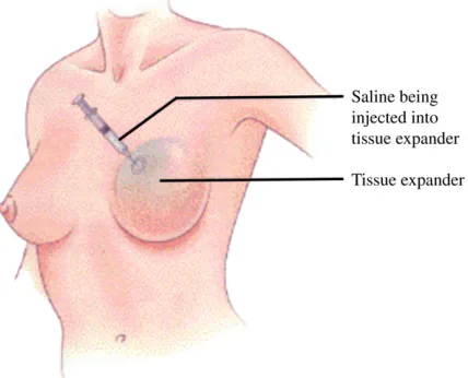
[image:5.432.110.324.400.573.2]The most common technique used for expander/implant placement is placing the expander in a subpectoral pocket (Fig. 1). For immediate tissue expander reconstructions, the goal is to obtain total submuscular coverage that protects the implant from becoming exposed if a minimal amount of skin necrosis occurs. To achieve this coverage, a portion of the serratus mus-cle is raised laterally and is plicated to the pectoralis major musmus-cle. Occa-sionally, the superior aspect of the rectus abdominis muscle must be elevated also. Overall, the pocket size should match the size of the expander (as determined preoperatively based on measurements of the patient’s chest wall). It is critical to not alter or undermine the inframammary fold, because this important landmark is difficult to reconstruct and is crucial to the long-term cosmetic result. If there is concern that the mastectomy flaps have com-promised vascular supply, the expander placement should be delayed. Typically, the expansion is done weekly, and the volume instilled depends on patient comfort and skin quality (eg, tightness, erythema). The expander typically is overexpanded by 25% to improve the skin drape over the im-plant, to allow for the skin recoil that occurs after expansion, and to allow for differences in the profile of the expander versus the implant.
Advantages/disadvantages
Advantages of this technique include the avoidance of donor site morbid-ity and the low overall functional impairments for the patient. Expander and subsequent implant placement often requires less operative time than autog-enous reconstruction, and the recovery period is shorter (typically patients are discharged on postoperative day one). One major disadvantage of this technique is the overall time required. Typically, tissue expansion is started 2 to 3 weeks postoperatively (if the wounds are healed), followed by weekly expansions in the clinic that can last several months before the tissue enve-lope is sufficiently expanded. Once expansion is complete, an additional 2 to 4 months are allowed for tissue equilibrium to occur before the skin enve-lope is ready for the expander to be exchanged for a permanent implant. The exchange of the expander for the implant also requires an additional, albeit relatively short, surgery. In addition, implants lack natural ptosis (or droop) and usually feel unnatural (especially saline implants).
Complications
Complications associated with expander/implant reconstructions can oc-cur in the acute and long-term settings. Acute complications that often re-quire removal of the expander or implant include exposure of the device, infection, malposition, or deflation. In addition, a hematoma or seroma may occur. Long-term complications include capsular contracture (scar tis-sue around the implant causing visible deformity and/or discomfort), visible wrinkling of the implant (especially with saline implants in thin women), and implant deflation (the devices typically last 10–15 years). The rate of re-ported complications with tissue expander/implant reconstructions in the setting of radiation approaches 50%[17–19].
Pedicled transverse rectus myocutaneous flap
Indications/contraindications
directly associated with a higher body mass index[10,22]. With increased ab-dominal fat, the blood supply to the skin and subcutaneous fat becomes un-reliable, leading to partial flap loss or fat necrosis. Other relative contraindications include severe comorbidities (eg, vascular disease, chronic obstructive pulmonary disease) or active use of nicotine.
Technique
[image:7.432.110.321.385.575.2]The blood supply to the TRAM flap is considered bipedicled (double), from the superior and inferior epigastric arteries, with the most direct supply from the inferior epigastric artery. In a pedicled TRAM flap, the inferior epigastric artery is severed, and the rectus muscle and overlying skin and subcutaneous tissue are rotated into the mastectomy defect based on the su-perior epigastric artery and the periumbilical perforators (Fig. 2). When this superior pedicle is used, the blood flows through the choke vessels (vessels that dilate based on need,Fig. 3) between the two pedicles before reaching the skin through perforating arteries. When the blood supply may be more tenuous (as in obese patients and patients who use nicotine), or when a large amount of tissue is needed for the reconstruction, the surgeon can dilate the choke vessels by severing the inferior epigastric artery 2 to 3 weeks before the actual reconstruction. This surgical-delay procedure is thought to im-prove the blood flow through these choke vessels and can be done at the time of the general surgeon’s sentinel lymph node biopsy. Another way to improve the arterial inflow and venous outflow to the pedicled TRAM flap is to take the inferior epigastric vessels and anastomose it
microsurgically to the thoracodorsal or internal mammary vessels, a proce-dure termed ‘‘supercharging.’’
Advantages/disadvantages
Advantages of the TRAM flap include a natural-appearing and -feeling reconstruction that will have an aging process similar to an unreconstructed breast. Disadvantages of this type of reconstruction include a long operative time (4–6 hours), relatively long hospitalization (3–5 days), and long postop-erative recovery. It takes most women 2 to 4 months to return to their pre-operative physical functioning.
Complications
Complications in the acute period include infection (12%), hematoma or seroma of the breast or abdomen (4%), umbilical necrosis, and partial (16%) or total flap loss (1%)[10]. In the long term, potential complications include abdominal wall laxity or hernia (8%)[10].
Microsurgical transverse rectus myocutaneous flap
[image:8.432.139.294.71.272.2]Another option with the TRAM flap is to perform a microsurgical or ‘‘free’’ transfer of the abdominal tissue to the mastectomy defect (Fig. 4). In this procedure, the blood supply is the deep inferior epigastric artery and its venae comitantes, which are severed at their origin. These vessels are anastomosed microsurgically to the thoracodorsal or internal mammary vessels. Relative indications for this procedure are similar to those for the pedicled TRAM flap. Unlike the pedicled TRAM flap, however, this
technique can be used when the superior epigastric artery has been divided (eg, in a patient who has had a previous open cholecystectomy). Some sur-geons also believe that this procedure provides a more robust blood supply than obtained with a pedicled technique. Disadvantages of this technique include a potentially longer operating time and the need for microsurgical ex-pertise. Complications in the acute period include infection (18%), hematoma or seroma of the breast or abdomen (4.5%–9%), umbilical necrosis, and par-tial (15%) or total flap loss (1.5%)[10]. In the long term, potential complica-tions include abdominal wall laxity or hernia (12%) [10]. Although the microsurgical technique often is considered in obese patients, these patients still have a significantly higher risk of certain complications (total flap loss, flap hematoma, flap seroma, mastectomy skin flap necrosis, donor-site infection, donor-site seroma, and hernia) than normal-weight patients[23].
Perforator flaps
Deep inferior epigastric perforator
[image:9.432.63.368.75.264.2]A more recently described technique, the deep inferior epigastric perfora-tor (DIEP) flap, is similar to the free TRAM flap, but the blood supply to this flap is based on only one or two of the perforator arteries off of the deep inferior epigastric artery. This procedure does not require harvest of the rectus abdominis muscle, resulting in less abdominal wall morbidity. Specifically, the incidence of abdominal wall laxity or hernia is less than with techniques that remove abdominal wall fascia with the rectus muscle. A recent study also has reported a shorter hospitalization and faster
recovery because there is less abdominal wall pain with perforator flaps than with traditional TRAM flap reconstructions [24]. This technique still has disadvantages. Significant microsurgical expertise is required, operative times are longer, and the incidence of partial or total flap loss is higher than with traditional TRAM procedures [25]. Earlier studies showed that the DIEP flap had a less robust blood supply, leading to an increased risk for fat necrosis [25]. More recent studies, however, suggest that the rates of fat necrosis or partial flap loss are no higher with perforator flaps than with pedicle TRAM procedures [15]. Nevertheless, the choice between the free TRAM and the DIEP flap should be based on the patient’s weight, the breast volume required, the amount of abdominal fat available, and on the number, caliber, and location of the perforating vessels [26]. For the properly selected patient, some microsurgeons now prefer the DIEP flap to the free TRAM flap.
Superficial inferior epigastric artery flap
Another option that is used less frequently is the superficial inferior epi-gastric artery (SIEA) flap. This flap option was presented by Allen[27] in 1990 but was dismissed at that time because of a high flap failure rate (in three of seven clinical cases). A more recent prospective study comparing the SIEA flap with the DIEP and free TRAM flaps found a 2% flap loss with the SIEA [28]. Advantages of the SIEA flap include minimal donor-site morbidity because the rectus abdominis fascia and muscle are not vio-lated and less postoperative pain[28].
Gluteal artery perforator flap
For patients who do not have sufficient abdominal tissue for breast re-construction but still prefer the use of autologous tissue, an option is the use of the buttock as donor tissue. This donor site can also be used for uni-lateral or biuni-lateral breast reconstructions. There are two options for blood supply to the flap. When the superior gluteal artery is used, the flap is called a ‘‘superior gluteal artery perforator’’ (SGAP) flap, and the upper buttock tissue is used. The scar lies in the upper buttock region and is hidden easily with underwear. When the inferior gluteal artery is used, the flap is called an ‘‘inferior gluteal artery perforator’’ (IGAP) flap, and the lower buttock tis-sue is used. The scar lies within the lower buttock crease.
Latissimus dorsi flap
Indications/contraindications
Another reliable workhorse for breast reconstruction is the latissimus dorsi muscle or myocutaneous (muscle and skin) flap. Indications for this flap include previous implant or TRAM flap failure, need to reconstruct a partial mastectomy or lumpectomy defect, abdominal obesity, or extreme thinness resulting in inadequate infraumbilical soft tissue. Contraindications to this technique include prior surgery that may have interrupted the blood supply (eg, posterior thoracotomy), the inability of the patient to be posi-tioned on her side, severe comorbidities, and the patient not desiring implant placement.
Technique
The dominant blood supply to this flap is the thoracodorsal artery (off the subscapular artery), with segmental blood supply from the posterior in-tercostals and lumbar vessels (Fig. 5). If the thoracodorsal artery is damaged more proximally during the mastectomy, this flap also may survive on ret-rograde blood flow from the serratus branch off the thoracodorsal artery. This flap can be transferred either on its pedicle or as a free tissue microsur-gical transfer.
Advantages/disadvantages
[image:11.432.68.359.431.574.2]The advantages of this technique include the ability to provide single-stage implant reconstruction (the latissimus muscle is excellent soft tissue coverage of the implant) and its reliability. Most plastic surgeons, however, believe that a better aesthetic result is obtained by first using a tissue ex-pander under the flap and then replacing the exex-pander with a permanent im-plant. Disadvantages of this flap include a significant donor-site scar
(especially if skin is also harvested) and the frequent need for implant and/or tissue expander placement because of insufficient tissue.
Complications
Most complications related to this procedure are related either to the im-plant or the donor site. Imim-plants have a risk of rupture, displacement, con-tracture, or infection. The donor site is at risk of hematoma, seroma, infection, and hypertrophic scarring. There also is a risk of flap necrosis at the recipient site.
Treatment of the contralateral breast
Once the mastectomy site has been reconstructed, often the next chal-lenge for the plastic surgeon is to create symmetry with the contralateral breast. Ideally, the contralateral breast should be evaluated preoperatively (at the same time as consultation for reconstruction), and discussion with the patient should elicit her preferences and explain her realistic options. Surgery to achieve contralateral symmetry can be performed at the time of the initial reconstruction or later. In addition, surgery to create contralateral symmetry can be considered in patients who undergo breast-conservation treatment. The 1998 WHCRA mandates that alteration of the contralateral breast in cases of breast cancer reconstruction be covered by insurance.
Options for symmetry procedures include breast reduction, breast augmentation, mastopexy (breast lift), or a combination of the procedures
[30]. For example, in a woman who has very large breasts and undergoes a mastectomy with reconstruction, a contralateral breast reduction would improve symmetry and patient comfort. A woman who has small breasts may require an augmentation on the contralateral side for symmetry. Al-though autogenous reconstruction often provides a better overall outcome when contralateral surgery is not performed, breast reconstruction rarely produces a breast that is symmetrical to the contralateral breast.
Nipple and areolar reconstruction
include the contralateral areola (if large), remnant excess abdominal tissue at the incision after a TRAM is performed, the medial thigh, or the mastec-tomy scar. A favorable aesthetic outcome also can be achieved with medical tattooing alone.
The type of papule reconstruction often is based on surgeon preference and the patient’s size preference. Typically, local tissue is raised to create flaps for papule projection. Nipple projection decreases postoperatively, re-quiring a 50% overcorrection at the time of surgery. Another option in-cludes a nipple-sharing technique that uses a papule graft from the contralateral nipple. There is, however, a risk of complications at the site of the contralateral nipple, including scarring and loss of nipple sensation, and thus use of local skin flaps is often the procedure of choice.
Nonsurgical options
Some women may choose not to have breast reconstruction or are poor surgical candidates for reconstruction. For these women, a breast prosthesis is an option. The advantages of not wearing a breast prosthesis include sim-plicity, comfort, and convenience[32]. The disadvantages include a feeling of imbalance and difficulty wearing certain clothes[32]. Prostheses can be purchased at surgical supply stores, pharmacies, custom lingerie shops, or through a private home shopping service[32]. Some stores have trained fit-ters who can help the woman find the appropriate prosthesis that fits her chest and matches the contralateral breast. Specialty clothing is available with pockets to hold the prosthesis in place, and some prostheses come with adhesive Velcro patches to keep the prosthesis in place on the chest. Most insurances cover a new prosthesis every 2 years and two brassieres with a prosthesis pocket each year[31].
References
[1] Wilkins E, Cederna P, Lowery J, et al. Prospective analysis of psychosocial outcomes in breast reconstruction: one year postoperative results from the Michigan Breast Reconstruc-tion Outcome Study. Plast Reconstr Surg 2000;106(6):1014–125.
[2] Cronin TD, Gerow FJ. Augmentation mammaplasty: a new ‘‘natural feel’’ prosthesis. Ex-cerpta Medica International Congress Series 1963;66:41.
[3] Food and Drug Administration. Available at:http://www.fda.gov/bbs/topics/NEWS/2006/ NEW01512.htm. Accessed February 20, 2006.
[4] Hollos P. Breast augmentation with autologous tissue: an alternative to implants. Plast Re-constr Surg 1995;96:381–4.
[5] Radovan C. Breast reconstruction after mastectomy using the temporary expander. Plast Reconstr Surg 1982;69(2):195–208.
[6] Hartrampf CR, Scheflan M, Black PW. Breast reconstruction with a transverse abdominal island flap. Plast Reconstr Surg 1982;69(2):216–25.
[8] Allen RJ, Tucker C Jr. Superior gluteal artery perforator free flap for breast reconstruction. Plast Reconstr Surg 1995;95:1207–12.
[9] US Department of Labor. Your rights after a mastectomy.Women’s Health & Cancer
Rights Act of 1998. Available at:http://www.dol.gov/ebsa/publications/whcra. Accessed September 14, 2006.
[10] Alderman AK, Wilkins E, Kim M, et al. Complications in post-mastectomy breast recon-struction: two year results of the Michigan breast reconstruction outcome study. Plast Re-constr Surg 2002;109:2265–74.
[11] Wilson CR, Brown IM, Weiller-Mithoff E, et al. Immediate breast reconstruction does not lead to a delay in the delivery of adjuvant chemotherapy. Eur J Surg Oncol 2004;30(6):624–7. [12] Newman LA, Kuerer HM, Hunt KK, et al. Presentation, treatment, and outcome of local recurrence after skin-sparing mastectomy and immediate breast reconstruction. Ann Surg Oncol 1998;5(7):620–6.
[13] Al-Ghazal SK, Sully L, Fallowfield L, et al. The psychological impact of immediate rather than delayed breast reconstruction. Eur J Surg Oncol 2000;26:17–9.
[14] Slavin SA, Schnitt SJ, Duda RB, et al. Skin-sparing mastectomy and immediate reconstruc-tion: oncologic risks and aesthetic results in patients with early-stage breast cancer. Plast Re-constr Surg 1998;102(1):49–62.
[15] Ramon Y, Ullman Y, Moscona R, et al. Aesthetic results and patient satisfaction with im-mediate breast reconstruction using tissue expansion: a follow-up study. Plast Reconstr Surg 1997;99(3):686–91.
[16] Khoo A, Kroll SS, Reece GP, et al. A comparison of resource costs of immediate and delayed breast reconstruction. Plast Reconstr Surg 1998;101(4):964–8.
[17] Chawla A, Kachnic L, Taghian A, et al. Radiotherapy and breast reconstruction: complica-tions and cosmesis with TRAM versus tissue expander/implant. Int J Radiat Oncol Biol Phys 2002;54(2):520–6.
[18] Spear SL, Onyewu C. Staged breast reconstruction with saline-filled implants in the irradi-ated breast: recent trends and therapeutic implications. Plast Reconstr Surg 2000;105(3): 930–42.
[19] Tallet AV, Salem N, Moutardier V, et al. Radiotherapy and immediate two-stage breast re-construction with a tissue expander and implant: complications and esthetic results. Int J Ra-diat Oncol Biol Phys 2003;57(1):136–42.
[20] Alderman AK, Wilkins E, Lowery J, et al. Determinants of patient satisfaction in post-mas-tectomy breast reconstruction. Plast Reconstr Surg 2000;106:769–76.
[21] Cederna PS, Yates WR, Chang P, et al. Postmastectomy reconstruction: comparative anal-ysis of the psychosocial, functional, and cosmetic effects of transverse rectus abdominis mus-culocutaneous versus breast implant reconstruction. Ann Plast Surg 1995;35:458–68. [22] Kroll SS, Netscher DT. Complications of TRAM flap breast reconstruction in obese
pa-tients. Plast Reconstr Surg 1989;84:886–92.
[23] Chang DW, Wang B, Robb GL, et al. Effect of obesity on flap and donor-site complications in free transverse rectus abdominis myocutaneous flap breast reconstruction. Plast Reconstr Surg 2000;105(5):1640–8.
[24] Garvey PB, Buchel EW, Pockaj BA, et al. DIEP and pedicled TRAM flaps: a comparison of outcomes. Plast Reconstr Surg 2006;117(6):1711–9.
[25] Kroll SS. Fat necrosis in free transverse rectus abdominis myocutaneous and deep inferior epigastric perforator flaps. Plast Reconstr Surg 2000;106(3):576–83.
[26] Nahabedian MY, Momen B, Galdino G, et al. Breast reconstruction with the free TRAM or DIEP flap: patient selection, choice of flap, and outcome. Plast Reconstr Surg 2002;110(2): 466–75.
[28] Chevray PM. Breast reconstruction with superficial inferior epigastric artery flaps: a prospec-tive comparison with TRAM and DIEP flaps. Plast Reconstr Surg 2004;114(5):1077–83. [29] Allen RJ, Levine JL, Granzow JW. The in-the-crease inferior gluteal artery perforator flap
for breast reconstruction. Plast Reconstr Surg 2006;118(2):333–9.
[30] Kroll SS. Options for the contralateral breast in breast reconstruction. In: Spear SL, editor. Surgery of the breast: principles and art. Philadelphia: Lippincott-Raven; 1998.
[31] Bhatty MA, Berry RB. Nipple-areola reconstruction by tattooing and nipple sharing. Br J Plast Surg 1997;50(5):331–4.