The Decellularized Vascular Allograft as an Experimental
Platform for Developing a Biocompatible Small-Diameter Graft
Conduit in a Rat Surgical Model
Seong-Jun Hwang,
1Seong Who Kim,
2Suk Jung Choo,
3Byoung Wook Lee,
2,3I-rang Im,
2Hye Joo Yun,
3Sang Kwon Lee,
5Hyun Song,
4Won Chul Cho,
3and Jae Won Lee
3Departments of 1Anatomy and Cell Biology and 2Biochemistry and Molecular Biology, and 3Thoracic and Cardiovascular Surgery, Asan Medical Center, University of Ulsan College of Medicine, Seoul; 4Department of Thoracic and Cardiovascular Surgery, Seoul St. Maryʼs Hospital,
The Catholic University of Korea, Seoul; 5Department of Thoracic and Cardiovascular Surgery, Yangsan Hospital, Pusan National University School of Medicine, Busan, Korea.
Received: March 23, 2010 Revised: June 28, 2010 Accepted: July 2, 2010
Corresponding author: Dr. Jae Won Lee, Department of Thoracic and Cardiovascular Surgery, Asan Medical Center, University of Ulsan College of Medicine,
86 Asanbyeongwon-gil, Songpa-gu, Seoul 138-736, Korea.
Tel: 82-2-3010-3584, Fax: 82-2-3010-6966 E-mail: [email protected]
∙ The authors have no financial conflicts of interest.
© Copyright:
Yonsei University College of Medicine 2011
This is an Open Access article distributed under the terms of the Creative Commons Attribution Non-Commercial License (http://creativecommons.org/ licenses/by-nc/3.0) which permits unrestricted non-commercial use, distribution, and reproduction in any medium, provided the original work is properly cited.
Purpose: The present study was aimed to assess the feasibility of using decellular-ized aortic allograft in a rat small animal surgical model for conducting small diam-eter vascular tissue engineering research. Materials and Methods:Decellularized aortic allografts were infra-renally implanted in 12 Sprague-Dawley (SD) adult rats. The conduits were harvested at 2 (n = 6) and 8 weeks (n = 6), and assessed by he-matoxylin and eosin (H&E), van Gieson, Masson Trichrome staining, and immu-nohistochemistry for von Willebrand factor, CD 31+, and actin. Results:Consistent, predictable, and reproducible results were produced by means of a standardized sur-gical procedure. All animals survived without major complications. Inflammatory immune reaction was minimal, and there was no evidence of aneurysmal degenera-tion or rupture of the decellularized vascular implants. However, the aortic wall ap-peared thinner and the elastic fibers in the medial layer showed decreased undula-tion compared to the normal aorta. There was also minimal cellular repopulaundula-tion of the vascular media. The remodeling appeared progressive from 2 to 8 weeks with increased intimal thickening and accumulation of both collagen and cells staining for actin. Although the endothelial like cells appeared largely confluent at 8 weeks, they were not as concentrated in appearance as in the normal aorta. Conclusion:
The results showed the present rat animal model using decellularized vascular al-lograft implants to be a potentially durable and effective experimental platform for conducting further research on small diameter vascular tissue engineering.
Key Words: Tissue engineering, vessel, rat, animal model
INTRODUCTION
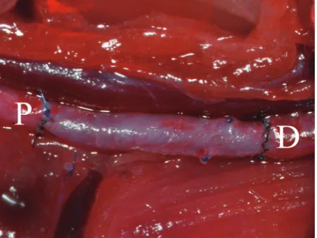
ration. IVC heparin (10 IU/100 g body weight) injection was performed after which the aorta was clamped just dis-tally to the renal artery and proximally to the iliac bifurca-tion. The mid portion of the isolated aorta was transected. The four opposing edges of the decellularized graft (two proximal and two distal) were sutured to the corresponding edges of the native vessel with #8-0 polypropylene sutures. Mosquito clamps were applied to each suture arm for trac-tion. The anterior margins of the proximal and distal native aorta were anastomosed with the graft using multiple inter-rupted Nylon #9-0 sutures. The aorta was then rotated 180 degrees by reversing the traction sutures from left to right which resulted in exposure of the unsutured posterior mar-gins. Anastomosis was performed in an identical manner with Nylon #9-0 sutures (Fig. 1). No additional anticoagu-lation was performed postoperatively. Ketoprofen 5 mg/kg sc daily and Amoxicillin 150 mg/kg sc x twice daily were administered for 3 days postoperatively for pain and pro-phylaxis against infection. All surgeries were performed in an environment that was largely specific pathogen-free (SPF). Animals were managed according to the “Revised Guide for the Care and Use of Laboratory Animals” pre-pared by the Institute of Laboratory Animal Resources and published by the National Institutes of Health, revised and published in 1996.8
Tissue decellularization
The rat infrarenal abdominal aorta was harvested for decellu-larization as described above. The harvested specimens, ap-proximately 1.5 cm each in length, were thoroughly washed two or three times in Hank’s balanced salt solution (HBSS) artery bypass grafting (CABG) but also for peripheral
vas-cular surgery.1-3 However, autologous SVGs are often limit-ed in availability. Various prosthetic small-diameter vascu-lar conduits have been used alternatively, but the outcomes have thus far been disappointing, including those associated with arteriovenous fistula (AVF) access creation for hemo-dialysis, which comprise the bulk of the demand for small-diameter vascular conduits.4-7
Vascular remodeling-induced premature graft failure is a major limitation of small-diameter vascular conduits. Decel-lularization is potentially an excellent tissue processing meth-od for increasing the availability of non autologous biologi-cal small diameter vascular conduits. Removal of antigenic determinants may enhance the utility of vascular allograft conduits such as cadaveric radial arteries or the saphenous vein. However, the propensity for structural deterioration and adverse vascular remodeling remain limitations which must be resolved through further research. Studies using large ani-mal models inherently pose difficulties related to higher cost and logistical problems. Therefore, the present study was conducted to investigate the feasibility of developing a small animal surgical model using the rat as a platform for con-ducting small diameter vascular tissue engineering research.
MATERIALS AND METHODS
Animal model and surgical procedure
A total of 24 Sprague-Dawley (SD) female rats, 12 donors and 12 recipients, each weighing 250 to 300 g, were used. Each animal was anesthetized with intra-peritoneal injec-tion of 0.25 mL of a 2 : 1 : 2 (v/v/v) mixture of Zoletil 50 (40 mg/kg, Virbac Laboratories, Carros, France), Xylazine (10 mg/kg Bayer, Leverkusen, Germany) and 0.9% (w/v) physiologic saline. For donor preparation, the abdominal aorta was approached via a midline laparotomy incision, af-ter which the aorta side branches were ligated using poly-propylene #8-0 monofilament sutures. The aorta was thus mobilized from a point just distal to the left renal artery take-off to the iliac bifurcation. To prevent clotting, heparin (10 IU/100 g body weight) was injected into the inferior vena cava (IVC). The aorta was then resected just distally to the renal artery and proximally to the iliac bifurcation. The har-vested conduit was then flushed with normal saline and transferred for decellularization.
For recipient preparation, anesthesia and aortic mobiliza-tion were performed in a manner identical to donor
[image:2.595.300.526.519.689.2]Willebrand factor was performed using goat anti-PECAM-1 antibody (1 : 100, catalog no. sc-1506; Santa Cruz Biotech-nology Inc.) and rabbit anti-von Willebrand factor. Antibody reaction was assessed using a rabbit anti-Goat IgG conjugat-ed Cy3 (catalog no. 305-165-006, Jackson immune research laboratories inc., West Grove, Pennsylvania, USA) and goat anti-rabbit IgG conjugated alexa 488(catalog no. A-11034, Invitrogen inc., Eugene, OR, USA). Samples were subse-quently counterstained with DAPI (catalog no. D21490, In-vitrogen Inc., Eugene, OR, USA).
RESULTS
Development and efficacy of animal model
A pre-study pilot trial was conducted to develop the current rat surgical model. High operative and late mortalities were encountered during this period due to various forms of tech-nical failures resulting from inexperience. Surgical bleeding accounted for 15 early deaths and suture occlusion of the ureter causing a large intra-abdominal renal cyst resulted in 1 late death. Accidental suturing of the posterior and anterior walls accounted for graft failure in another 6 animals. How-ever, after completion of this developmental phase, a stan-dardized surgical protocol which resulted in the predictable and reliable outcomes of the present study was established. Accordingly, the data for the present study were collected from animals operated on after this period, and therefore, did not include the animals from the pre-study trial. In the present series, there were no operative and late deaths, and surgery was conducted consecutively in which all animals survived the experiment.
Assessment of decellularization efficacy
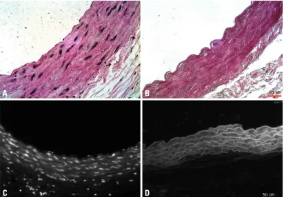
The present decellularization protocol was considered to be effective based on the absence of cellular or nucleic compo-nents in both the luminal surface and the underlying matrix scaffolding under high powered light microscopic examina-tion (400× magnificaexamina-tion) of the H&E-stained specimens (Fig. 2). The absence of DNA content was also confirmed through DAPI staining (Fig. 2C and D)
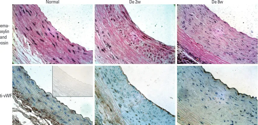
Vascular remodeling
Intimal hyperplasia, which appeared as early as 2 weeks post-implantation, was characterized by the accumulation of collagen and alpha actin-staining smooth muscle cells which appeared to be progressive up to 8 weeks. As a result, the in-and incubated overnight (15-18 hours) at 37ºC in 10 mL of
physiologic saline containing 0.25% (v/v) Triton X-100 (Sigma, St. Louis, MO, USA) and 0.25% (w/v) sodium de-oxycholate in a 10 cm-diameter Petri dish. The aortic seg-ments were immersed in pre-chilled HBSS and shaken vig-orously, with solution replacement every 2 hours. To ensure detergent elimination, this procedure was repeated three times. Each sample was incubated for 15-18 hours at 37ºC in 10 mL of PBS containing 150 IU/mL DNase I, 100 µg/ mL RNase A, and 50 mM MgCl2. Samples were again shaken in pre-chilled HBSS, with solution replacement ev-ery 2 hours, as above. Each decellularized aorta was stored at 4ºC in physiologic saline until use. Decellularization effi-cacy was evaluated by high-power light microscopic obser-vation of hematoxylin and eosin (H&E)-stained specimens.
Preparation for microscopic study
The implanted grafts were harvested at 2 (n = 6) and 8 (n = 6) weeks. To avoid endothelial cell sloughing by mechani-cal trauma, the graft specimens were fixed in 4% (v/v) buff-ered para-formaldehyde with gentle continuous perfusion at 60 mmHg prior to extraction. Each of the fixed infrarenal aortic specimens was removed en-bloc with a generous at-tachment of muscle and surrounding tissue. Stretching or otherwise traumatic instrumentation of the aorta was avoid-ed as much as possible. Excess muscle and surrounding tis-sue were gently trimmed using a surgical microscope. Prior to paraffin-embedding, specimens were post-fixed in for-malin for at least 3 hours. Basic light microscopic staining consisted of H&E, Elastica von Gieson (EVG), and Mas-son Trichrome (MT).
Immunohistochemistry
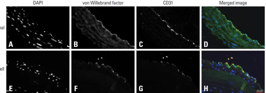
the von Willebrand factor staining endothelial like cells to stain for CD 31+ as well (Fig. 5).
DISCUSSION
The development of a biologically compatible small-diam-eter vascular conduit may have far-reaching clinical impli-cations, especially in situations where autologous conduits are either unusable or unavailable. In contrast to the central hemodynamic flow conditions of “large-diameter” arterial graft conduits, the decreased flow velocity, shear stress, and higher vascular resistance of the peripheral vascular system contribute to the poorer long term patency of small-diame-ter vascular conduits.9 Hemodynamic flow conditions have been shown to influence vascular remodeling and graft pa-tency, but properties specifically related to the graft material have also been reported to affect graft thrombosis, intimal hyperplasia, aneurysm formation, infection, and progres-sion of atherosclerosis.10 Therefore, improvement of the bio-compatibility of vascular scaffolding remains an impor-tant research objective.
Expanded polytetrafluoroethylene (ePTFE) was widely tima at 8 weeks was noticeably thicker with greater
intersti-tial deposition of collagen in the vascular media. Elastic fi-bers in the normal vascular media appeared to have an undulating pattern while those in the decellularized vascular implants appeared more straightened with an overall appear-ance of being more compressed than normal. With regards to cellularity, the normal aortic wall media appeared highly cellular, whereas the decellularized media appeared sparsely cellular with little improvement over time (Fig. 3).
Immunohistochemical staining
[image:4.595.85.498.67.353.2]In the normal aorta, a confluent layer of flattened endothelial like cells staining for von Willebrand factor were observed on the luminal surface. At 2 weeks after implantation of the decellularized vascular implant, however, the surface was almost devoid of endothelial like cells. At 8 weeks, the en-dothelial like cells were more prominent, but showed fewer cells than in the normal aortic surface. Similarly, the von Willebrand factor staining was more prominent at 8 weeks than at 2 weeks, but with noticeably weaker activity than in the normal aorta (Fig. 4). To further assess the characteris-tics of the endothelial like cells, CD 31+ was stained in ad-dition to that for von Willebrand factor. The results showed
Fig. 2. Histologic assessment of the decellularized scaffold. (A) Normal aortic wall with a confluent endothelial cell lining and abundant cellularity in the native vascular wall matrix. (B) Decellularized vascular scaffold with absence of endothelial cell lining and cellularity or nuclear staining within vascular matrix. (C) DAPI staining against DNA content in the aorta with dense cellularity. (D) Post decellularized aorta showing only the supporting matrix scaffold with complete absence of DNA content.
A
C
B
come these limitations included coating with heparin or recombinant human thrombomodulin. Nevertheless, the outcomes with these methods have not yet been substantiat-ed through long term studies.12,13
used as an artificial substitute for autologous saphenous vein grafts, however, the results have generally been disap-pointing due to the tendency for accelerated neointimal hy-perplasia and premature graft failure.3-5,11 Efforts to
over-Fig. 3. Von Gieson’s stain (black, far left column) shows absence of elastin fibers within the intimal proliferative layer. Collagen accumu-lation (middle column) and actin-positive smooth muscle cells (far right column) are present at 2 weeks. Both the thickness of collage-nous matrix layer and cells staining positively for actin are increased at 8 weeks. Insert image shows staining without primary antibody.
Van gieson and verhoeff iron hematoxylin stain Masson’s trichrome stain α-smooth muscle actin
Normal
De 2w
[image:5.595.75.509.136.446.2]De 8w
Fig. 4. Luminal surfaces of normal and decellularized aortic implant at 2 and 8 weeks. Greater numbers of endothelial cells are seen at 8 weeks, compared to 2 weeks, but in fewer concentrations than the normal aorta (upper row). Factor VIII staining shows a similar pattern correlating with the degree of endothelialization (lower row). Insert shows image of staining without primary antibody.
Normal De 2w De 8w
Hema-toxylin and eosin
[image:5.595.80.509.493.701.2]conditions. The relatively larger size and the anticipated greater ease of handling for surgery further support the use-fulness of the present model. The ability to accommodate a longer conduit than would be possible in the mouse, the amenability to procedural standardization, the ability to generate consistent, predictable, and reproducible operative outcomes, and the simplification of the anesthesia protocol which obviates ventilator requirements, all endorse the rat as being the more favorable small animal model. The dem-onstration of the ability of human mesenchymal stem cells (MSCs) to differentiate, survive, and function in a xenoge-neic non-immune compromised rat environment21 suggest further possibilities for using human-origin mesenchymal stem cells in a rat vascular implant environment.
For immunohistochemical assessment, several antibodies including human antibodies were (vWF and anti-SMA) used. These anti-human antibodies, which have gen-erally been adopted in many other animal studies,22,23 led us to follow their experimental protocols. In addition, accord-ing to the manufacturer’s protocols, the antibodies have been verified to cross-react against several mammalian spe-cies such as mouse, rat and chicken, and therefore, have been recommended for use with animal tissues.
From a technical standpoint, the rat aorta, despite its small size, does not represent the hemodynamic environment of the peripheral vascular system. However, as a platform for conducting further research to enhance the utility of decel-lularized small diameter vascular conduits, the present model was suitable for this purpose. Thinning and dilata-tion with eventual development of aneurysmal changes are prominent degenerative findings of small diameter vascular conduits. Therefore, we speculated the more densely packed Conceptually, decellularization may minimize the
inci-dence of graft failures and complications related to immune rejection.14 This contention is supported by the decreased mononuclear cell infiltrates observed in decellularized vein allograft implants at 2 weeks,15 contrasting with the maxi-mal inflammatory reaction occurring in fresh venous al-lografts harvested at similar time points.16 The suitability of decellularized vein allograft implants as scaffolding for vas-cular tissue engineering was suggested earlier by Martin et al., showing repopulation of the medial matrix by recipient cells at 8 weeks post implantation with excellent retention of strength and structural integrity.15 Accordingly, decellu-larization was considered to be a potentially good method for increasing the availability and utility of allograft vein grafts, however, it has thus far shown dismal results sec-ondary to graft rejection and propensity for degenerative destruction.17-19 Considering the importance of a functional endothelial-cell layer in preventing thrombosis and adverse vascular remodeling, further efforts to enhance re-endothe-lialization and repopulation of the decellularized matrix ap-pear warranted.
Foremost is the need to develop a surgical model condu-cive to efficient and effective data generation and compe-tence assessment. An animal model using a severely im-mune-compromised mouse model was previously described for such a purpose.20 However, the possibility of graft throm-bosis and other unforeseen factors that may have been oth-erwise independently affected by the influence of innate platelet dysfunction present in the immune-compromised mouse could not be ruled out. Accordingly, the present mod-el using rats with no such deficiencies may be viewed as more accurately representing the normal in vivo biological
A
E
B
F
C
G
D
[image:6.595.75.498.67.216.2]H
Fig. 5. DAPI study at 8 weeks showed evenly distributed cellular nuclei in the media of the normal aorta (A and E). In the De-cell specimen it was evident mostly in the neo intima layer with sparse cellularity in the media layer. Both the normal and De-cell specimen shows positive staining for von WIllebrandt factor (B and F) and CD 31+ (C and G). These signals were clearly co-localized on the innermost confluent cell layer in both groups albeit more strongly present in the normal aorta compared with the decellularized graft. Merging of the von Willebrand factor and CD 31 stains enhanced visualization of the endothelial cell markers (D and H).
DAPI
Normal
De-cell 8w
CD31
mals available. American Physiological Society. Physiologist 1996;39:199, 208-11.
9. Isenberg BC, Williams C, Tranquillo RT. Small-diameter artificial arteries engineered in vitro. Circ Res 2006;98:25-35.
10. Conte MS. The ideal small arterial substitute: a search for the Holy Grail? FASEB J 1998;12:43-5.
11. Quiñones-Baldrich WJ, Prego AA, Ucelay-Gomez R, Freischlag JA, Ahn SS, Baker JD, et al. Long-term results of infrainguinal re-vascularization with polytetrafluoroethylene: a ten-year experi-ence. J Vasc Surg 1992;16:209-17.
12. Ritter EF, Fata MM, Rudner AM, Klitzman B. Heparin bonding increases patency of long microvascular prostheses. Plast Reconstr Surg 1998;101:142-6.
13. Wong G, Li JM, Hendricks G, Eslami MH, Rohrer MJ, Cutler BS. Inhibition of experimental neointimal hyperplasia by recombinant human thrombomodulin coated ePTFE stent grafts. J Vasc Surg 2008;47:608-15.
14. Schaner PJ, Martin ND, Tulenko TN, Shapiro IM, Tarola NA, Leichter RF, et al. Decellularized vein as a potential scaffold for vascular tissue engineering. J Vasc Surg 2004;40:146-53. 15. Martin ND, Schaner PJ, Tulenko TN, Shapiro IM, Dimatteo CA,
Williams TK, et al. In vivo behavior of decellularized vein al-lograft. J Surg Res 2005;129:17-23.
16. Wagner E, Roy R, Marois Y, Douville Y, Guidoin R. Fresh venous allografts in peripheral arterial reconstruction in dogs. Effects of histocompatibility and of short-term immunosuppression with cy-closporine A and mycophenolate mofetil. J Thorac Cardiovasc Surg 1995;110:1732-44.
17. Allaire E, Guettier C, Bruneval P, Plissonnier D, Michel JB. Cell-free arterial grafts: morphologic characteristics of aortic isografts, allografts, and xenografts in rats. J Vasc Surg 1994;19:446-56. 18. Allaire E, Bruneval P, Mandet C, Becquemin JP, Michel JB. The
immunogenicity of the extracellular matrix in arterial xenografts. Surgery 1997;122:73-81.
19. Iaffaldano RA, Lewis BE, Johnson SA, Piffare R, McKiernan TL. Patency of cryopreserved saphenous vein grafts as conduits for coronary artery bypass surgery. Chest 1995;108:725-9.
20. Lopez-Soler RI, Brennan MP, Goyal A, Wang Y, Fong P, Tellides G, et al. Development of a mouse model for evaluation of small diameter vascular grafts. J Surg Res 2007;139:1-6.
21. Atoui R, Asenjo JF, Duong M, Chen G, Chiu RC, Shum-Tim D. Marrow stromal cells as universal donor cells for myocardial re-generative therapy: their unique immune tolerance. Ann Thorac Surg 2008;85:571-9.
22. Traktuev DO, Tsokolaeva ZI, Shevelev AA, Talitskiy KA, Stepa-nova VV, Johnstone BH, et al. Urokinase gene transfer augments angiogenesis in ischemic skeletal and myocardial muscle. Mol Ther 2007;15:1939-46.
23. Pascual G, Martínez S, Rodríguez M, Serrano N, Bellón JM, Buján J. Patency and structural changes in cryopreserved arterial grafts used as vessel substitutes in the rat. J Surg Res 2005;124:297-304. 24. Lesauskaite V, Tanganelli P, Sassi C, Neri E, Diciolla F,
Ivanovi-ene L, et al. Smooth muscle cells of the media in the dilatative pa-thology of ascending thoracic aorta: morphology, immunoreactiv-ity for osteopontin, matrix metalloproteinases, and their inhibitors. Hum Pathol 2001;32:1003-11.
25. Ikonomidis JS, Gibson WC, Gardner J, Sweterlitsch S, Thompson RP, Mukherjee R, et al. A murine model of thoracic aortic aneu-rysms. J Surg Res 2003;115:157-63.
appearance of the elastic fibers in the aortic implants to in-dicate early degenerative changes which may suggest chang-es reprchang-esenting aneurysmal progrchang-ession. Although findings which are commonly present in established aneurysmal transformation such as disruption and fragmentation of the elastin fibers were not observed, further long term studies are nevertheless warranted to resolve the arguments related to this issue.24,25
In conclusion, the present rat small animal model was found to be an effective and efficient animal model for con-ducting vascular tissue engineering research, aimed at en-hancing the availability and utility of decellularized allograft small diameter vascular conduits.
ACKNOWLEDGEMENTS
This work was supported in part by a grant from the Asan Institute for Life Sciences (#2006-424) and the National Research-Foundation (NRF) grant funded by the Korean government A (R13-2008-023-01002).
REFERENCES
1. Fitzgibbon GM, Kafka HP, Leach AJ, Keon WJ, Hooper GD, Burton JR. Coronary bypass graft fate and patient outcome: angio-graphic follow-up of 5,065 grafts related to survival and reopera-tion in 1,388 patients during 25 years. J Am Coll Cardiol 1996;28: 616-26.
2. Taylor LM Jr, Edwards JM, Porter JM. Present status of reversed vein bypass grafting: five-year results of a modern series. J Vasc Surg 1990;11:193-205.
3. Veith FJ, Gupta SK, Ascer E, White-Flores S, Samson RH, Scher LA, et al. Six-year prospective multicenter randomized compari-son of autologous saphenous vein and expanded polytetrafluoro-ethylene grafts in infrainguinal arterial reconstructions. J Vasc Surg 1986;3:104-14.
4. Klinkert P, Post PN, Breslau PJ, van Bockel JH. Saphenous vein versus PTFE for above-knee femoropopliteal bypass. A review of the literature. Eur J Vasc Endovasc Surg 2004;27:357-62. 5. Kashyap VS, Ahn SS, Quinones-Baldrich WJ, Choi BU, Dorey F,
Reil TD, et al. Infrapopliteal-lower extremity revascularization with prosthetic conduit: a 20-year experience. Vasc Endovascular Surg 2002;36:255-62.
6. Coburn MC, Carney WI Jr. Comparison of basilic vein and polytet-rafluoroethylene for brachial arteriovenous fistula. J Vasc Surg 1994;20:896-902.
7. Kherlakian GM, Roedersheimer LR, Arbaugh JJ, Newmark KJ, King LR. Comparison of autogenous fistula versus expanded polytetrafluoroethylene graft fistula for angioaccess in hemodialy-sis. Am J Surg 1986;152:238-43.