STRUCTURAL STUDIES OF THE E.COLI METHIONINE ABC TRANSPORTER AND ITS COGNATE BINDING PROTEIN
Thesis By
Neena Sujata Kadaba
In Partial Fulfillment of the Requirements for the degree of
Doctor of Philosophy
CALIFORNIA INSTITUTE OF TECHNOLOGY
Pasadena, California
2008
© 2008 Neena Sujata Kadaba
Acknowledgements.
I would first like to thank Doug Rees and members of the Rees lab who were extraordinarily helpful throughout the past few years. In particular, Heather Pinkett and Zhenfeng Liu were an amazing resource of information at every step, and I could not have accomplished much without their help. Additionally, Allen Lee, Mika Walton and Jeff Lai were a tremendous source of technical help, and I appreciate their patience in answering questions.
Many friends have also been a source of support over the past few years. In particular, I would like to extend a tremendous amount of thanks to Daniel Feldman for his encouragement and friendship over the past several years—it has made this process so much easier.
Abstract.
ATP binding cassette (ABC) transporters use ATP hydrolysis to facilitate the transfer of diverse substrates across the membrane. Members of the methionine uptake transporter family, thought to be of considerable biological interest, have not been structurally characterized thus far. The crystal structure of the methionine importer MetNI from Escherichia coli has been solved to 3.7 Å resolution. The inward-facing conformation of this transporter adopts a more extreme arrangement than seen
previously. While the permease domain consists of just five transmembrane helices per monomer, the ATP-binding cassette domain possesses a C-terminal domain in addition to the conserved architecture shared amongst this family. Analysis of the C-terminal extension has revealed a regulatory domain found in other proteins involved in amino acid metabolism, and further classifies this protein as part of the ACT family.
Methionine binding in this region suggests a novel mechanism for regulation of transport that possibly stabilizes the inactive conformation of this family of
Table of Contents.
Acknowledgements... iii
Abstract. ...iv
Table of Contents. ...v
List of Figures and Tables. ...vi
Abbreviations. ...ix
Chapter One. Introduction...1
1.1 Overview...1
1.2 ABC Transporter Structures ...4
1.3 The Catalytic Cycle of ABC Transporters ... 16
1.4 Regulatory Mechanisms: The ACT Domain ... 20
Chapter Two. MetNI. ... 24
2.1 Introduction... 24
2.2 Structure of the MetNI Transporter... 29
Appendix One. Methods and Data Relevant to Chapter Two. ... 49
A1.1. Materials and Methods... 49
Chapter Three. MetQ... 62
3.1 Introduction... 62
3.2 Methionine Binding Proteins ... 67
3.3 Structural Analysis of MetQ ... 70
Appendix Two. Methods and Data Relevant to Chapter Three. ... 79
A2.1. Materials and Methods... 79
Chapter Four. Conclusions... 85
References... 87
About the Author. ... 106
List of Figures and Tables.
Chapter One.
Figure 1.1—A conceptual diagram of ABC exporters and importers 2 Figure 1.2—The structure of the histidine ATP binding cassette 4 Figure 1.3—The structure of the vitamin B12 ABC transporter 5 Figure 1.4—The structure of the BtuCD-BtuF transporter complex 7 Figure 1.5—The structure of the metal-chelate type ABC transporter 9 Figure 1.6—The structure of the ModA-ModBC transporter complex 10 Figure 1.7—The structure of the maltose transporter complex 12 Figure 1.8—The structure of the multidrug exporter SAV1866 14 Figure 1.9—A comparison of two MalK structures 17 Figure 1.10—A conceptual diagram of the action of “coupling helices” 18 Figure 1.11—A schematic of the mechanism of ABC transporters 19 Figure 1.12—Two views of the ferredoxin-like fold 21 Figure 1.13—The ACT domain of 3-phosphoglycerate dehydrogenase 22
Chapter Two.
Figure 2.1—The structure of L-methionine 25
Figure 2.8—The differences between TMD domains 36 Figure 2.9—The coupling helix and surrounding regions 37 Figure 2.10—The structural motifs of the ATPase cassette 39
Figure 2.11—View of the MalK and MetN NBDs 40
Figure 2.12—Another view of the MalK and MetN NBDs 41 Figure 2.13—A comparison of the linker regions 44
Figure 2.14—A view of the ACT domain 45
Figure 2.15—Another view of the ACT domain 46
Figure 2.16—A bottom-up view of the ACT domain 47
Appendix One.
Figure A1.1—Sequence alignment of the TMDs 53
Figure A1.2—Sequence alignment of the NBDs 54
Figure A1.3—Purification data for MetNI 55
Figure A1.4—Crystals of MetNI 56
Figure A1.5—Initial density for MetNI 57
Table A1.1—Summary of secondary structure for MetN 58 Table A1.2— Summary of secondary structure for MetI 59 Table A1.3—Data processing statistics: Xe, native, and Se soak 60 Table A1.4—Data processing statistics: Selenomethionine MetNI 61
Chapter Three.
Figure 3.1—The structure of tp32 68
Figure 3.2—The structure of pg110 69
Figure 3.3—The structure of MetQ 71
Figure 3.5—The ligand binding site of MetQ 73
Figure 3.6— An overlay of MetQ and LAOBP 74
Figure 3.7— The surface electrostatics of MetQ 75 Figure 3.8— A comparison of the connector regions 76
Appendix Two.
Figure A2.1—Sequence alignment of methionine binding proteins 81
Figure A2.2—Crystals of MetQ 82
Table A2.1—Summary of secondary structure for MetQ 83
Abbreviations.
ABC – ATP binding cassette ATP – Adenosine tri-phosphate E. coli – Escherichia coli
MetI – TMD for the methionine uptake transporter MetN – NBD for the methionine uptake transporter MetNI – Methionine uptake transporter
Chapter One. Introduction.
1.1
Overview
Cell membranes play a vital role in the survival of a cell in its role as a barrier. Proteins embedded within these membranes, however, are responsible for the more specific functions of a cell membrane, and typically account for 50% of the total membrane by weight (Alberts et al., 2002). Transmembrane, or integral membrane proteins, are proteins that are able to traverse the membrane, possessing regions that are hydrophobic within the membrane and hydrophilic on either side of the membrane. Transport proteins, a class of membrane proteins, are responsible for the transfer of various molecules across the cell membrane.
ATP Binding Cassette (ABC) transporters are integral membrane proteins that utilize ATP hydrolysis to facilitate the unidirectional transfer of diverse substrates across the periplasm (Figure 1.1). Substrates for this family of transporters include amino acids, carbohydrates, metals, ions, lipids, drugs, toxins, peptides, osmolytes,
siderophores, and vitamins (Higgins, 1992). ABC importers assist in the uptake of nutrients and lipids after receiving the substrate from a substrate-specific binding protein within the periplasm. These cognate binding proteins ensure substrate specificity and unidirectional transport of the transporter. Exporters, however, are typically responsible for the excretion of drugs and toxins, and play a role in the
presentation of antigens to cytotoxic T cells. A rising number of medical conditions are linked to ABC transporter functions; the dysfunction of certain human ABC transporters leads to genetic diseases such as cystic fibrosis, while ABC exporters are responsible for multidrug resistance in tumor cells (Biemans-Oldehinkel, 2006). In addition to
ranging from immune deficiency to hypoglycemia, among others. A study of the E. coli genome revealed that the ABC transporters are the largest family of paralogous
proteins, comprising almost 5% of the E. coli genome (Hung et al., 1998; Higgins, 2001). This corresponds to approximately 70 different ABC transporters encoded by E. coli.
Architecturally, an ABC transporter comprises four domains (Figure 1.1): two cytoplasmic nucleotide binding domains (NBDs) responsible for hydrolyzing ATP, and two transmembrane domains (TMDs) that form a translocation pathway (Higgins, 1992). These can be encoded as 4 separate proteins, as a tetramer formed by 2 NBDs and 2 TMDs, by 2 half-transporters consisting of one chain that contains one TMD and
cytoplasm
periplasm
[image:11.612.112.526.359.600.2]exporter
importer
NBD, or alternatively by one long polypeptide chain encompassing all components (Higgins, 2001). Transmembrane domains are structurally and sequentially diverse, containing between 8 and 20 core helices, thought to reflect the diversity of substrates transported, as these substrates bind within the translocation pathway. The NBDs, however, possess certain sequence motifs. The conservation of a ~215 residue core that comprises the NBD is important in the definition of this family. The most characteristic features in the NBDs are the P- and Q-loops, present in many
ATP-binding proteins, and the ABC signature sequence, LSGGQ (Davidson and Chen, 2004). These conserved motifs, essential to ATP-binding and hydrolysis, suggest an archetypal mechanism for alternating between the inward- and outward-facing conformations required for substrate translocation.
The first ATP-binding cassette structure was solved in 1998 (Figure 1.2). This structure, of the E. coli histidine transporter (PDB ID: 1b0u), revealed the shared features present in this family of nucleotide binding domains (Hung et al., 1998). The “phosphate binding loop”, or P-loop, common to other ATP binding proteins was identified in this protein as part of the ATP-binding pocket, and was found to
correspond to the Walker A motif. The P-loop was identified as wrapping around the
β-phosphate of the ATP molecule, and provided hydrogen bonding for the hydrophilic
regions of ATP as well as hydrophobic interaction with the adenine base. The
structurally characterized in multiple nucleotide-bound and nucleotide-free states within the reaction mechanism. However, the transport mechanism is best understood with information from a combination of biochemical and structural studies; in that regard, a discussion of the full ABC transporter structures within the literature (as of March 2008) will be presented first.
1.2
ABC Transporter Structures
[image:13.612.170.469.81.387.2]To date, there have been five distinct structures of ABC importer proteins published. Additionally, there have been structural studies of two ABC exporters in various conformations. The following is an attempt to summarize the findings of each
of these novel structures and place them within a framework that illustrates the various conformations experienced during the transport process.
The Vitamin B12 ABC Transporter
[image:14.612.190.445.299.653.2]The first structure of a complete ABC transporter was solved in 2002 by Locher et al. (2002). This structure (PDB ID: 1l7v), of the vitamin B12 ABC transporter from E. coli, was solved to 3.2 Å resolution and revealed an interesting conformation of the four subunits (Figure 1.3). In this structure, the two ATPase subunits are in close contact to one another, and many features on the individual BtuD subunit resemble those found in
HisP. Interestingly, there is no nucleotide bound. As with other published ATP binding cassette structures, BtuD consists of a six-stranded β-sheet surrounded by nine α -helices and a smaller β-sheet made up of 3 β-strands. The dimer, as with other ATP binding cassette dimers, shows the cassettes aligned in such a way that they are
opposite one another in a head-to-tail fashion. Two ATP binding sites are created in the clefts between the P-loop and signature motifs of different subunits. Most of the
interface between these two identical subunits is formed by highly conserved residues, frequently those that are part of the important sequence motifs for this family.
Conversely, the novel (at the time) transmembrane region consists of ten helices per monomer; each monomer is also in close contact with its counterpart. The
translocation pathway formed by these two transmembrane domains is closed to the cytoplasm, open to the periplasm, and is lined with hydrophobic residues. Another interesting feature revealed by this structure concerns the interface between the transmembrane subunit and the ATPase domain. There is a cytoplasmic loop between TM6 and TM7 within the BtuC transmembrane domain that folds into two short helices that each have significant contacts with the BtuD ATPase domain. Further sequence analysis revealed that this sequence is well conserved amongst ABC transporters, and was further named the “EAA” loop (Saurin et al., 1994). On the ATPase domain,
residues from the Q-loop are responsible for contacts with the transmembrane domain. Overall, this structure was a critical turning point in the study of ABC importers, as the techniques used in this structure were useful in the preparation and study of other ABC transporters (Locher, 2004).
resolution structure of the BtuCD transporter (PDB ID: 2qi9) in complex with its binding protein possesses features not found in the individual structures, and was able to shed light on the process of how this complex is formed. In this structure, vitamin B12 is not present, and the vitamin B12 binding protein BtuF, a bi-lobed structure, has its lobes spread further apart than in the vitamin B12 structures previously solved
(Karpowich et al., 2003). Furthermore, the structure of BtuCD in this complex differs from the previously characterized structure. The transporter is closed to both the cytoplasm and the periplasm, and the two monomers of BtuC, the transmembrane
domain, are in slightly different conformations. The two-fold symmetry present in the first BtuCD structure is not present, and this asymmetry is most striking on the
cytoplasmic-facing portion of the membrane spanning subunit. The interface of BtuC and BtuF confirms previous predictions (Borths et al., 2002) that salt bridges between oppositely charged residues play an important role in the attraction between these two proteins. Loops from BtuC insert into the BtuF structure, possibly forcing the substrate into the translocation path. The nucleotide binding domains are in a nucleotide-free open state, but each individual subunit is structurally similar to the previously studied form. The changes between these two structures are a result of a flexible movement of the two BtuD monomers relative to one another, not within each monomer. As a result, the authors believe this structure is reflective of the transporter in a post-translocation stage in the transport mechanism. To further confirm their observations, they pursued a spin-labeling study that allowed them to replace all native cysteines with serine residues, and engineered specific cysteine residues within the TM5 helix in BtuC to use in spin labeling. The spin-labeled cysteines were studied using continuous wave EPR spectra to monitor the environment of the label; studies showed that BtuCD had considerably more flexibility in this region than the BtuCD-BtuF complex. Other spin-labeling studies further supported the structural observations, suggesting that this in fact was a rigid, asymmetric conformation of the BtuCD-BtuF complex.
A Metal-Chelate Type Transporter
conformation as compared to the outward-facing conformation of BtuCD, despite the fact that both structures are nucleotide-free. The relatively small movements illustrated between these two structures are interesting, as the difference between the inward- and outward-facing conformations of these homologs appears to be accomplished with slight shifts in the transmembrane domains. More specifically, the transmembrane domains must undergo a translational movement in the plane of the membrane to shift between these two states. This difference causes the HI1470/1 structure to be closed to the periplasm, while the BtuCD structure is more open to the periplasm. Similarly, the nucleotide binding domains of these two structures exhibit a slight difference; the
HI1470 dimer has a slightly more open conformation than the BtuD dimer. A comparison of these two structures has highlighted certain structural elements that may be important in substrate translocation, ATP binding and hydrolysis, as well as the movement that occurs in both of these domains during the substrate translocation process.
The Molybdate Transporter
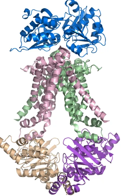
[image:19.612.232.437.287.620.2]The first structure of an ABC transporter in complex with its binding protein was the structure of the molybdate transporter ModBC (Hollenstein et al., 2007). This
3.1 Å resolution structure (PDB ID: 2onk) reveals a nucleotide-free, open conformation within the nucleotide binding domains ModC (Figure 1.6). The membrane-spanning domain, ModB, revealed a novel, considerably smaller set of six helices per monomer that shape the inward-facing conformation. Interestingly, one helix in each monomer crosses over to interact with the helices of the opposing monomer. In contrast to other structures of the transporter-binding protein complex, the binding protein in this structure contains a bound substrate, and is in a closed form; the overall structure of this complex indicates it is in a conformation that precedes transport. The substrate binding pocket of the binding protein is aligned with the translocation pathway, suggesting that ATP binding is necessary to promote active transport. Small helical regions (“coupling helices”) on a loop from the transmembrane domain mitigate movement between the nucleotide binding domain and the transmembrane domain, as seen in other structures. This region contains the conserved “EAA” motif and is thought to play an important role in communicating movement between the nucleotide binding domain and the transmembrane subunit. This structure fortifies the hypothesis that each cycle of transport translocates one substrate molecule across the membrane, yet requires two ATP molecules, suggesting a stoichiometrical relationship of 1 substrate to two ATP molecules. Mechanistic analysis suggests that transporters share a common “alternating access and release” mechanism as they move between the inward-and outward-facing conformations as a result of binding protein docking, substrate binding, ATP binding, and subsequent ATP hydrolysis.
The Maltose Transporter
unusual of all ABC transporter structures, as it shows the maltose ligand within the translocation pathway, an observation heretofore unseen (Figure 1.7). The ATP-bound
Figure 1.7. The structure of the complete maltose transporter, MalFGK, in complex with the maltose binding protein. This fascinating structure revealed the first substrate binding site within a
structure was obtained using a mutation in the Walker B region that prevents ATP hydrolysis, thereby trapping the transporter in a “closed” state. The MalFGK2 structure revealed the first ABC transporter with two different transmembrane domains
comprising the translocation pathway. MalF and MalG share a common core of six helices, in an arrangement very similar to that of the ModBC structure; in this
arrangement, one helix from each subunit crosses over and interacts with the opposite subunit. However, MalG and MalF share only 13% sequence identity, and the much longer MalF possesses a C-terminal segment that packs against the MalK subunit and is not present in MalG. The interface between the two transmembrane domains and MalK also shares similarities to the other published structures. The “EAA”,
represents the transition state of ligand transport much better than any previously published structure, providing a look at the ability of the translocation pathway to bind and transport this carbohydrate ligand.
ABC Exporters
[image:23.612.237.426.57.415.2]Structural studies of certain ABC exporters have also been pursued; while these exporters have significant differences from ABC importers, there are common features between these two groups. Perhaps the most striking similarity is between exporter and
importer ATP binding cassette domains, which possess conserved structural characteristics. The crystal structure of SAV1866, a multidrug exporter from
Staphylococcus aureus, was solved by Dawson and Locher (2006) to 3.0 Å resolution, and shortly afterwards, a structure with bound AMP-PNP was solved to 3.4 Å resolution (Dawson and Locher, 2007). The structure of SAV1866 (PDB ID: 2hyd and 2onj)
reveals two half-transporters that are intertwined to make up the active transporter. Each half-transporter consists of a transmembrane (N-terminal) domain and a nucleotide binding (C-terminal) domain, and are twisted around each other in the structure (Figure 1.8). Each transmembrane domain crosses the membrane lipid bilayer six times, resulting in a total of 12 TM helices for the full transporter. The NBD contains the typical ATP-binding cassette motifs and closely resembles the previously published structure of MJ0796 (Smith et al., 2002). While ADP is bound in the ATP binding cassette of SAV1866, Dawson and Locher have hypothesized that the
transporter is actually in the ATP bound state, based on the tight binding of the nucleotide and a comparison to the previously published structure of BtuCD. In this structure, it is apparent that there are a few loops in the TMD that allow
communication between the NBD and TMD. These loops (which actually have helical regions) are named “coupling helices”, to refer to the fact that they couple the
access to the substrate binding domain, and an outward-facing conformation with external access to release the unwanted substrate. SAV1866 is hypothesized to be in the ATP-bound, outward-facing state, where the substrates would be released into the lipid bilayer or the surrounding medium. The hydrolysis of ATP would convert the transporter back to its inward-facing conformation, in what Dawson and Locher have described as an “alternating access and release” mechanism. In addition to the
SAV1866 multidrug exporter, structures in various conformations of the lipid flippase MsbA from three different organisms (E. coli, V. cholerae, and S. typhimurium) have been published (Ward et al., 2007). These structures (PDB ID: 3b60, 3b5w, 3b5x, 3b5y, 3b5z) also possess the characteristic ATPase domain and show different states of homologous transporters, despite the controversy associated with the original crystal structure analyses of these structures.
1.3
The Catalytic Cycle of ABC Transporters
Attempts to understand the catalytic cycle of ABC transporters have been
and the ABC signature loop stabilizes the ATP molecule bound to the P-loop of the opposing NBD. Similarly, many NBDs do not form dimers in the absence of nucleotide. MalK is the exception to this, as it does form a dimer without nucleotide present,
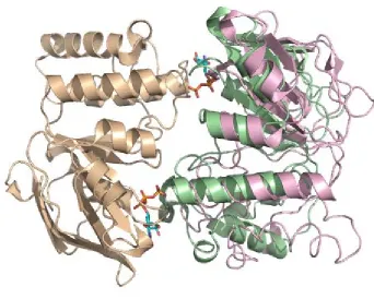
[image:26.612.149.491.77.351.2]presumably due to the C-terminal extension beyond the homologous NBD region that is thought to be a regulatory domain. Intact transporters appear to be more stable in the absence of nucleotide, perhaps due to the TMDs, as evidenced by the number of nucleotide-free transporter structures. The study of the MalK dimers in various conformations (PDB ID: 1q12, 1q1b, 2awo) revealed a tweezer-like motion between the two dimers upon ATP binding (Davidson and Chen, 2004). The C-terminal “regulatory domain” remains in contact regardless of the nucleotide present, but the homologous nucleotide binding domain experiences considerable changes (Figure 1.9).
Figure 1.9. A comparison of two conformations of the MalK dimer. The overlay of one subunit (shown in tan) allows the differences in the relative placement of the opposing subunit (ATP-bound state in green, non-nucleotide form in pink) to be highlighted. The ATP-(ATP-bound
A common mechanism has been proposed for both ABC importers and exporters. It has recently been suggested that the binding of ATP promotes the
outward-facing conformation, while the production and release of ADP and phosphate stabilizes the inward-facing conformation (Dawson and Locher, 2007). In most
descriptions of the catalytic cycle, the P-loop and ABC signature motif are deemed important for interactions with ATP, while the Q-loops are important for the NBD-TMD interface. The “head-to-tail” arrangement of its NBD dimer is a critical element of ABC transporters, as this organization forces the opposing dimers to form two nucleotide binding sites using the conserved sequence motifs. The P-loop of one NBD forms a nucleotide binding site with the ABC signature motif from the opposite NBD, a concept first identified with the structure of the Rad50 protein (Hopfner et al., 2000). Nucleotide-free structures impose fewer constraints on the structure, as evidenced by the variations observed between nucleotide-free structures; the various conformations of these transporters have been classified in “open” or “closed” states. The “coupling
cytoplasm
periplasm
[image:28.612.115.534.73.584.2]resting state
transition state
helices”, described as the important part of the TMD and its interface with the NBD, comprise the key shared feature amongst transmembrane regions. Movement of these helices towards or away from the translocation pathway appears to be important for the transmission of information between these two domains (Figure 1.10). Mutations in this area result in a decrease in transport or no transport at all (Hollenstein et al., 2007).
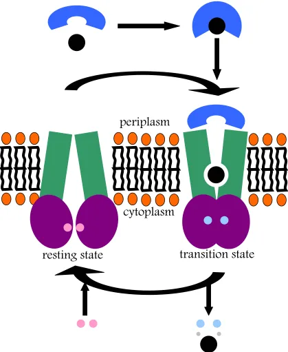
The full catalytic cycle may be summarized through a few different states (Figure 1.11). In the “open” state, NBDs are apart. Examples of this state include the ModBC-ModA or HI1470/1 structures. Once ATP binding occurs, the transporter can shift into a “closed” conformation, where the ATP is bound tightly between the NBD motifs previously mentioned, such as the P-loop and the ABC signature loop. Examples of an ATP-bound structure would include the MalFGK2 structure, as well as the SAV1866 exporter structure. In addition to the movement of NBDs, a key difference between these two conformations is the movement of the “coupling helices” and the subsequent shift of transmembrane helices. In moving from the “open” to “closed” states, the transmembrane helices shift towards each other. Once hydrolysis of ATP occurs, the transporter shifts back to the “open” conformation, its resting state. The stoichiometry of transport has been established; studies of glycine transporter OpuA (Patzlaff et al., 2003) as well as studies of the maltose transporter MalK have revealed that there appear to be two ATP molecules required per one molecule of substrate transported (Dawson and Locher, 2007). Overall, this mechanism corresponds to the “alternating access and release” mechanism first postulated by Jardetzky in 1966.
1.4
Regulatory Mechanisms: The ACT Domain
domains that are proposed to function as regulatory domains (Biemans-Oldehinkel, 2006). These “autoregulatory” domains are present in a variety of ABC transporters: MalK, OpuA, CFTR, and others. Each of these domains, however, is hypothesized to act in different ways. One such mechanism predicted to be important for some amino acid ABC transporters is the ACT domain.
The ACT domain, initially identified by Aravind and Koonin (1999), is a novel ligand-binding domain based upon a similarity in three different structures:
aspartokinase, chorismate mutase, and TyrA (ACT). This shared domain of unknown function was identified in aspartokinases, chorismate mutases, prephenate
dehydrogenases, prephenate dehydratases, homoserine dehydrogenases, malate dehydrogenases, phosphoglyerate dehydrogenases, phenylalanine and tryptophan-4-monooxygenases, phosphoribosylformylglycinamidine synthase, uridylyl transferase and removing enzyme, GTP pyrophosphokinase/phosphohydrolase, tyrosine and
phenol metabolism operon regulators, as well as several uncharacterized proteins from archaea, bacteria, and plants that contain from one to four copies of this domain
(Aravind and Koonin, 1999). Luckily, some of these proteins had been structurally
characterized, allowing a further analysis of this domain. The structure of 3-phosphoglycerate dehydrogenase (PDBID: 1psd) revealed this domain to be the C- terminal regulatory domain of 70-80 amino acids that formed a β-sheet with strand order 4-1-3-2 surrounded by helices (Figure 1.12). Upon further analysis, this domain was identified as having the ferredoxin-like fold, according to SCOP. A ferredoxin-like fold is defined as having the βαββαβ topology (Chipman and Shaanan, 2001). In the structure of 3-phosphoglycerate dehydrogenase, this domain binds L-serine, the final product of this biosynthetic pathway, and an allosteric regulator of this enzyme. The ACT domain is thought to possess a common ligand-binding area, critical to the function of this fold in regulation; many of the enzymes possessing this domain are known as “classic” allosteric regulators. Further analysis of this domain revealed that this domain forms a dimer, resulting in an 8-stranded β-sheet, with 2 ligand molecules
bound at the interface, suggesting that this is a conserved regulatory fold. While certain ACT family members show sequence homology, others have more divergent sequences while maintaining the same topology and ligand binding site (Chipman and Shaanan, 2001). A study of the types of proteins possessing ACT domains revealed that the majority of these are involved in amino acid and purine biosynthesis, and were previously known to be allosterically regulated proteins (Liberles et al., 2005). While the mechanism of action of this domain is not fully understood, it has been suggested that ligand binding leads to low flexibility within this domain and the rest of the structure, as rotations to allow for normal activity are blocked (Grant, 2006). As ACT domains have been identified in Archea, Bacteria, and Eukarya, they have been
Chapter Two. MetNI.
2.1
Introduction
The high-affinity methionine transport system MetD of E. coli consists of MetN, the ATPase domain, MetI, the transmembrane domain, and MetQ, the cognate binding protein located in the periplasm. The progression of study that resulted in the
The first studies by Kadner on this system described a temperature-dependent methionine uptake system that allowed for the accumulation of methionine (Figure 2.1) against a gradient. Kadner (1974) reported that the internal pool of methionine is estimated to be 0.1-0.3 mM, as the vast majority of intracellular methionine is converted to other metabolites such as spermine and spermidine within five minutes after uptake. Kadner suggested that this uptake was somehow dependent on energy, but was unsure whether this energy source came from glycolysis or oxidative
phosphorylation. He was further able to identify two separate methionine transport systems: a high-affinity system (with a Kt of approximately 7.5 x 10⁻⁸ M) and a low-affinity system (with a Kt of approximately 40 x 10-6 M) that differed over 400-fold in their ability to transport L-methionine. In his initial characterization, he was also able to identify certain methionine derivatives or dipeptides that acted as inhibitors to the methionine uptake system, and to determine that D-methionine, ethionine, and other
amino acids had low inhibitory activity. However, further studies began to slightly contradict his initial paper. Later the same year, he published a paper (Kadner and Watson, 1974) further describing the two uptake systems for methionine, named MetD and MetP. These studies suggested that the higher-affinity system, MetD, was in fact able to transport D-methionine as well as L-selenomethionine. Kadner went on to study and better understand the MetD and MetP mutants and the effects of these mutants on methionine activity; mutants of MetD lack high-affinity transport for L-methionine and low-affinity uptake for D-L-methionine (Kadner, 1977).
As Kadner progressed in his study of the methionine uptake system, he
encountered a mechanism of regulation in E. coli. In 1975, Kadner described that the activity of the methionine uptake transporter is regulated by the concentration of the internal methionine pool. He found that the activity of the transporter is depressed when cells are grown in the presence of methionine, but not methionine mimetics such as α-keto-γ-methiol-butyrate, D-methionine, or methionine sulfoxide. His research additionally suggested that this transport system was regulated by gene repression. Studies of the MetD system by Kadner revealed that D-methionine was a useful
methionine source for E. coli and was transported by the MetD system (Kadner, 1977). He further hypothesized that there were two substrate binding domains within the MetD system for both stereoisomers. Energetics studies suggested that transport was driven by phosphate bond energy (Kadner, 1975), but the use of ATP was not
triphosphatase mutants were created that affected transport and supported the idea that methionine transport utilizes ATP (Kadner and Winkler, 1975).
It was not until 2002 that the genes for the MetD system were identified and described as possibly comprising an ABC transporter (Merlin et al., 2002). Within the gene locus initially identified by Kadner, Merlin et al. were able to find six genes, of which four were unidentified and two (Abc and YaeE) were thought to be an ABC transporter ATPase and an ABC transporter permease, respectively. These two genes, Abc and YaeE, were labeled as an ABC transporter due to sequence homology,
particularly the Abc protein that contained many ABC transporter sequence motifs. The permease, YaeE, was hypothesized to possess five transmembrane helices. Further analysis using the BLAST tool identified YaeC as a possible methionine binding protein; the three genes were subsequently renamed to MetN, MetI, and MetQ to represent the ATPase, transmembrane domain, and binding protein, respectively. Merlin et al. also studied the genetic organization of the MetD locus within a variety of other sequenced bacteria. A similar organization was found in S. typhimurium, S. typhi, Y. pestis, V. cholerae, P. multocida, H. influenzae, A. tumefaciens, S. meliloti, B. melitensis, and M. loti, suggesting that each of these organisms has a similar methionine uptake system. Their studies described MetD deletants that were able to grow on a media
Building further on the concept that a similar organization for this gene is present in multiple organisms, Zhang et al. (2003) redefined this system as a
methionine uptake transporter (MUT). Their phylogenetic analysis revealed that this methionine uptake system was present in multiple organisms in the form of sequence-divergent primordial paralogs. These proteins make up the methionine uptake
transporter family, one of the first functionally characterized novel families of bacterial ABC permeases. An analysis of MetD mutations in S. typhimurium led to the mapping of the MetD gene near 4.8 minutes on the E .coli genome, which coincides with an ABC transporter mapping (Zhang et al., 2003). Regulation by these genes and other
methionine biosynthetic genes by the MetJ repressor has been established (Weissbach and Brot, 1991; Greene and Spiegelman, 1996; Sekowsha et al., 2000), along with a MetJ DNA binding site consensus sequence (Saint-Girons et al., 1984) and the identification of a MetJ binding site in the promoter region of the MetD ABC
transporter locus (Liu et al., 2001). Homologues of each of the three proteins within the MUT family share a similar size, and the transmembrane regions for all of them are hypothesized to have five transmembrane segments. The relationships revealed in the phylogenetic analysis suggest that these genetic duplications occurred early during evolutionary history, as the topology of the phylogenetic trees does not follow the phylogenies of these organisms. It has been hypothesized that there were several sequence-divergent primordial paralogs, which would account for the topology seen.
Furthermore, the use of energy poisons such as sodium arsenate abolished uptake of methionine, while others were less effective, suggesting that ATP is in fact the energy source for transport, as believed.
Studies have also elucidated possible pharmaceutical interest in this family of transporters, as several members of the MUT family are required for bacterial pathogenicity. It has been found that the MUT family member of S. typhimurium is essential for infection in mouse models (Pattery et al., 1999). A mutation in the MUT in the organism H. influenzae leads to reduced infection in rats (Chanyangam et al., 1991). The H. pylori MUT plays a role in the production of urease, which is essential for colonization of this organism (Hendricks and Mobley, 1997). Strains of Y. pestis frequently depend upon exogenous methionine for survival, and may also make MUTs important in this organism (Brubaker, 1972). The importance of this family of
transporters is not fully understood, but the implications within the pharmaceutical field are clearly growing, necessitating further study of this system.
To further investigate this system, we have determined the structure of the E. coli methionine ABC transporter MetNI following overexpression and affinity purification with dodecyl maltoside.
2.2
Structure of the MetNI Transporter
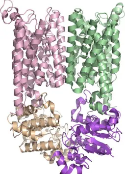
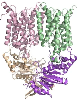
Crystallographic analysis revealed a nucleotide-free state (Figure 2.2 and Figure 2.3) that was refined to 3.7 Å after phasing with single isomorphous replacement and anomalous scattering from a xenon derivative (Figure 2.4). The sequence register was verified with a difference Fourier density map from data collected on
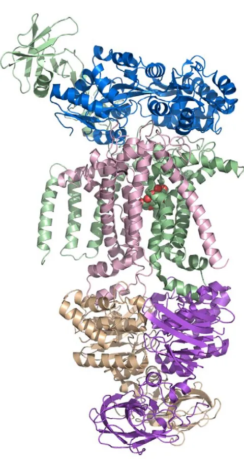
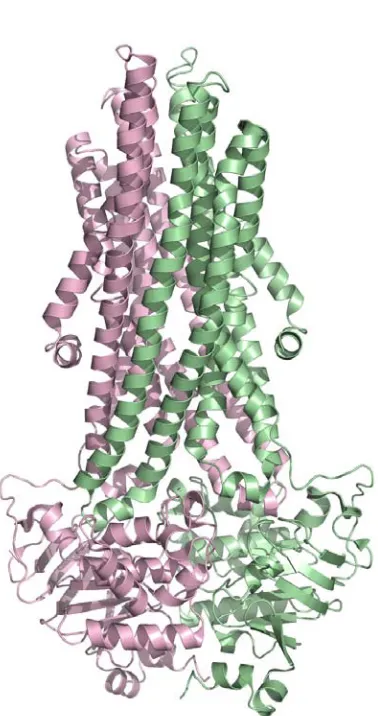
The overall architecture of MetNI reveals two copies of the ATPase MetN in complex with two copies of the transmembrane domain MetI. The methionine
transporter has a smaller transmembrane domain than other published ABC transporter structures, consisting of only five transmembrane helices per monomer. The
cytoplasmic ATPase contains the expected conserved ABC region, but possesses a C-terminal extension of approximately 100 amino acids that comprises an ACT regulatory domain that has not been seen previously as part of an ABC transporter. The relative positions of the two ATPase subunits and their corresponding transmembrane domains illustrate a distinct inward-facing orientation. Additionally, each of the two copies of MetN in the transporter adopts slightly different conformations, resulting in an asymmetrical organization of the MetN dimer due to a proposed hinge between the conserved ABC region and the C-terminal domain.
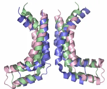
Figure 2.2. The E.coli methionine transporter MetN2I2 is a tetramer consisting of two copies of the transmembrane (MetI) permease protein (pink and green) in complex with two copies of an ATP binding cassette (MetN) protein (tan and purple). The transmembrane protein consists of five
Figure 2.4. The initial chain trace of the MetNI transporter (black) with the Xenon binding sites (red spheres) is shown above. There were 17 total Xe atoms bound to the two transporters within the asymmetric unit.
Figure 2.5. The asymmetric unit of the MetNI crystal form is shown below. The two intact
noticeable twist in the position of the ACT domain that makes up the C-terminal
extension. This movement may be indicative of the types of changes accompanying the large scale transition between the inward- and outward-facing conformations.
Transmembrane Domain
Each set of the membrane-spanning subunits of MetI are related by an
approximate two-fold rotation axis (~180˚) and are otherwise essentially identical. A subunit of the transmembrane domain MetI consists of 5 transmembrane helices
organized similarly to the core of the membrane-spanning subunits of ModB, MalF, and MalG, as ModB, MalF, and MalG contain an N-terminal helix not seen in MetI (Figure 2.7). As a result, the N-terminus of MetI lies in the periplasm, while the C-terminus
protrudes into the cytoplasm, a difference from other observed structures that all possess an even number of transmembrane helices per monomer and consequently have both termini in the cytoplasm. Helix TM 1 begins in the periplasm and forms a diagonal across the outside face of the other four helices, while TM 2 is a kinked helix that traverses from the cytoplasmic edge of the membrane back towards the periplasm. A loop helps to transition into TM 3, which leads to the short “coupling helix” that interfaces with the ATPase domain and, along with TM 4 makes up the central core of the translocation pathway. The C-terminal TM 5 helix traverses the membrane once
more, ending in the cytoplasm, on the opposite side from TM2, as these two helices form the outer core of the helices close their counterparts on the other monomer.
Comparisons to other ABC transporters such as BtuCD and HI1470/1 are
difficult, as the helical structure of these 10-helix domains differs significantly from the MetI structure, resulting in distinct organizations. When comparing individual
subunits, the root mean square deviation (r.m.s.d.) of structurally equivalent Cα atoms of ModB and MetI is ~3.4 Å, that of MalG and MetI is ~2.7 Å, and that of MalF and MetI is ~2.6 Å (Figure 2.7). The relative positions of the two copies of MetI
[image:45.612.137.512.74.388.2]demonstrate a wider, more inward-facing conformation than either the ModB or the MalFG dimers. Interestingly, the region of highest sequence similarity between these
structures consists of the previously named “coupling helix” and the two
transmembrane helices on either side, the region which forms the central core of the translocation pathway (Table A1.1).
[image:46.612.183.466.69.434.2]The coupling helix and the neighboring transmembrane helices display the most significant change between the nucleotide-free, inward-facing MetI conformation and the ATP-bound, substrate-occluded MalFG conformation. For MetI, these elements correspond to TM3-coupling helix-TM4, while on MalF this region corresponds to TM6-coupling helix-TM7, and for MalG and ModB it is TM4-coupling helix-TM5, due
to their N-terminal extensions beyond the 5-TM core shared by all four proteins. As a whole, this region in MalFG exhibits the greatest shift toward the translocation
pathway, as the two transmembrane helices are pushed closer to the central axis of the translocation pathway than those of MetI or ModB (Figure 2.8 and Figure 2.9). This movement is presumably crucial to activating the permease pathway for transport; the communication between the ATPase domain and this transmembrane region is
accomplished through a translation of the coupling helix toward the translocation pathway to allow for ATP hydrolysis and transport. Superposition of this region of the MalFG dimer with the corresponding portion of the MetI dimer illustrates this tilt of the cytoplasmic-proximate region of the MalFG transmembrane domain towards the center of the translocation pathway as compared with MetI2, with ModB2 appearing as an intermediate between these two states. The TM3 helix on MetI exhibits a rotation of approximately 22˚ away from the translocation pathway as compared with TM6 (TM 6) of MalG (MalF), while the TM4 helix of MetI is similarly rotated approximately 24˚
away from MalFG, with the greatest distance on the cytoplasmic-facing region of these helices being roughly 10 Å. The difference in positions of the coupling helices
the hypothesis that movement of these transmembrane domains is tweezer-like depending on the state of nucleotide binding. Additionally, the similarities extend to TM2 of MetI (and its equivalents in ModB, MalF, and MalG), particularly with respect to the shared helix length (a short 15 residues) and a kink in the helix caused by a proline residue (Pro67 in MetI).
ATPase Domain
The MetN ATP binding cassette protein consists of two domains, the
[image:48.612.119.531.316.593.2]approximately 225 amino-acid ATPase domain that is homologous with many other ABC transporters, and a C-terminal domain, comprised of the last 118 residues. The
ATPase domain of the methionine transporter possesses the characteristic features shared by many ABC transporters that were first identified in the structure of HisP (Hung et al., 1998). Similar to other characterized ATPase domains, the MetN
monomer possesses a six-stranded β-sheet with strand order 2-3-4-1-5-6, with strand 6 being antiparallel to the five other strands. This domain possesses another β-sheet, comprised of four β-strands in an antiparallel structure with strand order 2-1-3-4. The two sheets surround one of the α-helices, and are bordered by another 8 helices that make
[image:49.612.140.511.407.645.2]up this domain. The features found in this structure are similar to those described in the structure of HisP, which further divided the ATPase structure into two separate domains consisting of an α- plus β- type structure in one domain and a primarily helical second domain. Interestingly, when this overall fold was first identified, it was
shared by no other structures within the PDB (Hung et al., 1998). Since that time, however, it has been established as a common, homologous fold amongst the ABC transporter family, as both importers and exporters share these features. The HisP and subsequent ABC transporter ATPase structures also elucidated certain motifs present within this family, which have been identified within the MetN structure. The Walker A/B motif includes residues 38-45 and 161-165, while the signature sequence extends from 141-145 (Figure 2.10, Figure 2.11, and Figure 2.12).
[image:50.612.140.513.78.331.2]When comparing the homologous regions of various transporter structures to MetN, r.m.s.d. values of structurally equivalent Cα atoms for the individual subunits were 1.7 Å with HI1470, 1.8 Å with ModC, 2.3 Å with BtuC, and 2.6 Å with MalK. Interestingly, the monomer with the highest level of sequence homology to MetN is HisP, which has not been solved as a full transporter. HisP and MetN have an r.m.s.d.
of 1.8 Å. While each of the 5 ATP binding cassettes from the structures of full
transporters are relatively closely related in structure, the positions of the components within the respective dimers varies significantly depending on the overall transporter conformation. The MetNI dimer reveals a more pronounced inward-facing
transporter, with a much greater distance between the ATPase subunits than any other transporter structure solved at present. While the recently published MalK structure depicts the conformation of the ATP-bound state (Oldham et al., 2007), the
organization of MetN portrays an inward-facing conformation with widely separated NBDs. The contrasting relationship between the MalK dimer and the MetN dimer suggests that a tweezer-like swinging motion brings the two subunits together (Figure 2.12). The rotational and translational shift between the MetN closed state and the substrate-bound conformation of MalK is more significant than the differences observed between other inward- and outward-facing conformations, such as the changes between the conformations of HI1470/1 and BtuCD. The regions closest to the membrane appear to experience the most significant movement between the two states; this corresponds with the cytoplasmic-facing rearrangement between the transmembrane helices, suggesting that the central portion of the transporter undergoes the greatest shift through the transport process. More simply, the ATP binding cassette dimer moves between open and closed states in a tweezer-like
swinging motion that is similar to the motion of the transmembrane domain yet in the opposite direction. The differences in distance between the motifs of the catalytic core on each subunit of the MetN dimer versus the MalK dimer are considerable, as the distances between corresponding residues on MalK are 10 Å closer together than those on MetN, on average. Using residues of the P-loop and the signature motif, the
approximately 28 Å. In contrast, these differences shrink to 11 Å and 16 Å for the maltose and molybdate structures and to 14 Å and 16 Å for BtuCD and HI1470/1 (Pinkett et al., 2007). The swinging motion is more apparent when residues on the periplasmic-proximate and the membrane-proximate regions of these two dimers are considered: distances between residues on the periplasmic face are considerably shorter than distances on the membrane facing region. The MetN conformation is clearly only open to the cytoplasm, and this arrangement may be modulated by the C-terminal domain of the ATP binding cassette.
Regulatory Domain
The C-terminal domain of the MetN subunit consists of two elements: a
catalytic domains of 3-phosphoglycerate dehydrogenase are separate, just as the ATP binding cassette and ACT domain of MetN are clearly divided by a linker region.
In MetN, movement of these two domains relative to one another is
accomplished through a hinge mechanism within the linker region. The linker itself comprises the 40 residues in between the regulatory and ATPase domains, and
[image:53.612.139.513.91.470.2]possesses four short α-helices and five loop regions. The transporter structure has revealed two different conformations of this linker region, resulting in an asymmetrical
relationship of the two MetN chains (Figure 2.13). The ATP binding cassette region as well as the ferredoxin-like fold of the C-terminal extension exhibit approximate two- fold rotational symmetry between the two chains, but the rotation axes for the C-terminal extension and the ATP binding cassette differ in orientation by approximately 20˚. These two orientations account for the asymmetry seen in the structure, and are the result of two different conformations of the linker region. This hinge mechanism, observed in MetN through differing conformations of the linker region between the two domains, is most likely responsible for the movement between varying states of the ACT
domain. The two ACT domains are in closer proximity to one another than the remainder of MetN, suggesting that this conformation is keeping the ATP binding cassettes from interacting, thereby blocking ATP binding and methionine transport (Figure 2.14, Figure 2.15, and Figure 2.16). While the methionine binding site was not well characterized at this resolution, the binding sites are located in close proximity to Met301 and Met312. This unusual feature could be an indication that there is
competition between intercellular and intracellular methionine binding at this site. Biochemical studies corroborate that high methionine levels inhibit transport (Zhang, 2003). Interestingly, no selenomethionine was bound in any region of the
transmembrane domain, suggesting that the ACT domain has a higher affinity for selenomethionine than the translocation pathway. The presence of a regulatory domain may indicate that transport in these regulated ABC transporters is substrate
[image:56.612.128.524.71.466.2]need-dependent, as MetNI is inactivated with high levels of intracellular methionine. This regulatory activity adds a further dimension to the current models of ABC transporter mechanisms, suggesting that transport is primarily dependent upon the need for
substrate rather than simply the presence of ATP and substrate delivered by a cognate binding protein.
In conclusion, this structure has provided insights onto the structural organization of ABC importers. First, this structure has identified the smallest
transmembrane domain characterized thus far, suggesting that these 5 helices form a minimal core for an adequate translocation pathway. Second, the ATPase domains are stabilized in a conformation with a much greater distance between the two subunits than seen before. Third, the regulation of transport is modulated by a previously
identified regulatory domain, and in cases of ligand binding in this area, the transporter is stabilized in an inward-facing conformation that precludes ATP binding. This
mechanism supports earlier findings by Kadner and allows for insight into the
Appendix One. Methods and Data Relevant to Chapter Two.
A1.1.
Materials and Methods
Cloning and Expression
The E.coli methionine ABC transporter operon was identified from the NCBI website. Following a similar strategy used in the BtuCD and HI1470/71 structure determinations (Locher et al. 2002, Pinkett et al. 2007), metN and MetI were cloned from E. coli K-12 genomic DNA (American Type Culture Collection) into a modified fusion pET21b(+)/pET19b vector that can co-express the metI and metN genes simultaneously using separate T7 promoters. The entire MetNIQ operon was isolated from the genomic DNA by PCR and inserted into a pCR-Blunt II-TOPO vector (Invitrogen). The metN (ATPase subunit) gene was subsequently isolated by PCR using two oligonucleotides which inserted NdeI and XhoI sites. The PCR fragment was ligated into a pET19b vector (Novagen/EMD) using the NdeI and XhoI sites to introduce an N-terminal 10xhistidine tag. Similarly, the metI (transmembrane subunit) gene was isolated by PCR using two oligonucleotides which inserted NdeI and XhoI sites and removed the STOP codon from the end of the gene. The PCR fragment was ligated into a pET21b (+) vector (Novagen/EMD) using the NdeI and XhoI sites to introduce a C-terminal 6xhistidine tag.
modified pET19b vector can express the metI and metN genes simultaneously using separate T7 promoters. A mutagenesis reaction was then performed on the metNI -pET19b plasmid to insert a STOP codon immediately after the metI gene so that the C-terminal 6xhistidine tag will not be expressed. The reaction was performed using the Quickchange Mutagenesis Kit (Stratagene). As a result, the final plasmid contains an ATPase subunit with an N-terminal MGHHHHHHHHHHSSGHIDDDDKH sequence, while the transmembrane subunit has no additional residues. The cloning was performed by Allen Lee.
The cloned plasmid was transformed into BL21 (gold) cells (Novagen) and protein expression was carried out at 37˚C in Terrific Broth with 0.2 mg/mL ampicillin and 1% glucose instead of glycerol in an 80 L New Brunswick fermentor containing 60 liters of medium. The cells were induced at OD 600=4.0 with 2 mM IPTG for 2 hours before harvesting and storing at -80˚C.
Purification and Crystallization
In the purification of MetNI, 10 g of cell paste is re-suspended in 50 mL of a 50 mM Tris pH 7.5, 20 mM imidazole, and 1 M sodium chloride solution with 40 mL dH2O added. The solution is clarified using a homogenizer, after which 10 mL of a 10% n-dodecyl-β-D-maltopyranoside (Anatrace) solution and a protease inhibitor tablet (Roche) is added, bringing the final concentration to 1% n-dodecyl- β-D-maltopyranoside.
at 4˚C before centrifugation at 39,700 x g for 30 minutes. The supernatant was loaded and equilibrated onto a 5 g Ni-sepharose affinity chromatography column
(Amersham), washed with 92 mM imidazole, and eluted with 250 mM imidazole, 25 mM Tris pH 7.5, 500 mM NaCl, and 0.1% DDM. The eluate was dialyzed overnight into a solution containing 0.1% DDM, 25 mM Tris pH 7.5, 500 mM NaCl, 5 mM dithiothreitol, 2.5 mM MgCl2, and 2.5 mM ATP before it was concentrated to 20 mg/ml using an Amicon Ultra MWCO100 concentrator (Millipore). Protein purity was estimated to be greater than 90% using SDS gel electrophoresis.
Selenomethionine substituted protein was made using E. coli B834 (DE3) cells (EMD Biosciences). Cells were grown in M9 medium supplemented with seleno-D,L-methionine. Procedures for purification and crystallization of the selenomethionine-substituted protein were identical to that of the native protein.
Crystallization experiments were carried out with the hanging drop method at 20˚C, using 2 μL protein plus 2 μL precipitant solution. Chunky, diamond-shaped crystals were obtained using solutions in the range of 28-32% PEG 400, 0.05 M Li2SO4, 0.2 M (NH4)2HPO4, 0.05-0.15 M (NH2)2SO4, and 0.1 M Sodium Citrate pH 4.2-4.5. Crystals grew to 0.3 x 0.3 x 0.4 mm3 in two to three weeks, and were frozen in liquid nitrogen prior to data collection.
Xenon-derivatized crystals were formed by exposing the native crystal to 100psi Xe gas for 10 minutes in a pressure chamber. Selenomethionine-soaked crystals were obtained by soaking native crystals in 1-20 mM L-selenomethionine for 30 minutes to 3 hours.
Data Collection and Structure Determination.
1992). A 3.8 Å resolution Xenon-derivatized dataset (1.5 Å wavelength) was collected at the Stanford Synchrotron Radiation Laboratory beam line 9-2 and processed using MOSFLM and SCALA. A 3-wavelength 5.5 Å resolution dataset using
selenomethionine-substituted crystals was collected at the Advanced Light Source beam line 8.2.1 and processed using MOSFLM and SCALA (Leslie, 1992). A peak wavelength 4.5 Å resolution dataset of an L-selenomethionine-soaked crystal was collected at Stanford Synchrotron Radiation Laboratory beam line 9-2 and processed using MOSFLM and SCALA.
Initial protein phases and heavy atom binding sites were found from the Xe-derivatized data using single isomorphous replacement in the program SHELX
Figure A1.1. The sequence alignment of MetI, the transmembrane domain, with the transmembrane domains of MalF, MalG, and ModB, is shown above. The highest level of sequence similarity is within the segment containing the coupling helix and extending to the helices on either side of this
Table A1.1: Summary of Secondary Structure for MetN
Secondary Structure Residue Range
β1 1-11
β2 17-28
β3 32-39
β4 59-65
β5 67-70
β6 84-89
β7 160-170
β8 193-199
β9 209-215
β10 218-223
β11 266-273
β12 295-303
β13 309-318
β14 334-343
α1 42-62
α2 75-82
α3 99-108
α4 114-127
α5 130-135
α6 144-156
α7 174-190
α8 201-206
α9 234-239
α10 242-249
α11 252-258
α12 281-290
Table A1.2: Summary of Secondary Structure for MetI Secondary Structure Residue Range
α1 (TM 1) 8-41
α2 (TM 2) 52-65
α3 (TM 2) 68-82
α4 (TM 3) 89-114
α5 (coupling helix) 118-126
α4 (TM 4) 131-166
α5 (loop) 172-183
Table A1.3: Processing, Phasing, and Refinement Statistics for Xe, Native and Se soak Data processing
Dataset Xenon Native SeMet soak
Spacegroup P 21 21 21 P 21 21 21 P 21 21 21 Unit Cell dimensions (Å) a= 97.74
b= 169.20 c= 290.60 a= 97.77 b= 165.81 c= 288.92 a= 97.74 b= 169.20 c= 290.60
Wavelength (Å) 1.5 1 .979
Resolution (Å) 40-3.8 40-3.7 40-4.7
Unique reflections 61583 68389 28494
redundancy 8.0 (7.8) 11.3 (12.2) 7.4 (6.8)
aCompleteness 99.9 (99.9) 94.3 (100.0) 97.5 (95.8)
bRmerge .11 (.81) .11 (.92) .07 (.59)
Phasing power from Xe dataset
Isomorphous phasing power from (centric/acentric)
.674/.771
Anomalous phasing power .744
Figure of Merit (centric/acentric)
.186/.257
Figure of Merit after DM .74
Refinementc
Resolution (Å) 40-3.7
Reflections used 49914
Test reflections 5048
dRWork (%) 31.0
dRfree (%) 35.4
Average B factor (Å2) 169.67
Rmsd bond length (Å) 0.02
Rmsd bond angle (°) 2.78
Ramachandran (%)
Most favored 91.0
Allowed 4.2 Disallowed 4.8
aNumbers in parentheses refer to the highest resolution shell only.
bRmerge=Σ|Ih-<I>h|/ΣIh, where <I>h is average intensity over symmetry equivalents.
Table A1.4: Processing, Phasing, and Refinement Statistics from Semet protein Data processing
Dataset Peak Inflection Remote
Spacegroup P 21 21 21 P 21 21 21 P 21 21 21 Unit Cell dimensions (Å) a= 93.79 b= 159.59 c= 294.33
Wavelength (Å) .9796 .9795 .972
Resolution (Å) 30-6.0 30-6.0 30-6.0
Unique reflections 22391 22386 22387
redundancy 12.1 (12.6) 12.1 (12.6) 11.9 (12.2)
aCompleteness 99.9 (100.0) 99.9 (100.0) 99.9 (100.0)
bRmerge .153 (.47) .148 (.51) .149 (.50)
aNumbers in parentheses refer to the highest resolution shell only.
Chapter Three. MetQ.
3.1
Introduction
An integral component of the complete ABC importer system is the substrate binding protein; the substrate binding protein is responsible for the delivery of the substrate to the ABC importer. In Gram-negative bacteria, this substrate binding protein is secreted and contained within the periplasm by the outer membrane. In Gram-positive organisms, the substrate binding proteins are typically tethered to the plasma membrane in some manner, through a lipid anchor or through fusion to the membrane-spanning region of the transporter (Williams et al., 2004). These substrate binding proteins (whether soluble proteins or lipoproteins) are responsible for binding a specific ligand and associating with the transmembrane domain of the ABC
transporter to facilitate the transport of the substrate across the periplasm. This mechanism of delivery helps to confer substrate specificity and affinity upon the ABC transporter (Tam and Saier, 1993). Without its substrate binding protein, an ABC transporter is typically unable to retain its full activity and becomes a low-efficiency transporter. Further supporting this idea is the evidence that Kd values for most substrate binding proteins are within an order of magnitude of the Km of the
corresponding ABC transporter, suggesting that the binding protein delivery system is responsible for much of the transporter’s substrate affinity (Tam and Saier, 1993).
studies concerning these unidentified proteins, leading to a hypothesis that they were important for bacterial transport (Barash and Halpern, 1971). However, more definite genetic and biochemical proof of a link between these binding proteins and their involvement in transport was provided via a study of the galactose transport system. This study established that the galactose binding protein was a vital part of the galactose transport system by identifying a mutation that selectively disabled the galactose binding protein but did not affect the entire gene locus (Boos, 1972). In the following years, X-ray structures of various periplasmic binding proteins were solved, although a further biochemical understanding of these systems was not yet achieved. A study comparing the biochemical activities of a variety of periplasmic carbohydrate binding proteins suggested that the high affinity these proteins have for their respective substrates is responsible for a rapid response in binding. Furthermore, it was proposed that these binding proteins confer the high specificity and sensitivity to their
corresponding transport systems (Miller et al., 1983). This concept, combined with the hypothesis that these proteins interact with their membrane-bound counterparts
(Higgins, 1992), suggested a system that had multiple components working together to bind a substrate with high specificity and transport it across the membrane.
Interestingly, many of these substrate binding proteins share structural similarity, as the structures have been revealed to be bi-lobed structures with two nonequivalent domains surrounding the ligand. This is thought to be tied to
Mechanistically, these proteins are thought to be similar as well, utilizing a conserved mechanism named the “Venus flytrap mechanism,” according to Mao et al. (1982). In this mechanism, the ligand-free protein adopts an open conformation, with the substrate binding pocket exposed to the solvent. Substrate can subsequently bind, allowing for the substrate binding protein to move to a closed conformation with a tightly bound ligand that is shielded from solvent. As the substrate binding protein docks onto the transmembrane permease protein, conformational changes resulting from the ATPase activity of the ABC transporter are thought to lead to movement of the transmembrane region, which is thought to pull apart the substrate binding domain, releasing the substrate into the translocation pathway of the permease (Williams et al., 2004). Originally, this hypothesis proposed that the “open” or non-ligand-bound state of the binding protein adopted a conformation in which the two lobes of the binding protein were 20˚ further apart than in the bound state (Mao et al., 1982).
Interestingly, there are few binding protein structures that have been solved in both the ligand-unbound and ligand-bound conformations. The first example was the maltose binding protein, which revealed a 35˚ rotation about the hinge upon ligand binding (Sharff et al., 1992). Another comparison has been made for the vitamin B12 binding protein BtuF; in this case, there is relatively little structural change upon ligand binding, although substrate binding was shown to cause a decrease in mobility within the protein (Karpowich et al., 2003).
hypothesis. The binding protein in this structure is seen in an open conformation, with the two lobes spread apart. At the interface, loops from the BtuC permease insert into the B12 binding site, resulting in the open conformation for BtuF (Hvorup et al., 2007). This notion is further supported by the structure of the maltose binding protein in complex with the maltose transporter. In this structure, the substrate maltose is present, but within the permease translocation pathway, not the binding protein. Interestingly, the maltose binding protein is an open state as well, and a loop from the MalG permease inserts into the binding site. It has been hypothesized that this loop acts as a scoop, as it is responsible for dislodging the sugar and forcing it to transfer into the translocation pathway (Oldham et al., 2007). The third structure of an intact
transporter-binding protein complex, ModABC, possesses a ligand within the binding protein, suggesting that this conformation is pre-transport. This interaction however, supports the notion that docking is mediated by attractions between oppositely charged amino acid side chains on both the permease and binding protein (Hollenstein et al., 2007).
structural features as well: both domains participate in the ligand binding, water molecules are frequently present in the ligand binding site, and a hinge-bending motion is thought to be responsible for the difference between the open (apo) and closed (ligand-bound) states of these binding proteins (Quicho et al., 1996).
3.2
Methionine Binding Proteins
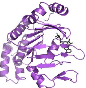
The structure of a methionine binding protein from the MUT family from Treponema pallidum, the organism responsible for the disease syphilis, has been solved previously to 1.85 Å resolution (Deka et al., 2004). The structure of this protein (PDB ID: 1XS5), called tp32, resembles other periplasmic ligand-binding proteins and is thought to be a lipoprotein in physiologic conditions (Figure 3.1). As is common for these binding proteins, tp32 possesses two α/β domains that are linked by two crossovers. The binding pocket is located between these two domains, and an
L-methionine residue was present in the electron density map. Each domain consists of a central core, comprised of a five stranded β-sheet with α-helices on either side. In each
β-sheet, there is one anti-parallel strand. While one of the regions connecting the two