Antimicrobial Evaluation of Selected
Medicinal Plants Using Molecular Approach
ABDULLAH GHANIM QADDOORI
Antimicrobial Evaluation of Selected
Medicinal Plants Using Molecular Approach
ABDULLAH GHANIM QADDOORI
Biomedical Research Centre (BRC)
School of Environment & Life Sciences
College of Science & Technology
University of Salford
Manchester, UK
A Thesis Submitted in Partial Fulfilment of the
Requirements for the Degree of Doctor of Philosophy
i
Table of Contents i
List of Tables v
List of Figures vi
Abstract ix
Acknowledgement x
Dedication xi
Declaration xii
Abbreviations xiii
Chapter 1 - Introduction 1
1.1 Project background 2
1.2 Aims and objectives of the present study 8
1.3 Significance of the study 9
Chapter 2 - Literature Review 11
2.1 Antibiotics 12
2.1.1 Antibiotic resistance of bacterial pathogens 14
2.1.2 The Basic characteristics of antibiotics 15
2.1.3 Mechanisms of antibiotic actions 16
2.1.4 Mechanisms of antibiotic resistance 17
2.2 Methicillin-resistant staphylococcus aureus (MRSA) 21
2.2.1 Pathogenesis of Methicillin-resistant Staphylococcus aureus 22
2.3 Proteomics 25
2.3.1 Origin, definition and significance 25
2.3.2 Proteomics in Microbiology 28
2.3.3 Proteomics as a tool in drug discovery 30
2.3.4 Proteomic analysis techniques 33
2.3.4.1 Protein separation using gel based techniques 34
2.3.4.2 Methods of protein quantitation and identification 36
2.4 Natural products 41
2.4.1 General introduction 41
2.4.2 Importance of medicinal plants 43
2.4.3 Extraction of plant-derived natural products 46
2.4.4 Isolation and purification of natural products 48
2.4.4.1 High-performance liquid chromatography (HPLC) 50
2.4.5 Structure elucidation of natural products 51
2.5 Medicinal plants used in the study 52
ii
2.5.1.3 Chemical constituents isolated from Centella asiatica (L.) 54
2.5.2 Imperata cylindrica (L.) 55
2.5.2.1 Description of the plant 55
2.5.2.2 Pharmacological properties of Imperata cylindrica (L.) 56
2.5.2.3 Chemical constituents isolated from Imperata cylindrica (L.) 57
2.5.3 Morinda Citrifolia (L.) 57
2.5.3.1 Description of the plant 57
2.5.3.2 Pharmacological properties of Morinda Citrifolia (L.) 58
2.5.3.3 Chemical constituents isolated from Morinda Citrifolia (L.) 59
2.5.4 Sauropus androgynus (L.) 60
2.5.4.1 Description of the plant 60
2.5.4.2 Pharmacological properties of Sauropus androgynus (L.) 61
2.5.4.3 Chemical constituents isolated from Sauropus androgynus (L.) 62
Chapter 3 - Research Methodology 63
3.1 Materials 64
3.1.1 Plant materials 64
3.1.2 Chemicals 64
3.1.3 Equipments 64
3.1.4 Culture media 64
3.1.5 Bacterial strains 65
3.2 Methods 66
3.2.1 Bacterial culture conditions and preparation of inoculums 66
3.2.2 In-vitro, antibacterial activity assays 67
3.2.2.1 Disc diffusion assay 67
3.2.2.2 MIC calculation using Bioscreen-C system 68
3.2.2.3 MBC calculation of tested molecules 71
3.2.3 Natural products techniques 71
3.2.3.1 Plant material extraction 71
3.2.3.2 Isolation and purification of natural products 72
3.2.3.3 Structure elucidation of active compounds 73
3.2.3.3.1 Nuclear magnetic resonance (NMR) spectroscopy 74
3.2.3.3.2 Purity check of the active compounds 74
3.2.3.3.3 Mass Spectroscopy (MS) of the active compounds 74
3.2.4 Proteomics analysis 75
3.2.4.1 Total bacterial protein extraction 75
iii
3.2.4.3.2 Isoelectric focusing (IEF) 78
3.2.4.3.3 Preparation of SDS- polyacrylamide gels 78
3.2.4.3.4 IPG strips equilibration 78
3.2.4.3.5 Second dimension gel electrophoresis 79
3.2.4.3.6 Protein staining and gel imaging 80
3.2.4.4 Separation of proteins by 1D SDS-PAGE 80
3.2.4.5 Label-free protein quantitation and proteomics analysis 80
3.2.4.6 LC-MS/MS analysis of digested portions 82
3.2.5 Statistical analysis 83
Chapter 4 - Bioassay Guided Extraction 84
4.1 Crude extracts 85
4.1.1 Introduction 85
4.1.2 Results 86
4.1.2.1 Plant material 86
4.1.2.2 Preliminary plant extraction 87
4.2 Antibacterial screening of crude extracts 88
4.2.1 Introduction 88
4.2.2 Results 89
4.2.2.1 Antibacterial activity of crude extracts (Disc Diffusion Assay) 89
4.2.2.2 Minimum inhibitory concentration (Bioscreening assay) 96
4.3 Discussion 102
4.4 Conclusion 108
Chapter 5 - Bioassay Guided Purification 109
5.1 Separation and purification of methanolic extracts by (HPLC) 110
5.1.1 Introduction 110
5.2 Results 110
5.2.1 Optimisation of the solvent system using analytical RP-HPLC 110
5.2.2 Separation of methanolic extracts using preparative RP-HPLC 116
5.2.3 Antibacterial testing of HPLC purified fractions 121
5.3 Discussion 129
5.4 Conclusion 132
Chapter 6 -Structure Elucidation of the Active Compounds 134
6.1 Sample purity analysis 135
6.1.1 Introduction 135
6.1.2 Results 136
iv
Chapter 7 - Elucidation of the aucubin Mechanism of Action 159
7.1 Introduction 160
7.2 Results 161
7.2.1 Label-free quantitative proteomics analysis 161
7.2.2 Aucubin pathway analysis 170
7.3 Discussion 175
7.4 Conclusion 185
Chapter 8 - General Conclusions and Future Perspectives 186
8.1 General conclusions 187
8.2 Future perspectives 190
List of References 191
v
Table 2.1 Summary of mechanisms of action and their targets of selected
classes of antibiotic. 17
Table 2.2 Typical solvents used to extract active components from medicinal
plants. 47
Table 2.3 Chemical constituents of Centella asiatica (L.). 55
Table 2.4 Examples of compounds isolated from Morinda citrifolia (L.). 60
Table 3.1 Bacterial strains used throughout this project 65
Table 3.2 Components of the rehydration solution. 77
Table 3.3 Components of (10x) electrophoresis buffer. 79
Table 3.4 Components of agarose-sealing solution. 79
Table 4.1 Total plant extracts produced by maceration extraction technique. 87
Table 4.2 Total plant extracts produced by Soxhlet extraction technique. 87
Table 4.3
Antibacterial activity of methanol, ethanol, chloroform, and hexane
crude extracts (maceration and Soxhlet) of Centella asiatica (L.)
leaves against E. coli, S. aureus, P. aeruginosa, and E. hirae bacteria.
90
Table 4.4
Antibacterial activity of methanol, ethanol, chloroform, and hexane
crude extracts (maceration and Soxhlet) of Imperata cylindrica (L.)
leaves against E. coli, S. aureus, P. aeruginosa, and E. hirae bacteria.
91
Table 4.5
Antibacterial activity of methanol, ethanol, chloroform, and hexane
crude extracts (maceration and Soxhlet) of Morinda Citrifolia (L.)
leaves against E. coli, S. aureus, P. aeruginosa, and E. hirae bacteria.
Table 4.6
Antibacterial activity of methanol, ethanol, chloroform, and hexane
crude extracts (maceration and Soxhlet) of Sauropus androgynus (L.)
leaves against E. coli, S. aureus, P. aeruginosa, and E. hirae bacteria.
Table 5.1 The retention time and weight of the compounds separated from the
methanolic extract of Centella asiatica L. by preparative HPLC. 117
Table 5.2 The retention time and weight of the compounds separated from the
methanolic extract of Imperata cylindrica L. by preparative HPLC. 118
Table 5.3 The retention time and weight of the compounds separated from the
methanolic extract of Morinda citrifolia L. by preparative HPLC. 119
Table 5.4 The retention time and weight of the compounds separated from the
methanolic extract of Sauropus androgynus L. by preparative HPLC. 120
Table 6.1 1H NMR peaks of the active fraction (CA7) in Centella asiatica L. 138
Table 6.2 1H NMR peaks of the active fraction (IC10) in Imperata cylindrica L. 140
Table 6.3 1H NMR peaks of the active fraction (MC4) in Morinda citrifoliaL. 142
Table 6.4 Structure of flavonoids with some examples. 155
Table 7.1 Pathway enrichment analysis by DAVID of the differentially
vi
Figure 2.1 Timeline of antibiotics deployment and the development of
antibiotic resistance. 15
Figure 2.2 Schematic diagram of major antibiotic mechanisms of resistance. 19
Figure 2.3 Biochemical and genetic aspects of antibiotic resistance
mechanisms in bacteria. 20
Figure 2.4 Scanning electron micrograph of MRSA. 21
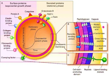
Figure 2.5 Pathogenic factors of Staphylococcus aureus. 23
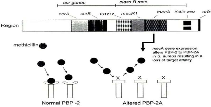
Figure 2.6 Structure of the staphylococcal cassette chromosome mec, with the
recombinase genes complex upstream of the mec complex. 24
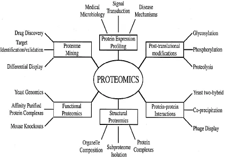
Figure 2.7 Type of proteomics and their applications to biology. 26
Figure 2.8 Schematic diagram showing the mechanisms of how a single gene
can lead to multiple gene products. 27
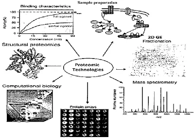
Figure 2.9 Partial view of various proteomic technologies in drug discovery. 31
Figure 2.10 2D-Electrophoresis. Protein spot result from two separations: first
by pI (IEF) and then by size (SDS-PAGE). 35
Figure 2.11 Steps of natural products discovery. 42
Figure 2.12 An example of natural product drug discovery process. 45
Figure 2.13 Schematic diagram illustrates analytical methodology used for
isolation and purification of natural bio-active compounds. 49
Figure 2.14 Aerial parts of Centella asiatica (L.). 53
Figure 2.15 Aerial parts of Imperata cylindrica (L.) in two different life stages. 56
Figure 2.16 Aerial parts of Morinda citrifolia (L.). 58
Figure 2.17 Aerial part of Sauropus androgynus (L.). 61
Figure 3.1 Whitley Automated Spiral Plater and Automatic Colony Counter. 66
Figure 3.2 Automated microbial growth curve analysis system (Bioscreen-C). 67
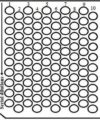
Figure 3.3 Schematic diagram of a 100 well plate used in Bioscreen-C system. 68
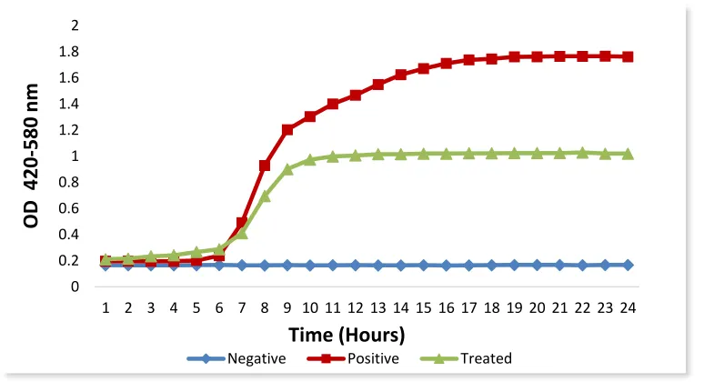
Figure 3.4 A typical Bioscreen growth curve (OD vs. Time). 69
Figure 3.5 A typical profile of fractional area (fa) vs. drug concentration. 70
Figure 3.6 Schematic diagram of Soxhlet extraction apparatus. 72
Figure 3.7 Standard curve for (BSA) using the microtiter plate protocol. 76
Figure 3.8 Experimental workflow of proteomics analysis including sample
preparation and data analysis. 83
Figure 4.1 Disc diffusion assay of Imperata cylindrica L. ethanolic extract in
S. aureus and E. coli. 94
Figure 4.2 Comparative analysis of all bacterial culture MICs of methanolic
Soxhlet crude extracts. 97
Figure 4.3 Comparative analysis of all bacterial culture MICs of ethanolic
Soxhlet crude extracts. 97
Figure 4.4 MBC values of methanolic Soxhlet crude extracts. 98
vii
Figure 4.7 Standard S. aureus growth curve of 1 x and ½ x MIC, activity of
methanolic Soxhlet crude extract of Morinda citrifolia L. 100
Figure 4.8 Standard P. aeruginosa growth curve of 1 x and ½ x MIC, activity
of methanolic Soxhlet crude extract of Morinda citrifolia L. 101
Figure 4.9 Standard E. hirae growth curve of 1 x and ½ x MIC, activity of
methanolic Soxhlet crude extract of Morinda citrifolia L. 101
Figure 5.1 Analytical HPLC profile of Centella asiatica methanolic extract. 112
Figure 5.2 Analytical HPLC profile of Imperata cylindrica methanolic extract. 113
Figure 5.3 Analytical HPLC profile of Morinda citrifolia methanolic extract. 114
Figure 5.4 Analytical HPLC profile of Sauropus androgynus methanolic
extract. 115
Figure 5.5 Preparative RP-HPLC chromatogram of Centella asiatica L.
methanolic extract. 117
Figure 5.6 Preparative RP-HPLC chromatogram of Imperata cylindrica L.
methanolic extract. 118
Figure 5.7 Preparative RP-HPLC chromatogram of Morinda citrifolia L.
methanolic extract. 119
Figure 5.8 Preparative RP-HPLC chromatogram of Sauropus androgynus L.
methanolic extract. 120
Figure 5.9 MIC values of Centella asiatica L. fractions (CA3, CA4 and CA7)
against six different bacterial strains. 122
Figure 5.10 MIC values of Imperata cylindrica L. fractions (IC6 and IC10)
against six different bacterial strains. 122
Figure 5.11 MIC values of Morinda citrifolia L. fractions (MC4, MC7 and
MC8) against six different bacterial strains. 124
Figure 5.12 MIC values of Sauropus androgynus L. fractions (SA2, SA3 and
SA7) against six different bacterial strains. 124
Figure 5.13 MIC values of the most active fractions CA7, IC10, MC4, and SA3,
against six different bacterial strains. 125
Figure 5.14 MBC values of the most active fractions CA7, IC10, MC4, and
SA3, against six different bacterial strains. 125
Figure 5.15 Standard Escherichia coli growth curve with 1 x and ½ x MIC
activity of MC4 fraction of Morinda citrifolia L. 126
Figure 5.16 Standard Staphylococcus aureus growth curve with 1 x and ½ x
MIC activity of MC4 fraction of Morinda citrifolia L. 126
Figure 5.17 Standard Pseudomonas aeruginosa growth curve with 1 x and ½ x
MIC activity of MC4 fraction of Morinda citrifolia L. 127
Figure 5.18 Standard Enterococcus hirae growth curve with 1 x and ½ x MIC
activity of MC4 fraction of Morinda citrifolia L. 127
Figure 5.19 Standard Staphylococcus aureus (MRSA) growth curve with 1 x
viii
Figure 6.1 q
1H NMR spectra of the purity check analysis of the active fraction
(CA7) in Centella asiatica L. 137
Figure 6.2 q
1H NMR spectra of the purity check analysis of the active fraction
(CA7) in Imperata cylindrica L. 139
Figure 6.3 q
1H NMR spectra of the purity check analysis of the active fraction
(CA7) in Morinda citrifolia L. 141
Figure 6.4 Chemical structures of asiatic acid from Centella asiatica L. 144
Figure 6.5 1H NMR spectra of CA7 fraction and standard asiatic acid. 144
Figure 6.6 13C NMR spectra of CA7 fraction and standard asiatic acid. 145
Figure 6.7 Chemical structures of quercetin from Imperata cylindrica L. 146
Figure 6.8 1H NMR spectra of IC10 fraction and standard quercetin. 146
Figure 6.9 13C NMR spectra of IC10 fraction and standard quercetin. 147
Figure 6.10 Chemical structures of aucubin from Morinda citrifolia L. 148
Figure 6.11 1H NMR spectra of MC4 fraction and standard aucubin. 148
Figure 6.12 13C NMR spectra of MC4 fraction and standard aucubin. 149
Figure 6.13 1H NMR spectra of SA3 fraction from Sauropus androgynus L. 149
Figure 6.14 13C NMR spectra of SA3 fraction from Sauropus androgynus L. 150
Figure 7.1 1D-SDS-PAGE separation of Staphylococcus aureus (MSSA) total
proteinsin two conditions. 162
Figure 7.2 Graphical views of the treated sample alignment used in Progenesis
QI for proteomics software. 163
Figure 7.3 Biplot of Principal Component Analysis (PCA) analysis of MSSA
proteome data sets. 164
Figure 7.4 Venn diagram of proteome comparison on control sample of
(MSSA) and aucubin-treated MSSA. 164
Figure 7.5 Gene ontology of the differentially expressed proteins in MSSA. 166
Figure 7.6 Gene ontology of total MSSA proteome in control sample. 168
Figure 7.7 Gene ontology of total MSSA proteome in treated sample. 169
Figure 7.8 Pyruvate metabolism pathway showing the up-regulated genes in
aucubin-traded MSSA relative to control MSSA. 173
Figure 7.9 Glycolysis/gluconeogenesis metabolism pathway showing the
up-regulated genes in aucubin-traded MSSA relative to control MSSA. 174
Figure 7.10 Schematic representation of the pyruvate metabolic pathways in
ix medication. It has lately become more dangerous because we can no longer be certain that any antibiotic chosen will work and because of the emergence of multidrug resistant bacteria. It is more and more clear that antimicrobial resistance is easy to make but hard to miss. Resistance is a major concern with any new agent, and will become even more important in the future if new classes of drugs are established. In pharmaceutical studies, natural products, either as pure compounds or as standardised extracts, provide limitless opportunities for novel drug leads because of the unmatched availability of chemical diversity. Hence, natural products require a powerful and deep assessment of their antibacterial qualities, to help in this unprecedented crisis.
Accordingly, the main objective of this project was concentrating on the development of natural products that can act as an antibacterial agent, with a special reference to the MRSA bacteria, and involved an investigation of the role and mechanism of action of
compounds derived from selected medicinal plants, these are Centella asiatica (L.),
Imperata cylindrica (L.), Morinda citrifolia (L.), and Sauropus androgynus (L.), these plants have previously been implicated as having antibacterial properties. It is a fundamental necessity to uncover and understand the mechanism of action of the tested drug, and its biological pathways in the cell to obtain information for optimisation of lead compounds. During the course of this study, bioassay-guided plant extraction, isolation protocols, and many supporting analytical and biological methodologies were used and developed in order to evaluate the antibacterial activity of the four medicinal plants. Crude extracts were screened against Gram-positive and Gram-negative bacteria, including resistant strains. The isolation procedures were performed using advanced chromatographic techniques including RP-HPLC, guided by the Bioscreen-C results, in order to purify the most active compounds of extracts. These compounds were later identified by employing Mass Spectrometry (MS)
and Nuclear Magnetic Resonance (NMR) techniques. Asiatic acid from Centella asiatica
(L.), Quercetin from Imperata cylindrica (L.), and Aucubin from Morinda citrifolia (L.),
were identified. Aucubin showed to have the highest activity among all other active compounds.
Results suggested that extracts/compounds, from the medicinal plants under investigation, have a potency as an antibacterial agent, with the lowest MIC observed in the
compound derived from Morinda citrifolia (L.) named as aucubin, which was later subjected
to the label-free quantitative proteomics and pathway analysis by using Staphylococcus
aureus (MSSA) as a drug discovery model. The label free proteomics technique conducted with an aid of several software packages, as an attempt to identify the fundamental principles of the mechanism of action of aucubin. Pathway enrichment analysis suggested that the bacterial central metabolism might be the main target of the aucubin, and the pyruvate
metabolism pathway showed the highest enrichment score followed by
glycolysis/gluconeogenesis pathway, these results yet to be validated. An antibacterial that have different mechanism of actions to conventional antibiotics are desirable as they will help slow the onset and spread of microbial drug resistance. Overall, current study suggested
that the compounds isolated from Centella asiatica L., Imperata cylindrica L., Morinda
x This work would not be possible without the help and support of many people. Completing this PhD has been an extremely rewarding and challenging experience, and has changed my outlook on my research career ahead.
First and foremost, I would like to thank all those who supervised my work since I started my PhD. I owe a huge debt of gratitude towards my supervisor Dr. David Pye, who has been my mentor and trainer for almost four years. I greatly appreciate all the hours spent reading my work and providing me with scathing but constructive criticism. Thank you Dave for your advice, patience, encouragement and your keen interest in the project, and above all, for your friendship and having confidence in me despite all the difficulties we faced throughout the project. I would also like to extend my gratitude to my co-supervisor, Dr. James Wilkinson, who has been supporting me throughout my research time. I would also like to thank the Higher Committee for Education Development in Iraq (HCED-Iraq) for sponsoring me to do my PhD in the UK. This research project would not have been possible without their financial support.
I would like to thank Dr. Paul Humphreys at the University of Huddersfield, Hygiene and Disinfection Centre, for allowing me to use his microbiology lab. I would also like to thank him for the invaluable advice he has given me throughout my research. I am grateful to Dr. Richard Beniston at Sheffield University, biOMICS Facility, for his support, advice, and help with the label-free proteomics technique. He spent a tremendous amount of time teaching me the laboratory techniques I needed for the proteomics work, as well as helping me interpret the results critically, and planning further experiments. My special thanks go to Kirit Amin, at University of Salford, Analytical Services (SAS), for his efforts at making the NMR experience as easy as possible. Kirit spent a considerable period of time teaching me how to prepare and run the samples. I would also like to thank Syed Bukhari from Nonlinear Dynamics for his training and help with the Progenesis QI proteomics software, and John Cottrell from Matrix Science for running my proteomics data on Mascot. A special thanks goes out to all the staff and research colleagues in the Biomedical Research Centre at University of Salford, past and present, that have made my PhD an enjoyable and rewarding experience.
Finally, my gratefulness goes to my wife. You are my rock and I will always be grateful for the support and encouragement that you have given me, I could never have done this without you. You and our children are my motivation. I am also eternally grateful to my mum and dad, for their prayers and emotional support, and teaching me to work hard to achieve my goals.
xi
Dedication
This thesis is dedicated to my precious family.
xii This thesis submitted under the University of Salford regulations for the award of a
Doctorate of Philosophy degree by research, and the work was performed under the
supervision of Dr. David Pye, at the University of Salford, Manchester, United Kingdom. I
hereby declare that this thesis and the work presented are my own work and has not been
previously submitted, in whole or in part, to meet requirements for another degree or
qualification in this or any other higher education institution. I also certify that to the best of
my knowledge, wherever contributions of others are involved, every effort is made to
indicate this clearly,and all the sources used have been duly acknowledged and referenced.
xiii
ABC Ammonium Bicarbonate
ACN Acetonitrile
ADP Adenosine Diphosphate
APS Ammonium Persulphate
ATCC American Type Culture Collection
ATP Adenosine Triphosphate
BSA Bovine Serum Albumin
CC Column Chromatography
CFU Colony forming unit
CHCl3 Chloroform
CID collision Induced Dissociation
COSY Correlation Spectroscopy
DAVID Database for Annotation, Visualization, and Integrated Discovery
DDA Data Dependent Acquisition
DIA Data Independent Acquisition
DMSO Dimethyl Sulfoxide
DNA Deoxyribonucleic Acid
DTT Dithiothreitol
EDT Electron Transfer Dissociation
ESI Electrospray Ionisation
EtOH Ethanol
FAME Fatty Acid Modifying Enzymes
FDR False Discovery Rate
FT-ICR Fourier Transform Ion Cyclotron Resonance
FT-IR Fourier Transform Infrared
GC Gas Chromatography
GO Gene Ontology
Hex Hexane
HMBC Heteronuclear Multiple-bond Correlation Spectroscopy HMQC Heteronuclear Multiple-Quantum Correlation
HPLC High-Performance Liquid Chromatography
HPTLC High Performance Thin Layer Chromatography
IEF Isoelectric Focusing
IPG Immobilised pH Gradients
iTRAQ Isotope Tagged Relative and Absolute Quantitation
KEGG Kyoto Encyclopaedia of Genes and Genomes
LC Liquid Chromatography
m/z mass-to-charge
MALDI Matrix-Assisted Laser Desorption Ionisation
xiv
mg Milligram
ml Millilitre
mM Millimolar
MRSA Methicillin-Resistant Staphylococcus Aureus
MS Mass Spectrometry
MS/MS Tandem Mass Spectrometry
MSSA Methicillin Sensitive Staphylococcus Aureus
MW Molecular weight
NCTC National Collection of Type Cultures
NMR Nuclear Magnetic Resonance
NOESY Nuclear Overhauser Effect Spectroscopy
NP-HPLC Normal Phase High-Performance Liquid Chromatography
OD Optical Density
PAGE Polyacrylamide Gel Electrophoresis
PANTHER Protein Analysis Through Evolutionary Relationships
PMS Peptide Mass Fingerprinting
ppm Parts Per Million
PTMs Post-Translational Modifications
qNMR Quantitative Nuclear Magnetic Resonance
RP-HPLC Reverse Phase High-Performance Liquid Chromatography
RNA Ribonucleic Acid
ROESY Rotating Frame Nuclear Overhauser Effect Spectroscopy
SDS Sodium Dodecyl Sulfate
SILAC Stable Isotopic Amino Acids in Culture
TEMED Tetramethylethylenediamine
TFA Trifluoroacetic Acid
TLC Thin-Layer Chromatography
TOCSY Total Correlation Spectroscopy
TOF Time of Flight
tR Retention Time
Tris Trisaminomethane
TSA Tryptone Soya Agar
TSB Tryptone Soya Broth
UHPLC Ultra-High-Performance Liquid Chromatography
UV Ultra Violet
VRSA Vancomycin Resistant Staphylococcus Aureus
WHO World Health Organization
μg Microgram
2
1.1 Project background
Bacteria, are the oldest forms of life on earth, they are remarkably diverse and exist in
surprising numbers. Bacteria are group of micro-organisms that are single cells and
approximately one micron in transverse diameter. Diseases caused by bacteria comprise some
of the most common infections in the world, it is considered one of the threatening issues in
medical field, past, present, and probably future (Relman, 2002). Bacteria have been classified
according to phenotype, including shape, size, staining properties and biochemical properties.
Recently, the classification has been dominated by genotype analysis, especially using
conserved molecules such as 16S ribosomal RNA. On the other hand, bacteria can be classified
as either pathogenic or nonpathogenic bacteria. Pathogenic bacteria cause bacterial infection,
whereas the others do not. Nonpathogenic are commonly called normal flora. Some species of
bacteria, such as Pseudomonas aeruginosa, are opportunistic pathogens and cause disease
mainly in people suffering from immunosuppression (Entenza et al., 2014).
Infections that are induced by bacteria are treated with drugs called antibiotics or
antibacterials. Antibiotics can be described as a compound that works to either stop bacteria
from growing (bacteriostatic agents) or by killing them entirely (bactericidal agents). The
effectiveness of these compounds against the survival of bacteria stems from their ability to
block critical bacterial cellular processes (Sommer & Dantas, 2011). The application of modern
antibiotics therapy (Post-world war II) has had a deep impact on our societies and changed the
features of the medicine and the patient care. Modern procedures in medicine rely on the use of
antibiotics to control infections, organ transplants, surgery, care of premature neonates and to
allow successful patient rehabilitation (WHO, 2014). The use of antibiotics has removed
infectious diseases being the top priority healthcare concern in the western countries over the
last few decades. However, infectious diseases remain the major causes of mortality in
low-income countries and the third highest cause of mortality worldwide (Thomas & Rima, 2011).
The treatment of bacterial infections has been obstructed by resistance to antibiotics. First
observed in Flemings' lab shortly after his discovery of penicillin (1928), the detection of
resistance to antibiotics continued throughout the golden age of antibiotic discovery (1950s)
where many new drugs and drug classes were discovered to keep up with the increasing rates of
3
hazards to global public health and the problem knows no boundaries. Drug-resistant microbes
of all kinds can move amongst people and animals, from one country to another without notice.
The problem is clearly more severe in developing countries where drug availability is limited
and resistance is high (Breu et al., 2008). The World Health Organisation (WHO) describes
antibiotic resistance as a serious threat that is no longer predictable. It is occurring in every part
of the world and has the potential to affect anyone, of any age, in any country (WHO, 2014).
The Infectious Diseases Society of America, which represents the infectious disease specialists,
who are in the front line of antibiotic use, has called for the delivery of a new antibiotic drugs
in the upcoming years, due to the growing threat of antibiotic resistant pathogens which
increasingly causes death and weakening disease (Venter, 2014). The new multi-drug
pathogenic strains which accumulating large numbers of resistance elements, greatly limiting
therapeutic options. Furthermore, the massive financial and logistical burden on health care
sectors across the globe (Wright, 2012).
Ultimately, our primary concern regarding resistance is that resistant bacteria are more
difficult to get rid of and that complications and deaths resulting from infections caused by them
will only increase in time. Very few really new antibiotics have been developed recent years’
term, and tradition natural sources of antibiotics such as soil bacteria and fungi may have given
up all their arsenal of antibiotics (Carlos, 2010). Hence we need to look elsewhere for new
sources of antibiotics, and plant material is an obvious choice. In pharmacognosy studies,
natural products, which are mainly derived from plants, have been widely exploited as a resource
in drug development. For thousands of years, natural products have played an important role
throughout the world in treating and preventing human diseases and once served humankind as
the source of all drugs. Higher plants provided most of these therapeutic agents. Until recently,
the use of traditional medicine was a common practice in poor or developing countries, where
it serves as an alternative medicine because of the lack of the modern health facilities
(Calixto et al., 1998).
In terms of the discovery of novel biologically active compounds, including antibacterial
agents, what is needed are molecules that are diverse in their structure (and consequently diverse
in their functions). Small organic molecules have always been of interest in pharmaceutical and
4
macromolecules that make up living systems (Ji, Li, & Zhang, 2009). A collection of such
compounds has the potential to provide hits against different biological targets. There are a
number of potential sources of small molecules for use in biological screening. Most of the
major categories of antibiotics in therapeutic use are natural products or semi-synthetic
derivatives. In addition, the natural products have the ability to interact with target proteins and
the hit rates in high-throughput screens are usually several times higher for natural products
compared to small molecule from traditional synthetic sources (Galloway et al., 2009).
Plants represent an inexhaustible source of novel chemical compounds, which are of
potential use in medicine and other applications. Plants consist of many active compounds such
as alkaloids, steroids, tannins, glycosides, volatile oils, fixed oils, resins, phenols and flavonoids,
which found in their specific parts such as leaves, flowers, bark, seeds, fruits, root, etc. The
beneficial medicinal effects of plant materials typically result from the combination of these
secondary products. These compounds selectively inhibit the function of biological targets
(Gupta, Naraniwal, & Kothari, 2012). Today, natural products (and their derivatives and
analogs) still represent over 50 % of all drugs in clinical use, with higher plant-derived natural
products representing 25 % of the total. Some relevant examples are galegine, from Galega
officinalis L., which was the model for the synthesis of metformin and other antidiabetic drugs;
papaverine from Papaver somniferum L., which formed the basis for verapamil used in the
treatment of hypertension. Opium is better known as being the source of painkillers such as
morphine and codeine (Cragg & Newman, 2014).
The World Health Organization (WHO) estimates that 80 % of the people in developing
countries rely on traditional medicine for their primary health care, and about 85 % of traditional
medicine involve the use of plant extracts. This means that about 3.5 to 4 billion people in the
world rely on plants as sources of drugs (Chandrawat et al., 2014). The use of plants for
medicinal purposes represents the largest use of biodiversity in the world. Many more species
of plants are used as medicines, than are used for food. This fact concludes a significant role for
natural products in the new drug discovery (Maridass, 2010). Recently, researchers are involved
in an intensive screening of plants used in traditional medicine in an attempt to discover new
5
Natural products have been a rich source of compounds in antibiotic drug discovery with
most antibiotic drugs being derived from a natural product or natural product lead. However,
the rapid onset of resistance to most antibiotics diminishes their effectiveness considerably in
the last two decades. (Maria, 2012). More reasons to add to the current crisis in antibiotic
development is the poor return of investment, which is substantial in drug development. Despite
this, smaller pharmaceutical companies are attempting to address the medical need for new
antibiotics. In addition, the structural complexity of many natural products has often been
understood as an obstacle, since it may impose serious challenges to chemical synthesis and
derivatisation during the lead optimisation process (Luzhetskyy et al., 2007). There is an urgent
necessitates of a constant supply of new antibiotics for effective treatment of infections.
In the development of new antibiotic agents from natural products, several issues need to
be addressed, including the establishment and selection of primary screening assays, which are
crucial to ensure a selection of extracts or molecules with relevant pharmacological action are
worthy to follow up. The assay must be easy to perform, speedy, uncomplicated, produce quick
results, and preferably at a low cost (Monteiro et al., 2012). In order to evaluate the plant
constituents for biological activity, the plant material must firstly be subjected to a suitable
extraction process. Extraction (as the term is pharmaceutically used) is the procedure used in
separation of medicinally active molecules of natural product using selective solvents through
standard processes. A wide range of technologies is available for the extraction of active
components. The crude extract is screened to obtain an evaluation of the potential biological
activities, the crude extracts are sequentially fractioned, and each fraction subjected to a further
suitable bio-assay tests (Bobzin, Yang, & Kasten, 2000).
There are various chromatographic techniques available to separate the bio-active
components from the crude mixture, such as Column Chromatography (CC), Liquid
Chromatography (LC), and Gas Chromatography (GC). Purification of the bio-active
compounds from plants by chemical and chromatographic approaches usually followed by
structural characterisation using spectroscopic methods including Ultra Violet (UV), Fourier
Transform Infrared (FT-IR), Nuclear Magnetic Resonance (NMR), and Mass Spectrometry
(MS). NMR and MS are the most powerful and widely used techniques for the structure
6
In the field of drug discovery, it is necessary to distinguish between the cause of the
disease and its mechanism at the molecular level. Protein molecules that linked to a given
disease are identified first. Alongside the determination of the target, we must understand the
mechanism of action of the tested drug and its biological pathways in the cell. Currently,
approaches to analyse how a drug works fall into these broad categories. First, includes several
strategies that rely on model organisms that are compatible with the genetic manipulations.
Second category involves affinity-based methods employed to identify proteins that stick to the
drug. (Wacker et al., 2012). Modern drug discovery seeks to identify new small molecules that
potently and selectively modulate the functions of target proteins (Monica et al., 2013). Good
drugs must have significant effects on a specific biological pathway and minimal effects on all
other pathways. Confirmation that a tested compound inhibits any intended target and the
identification of mainly secondary effects are among the main challenges in the development of
new drugs. Extensive methods that enable researchers to determine which genes or activities are
affected by a given drug might improve the efficiency of the drug discovery process by quickly
identifying potential protein targets (Marton et al., 1998).
Proteomics may be defined as the genome-scale analysis of the structure, abundance,
localisation, modification, and function of proteins present in a cell, organ, or organism at a
given time. Currently, proteomics has gained comprehensive attention in the field of drug
development and mechanism studies, since proteomic analysis provides a direct reflection of
gene expression. Identifying protein targets of bio-active compounds are an efficacious
approach to discover unknown protein functions, as well as dissect molecular mechanisms of
drug effectiveness. It is really important to systemically characterise the drug and its reactive
intermediate (Ji, 2015). Technically, proteomic analysis requires the combination of several
technologies for protein separation and processing such as two-dimensional polyacrylamide gel
electrophoresis (2D-PAGE), high-performance liquid chromatography (HPLC), and mass
spectrometry (MS) which is the primary tool for protein identification and characterisation.
There are many types of each technique and the researcher may choose the one useful for the
intended work. Bioinformatics used in the analysis of qualitative and quantitative proteomic
data, various search engines and software packages available for researchers, i.e. MaxQuant,
7
Most proteomic studies involve the high-resolution separation of proteins from a complex
protein mixture isolated under certain experimental conditions. Particular proteins of interest
are identified using MS data linked to a genome sequence database by specialised softwares.
The ultimate goal of proteomics is the complete identification and quantification of a specific
proteins, with aim of revealing proteins function and post-translational modifications (PTMs)
as part of a complex associated system. It is crucial to include proteomic study in the field of
drug discovery to reveal the mechanism of action and functional pathways of the drug under
investigation (Cooper & Carucci, 2004). There are a number of ways to perform comparative
analysis of proteins using MS techniques within samples, either using labeling techniques or
label-free techniques. Labeling of samples for subsequent mass spectrometric analysis allows
for direct comparison of individual protein abundance between two different conditions by
comparison of the mass shift induced in labeled peptides with their unlabeled counterpart (Alan
et al., 2005). Despite the success of labeling techniques in quantification of relative abundances
of proteins between two samples, the labeling reagents are expensive, and data analysis of mixed
samples is often complicated and time consuming. On the other hand, label-free techniques have
the advantage of comparing two samples with different conditions. In addition, label-free
approaches can offer the advantage of relatively uncomplicated data analysis and less costly
(Tuli & Ressom, 2009). Current study employed the label-free proteomics approach to
investigate the antibacterial mode of action of the compounds under investigation in
Staphylococcus aureus (MSSA) as a drug discovery model.
The noticeable developments in gene and protein sequence databases, provides a means
to identify proteins expressed in organisms. Bioinformatics tools, such as Mascot, make it much
easier to access these databases to search, match and identify a protein component. UniProt,
trEMBL ExPASy, Swiss-Prot and NCBInr are the widely used protein sequence databases
(Kambiz, Luc and Sylvia 2010). Identification of the cellular targets for the tested compounds
is a crucial step in understanding their true mode of action (Wang et al., 2011).
After a protein is synthesised, it can undergo posttranslational modifications (PTMs), the
post-translational modification of proteins plays a critical role in the regulation of a broad range
of cellular processes. The analysis of posttranslational modifications is particularly critical for
8
influence their structure, stability, function, protein-protein interactions, and localisation within
the cell. There are multiple assays available to measure post-translational modifications,
including western blots, ELISAs, and mass spectrometry (Freiberg et al., 2004; Brötz-Oesterhelt
et al., 2005).
1.2 Aims and objectives of the present study
The main aim of this PhD project was the identification and characterisation of natural
products that can act as antibacterial agents. The objective of this research was to investigate
the role and mechanism of action of compounds derived from four medicinal plants; Centella
asiatica L., Imperata cylindrica L., Morinda citrifolia L., and Sauropus androgynous L. These
plants have previously been implicated as having antibacterial properties and have previously
raised a significant interest of many researchers. In order to evaluate these plants therapeutically,
many lab techniques have been used and developed throughout this project. The outline
strategies followed in this project, are:
1. Extraction and purification of small molecules from Centella asiatica L., Imperata
Cylindrical L., Morinda citrifolia L. and Sauropus androgynous L. by using various
extraction and purification techniques.
2. In-vitro evaluation of the antibacterial activity of these compounds against various known pathogens by utilising convenient biological assays in order to investigate their
potential therapeutic activities, and determination of their Minimum Inhibitory
Concentration (MICs) and Minimum Bactericidal Concentration (MBCs).
3. Isolation and structure identification of the active compounds comprised in Centella
asiatica L., Imperata Cylindrical L., Morinda citrifolia L., and Sauropus androgynous
L. extracts that are responsible for inhibition of bacterial growth by standard
spectroscopy techniques.
4. Elucidation of the mechanism of action of selected compounds shown to have the potent
antibacterial activities via liable-free proteomics analysis and MS fingerprinting.
5. Identification of the targets on the bacteria that are bound to the active compounds
9
1.3 Significance of the study
Bacteria, which cause diseases, react to the antibiotics used as treatment by becoming
resistant to them, sooner or later, this natural process of adaptation and antimicrobial resistance
affect the lifespan of antibiotics. The antimicrobial resistance has increased recently due to
unnecessary use and inappropriate use of antibioticsespecially. Accordingly, this lead to hyper
mutation of many bacterial strains and spread of resistant bacteria (Chauhan et al., 2013). As
resistance and virulence increases, the cost and burden on society increases as well, the
damaging effects of the antimicrobial resistance are already manifesting themselves across the
world. Antimicrobial resistant infections currently claim at least 50,000 lives each year across
Europe and the US alone, with many hundreds of thousands more dying in other areas of the
world. Latest figures suggest that drug resistant infections could kill an extra 10 million people
across the world every year by 2050 if they are not tackled. World Health Organization (WHO)
is calling on governments and the pharmaceutical industry to work together in taking
comprehensive action against drug-resistant infections by increasing the investment in antibiotic
researches to meet global public health needs (Suraj & Sapkal, 2015).
New strategies have been implemented to combat this growing crisis and to help the
healthcare sector coping with antimicrobial resistance. Investigation of the antimicrobial activity
of the secondary metabolites from natural products represents a promising possible solution.
Currently, there is an increasing interest in the isolation, screening and exploitation of
antimicrobial activity of natural products of plants as an inexpensive and rich source of
secondary metabolites. In this project, different strategies were implemented to evaluate the
antibacterial activity of four medicinal plants, Centella asiatica L., Imperata cylindrica L.,
Morinda citrifolia L., and Sauropus androgynous L., in-vitro against various strains of
pathogenic bacteria.
Although the biological effects of Centella asiatica L., Imperata cylindrica L., Morinda
citrifolia L., and Sauropus androgynous L. have been studied, there is no comprehensive study
available to explain the actual role of active compounds from these plants and the mechanisms
of their action. Accordingly, this project conducted a screening of the effects of various extracts
of these plants to demonstrate their antibacterial activity. The extracts of interest were further
10
activities. The elucidation of possible mechanisms of action of the activity was also studied
using LC-MS/MS proteomics approach. Success in combatting antimicrobial resistance
requires sustainable commitment and partnership with the private sector, governments, and
12
2.1 Antibiotics
The word antibiotic is derived from the Greek words anti (against) and bios (life) and
means, in principle, a substance, which kills any living organism. However, in medical practice,
antibiotics are compounds produced by, or derived from, certain fungi, bacteria, and other
organisms, which used to combat bacterial infections by either prevent the bacteria from
growing (bacteriostatic agents) or to kill them outright (bactericidal agents) (Sommer & Dantas,
2011). The term antibiotic is used to refer to a drug that cures infections caused by bacteria,
while an antimicrobial agent is a general term mostly applied to substances working on bacteria
(antibacterial), but can also be applied to agents working on viruses (antivirals), fungi
(antifungals) and parasites (antiparasitic agents). Antimicrobials include antibiotics produced
by other organisms (e.g. penicillin, tetracycline, and erythromycin), chemically modified
antibiotics (e.g. doxycycline) as well as chemically produced substances (e.g. fluoroquinolones)
(Davies, J, & Davies, D., 2010).
There has been an ongoing conflict throughout history between humans and the
uncountable numbers of microorganisms that cause infection and disease. Around the middle of
the twentieth century, there was a major evolution in antibacterial drug development and other
means of infection control, which helped turn the tide in favour of humans. This development
was dramatically improved when penicillin became available for use in the early 1940s.
However, the euphoria of this potential victory over infectious diseases last only for a limited
time (Lynne, 2000). The first antibiotic, penicillin, was discovered by Alexander Fleming in
1928 when he observed that a common mold (Penicillum) produced a substance that lysed
colonies of Staphylococcus spp. The first major development after the introduction of penicillin
was ampicillin, which offered a broader spectrum of activity than the original penicillin.
Ampicillin has been used extensively to treat bacterial infections since 1961. In the following
decades, many new antibiotics with novel properties were discovered, including streptomycin,
chloramphenicol, and tetracycline. Modification of already known antibiotics has led to several
derivatives having different antibacterial activities, pharmacokinetic properties, and resistance
characteristics as compared to the older drugs (Sharma et al., 2013).
The effectiveness of these compounds ‘against life’ stems from their ability to block or
13 pathogen, which are different from that found in the host. For instance, the antibiotics
chloramphenicol and tetracycline inhibit the bacterial ribosome (serve as the site of mRNA
translation and protein synthesis), but not the structurally-different eukaryotic ribosome,
therefore it shows selective toxicity against bacteria only (Olgica et al., 2012).
Almost as soon as antibiotic drugs were rolled out for general use in the world’s
population, bacteria responded by developing various forms of resistance. As antibacterial usage
increased, so did the level and complexity of the resistance mechanisms exhibited by bacterial
pathogens (Nikaido, 2009). The struggle to control the infections continues to this day, and
surprisingly the number of scientists who are developing new antibiotic agents has begun to fall
back, even as bacteria evolve ever more advanced mechanisms of resistance. Nonetheless,
recent lobbying of governments, worldwide, has brought a change in attitudes toward antibiotic
research and more money is being made available to research centres to support this most urgent
experimentation (Tenover, 2006).
The number of new antibiotic agents coming on to the market is falling, for instance,
sixteen agents were approved for use between “1983-87” but only seven were approved between
“1998-2002” (Bush & Pucci 2011). Recently, only two new classes of antibiotics; daptomycin
and oxazolidinones have been utilised to treat Gram-positive infections, whereas, innovation to
address Gram-negative bacteria are still struggling. Furthermore, drugs with novel modes of
action are even fewer in number. For instance, linezolid, which was approved in 2003, had a
novel mechanism of action (protein synthesis inhibitor, which works by blocking the initiation
of protein production, and not one of the later steps), while the remainder were solely modified
structures of existing agents. (Miller et al., 2014).
A major problem of novel antibiotic development is that many drug companies see it as
an unattractive financial risk. This is due to huge production costs for compounds which require
relatively small doses and short treatment cycles to be effective and which may not have a long
clinical shelf life. Many 'Big Pharma' are focusing their efforts on compounds used in long-term
treatment plans, for chronic illnesses, obesity and quality of life drugs, all of which are more
probable to yield a larger profit than antibiotics. Still, many governments and international
authorities are aware of the situation and measures are being conducted to enhance awareness
14
2.1.1 Antibiotic resistance of bacterial pathogens
Antibiotics originally evolved within bacteria almost a billion years ago as organisms
competed with each other, long before mankind had discovered antibiotics. It is fair to presume
that resistance to antibiotics developed shortly afterwards, as susceptible bacteria developed
means to defend themselves against antibiotic-producing bacteria (Bradley, 2014). Since the
introduction of antibiotics into clinical use in the mid-1940s, microorganisms have shown a
remarkable ability to protect themselves by developing and acquiring antibiotic resistance. By
1942, penicillin resistance was reported after only a few months of limited clinical trials. With
the development of resistance to tetracycline, streptomycin, chloramphenicol and erythromycin,
other classes of antibiotics began to emerge (Mengiste, Hagos, & Moges, 2014).
Many antibiotics from the days of Alexander Fleming were used for healing diseases.
When microorganisms are exposed to antibiotics they adapt some alternative survival routes
related to their metabolism, enzyme production and gene transfers. Microbes found adaptation
to unfavourable environments and continuous doses of antibiotics. This phenomenon is
frequently found throughout the world where organisms come into contact with antibiotics, and
the occurrence of the resistance is more likely when patients being exposed to high dosages of
antibiotic, such as during hospitalisation (Smitha et al., 2014). Figure (2.1) summaries the
timeline of antibiotics deployment and their resistance development.
The overwhelming use of antibiotics has played a significant role in the emergence of
antibiotic resistant bacteria. Resistance of pathogenic organisms to approved antibiotics has
become one of the medical world’s biggest concerns with serious consequences for the treatment
of infectious diseases in patients. The main cause of this phenomenon is the increased use or
misuse of antibiotics. There has been a terrifying increase of antibiotic resistance in bacteria that
cause either community infections or hospital - acquired infections (Nosocomial infections).
There is a particular interest in multidrug resistant pathogens (MDR), and one of these in
particular is methicillin resistant staphylococcus aureus MRSA (Alekshun & Levy, 2007).
Hospital infections with methicillin resistant staphylococcus aureus raised serious public health
concerns recently, since the infection is resistant to numerous antibiotics, including methicillin,
amoxicillin, penicillin and oxacillin, thus making it challenging to treat the infection
15
Figure 2.1: Timeline of antibiotics deployment (top) and the development of antibiotic
resistance (bottom). Adapted from (Clatworthy et al., 2007)
Pharmaceutical companies have ignored or dropped dramatically investments in the
antibiotic research as mentioned earlier, and as a result the race to develop new classes of
antibiotics with novel modes of action has been compromised in the last decades (Fox, 2006).
In the absence of major new classes of antibiotics, we need to focus our efforts on new sources,
such as natural products, as an alternative provider of antimicrobial agents in order to address
this problem of mounting resistance. In addition, other strategies have to be considered to
minimise the development of resistance, such as judicious antibiotic use, narrow spectrum
antibiotics should also be used whenever possible, and antibiotics given for short courses and at
appropriate times (Breu, Guggenbichler, & Wollmann, 2008). Accordingly, in search of a better
drug, we have concentrated our search on natural product compounds derived from plants.
Recently, natural product compounds have attracted attention of medical researchers towards
their search for potential antibiotic.
2.1.2 The Basic characteristics of antibiotics
Nowadays, there are about 4000 compounds identified with antibiotic properties.
Antibiotics are derived from three main sources: moulds or fungi, bacteria and synthetic or
semi-synthetic compounds. They can be used either topically or internally, and the results of their
action are either inhibition of the growth of pathogens or their elimination. Therefore, antibiotics
16 is not absolute, and depends on the drug concentration, the bacterial species, and the phase of
growth. (Shamnas, Arya, & Deepak, 2013).
Antibiotics can be further classified on their ability to target Gram positive and Gram
negative bacteria, these classifications are broad and narrow spectrum antibiotics. Broad
spectrum antibiotics are active against both Gram-positive and Gram-negative organisms.
Examples include: tetracyclines, phenicols, and fluoroquinolones. Narrow spectrum
antibacterials have limited action and are mainly useful against particular species of
microorganisms. For instance, glycopeptides and bacitracin are only effective against
Gram-positive bacteria, whereas polymixins are usually only effective against Gram negative bacteria.
Aminoglycosides and sulfonamides are only effective against aerobic organisms, while
nitroimidazoles are generally only effective against anaerobes organisms (Shaikh et al., 2015).
2.1.3 Mechanisms of antibiotic actions
Antibiotics are used to treat bacterial infections by interrupting the physiological
mechanisms inside the bacterial cell that allow normal cellular function. Antibiotics agents act
selectively on vital bacterial functions with minimal effects, or without affecting host cell
mechanisms. Different antimicrobial agents act in different ways, the understanding of these
mechanisms as well as the chemical nature of the antimicrobial agents is crucial in the
understanding of the ways in which resistance against them develops (Kohanski et al., 2010).
As mentioned earlier, antibiotic agents may be described as either bacteriostatic or
bactericidal. Bacteriostatic antibacterial agents only inhibit the growth or multiplication of the
bacteria, giving the immune system the time to clear them. Complete elimination of the bacteria
in this case therefore is dependent on the efficiency of the immune system. Bactericidal agents
kill the bacteria with or without the assistance of the immune system of the host. However, the
mechanism of action of antimicrobial agents can be categorised further based on the structure
of the bacteria or the functions that are affected by the agents (Salih, Salimon, & Yousif, 2012).
Accordingly, antibiotics can be classified into five distinct classes based upon their mechanism
of action. Those classes are (1) inhibition of bacterial cell wall biosynthesis, (2) disruption of
bacterial cell membrane, (3) inhibition of protein biosynthesis, (4) inhibition of
deoxyribonucleic acid (DNA) or ribonucleic acid (RNA) synthesis, and (5) inhibition of folate
17
Mechanism of action Target Antibiotic families
Inhibition of cell wall synthesis
Penicillin binding proteins, D-alanyl-D-alanine, Muropeptide transport
β-lactam (Penicillins, Cephalosporins, Carbapenems, Monobactams); Glycopeptides; Cyclic lipopeptides (Daptomycin)
Inhibition of protein synthesis
30s and 50s subunits of the
ribosome
Tetracyclines; Aminoglycosides; Oxazolidonones (Linezolid); Streptogramins (Quinupristin-dalfopristin); Ketolides; Macrolides;
Lincosamides
Inhibition of DNA synthesis
DNA gyrase, DNA structure
integrity Fluoroquinolones (Ciprofoxacin)
Inhibition of RNA synthesis RNA polymerase Rifampin
Inhibition of folic acid pathway
Dihydrofolate reductase,
Dihydropteroate synthetase Sulfonamides; Trimethoprim
Disruption of
bacterial membrane Phospholipid structure Polymyxins (Polymyxin-B, Colistin)
Table2.1: Summary of mechanisms of action and their targets of selected classes of antibiotic.
Adapted from (Brotz-Oesterhelt & Brunner, 2008).
2.1.4 Mechanisms of antibiotic resistance
After the discovery and commercial introduction of antibiotics, it was soon noticed that
usually treatable infections were not affected by the treatment with antibiotics and had adapted
mechanisms of resistance. Alexander Fleming predicted that too low frequent doses of penicillin
would lead to the development of penicillin resistance. Surprisingly, bacterial resistance towards
penicillin was actually noticed before it was made widely available to use, after a while, it was
18
antibiotics were not restricted by any means (Hellen et al., 2015). Antibiotic resistance is a result
of the evolutionary pressure that bacteria undergo, resistance occurs for all antibiotics after their
clinical administration, and there is a limit to the number of antibiotic substances which fulfil
all pharmacokinetics demands. Because of this, much of the antibiotic work done after the 1960s
was focused on chemically modifying existing antibiotics to make them more potent to resistant
pathogens and to improve pharmacokinetics properties. Recently, researchers have tried to find
a new antibiotic candidate from natural products (e.g. medicinal plants) in a hope to meet the
health sector demands (Davies & Dorothy, 2010).
There are many methods by which bacteria can become resistant to antibiotics. The current
scale of the problem and the number of resistances against drugs across different classes is
unprecedented. Resistance can be caused by a variety of mechanisms and it can be summarised
as: (i) the presence of an enzyme that inactivates the antibiotic agent, bacteria can produce
enzymes that are capable of adding different chemical groups to antibiotics that prevents binding
between the antibiotic and its target in the bacterial cell; (ii) the presence of an alternative
enzyme for the enzyme that is inhibited by the antibiotic agent, for instance, Staphylococcus
aureus can acquire the resistance gene mecA and produce a new penicillin-binding protein, the
new penicillin-binding protein has low affinity to β-lactam antibiotics and results resistant to
the drugs, (iii) a mutation in the antibiotic agent’s target which results changes in the
composition or structure of the target in the bacterium and stop the antibiotic from interacting
with the target. Alternatively, the bacteria can add different chemical groups to the target
structure, in this way shielding it from the antibiotic; (iv) destroy the antibiotic through enzymes
that can inactivate antibiotics. One obvious example is β-lactamase that destroys the active
component (the lactam ring) of penicillin. Later, bacteria developed extended-spectrum
lactamases and become a major problem, due to the ability of destroying a wide spectrum of
β-lactam antibiotics; (v) reduced uptake of the antibiotic agent,bacteria achieve that by decrease
the permeability of the membrane that surrounds the bacterial cell which make it more difficult
to pass through; and (vi) efflux pumps, bacteria can produce pumps that are localised in their
membrane or cell wall. In some cases, mutations in the bacterial DNA can make the bacteria
produce more pumps to reduce the antibiotic concentration inside the bacterial cell (Fluit et al.,
19
Figure 2.2: Schematic diagram of major antibiotic mechanisms of resistance (Gullberg, 2014).
At least seventeen different classes of antibiotics have been produced to date.
Unfortunately, for each one of these classes at least one mechanism of resistance has developed
over the years. In fact, in some cases these bacteria have been able to develop simultaneous
resistance to two or more antibiotic classes, making the treatment of infections caused by these
microorganisms extremely difficult, very costly and in many cases associated with high
morbidity and mortality (Alanis, 2005).
Antibiotic resistance can be divided into natural resistance and acquired resistance.
Natural resistance means that the bacteria are intrinsically resistant, an example of this can be
due to increased efflux activity, a mechanism responsible for moving out toxic substances and
antibiotics outside the cell. Acquired resistance refers to bacteria that are usually sensitive to
antibiotics, but are liable to develop resistance. Acquired resistance is often caused by mutations
in chromosomal genes, or by the acquisition of mobile genetic elements, such as plasmids or