0095-1137/11/$12.00 doi:10.1128/JCM.01750-10
Copyright © 2011, American Society for Microbiology. All Rights Reserved.
Human and Swine Hosts Share Vancomycin-Resistant
Enterococcus faecium
CC17 and CC5 and
Enterococcus faecalis
CC2 Clonal Clusters
Harboring Tn
1546
on Indistinguishable Plasmids
䌤
†
Ana R. Freitas,
1,2Teresa M. Coque,
2,3,4* Carla Novais,
1,5Anette M. Hammerum,
6Camilla H. Lester,
6Marcus J. Zervos,
7,8Susan Donabedian,
7Lars B. Jensen,
9Maria Victoria Francia,
10Fernando Baquero,
2,3,4and Luísa Peixe
1REQUIMTE, Laborato´rio de Microbiologia, Faculdade de Farma´cia, Universidade do Porto, Porto, Portugal1; Servicio de Microbiología, Instituto Ramo´n y Cajal de Investigacio´n Sanitaria (IRYCIS), Madrid, Spain2; Unidad de Resistencia a Antibio´ticos y
Virulencia Bacteriana (RYC-CSIC), Madrid, Spain3; Ciber en Epidemiología y Salud Pu´blica (CIBER-ESP), Madrid, Spain4; CEBIMED, Faculdade Cieˆncias da Sau´de, Universidade Fernando Pessoa, Porto, Portugal5; Department of
Microbiological Surveillance and Research, Statens Serum Institut, Copenhagen, Denmark6; Henry Ford Health System, Detroit, Michigan7; Wayne State University School of Medicine, Detroit, Michigan8; Division of Microbiology and
Risk Assessment, National Food Institute, Danish Technical University, Copenhagen V, Denmark9; and Servicio de Microbiología, Hospital Marque´s de Valdecilla e Instituto de Formacio´n e
Investigacio´n Marque´s de Valdecilla (IFIMAV), Santander, Spain10
Received 27 August 2010/Returned for modification 10 October 2010/Accepted 21 December 2010
VRE isolates from pigs (nⴝ29) and healthy persons (nⴝ12) recovered during wide surveillance studies
performed in Portugal, Denmark, Spain, Switzerland, and the United States (1995 to 2008) were compared with
outbreak/prevalent VRE clinical strains (nⴝ190; 23 countries; 1986 to 2009). Thirty clonally related
Entero-coccus faeciumclonal complex 5 (CC5) isolates (17 sequence type 6 [ST6], 6 ST5, 5 ST185, 1 ST147, and 1 ST493) were obtained from feces of swine and healthy humans. This collection included isolates widespread among pigs of European Union (EU) countries since the mid-1990s. Each ST comprised isolates showing
similar pulsed-field gel electrophoresis (PFGE) patterns (<6 bands difference; >82% similarity). Some CC5
PFGE subtype strains from swine were indistinguishable from hospital vancomycin-resistant enterococci
(VRE) causing infections. A truncated variant of Tn1546(encoding resistance to vancomycin) andtcrB(coding
for resistance to copper) were consistently located on 150- to 190-kb plasmids (reppLG1).E. faecium CC17
(ST132) isolates from pig manure and two clinical samples showed identical PFGE profiles and contained a
60-kb mosaic plasmid (repInc18plus reppRUM) carrying diverse Tn1546-IS1216variants. The onlyEnterococcus
faecalisisolate obtained from pigs (CC2-ST6) corresponded to a multidrug-resistant clone widely disseminated in hospitals in Italy, Portugal, and Spain, and both animal and human isolates harbored an indistinguishable
100-kb mosaic plasmid (reppRE25plus reppCF10) containing the whole Tn1546backbone. The results indicate
a current intra- and international spread ofE. faeciumandE. faecalisclones and their plasmids among swine
and humans.
Vancomycin-resistant enterococci (VRE) are among the most common nosocomial pathogens in the United States and in several European Union (EU) countries (23, 50). They have frequently been isolated from farm animals, pets, and retail food products in Europe, but until very recently, the detection of VRE from either processing or production food animal environments in the United States was infrequent (11, 20, 42, 53). There is limited evidence as to the direct role of the food chain in the dissemination of VRE among humans. Despite this, the potential hazard has been widely recognized and led to the adoption of intervention measures, such as the ban on the growth-promoting use of antimicrobials in the EU. A re-markable reduction in the prevalence of VRE among animals
and humans has been observed after the EU withdrawal (see reference 42 and references therein). However, the role of nonhuman hosts as reservoirs of highly transmissible clones, the transient or permanent human fecal carriage of VRE of animal origin, and the consequent risk of gene transfer to resident human flora are issues still discussed and not fully addressed (41, 42).
Both Enterococcus faecium and Enterococcus faecalis are opportunistic pathogens comprising some host-specific lin-eages (30, 53). Strains from human-adapted clonal complexes (CCs) causing most enterococcal infections may eventually be recovered from farm and companion animals (e.g.,E. faecium
clonal complex 17 [CC17] and E. faecalisCC2), and strains from CCs commonly found among animals have also been isolated from humans (E. faecium CC5, E. faecalis sequence type 16 [ST16], or E. faecalis CC21) (4, 9, 13, 14, 28, 53). Documented cases of animal-human VRE transmission fre-quently involve healthy humans in close interaction (farming or petting) with animals, but most of these studies do not provide molecular characterization of either clones or their
* Corresponding author. Mailing address: Servicio de Microbiología, Hospital Universitario Ramo´n y Cajal, Carretera de Colmenar, km. 9.1, Madrid 28034, Spain. Phone: 34-913368330. Fax: 34-913368809. E-mail: [email protected].
† Supplemental material for this article may be found at http://jcm .asm.org.
䌤Published ahead of print on 12 January 2011.
925
on May 16, 2020 by guest
http://jcm.asm.org/
subcellular genetic elements (1, 3, 10, 17, 26, 28, 31, 33), de-spite the comprehensive epidemiological studies of Tn1546
(vanA) and Tn5382(vanB) (8, 24, 38, 52, 54).
In this work, a comparative multilayered molecular analysis of representative VRE strains from swine, healthy humans, and clinical isolates recovered from wide surveillance studies was carried out with the aim of identifying and characterizing epidemic VRE clones and plasmids shared by human and swine hosts.
MATERIALS AND METHODS
Bacterial strains and epidemiological background.The epidemiological back-ground of the 41 isolates analyzed in this study is shown in Table 1. It includes representative isolates of VRE recovered from swine and healthy humans in national surveillance studies conducted in Portugal and Denmark (1995 to 2008) (references 17, 26, 35, 37, and 38 and this study), strains widespread among swine from Switzerland and Spain (5, 22), and the first VRE isolates recently recovered from animals in the United States (11). For comparison, we included a large and well-typed collection of clinical VRE isolates (140E. faeciumand 50E. faecalis
isolates) recovered from 23 countries, including Portugal, Spain, Denmark, and the United States, during the last 3 decades, most of which had caused hospital outbreaks (12, 16). Testing of susceptibility to 12 antibiotics was performed either by E strip (bioMe´rieux, Solna, Sweden) or by a standard agar dilution method following recommended guidelines of the manufacturer or CLSI (6). The presence of genes coding for antimicrobial (glycopeptides, macrolides, tet-racyclines, and aminoglycosides) and copper (tcrB) resistance and putative vir-ulence traits (agg,gel,cyl,esp, andhylEfm) was analyzed by using different PCR schemes (18, 36).
Clonal relatedness.Clonal relatedness was established by pulsed-field gel elec-trophoresis (PFGE) and multilocus sequence typing (MLST), as described previ-ously (36, 48; http://www.mlst.net). Computer analysis of the PFGE banding patterns was performed with the Fingerprinting II Informatix software package (Bio-Rad Laboratories, Hercules, CA). The similarity of the PFGE banding patterns was analyzed by the Dice coefficient, and cluster analysis was performed by the un-weighted pair group method with arithmetic average (UPGMA). We applied a cutoff equivalent to 82% to group possibly genetically related isolates (34). Different PFGE types were designated by capital letters or numbers (35, 36, 38). Subtypes were designated by a number (indicating the number of bands that differed from the
index strain) and primes when necessary (to distinguish among subtypes with the same number of bands but showing different banding patterns) (48).
Characterization of glycopeptide resistance.The Tn1546backbone was ana-lyzed by PCR mapping, as previously described (38, 54). Conjugation experi-ments were performed by filter mating at a 1:1 donor-recipient ratio usingE. faeciumGE-1 and/or 64/3 andE. faecalisJH2-2 as recipient strains and vanco-mycin (6 mg/liter), rifampin (30 mg/liter), and fusidic acid (20 mg/liter) as antimicrobial selective markers. The genomic locations ofvanAandtcrBwere assessed by hybridization of I-CeuI and S1 nuclease (Takara Bio Inc., Shiga, Japan)-digested genomic DNA using intragenicvanA,tcrB, and 23S rRNA gene probes (16). Plasmid analysis included determination of size and content by PFGE of S1 nuclease-digested genomic DNA as previously described (16). Also, the identification of replication initiator proteins (rep) and maintenance systems (the toxin-antitoxin systems Axe-Txe and-ε-and the partition module
par-pAD1) was performed using recently developed PCR plasmid-typing methods, sequencing, and hybridization (25, 44). Restriction fragment length polymor-phism (RFLP) analysis using EcoRI or ClaI enzyme was performed for plasmids of⬍150 kb (13). The plasmids were designated with Roman numerals (E. faecalis) or capital letters (E. faecium), following the nomenclature of previous studies in which some of the strains were initially reported (13). Hybridization experiments were performed by using the Gene Images AlkPhos Direct labeling and detection system (Amersham GB, GE Healthcare Life Sciences UK Limited). We referred to the rep sequences according to the plasmid type in which they were identified, as well by as the numeric nomenclature used by Jensen et al. (25).
RESULTS
Thirty-two (31E. faecium[VREfm]; 1E. faecalis[VREfs]) of the 41 VRE isolates studied were grouped in PFGE types highly similar to those of strains causing infection in hospital-ized patients. They were identified asE. faeciumCC5 (n⫽30),
[image:2.585.40.545.90.324.2]E. faeciumCC17 (n⫽ 1), andE. faecalis CC2 (n⫽ 1). Five other fecal isolates from healthy humans were classified asE. faeciumST18 (CC17), which is one of the predominant STs of the polyclonal subcluster CC17, although no similar PFGE types were observed among hospital VRE. The other four isolates were recovered from healthy humans, but their PFGE
TABLE 1. Origins of VRE isolated from swine and healthy humans during national surveillance studies in Denmark, Portugal, Spain, Switzerland, and the United States (1995 to 2008)
Species Country
No. isolated from:
Origin Reference(s)
Swine Healthy humans
E. faecium(n⫽40) Denmark 15 3 15 VREfm from healthy pigs among 1,594E. faeciumfecal
isolates (DANMAPa, 1995–2006) 17, 26; this study 3 VREfm recovered from 525 community-dwelling human
samples (2002–2006)
17; this study
Portugal 4 9 4 VREfm from 84 fecal or environmental samples in different production piggeries (1997; 2006–2007); one isolate from 1997 belongs to the CC5 widespread clone recovered in different countries over years
14, 35
9 VREfm fecal isolates from 99 healthy volunteers living in different Portuguese cities (2001–2004)
37
United States 6 0 6 VREfm recently isolated from 55 swine (10.9%) in three Michigan counties (2008)
11
Spain 2 0 2 VREfm recovered from 900 pig fecal samples at slaughterhouses (9.7% of all pigs slaughtered in 1998) in Valencia and Murcia (1998–2000)
22, 35
Switzerland 1 0 1 VREfm recovered from samples of pig feces among 155 Enterococcusisolates obtained in 16 Swiss farms (1999–2000)
5, 35
E. faecalis(n⫽1) Portugal 1 0 1 VREfs from 84 fecal or environmental samples in different production piggeries (2006–2007)
13
aDANMAP, Danish Integrated Antimicrobial Resistance Monitoring and Research Programme.
on May 16, 2020 by guest
http://jcm.asm.org/
or MLST profiles were not related to animal or hospital VRE. A detailed analysis of the isolates found in swine and healthy and hospitalized humans is provided in Table 2.
CC5E. faeciumcarryingvanAon large plasmids (>150 kb) spread among human and swine hosts from the EU and the
United States.ThirtyE. faeciumisolates clustering in CC5 (17
ST6, 6 ST5, 5 ST185, 1 ST147, and 1 ST493) were obtained from swine samples from Denmark, Portugal, Spain, Switzer-land, and the United States, as well as from fecal samples from healthy Danes (1995 to 2008) (Table 2). Each ST comprised isolates recovered during a wide temporal and geographical frameshift that showed similar PFGE banding patterns (ⱕ6 bands difference,⬎82% similarity [Fig. 1]): ST6 (1995 to 2008; 5 countries; subtypes A, A1, A1⬘, A1⬙, A1⬙⬘, and A2), ST5 (2003 to 2008; 2 countries; A1⬙⬙, A3⬙, A3⬙⬘, and A5), ST147 (2003; 1 country; A5), ST185 (2001 to 2008; 3 countries; A3, A3⬘, A4, and A4⬘), and ST493 (2005; 1 country; A6). The isolates were classified as clonally related following interpre-tive criteria using PFGE (48) and taking into account the similarity among MLST profiles (all STs were single- or dou-ble-locus variants [SLVs or DLVs, respectively] of ST6, with the exception of ST493, which differs in three alleles). These strains were similar to a CC5 epidemic VRE clone widespread among swine from different EU countries since the mid-1990s, and they were considered to be the same clone (35). Compar-ison with large collections of hospital VRE revealed two clonally related isolates causing urinary tract infections in pa-tients from different Portuguese hospitals (ST5, subtype A3⬙⬙). These clinical isolates were not associated with a nosocomial outbreak.
All human and animal isolates expressedtetM-mediated tet-racycline resistance, and most isolates (25/30) were also resis-tant to erythromycin (ermB), while none contained the putative virulence geneesporhyl.They all carried a deleted variant of Tn1546previously designated type “D” and largely linked to swine hosts in different studies (26, 38, 54) and the genetcrB
coding for copper resistance, both located on large conjugative plasmids ranging from ca. 150 kb to 190 kb (see Fig. S1 in the supplemental material). AllvanA plasmids carried a protein homologous to RepA of the recently sequenced megaplasmid pLG1 (GenBank accession number HM565183), which seems to be widespread among hospitalE. faeciumstrains (29; A. R. Freitas and T. M. Coque, unpublished data).
CC17-ST132 E. faeciumcarrying Inc18-likevanAplasmids
spread among human and swine hosts from Portugal. One
ST132-VREfm isolate from swine (n⫽1) recovered in Portu-gal in 2007 showed a PFGE type identical to that of two Portuguese isolates causing urinary tract infections recovered in 2002 (14) (Table 2 and Fig. 1). ST132 is an SLV of ST18, a predominant clone of the CC17 polyclonal subcluster. Al-though ST132 is not a predominant CC17 clone, isolates be-longing to the ST have been recovered from unrelated patients hospitalized in Portugal and Tanzania, and it has been associ-ated with a nosocomial outbreak described in Spain during an 18-month period, suggesting potential transmissibility (14, 39, 43, 53; http://efaecium.mlst.net).
All three isolates expressed resistance to ampicillin, eryth-romycin, and glycopeptides and high levels of resistance (HLR) to gentamicin and kanamycin due to the presence of
ermB,vanA,aac(6⬘)-aph(2⬙), andaph(3⬘)-IIIagenes. They also
contained the esp gene, usually associated with a pathoge-nicity island ofE. faecium(Table 2). Similar Tn1546 back-bones identified in these isolates (all carried IS1216within the
vanX-vanYregion and differed only upstream ofvanR) were located on ca. 60-kb plasmids showing highly similar RFLP patterns (Fig. 2). Plasmid DNA hybridized with probes specific for homologues of replicases from the Inc18 plasmid pRE25 (rep2pRE25; GenBank accession no. X92945) and pRUM
(rep17pRUM; GenBank accession no. AF507977). The vanA
plasmid recovered from the clinical isolate contained a third rep highly homologous to pIP501 and other Inc18-like plasmids (rep1pIP501; GenBank accession no. AJ505823) and was the
only transferable vancomycin-resistant plasmid harbored by the clone.
Plasmids showing an RFLP pattern identical to that identi-fied in ST132 strains have also been observed among strains isolated from hospital sewage and the Douro river in Portugal over a long time. Although they belong to ST368 and ST369, which are SLVs of ST132, they showed different PFGE types (data not shown).
Although no clonal relationships with clinical or animal iso-lates were observed for the two PFGE types corresponding to the five E. faecium ST18 (CC17) isolates recovered from a healthy human during a 5-year period, it is of interest to high-light the relationship of the genetic elements of these strains with others described above. Both clones expressed resistance to ampicillin, erythromycin, and tetracycline, while they dif-fered in susceptibility to ciprofloxacin and resistance to high levels of gentamicin and streptomycin (Table 2). Two Tn1546
variants, one containing an ISEf1 insertion, which is usually recovered from hospital VRE (previously designated PP5) (39), and the other associated with swine (type D), were linked to each PFGE type (Table 2), probably reflecting different acquisition events. The rep gene content was similar to that described above for otherE. faeciumCC17 isolates.
CC2-ST6 E. faecalis strains carrying pheromone-likevanA
plasmids are disseminated among human and swine hosts in
Europe.The VREfs isolate recovered from Portuguese swine
showed a PFGE type identical to that of a multidrug-resistant ST6-CC2 vanA E. faecalis strain isolated from Portuguese, Italian, and Spanish hospitals since at least 1993 (15) (Fig. 1). They commonly expressed resistance to tetracycline, erythro-mycin, ciprofloxacin, and HLR to gentamicin, while HLR to kanamycin were seen and resistance to chloramphenicol was variable. ThevanA,tetM, andaac(6⬘)-aph(2⬙) genes were de-tected in all four isolates, with the exception of the pig isolate, which lackedermBbut still containedaph(3⬘)-IIIa(Table 2). Like the majority of VREfs from Portuguese hospitals, the swine isolate did not containesp(unpublished results).
A complete Tn1546backbone was identified in all CC2-ST6 isolates analyzed except the Portuguese clinical isolate, in which an ISEf1insertion was identified (36). Plasmids carrying Tn1546 ranged from 85 kb to 100 kb. The same ClaI-di-gested DNA pattern was observed among plasmids of ca. 100 kb from the Spanish clinical isolate (1999) and the Portuguese swine isolate (Fig. 2), which contained se-quences homologous to those of replicases linked to pRE25 (rep2pRE25) and to pheromone-responsive plasmids
pBEE99 and pTEF2 (rep9pCF10) (GenBank accession num-bers NC_013533 and NC_004671, respectively).
on May 16, 2020 by guest
http://jcm.asm.org/
TABLE 2. Epidemiological and genetic backgrounds of clonally related enterococcal isolates from humans and swine recovered in Denmark, Portugal, S pain, Switzerland, and the United States Species and CC ST PFGE type No. of isolates Source Country Yr of isolation Antibiotic resistance profile g Antibiotic resistance genes tcrB h
Putative virulence traits
Tn 1546 type f Mating h vanA plasmid Size (kb) RFLP i E. faecalis CC2 ST6 B3 ⬘ 1 Hospitalized patient (blood) Italy 1993 VAN, TEC, TET, ERY, CIP, GEN, CHL vanA , tetM , ermB , aac(6 ⬘ )-aph(2 ⬙ ) ⫺ agg , gel A ⫹ 100 VI B a 1 Hospitalized patient (blood) Portugal 1996 VAN, TEC, TET, ERY, CIP, GEN, KAN, CHL vanA , tetM , ermB , aac(6 ⬘ )-aph(2 ⬙ ) ⫺ agg , gel , cyl , esp PP-4 ⫹ 85 I B5 1 Hospitalized patient (blood) Spain 1999 VAN, TEC, TET, ERY, CIP, GEN, CHL vanA , tetM , ermB , aac(6 ⬘ )-aph(2 ⬙ ) ⫺ agg , gel A ⫹ 100 III B5 1 Piggery (manure) Portugal 2007 VAN, TEC, TET, ERY, CIP, GEN, KAN, CHL vanA , tetM , aac(6 ⬘ )-aph(2 ⬙ ) , aph(3 ⬘ )-IIIa ⫺ agg , gel A ⫹ 100 III E. faecium CC17 ST132 b 119 c 2 Hospitalized patient (UTI j) Portugal 2002 VAN, TEC, AMP, ERY, CIP, GEN, KAN, Q-D vanA , ermB , aac(6 ⬘ )-aph(2 ⬙ ) , aph(3 ⬘ )-IIIa ⫺ esp PP-13 ⫹ 65 P 119.5 1 Piggery (manure) Portugal 2007 VAN, TEC, AMP, ERY, GEN, KAN vanA , aac(6 ⬘ )-aph(2 ⬙ ) , aph(3 ⬘ )-IIIa ⫺ esp PP-31 ⫺ 60 P1 E. faecium CC5 ST5 A3 ⬙⬙ d 2 Hospitalized patient (UTI) Portugal 2001–2002 VC, TEC, TET, ERY vanA , tetM , ermB ⫹ D ⫹ 150 ND A1 ⬙⬙ ,A 3 ⬙ , A3 ⬙⬘ 4 Slaughterhouse (feces) Denmark 2003–2006 VAN, TEC, TET, ERY vanA , tetM , ermB ⫹ D( n ⫽ 2) ⫹ 150–170 ND A5 2 Piggery (animal feces) United States 2008 VAN, TEC, TET, ERY vanA , tetM , ermB ⫹ D ⫹ 170 ND ST6 A, A1 ⬘ , 〈 1 ⬙ , 〈 2 9 Slaughterhouse (feces) Denmark 1995–2006 VAN, TEC, TET, (ERY) vanA , tetM , (ermB) ⫹ D( n ⫽ 3) ⫹ 145–180 ND A 1 Healthy humans Denmark 2005 VAN, TEC, TET, ERY vanA , tetM , ermB ⫹ D ⫹ 175 ND A e 1 Slaughterhouse (feces) Portugal 1997 VAN, TEC, TET, ERY vanA , tetM , ermB ⫹ D ⫹ 175 ND A1 1 Piggery (feces) Switzerland 1999 VAN, TEC, TET, ERY vanA , tetM , ermB ⫹ D ⫹ 150 ND A1 ⬘ 2 Slaughterhouse (feces) Spain 1998–2000 VAN, TEC, TET, ERY, KAN, STR vanA , tetM , ermB , aph(3 ⬘ )-IIIa ⫹ D ⫹ 150 ND A, A1 ⬙⬘ 3 Piggery (animal feces) United States 2008 VAN, TEC, TET, ERY vanA , tetM , ermB ⫹ D( n ⫽ 2) ⫹ 170 ND ST147 A5 1 Healthy humans Denmark 2003 VAN, TEC, TET vanA , tetM ⫹ D ⫹ 175 ND ST185 A3, A4 ⬘ 2 Slaughterhouse (feces) Denmark 2001–2004 VAN, TEC, TET, (ERY) vanA , tetM , (ermB) ⫹ D ⫹ 160–180 ND A3 ⬘ 2 Piggery (soil) Portugal 2007 VAN, TEC, TET van , tetM ⫹ D ⫹ 150 ND A4 1 Piggery (animal feces) United States 2008 VAN, TEC, TET, ERY vanA , tetM , ermB ⫹ D ⫹ 170 ND ST493 A6 1 Healthy humans Denmark 2005 VAN, TEC, TET, ERY, KAN vanA , tetM , ermB , aph(3 ⬘ )-IIIa ⫹ D ⫹ 190 ND aRepresentative isolate from a hospital outbreak strain widespread in six Portuguese hospitals in dif ferent cities (1996–2008). bST132 is a single-locus variant of ST18. cRepresentative strain of two isolates causing single urinary tract infections in unrelated patients in one Porto hospital during 2002; only one was characterized. dClinical strain causing single urinary tract infections and disseminated in two hospitals during 2001–2002. eRepresentative isolates of a strain widespread in Europe since the mid-1990s (35). fThe Tn 1546 designation is based on PCR mapping as described previously (38, 54). Briefly, type A corresponds to the whole Tn 1546 backbone; type D contains alterations within orf1 and a point mutation at position 8234 in vanX ; PP-4 contains an IS Ef1 insertion within the vanX -vanY region; PP-13 and PP-31 have an IS 1216 insertion within the vanX-vanY region (PP-31 dif fers in the region upstream of vanR ). The number of isolates included in the Tn 1546 typing is indicated when appropriate. gVAN, vancomycin; TEC, teicoplanin; AMP, ampicillin; TET, tetracycline; ERY, erythromycin; CIP, ciprofloxacin; GEN, high-level resistance to gent amicin; KAN, high-level resistance to kanamycin; STR, high-level resistance to streptomycin; CHL, chloramphenicol; Q-D, quinupristin-dalfupristin. Antibiotic designations in parentheses represent variable resistance to the indicated antibiotic. h⫺ , negative; ⫹ , positive. iND, not done. jUTI, urinary tract infection.
on May 16, 2020 by guest
http://jcm.asm.org/
[image:4.585.77.492.71.731.2]DISCUSSION
The present study suggests interhost transmission of partic-ular VRE strains belonging to predominant enterococcal clonal complexes associated with hospital-acquired human in-fections (E. faeciumCC17 andE. faecalisCC2) or swine col-onization (E. faeciumCC5) in several European countries. The recovery from swine in Europe and the United States of anE. faeciumCC5 clone with the ability to either colonize humans or cause human infections is of concern, and it might be added to the list of clonal lineages of Gram-positive organisms, such as methicillin-resistantStaphylococcus aureusST398 or Staph-ylococcus pseudintermedius ST71, which are increasingly re-ported among animals and humans (46, 47). Although the origin and transmission routes of swine colonized by clonally relatedE. faeciumCC5 isolates could not be established, inter-and intracountry food trade or dispersal of contaminated ani-mals for food production cannot be ruled out. Contaminated imported chickens were suggested as the cause for clonal ex-pansion of VREfs among poultry and pets in Japan and New Zealand during the last decade, although the isolates were not studied in detail at the genetic level (33, 40). Besides dispersal of animals in global markets, the selective pressure exerted by antibiotics (e.g., tetracyclines and-lactams) or metals (e.g., copper) heavily used in veterinary medicine and husbandry may have contributed to the maintenance of VRE among swine farms and facilitated the horizontal transfer of
conjuga-tive plasmids to other enterococcal hosts (or other Gram-positive species serving as intermediates in the processes of horizontal gene transfer). Similarly to that described for E. faeciumCC5, widespread clones of the predominant human enterococcal lineageE. faeciumCC17 orE. faecalisCC2 have been recovered from companion and farm animals (4, 9, 13, 14).
Although some of the most common STs associated withE. faeciumCC5 (ST5 and ST6),E. faeciumCC17 (ST18), andE. faecalisCC2 (ST6) were detected, an unexpected diversity of STs and PFGE subtypes was observed within closely relatedE. faecium isolates belonging to CC5 and CC17. Examples of isolates showing the same or similar PFGE types but clustering in different STs have previously been described for bothE. faecalis (13, 27) and E. faecium (7). It is known that large plasmids or integrative conjugative elements (ICEs), which are frequent in E. faecium (16, 21, 29, 55), can affect digested genomic DNA banding patterns (34, 49). In addition, a recent paper shows that mobilization of ICEs mediated by plasmids may also contribute to the diversity of housekeeping genes included in the MLST scheme (32). All these observations highlight not only the plasticity of enterococcal clones, which are able to evolve by diverse lateral transfer or mutational events, but also the potential difficulties in establishing epide-miological links among strains in some instances.
[image:5.585.71.509.73.345.2]Confirming previous observations, specific Tn1546variants
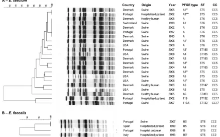
FIG. 1. Computer analysis of SmaI-digested genomic DNAs of representative vancomycin-resistantE. faecium(A) andE. faecalis(B) isolates from humans and swine. PFGE subtypes were established by visual analysis following the criteria of Tenover et al. (48). Computer analysis was performed with the Fingerprinting II Informatix software package (Bio-Rad Laboratories, Hercules, CA). The acquisition of the image was performed by using a Gel Doc XR camera (Bio-Rad Laboratories Inc.). The image was normalized by using four reference lanes of the low-range PFGE marker (0.13 kb to 1,018.5 kb; New England BioLabs, La Jolla, CA) as an external DNA size marker. The phylogenetic tree was subsequently constructed by use of the Dice coefficient and UPGMA clustering (optimization, 0.5%; band tolerance, 1.5%; threshold cutoff value set at 82%) (36). Year, year of isolation.
on May 16, 2020 by guest
http://jcm.asm.org/
were associated with VRE from humans or animals (10, 24, 38, 52, 54). The linking of such Tn1546variants with particular plasmid types fromE. faecium(megaplasmids and mosaic plas-mids containing replication proteins belonging to the RepA_N family, such as those of pLG1 or pRUM, and Inc18) orE. faecalis(pheromone-responsive plasmids) is also in agreement with some recent studies (12, 25, 29, 44, 56). Rosvoll et al. recently described the presence ofvanA-Inc18 plasmids con-taining one or two rep (rep1and/or rep2) amongE. faecium
strains from poultry and farmers in Norway and Italy (44), some of them closely related to the firstvanA-Inc18 plasmid recovered in France in 1986 from a clinical isolate (45). Other studies have also described the presence of large plasmids carrying Tn1546amongE. faeciumisolates from hospitalized humans (2, 51) and swine (19). The recovery of resistant plas-mids from clonally unrelated isolates from different sources over an extended period of time, such as that carried by the
ST132 clone, is of concern, since it would reflect a wide spread and maintenance in both human and nonhuman hosts.
In summary, this study documents that enterococcal clones belonging to host-adapted clonal complexes of E. faecium
(CC5 and CC17) andE. faecalis(CC2) are shared by swine and humans. The fact that these clones are able to colonize and cause human infections (and, in some instances, nosocomial outbreaks, such asE. faeciumST132 orE. faecalisST6) con-firms the relevance of reverse and alternative routes for dis-semination of commensal and opportunistic bacteria. Multilay-ered molecular epidemiology studies will be required to understand the spread and evolution of clones and genetic elements encoding vancomycin resistance and overcoming the species barriers between humans and swine.
ACKNOWLEDGMENTS
Ana R. Freitas was supported by fellowships from Fundac¸a˜o para a Cieˆncia e Tecnologia of Portugal (SFRH/BD/24604/2005) and the European Union (LSHE-2007-037410). Research on enterococci was supported by grants from the European Union (LSHE-2007-037410), from the Ministry of Science and Innovation of Spain (PI07/1441, PS09/02381), and from Fundac¸a˜o para a Cieˆncia e Tecnologia of Portugal (POCI/AMB/61814/2004).
We thank Maria del Grosso and Annalisa Pantosti (Instituto Supe-riore di Sanita`, Rome, Italy), Patrice Boerlin (Veterinary College, University of Guelph, Guelph, Ontario, Canada), Patricia Ruiz-Gar-bajosa (Hospital Ramo´n y Cajal, Madrid, Spain), and Inmaculada Herrero and Miguel A. Moreno (Veterinary School, Complutense University of Madrid, Madrid, Spain) for the gift of strains. We are also grateful to the Spanish Network for the Study of Plasmids and Extrachromosomal Elements (REDEEX) for encouraging and fund-ing cooperation among Spanish microbiologists workfund-ing on the biology of mobile genetic elements (Ministry of Science and Innovation of Spain, BFU 2008-0079-E/BMC).
REFERENCES
1.Agersø, Y., et al.2008. Vancomycin-resistantEnterococcus faecalisisolates from a Danish patient and two healthy human volunteers are possibly related to isolates from imported turkey meat. J. Antimicrob. Chemother.62:844– 845.
2.Arias, C. A., D. Panesso, K. V. Singh, L. B. Rice, and B. E. Murray.2009. Cotransfer of antibiotic resistance genes and ahylEfm-containing virulence plasmid inEnterococcus faecium. Antimicrob. Agents Chemother.53:4240– 4246.
3.Bates, J., J. Z. Jordens, and D. T. Griffiths.1994. Farm animals as a putative reservoir for vancomycin-resistant enterococcal infection in man. J. Antimi-crob. Chemother.34:507–514.
4.Biavasco, F., et al.2007. VanA-type enterococci from humans, animals, and food: species distribution, population structure, Tn1546typing and location, and virulence determinants. Appl. Environ. Microbiol.73:3307–3319. 5.Boerlin, P., A. Wissing, F. M. Aarestrup, J. Frey, and J. Nicolet.2001.
Antimicrobial growth promoter ban and resistance to macrolides and van-comycin in enterococci from pigs. J. Clin. Microbiol.39:4193–4195. 6.Clinical Laboratory Standards Institute.2010. Performance standards for
antimicrobial susceptibility testing; 20th informational supplement M100-S20, vol. 30. Clinical and Laboratory Standards Institute, Wayne, PA. 7.Coque, T. M., et al.2005. Population structure ofEnterococcus faecium
causing bacteremia in a Spanish university hospital: setting a scene for a future increase in vancomycin resistance? Antimicrob. Agents Chemother.
49:2693–2700.
8.Dahl, K. H., G. S. Simonsen, O. Olsvik, and A. Sundsfjord.1999. Hetero-geneity in thevanBgene cluster of genomically diverse clinical strains of vancomycin-resistant enterococci. Antimicrob. Agents Chemother.43:1105– 1110.
9.Damborg, P., et al.2009. Dogs are a reservoir of ampicillin-resistant Entero-coccus faeciumlineages associated with human infections. Appl. Environ. Microbiol.75:2360–2365.
10.Descheemaeker, P. R. M., et al.1999. Comparison of glycopeptide-resistant
Enterococcus faeciumisolates and glycopeptide resistant genes of human and animal origins. Antimicrob. Agents Chemother.43:2032–2037.
11.Donabedian, S. M., et al.2010. Characterization of vancomycin-resistant
[image:6.585.91.234.70.408.2]Enterococcus faeciumisolated from swine in three Michigan counties. J. Clin. Microbiol.48:4156–4160.
FIG. 2. ClaI-digested plasmid DNAs of VRE isolates recovered from hospitals and swine from different origins. Lane M, molecular marker ladder EcoT 14 I/BglII (Takara Bio Inc., Otsu, Japan); lane 1, E. faecalisST6-CC2 (PFGE B5) from an outbreak strain recovered in Spain in 1999; lane 2,E. faecalisST6-CC2 (PFGE B5) from a swine isolate recovered in Portugal in 2007; lane 3,E. faeciumST132-CC17 (PFGE 119.5) from a swine isolate recovered in Portugal in 2007; lane 4,E. faeciumST132-CC17 (PFGE 119) from a clinical isolate recov-ered in Portugal in 2002 that is representative of strains causing urinary tract infections in two unrelated patients during 2002.
on May 16, 2020 by guest
http://jcm.asm.org/
12.Freitas, A. R., et al.2008. Plasmid characterization of vancomycin-resistant
Enterococcus faecalisstrains from different continents (1989–2004), abstr. 2112, p. S624. Abstr. 18th Eur. Congr. Clin. Microbiol. Infect. Dis. (Barce-lona, Spain).
13.Freitas, A. R., C. Novais, P. Ruiz-Garbajosa, T. M. Coque, and L. Peixe.
2009. Clonal expansion within clonal complex 2 and spread of vancomycin-resistant plasmids among different genetic lineages ofEnterococcus faecalis
from Portugal. J. Antimicrob. Chemother.63:1104–1111.
14.Freitas, A. R., C. Novais, P. Ruiz-Garbajosa, T. M. Coque, and L. Peixe.
2009. Dispersion of multidrug-resistantEnterococcus faeciumisolates be-longing to major clonal complexes in different Portuguese settings. Appl. Environ. Microbiol.75:4904–4908.
15.Freitas, A. R., et al.2008. Genetic dynamics of a successfulEnterococcus faecalisclone persistently recovered from European hospitals during the last decade (1993–2008), abstr. C2-1996. Abstr. 48th Intersci. Conf. Antimicrob. Agents Chemother. ASM Press, Washington, DC.
16.Freitas, A. R., et al.2010. Global spread of thehylEfmcolonization-virulence gene in megaplasmids of theEnterococcus faeciumpolyclonal subcluster. Antimicrob. Agents Chemother.54:2660–2665.
17.Hammerum, A. M., et al.2004. A vancomycin-resistantEnterococcus faecium
isolate from a Danish healthy volunteer, detected 7 years after the ban of avoparcin, is possibly related to pig isolates. J. Antimicrob. Chemother.
53:547–549.
18.Hasman, H., et al.2006. Copper resistance inEnterococcus faecium, medi-ated by thetcrBgene, is selected by supplementation of pig feed with copper sulfate. Appl. Environ. Microbiol.72:5784–5789.
19.Hasman, H., A. G. Villadsen, and F. M. Aarestrup.2005. Diversity and stability of plasmids from glycopeptide-resistant Enterococcus faecium
(GRE) isolated from pigs in Denmark. Microb. Drug Resist.11:178–184. 20.Hayes, J. R., et al.2003. Prevalence and antimicrobial resistance of
Entero-coccusspecies isolated from retail meats. Appl. Environ. Microbiol.69:7153– 7160.
21.Hegstad, K., T. Mikalsen, T. M. Coque, G. Werner, and A. Sundsfjord.2010. Mobile genetic elements and their contribution to the emergence of antimi-crobial resistantEnterococcus faecalisandEnterococcus faecium.Clin. Mi-crobiol. Infect.16:541–554.
22.Herrero, I. A., T. Teshager, J. Garde, M. A. Moreno, and L. Dominguez.
2000. Prevalence of vancomycin-resistantEnterococcus faecium(VREF) in pig faeces from salughterhouses in Spain. Prev. Vet. Med.47:255–262. 23.Hidron, A. I., et al. 2008. NHSN annual update: antimicrobial-resistant
pathogens associated with healthcare-associated infections: annual summary of data reported to the National Healthcare Safety Network at the Centers for Disease Control and Prevention, 2006–2007. Infect. Control Hosp. Epi-demiol.29:996–1011.
24.Jensen, L. B., et al.1998. Molecular analysis of Tn1546inEnterococcus faeciumisolated from animals and humans. J. Clin. Microbiol.36:437–442. 25.Jensen, L. B., et al.2010. A classification system for plasmids from entero-cocci and other Gram-positive bacteria. J. Microbiol. Methods80:25–43. 26.Jensen, L. B., A. M. Hammerum, R. L. Poulsen, and H. Westh.1999.
Van-comycin-resistantEnterococcus faeciumstrains with highly similar pulsed-field gel electrophoresis patterns containing similar Tn1546-like elements isolated from a hospitalized patient and pigs in Denmark. Antimicrob. Agents Chemother.43:724–725.
27.Kawalec, M., et al.2007. Clonal structure ofEnterococcus faecalisisolated from Polish hospitals: characterization of epidemic clones. J. Clin. Microbiol.
45:147–153.
28.Larsen, J., et al.2010. Porcine-origin gentamicin-resistantEnterococcus fae-calisin humans, Denmark. Emerg. Infect. Dis.16:682–684.
29.Laverde Gomez, J. A., et al.2011. A multiresistance megaplasmid pLG1 bearing ahylEfmgenomic island in hospitalEnterococcus faeciumisolates. Int. J. Med. Microbiol.301:165–175.
30.Leavis, H. L., M. J. Bonten, and R. J. Willems. 2006. Identification of high-risk enterococcal clonal complexes: global dispersion and antibiotic resistance. Curr. Opin. Microbiol.9:454–460.
31.Lu, H. Z., et al.2002.Enterococcus faecium-related outbreak with molecular evidence of transmission from pigs to humans. J. Clin. Microbiol.40:913– 917.
32.Manson, J. M., L. E. Hancock, and M. S. Gilmore.2010. Mechanism of chromosomal transfer ofEnterococcus faecalispathogenicity island, capsule, antimicrobial resistance, and other traits. Proc. Natl. Acad. Sci. U. S. A.
107:12269–12274.
33.Manson, J. M., S. Keis, J. M. Smith, and G. M. Cook.2003. Characterization of a vancomycin-resistantEnterococcus faecalis(VREF) isolate from a dog
with mastitis: further evidence of a clonal lineage of VREF in New Zealand. J. Clin. Microbiol.41:3331–3333.
34.Morrison, D., N. Woodford, S. P. Barrett, P. Sisson, and B. D. Cookson.
1999. DNA banding pattern polymorphism in vancomycin-resistant Entero-coccus faeciumand criteria for defining strains. J. Clin. Microbiol.37:1084– 1091.
35.Novais, C., et al.2005. Vancomycin-resistantEnterococcus faeciumclone in swine, Eur. Emerg. Infect. Dis.11:1985–1987.
36.Novais, C., T. M. Coque, J. C. Sousa, F. Baquero, and L. Peixe.2004. Local genetic patterns within a vancomycin-resistantEnterococcus faecalisclone isolated in three hospitals in Portugal. Antimicrob. Agents Chemother.48:
3613–3617.
37.Novais, C., T. M. Coque, J. C. Sousa, and L. V. Peixe.2006. Antimicrobial resistance among faecal enterococci from healthy individuals in Portugal. Clin. Microbiol. Infect.12:1131–1134.
38.Novais, C., et al.2008. Diversity of Tn1546and its role in the dissemination of vancomycin-resistant enterococci in Portugal. Antimicrob. Agents Che-mother.52:1001–1008.
39.Novais, C., J. C. Sousa, T. M. Coque, and L. V. Peixe.2005. Molecular characterization of glycopeptide-resistantEnterococcus faeciumisolates from Portuguese hospitals. Antimicrob. Agents Chemother.49:3073–3079. 40.Ozawa, Y., et al.2002. Vancomycin-resistant enterococci in humans and
imported chickens in Japan. Appl. Environ. Microbiol.68:6457–6461. 41.Phillips, I., et al.2004. Does the use of antibiotics in food animals pose a risk
to human health? A critical review of published data. J. Antimicrob. Che-mother.53:28–51.
42.Phillips, I.2007. Withdrawal of growth-promoting antibiotics in Europe and its effects in relation to human health. Int. J. Antimicrob. Agents30:101–107. 43.Romo, M., H. Tomita, Y. Ike, L. Martínez-Martínez, and M. Francia.2007. Emergence of worldwide epidemic clones of vancomycin-resistant Entero-coccus faeciumin a Northern Spain hospital, abstr. 681, p. S163. Abstr. 17th Eur. Congr. Clin. Microbiol. Infect. Dis. (Munich, Germany).
44.Rosvoll, T. C., et al.2010. PCR-based plasmid typing inEnterococcus faecium
strains reveals widely distributed pRE25-, pRUM-, pIP501-, and pHTbeta-related replicons associated with glycopeptide resistance and stabilizing toxin-antitoxin systems. FEMS Immunol. Med. Microbiol.58:254–268. 45.Sletvold, H., et al.2010. Tn1546is part of a larger plasmid-encoded genetic
unit horizontally disseminated among clonalEnterococcus faeciumlineages. J. Antimicrob. Chemother.65:1894–1906.
46.Smith, T. C., et al.2009. Methicillin-resistantStaphylococcus aureus(MRSA) strain ST398 is present in Midwestern U.S. swine and swine workers. PLoS One4:e4258.
47.Stegmann, R., A. Burnens, C. A. Maranta, and V. Perreten.2010. Human infection associated with methicillin-resistantStaphylococcus pseudinterme-diusST71. J. Antimicrob. Chemother.65:2047–2048.
48.Tenover, F. C., et al.1995. Interpreting chromosomal DNA restriction pat-terns produced by pulsed-field gel electrophoresis: criteria for bacterial strain typing. J. Clin. Microbiol.33:2233–2239.
49.Thal, L. A., J. Silverman, S. Donabedian, and M. J. Zervos.1997. The effect of Tn916insertions on contour-clamped homogeneous electrophoresis pat-terns ofEnterococcus faecalis. J. Clin. Microbiol.35:969–972.
50.Werner, G., et al.2008. Emergence and spread of vancomycin resistance among enterococci in Europe. Euro Surveill.13:ii⫽19046.
51.Werner, G., I. Klare, and W. Witte.1999. Large conjugativevanAplasmids in vancomycin-resistantE. faecium. J. Clin. Microbiol.37:2383–2384. 52.Willems, R. J., et al.1999. Molecular diversity and evolutionary relationships
of Tn1546-like elements in enterococci from humans and animals. Antimi-crob. Agents Chemother.43:483–491.
53.Willems, R. J., and W. van Schaik.2009. Transition ofEnterococcus faecium
from commensal organism to nosocomial pathogen. Future Microbiol.
4:1125–1135.
54.Woodford, N., A. M. A. Adebiyi, M. F. I. Palepou, and B. D. Cookson.1998. Diversity of VanA glycopeptide resistance elements in enterococci from humans and nonhuman sources. Antimicrob. Agents Chemother.42:502– 508.
55.Zhang, X., J. E. Vrijenhoek, M. J. Bonten, R. J. Willems, and W. Van Schaik.
2010. A genetic element present on megaplasmids allowsEnterococcus fae-ciumto use raffinose as carbon source. Environ. Microbiol. doi:10.1111/ j.1462–2920.2010.02355.x.
56.Zhu, W., et al.2010. Dissemination of anEnterococcus Inc18-LikevanA
plasmid, associated with vancomycin-resistantStaphylococcus aureus. Anti-microb. Agents Chemother.54:4314–4320.