Nijmegen
The following full text is a publisher's version.
For additional information about this publication click this link.
http://hdl.handle.net/2066/200447
Please be advised that this information was generated on 2019-06-02 and may be subject to
change.
doi: 10.3389/fmicb.2018.00603
Edited by: Sven Hammerschmidt, University of Greifswald, Germany Reviewed by: Konstantinos Papadimitriou, Agricultural University of Athens, Greece Marcelo Gottschalk, Université de Montréal, Canada *Correspondence: Christoph Jans [email protected]
Specialty section: This article was submitted to Infectious Diseases, a section of the journal Frontiers in Microbiology Received:14 December 2017 Accepted:15 March 2018 Published:10 April 2018 Citation: Jans C and Boleij A (2018) The Road to Infection: Host-Microbe Interactions Defining the Pathogenicity of Streptococcus bovis/Streptococcus equinus Complex Members. Front. Microbiol. 9:603. doi: 10.3389/fmicb.2018.00603
The Road to Infection: Host-Microbe
Interactions Defining the
Pathogenicity of
Streptococcus
bovis/Streptococcus equinus
Complex Members
Christoph Jans1* and Annemarie Boleij2
1Laboratory of Food Biotechnology, Institute of Food Nutrition and Health, Department of Health Science and Technology,
ETH Zurich, Zurich, Switzerland,2Department of Pathology, Radboud Institute for Molecular Life Sciences, Radboudumc,
Nijmegen, Netherlands
TheStreptococcus bovis/Streptococcus equinuscomplex (SBSEC) comprises several species inhabiting the animal and human gastrointestinal tract (GIT). They match the pathobiont description, are potential zoonotic agents and technological organisms in fermented foods. SBSEC members are associated with multiple diseases in humans and animals including ruminal acidosis, infective endocarditis (IE) and colorectal cancer (CRC). Therefore, this review aims to re-evaluate adhesion and colonization abilities of SBSEC members of animal, human and food origin paired with genomic and functional host-microbe interaction data on their road from colonization to infection. SBSEC seem to be a marginal population during GIT symbiosis that can proliferate as opportunistic pathogens. Risk factors for human colonization are considered living in rural areas and animal-feces contact. Niche adaptation plays a pivotal role
where Streptococcus gallolyticus subsp. gallolyticus (SGG) retained the ability to
proliferate in various environments. Other SBSEC members have undergone genome reduction and niche-specific gene gain to yield important commensal, pathobiont and
technological species. Selective colonization of CRC tissue is suggested for SGG,
possibly related to increased adhesion to cancerous cell types featuring enhanced
collagen IV accessibility. SGG can colonize, proliferate and may shape the tumor
microenvironment to their benefit by tumor promotion upon initial neoplasia development. Bacteria cell surface structures including lipotheichoic acids, capsular polysaccharides
and pilus loci (pil1, pil2, and pil3) govern adhesion. Only human blood-derived SGG
contain complete pilus loci and other disease-associated surface proteins. Rumen
or feces-derived SGG and other SBSEC members lack or harbor mutated pili. Pili
also contribute to binding to fibrinogen upon invasion and translocation of cells from the GIT into the blood system, subsequent immune evasion, human contact
system activation and collagen-I-binding on damaged heart valves. OnlySGGcarrying
complete pilus loci seem to have highest IE potential in humans with significant
links between SGG bacteremia/IE and underlying diseases including CRC. Other
Comparative genome data of blood, commensal and food isolates are limited but required to elucidate the role of pili and other virulence factors, understand pathogenicity mechanisms, host specificity and estimate health risks for animals, humans and food alike.
Keywords:Streptococcus gallolyticus, Streptococcus lutetiensis, Streptococcus infantarius, virulence, infective endocarditis, colorectal cancer, pilus, microbiota
GENERAL INTRODUCTION TO THE
RELEVANCE OF THE
STREPTOCOCCUS
BOVIS/STREPTOCOCCUS EQUINUS
COMPLEX
Streptococcus bovis/Streptococcus equinus complex (SBSEC)
bacteria are Gram-positive species that inhabit the gastrointestinal tract (GIT) of animals and humans. Most SBSEC have been described as commensal bacteria, but some cause serious infections such as bacteremia and infective endocarditis (IE) in humans and animals and match the pathobiont description (Chow et al., 2011; Boleij and Tjalsma, 2013; Jans et al., 2015, 2016). They are associated with underlying conditions including occult colorectal cancer (CRC) (Boleij et al., 2011c), which highlights the importance of SBSEC members in public- and animal health alike.
Furthermore, SBSEC members are detected in food products including fermented milk in sub-Saharan Africa, Asia and Southern Europe, fermented fish in Asia and fermented plants in sub-Saharan Africa and Latin America suggesting a range of habitats and adaptability to different environments for these bacteria (Jans et al., 2015, 2017). Recent advances in phenotypic and molecular technologies provide more detailed classification abilities at various levels from species to sequence type. This advanced classification scheme helps to elucidate the SBSEC population structure, disease associations, transmission routes and host specificity (Dumke et al., 2014; Shibata et al., 2014; Jans et al., 2016). It is still unclear how SBSEC members establish from commensal organisms to pathogens, particularly relating to survival, colonization, adhesion, invasion and interaction with the host immune system. Furthermore, the causality of SBSEC in CRC is not yet proven which leaves the bacterial-driver-passenger model as important theory to describe the potential mechanisms of host-microbe interaction (Tjalsma et al., 2012).
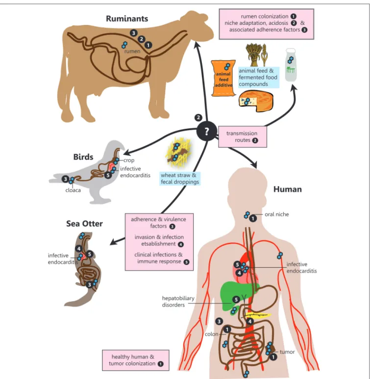
Therefore, this narrative review aims to provide a comprehensive overview for the following questions in relation to SBSEC members on their road to infection regarding prevalence, transmission, niche colonization and mechanisms for adhesion, invasion and infection establishment within the human-animal-food system (Figure 1):
(1) What abilities help SBSEC members to colonize different body sites or ecological niches and facilitate transmission? (2) Which factors determine SBSEC to evolve from commensal
to pathogen?
(3) What is the prevalence of SBSEC members in different habitats in relation to clinical manifestations and infections?
(4) Which genetic factors are known to encode for these abilities and can be linked to experimental studies?
BASIC TAXONOMY AND IDENTIFICATION
OF THE SBSEC
SBSEC members are group D streptococci, although the Lancefield group D antiserum reaction is not ubiquitous (Beck et al., 2008). The SBSEC is comprised of several different species, which in this review will be used according to following nomenclature: Streptococcus equinus (SE), Streptococcus
infantarius subsp. infantarius (SII), Streptococcus lutetiensis
(SL), Streptococcus alactolyticus and three subspecies of the
clade Streptococcus gallolyticus, namely gallolyticus (SGG),
macedonicus (SGM) and pasteurianus (SGP). The taxonomic
assignment ofSL as separate species is not fully agreed upon and also referred to as Streptococcus infantarius subsp. coli (Dekker and Lau, 2016). When no distinction was made between subspecies, the old nomenclatureStreptococcus bovis (SB) was used (Schlegel et al., 2000, 2003; Poyart et al., 2002; Jans et al., 2015). Biotype differentiation is based on the ability to produce acid from mannitol (biotype I=SGG) or not (biotype II=SE, SGP,SL, andSII) (Schlegel et al., 2003; Jans et al., 2015). For full phenotype descriptions, we refer to Bergey’s Manual and their implementations in API and VITEK identification approaches (Whiley and Hardie, 2009).
Alternative identification approaches utilize Matrix Assisted Laser Desorption/Ionization Time Of Flight Mass Spectrometry (MALDI-TOF MS) to identify and discriminate species of the SBSEC but results in unreliable identification of SGM and a roughly 80% identification rate for SGG, SGP and SL (Hinse et al., 2011a; Ben-Chetrit et al., 2017). Furthermore, DNA-based approaches are widely applied using single gene PCR and qPCR assays on 16S rRNA gene (Jans et al., 2012b),sodA(Poyart et al.,
1998),groES/groEL(Chen et al., 2008; Lazarovitch et al., 2013;
Sheng et al., 2014),recNandgyrB(Lopes et al., 2014) as well as multi locus sequence typing (Dumke et al., 2014; Shibata et al., 2014; Jans et al., 2016).
PREVALENCE AND COLONIZATION OF
SBSEC IN ANIMALS AND HUMANS
Prevalence and Colonization in Animals
SBSEC are mainly described as colonizers of the rumen, crop and cloaca of animals and colon of humans. SBSEC members have been isolated from the GIT or blood system of birds, companion animals, livestock (ruminants, poultry and pigs), marsupials,FIGURE 1 |SBSEC - the road to infection. Graphical overview of the different niches inhabited by SBSEC members as well as relevant aspects of host colonization, adherence, invasion and infection covered in the corresponding chapters indicated by bullet point numbers. 1. Prevalence and colonization of SBSEC in animals and humans, 2. Transmission and niche adaptation of SBSEC members, 3. Mechanisms and virulence factors responsible for adhesion and host colonization by SBSEC members, 4. Invasion and infection establishment, 5. Clinical infections and host-immune response due to SBSEC in animals and humans.
aquatic mammals and game (Jans et al., 2015), but prevalence data is limited to birds, cattle and lamb.SGGwas found in over 90% of fecal droppings in turkey flocks and reached up to 80% prevalence in pigeon crop and cloaca samples (De Herdt et al., 1994a,b; Schulz et al., 2015). SBSEC members were also isolated
from chicken crops, but less frequently and not as predominant bacteria (Baele et al., 2001).
In ruminants, SBSEC members are considered as aerotolerant components of the rumen epithelial surface (epimural) microbiota (Mead and Jones, 1981). SBSEC members are
estimated to contribute 106 to 107cells per milliliter of rumen content (Hudson et al., 2000). Their prevalence in cattle is estimated between 20 and 90% with lowest prevalence at early ages (Jans et al., 2015) while in lambs an early live predominance of SBSEC is suggested (Mueller et al., 1984). Colonization and predominance is likely affected by feed composition as shown for reindeers where high SBSEC colonization correlates with higher quantities of starch-rich feed during the summer months (Orpin et al., 1985). SBSEC members are also supplied directly to young calves and goats as probiotics to support the establishment of an anaerobic rumen microbiota and to benefit from consistent
α-amylase activity for feed digestion (Kmet et al., 1993; Kumar et al., 2016). SBSEC members therefore seem to form an integral part of the GIT microbiota of birds whereas ruminants feature age-dependent and possible host-associated SBSEC prevalence.
Prevalence in Healthy Humans
The fecal carriage rate of SBSEC member in humans is varying from five to over 60% (Table 1) (Klein et al., 1977; Noble, 1978; Potter et al., 1998; Abdulamir et al., 2010; Al-Jashamy et al., 2010; Chirouze et al., 2013; Lopes et al., 2014; Boltin et al., 2015; Dumke et al., 2017; Kaindi et al., 2017). This variation might depend on detection techniques and regional differences. Furthermore, most studies were conducted using a hospital-derived population with possible differences in age, sex and underlying diseases that limits extrapolation to the general population. (Huang et al., 2008; Chirouze et al., 2013; Lopes et al., 2014; Dumke et al., 2017; Harris et al., 2017).Oral niche colonization in humans seems to be infrequent, but isolation ofSII,SGP, andSEwas confirmed from dental plaques and root caries lesions (Sissons et al., 1988; Shen et al., 2005; Arul and Palanivelu, 2014).
Selective Colonization of CRC Patients and
Preliminary Causal Evidence
Selective Colonization of CRC Patients
Fecal carriage of SBSEC members associated with CRC was initially reported to be 56% in 63 CRC patients compared to 11% in 105 healthy controls (Table 1) (Klein et al., 1977). This association has since been reported in a range of 6– 46% in patients with adenomas and CRC vs. 7–14% in control patients (Abdulamir et al., 2010; Al-Jashamy et al., 2010; Chirouze et al., 2013; Boltin et al., 2015; Kaindi et al., 2017). Of these five studies, only two observed a significant carriage difference between healthy and neoplasia patients but one suggested a novel association of SII prevalence with hemorrhoids (Al-Jashamy et al., 2010; Kaindi et al., 2017).
The selective association with CRC tissue is also controversial. While no specific association of SBSEC members with CRC tissue was observed on small sample sets based on phenotypic identification (Norfleet and Mitchell, 1993; Potter et al., 1998), more recent DNA-based approaches approve of such an association ranging from 0 to 2% in controls, 47% in normal tissue of cancer patients and 3–74% in tumor tissues (Table 1) (Abdulamir et al., 2010; Andres-Franch et al., 2017; Kumar et al., 2017). Interestingly, tumor tissue of patients with bacteremia was more often positive forSGG(48.7%) than that of patients without bacteremia (32.7%) based onsodAgene PCR, which increased the detection rate of SGG from 27% and 16%, respectively,
based on selective culturing. (Abdulamir et al., 2010). Tumor tissues harboring SGG were also significantly associated with co-infection by Epstein-Barr virus (OR: 9.49; 95% CI: 1.1–82.9) (Andres-Franch et al., 2017). Nevertheless, two other studies using a culture-based and qPCR-based approach still suggested no association between tumorous (0–15%) and non-tumorous tissues (0–12%) for SB/SGG(Boltin et al., 2015; Viljoen et al., 2015).
These contradicting data on SBSEC tumor tissue colonization might relate to patients with clinical infections of SBSEC (Abdulamir et al., 2010) or without any clinical symptoms (Viljoen et al., 2015; Andres-Franch et al., 2017), differences in detection techniques (culture-independent vs. selective culturing) or study population. This also indicates a non-obligatory relationship ofSGGor SBSEC with CRC. SBSEC might only proliferate if certain requirements to facilitate colonization are met to become passengers as hypothesized in the bacterial-driver-passenger model (Tjalsma et al., 2012).
Preliminary Causality of SBSEC in CRC
Only minimal evidence for a causal relationship between SBSEC and CRC exists. Wall-extracted antigens fromSGGNCTC8133, a human fecal isolate with controversial earlier classification as SII orSE, and its active S300 fraction are suggested to induce cell proliferation, polyamines and aberrant crypt foci in the distal colon of azoxymethane-treated rats (Ellmerich et al., 2000b; Biarc et al., 2004; Jans et al., 2016). In human CRC cell lines, stationary SGGstrains TX20005 and TX20030 increased cell proliferation of HCT116, HT-29 and LoVo but not of SW480, SW1116, human lung carcinoma cell line A549, kidney epithelial cell line HEK293, human normal colon epithelial cell lines CCD841, CoN, and FHC suggesting the need for specific conditions to facilitate cell proliferation (Kumar et al., 2017).
The adenomatous polyposis coli tumorsuppressor geneAPC is inactivated in many CRCs. This leads to an accumulation ofβ-catenin in the nucleus (He et al., 1998) thereby activating the c-Myc oncogene. Increased levels ofβ-catenin and c-Myc were detected in CRC cells after SGG incubation. This effect was not observed for SII, SGP, and SGM or for live SGG separated by trans-well membranes from cells, nor with bacterial supernatants, heat-killed bacteria or bacterial lysates suggesting a necessity of liveSGGand directSGG-cell contact (Kumar et al., 2017).
In azoxymethane-treated mice, inoculation withSGGled to increased cell proliferation,β-catenin accumulation in colonic crypts and higher numbers of tumors and dysplasia grade supporting a potential tumor-promoting role forSGG (Kumar et al., 2017). This effect was only observed for SGG strains able to induce cell proliferationin vitro (Kumar et al., 2018). The increase in cell proliferation was correlated with increased adhesion abilities in vitro. In vivo such strains also had an increased ability to colonize the mucosa in both C57BL6 as A/J mice. Therefore, the original bacterial driver-passenger model (Tjalsma et al., 2012) might need to be updated regarding the potential driving role of SGG. If SGG contributes to tumor development it would exert its effects probably after colonization of neoplastic sites as a passenger, which might depend on the specific strain and contribute to tumor progression rather than
T A B L E 1 | S B S E C p re va le n c e in th e G IT in st u d ie s in c lu d in g h e a lth y p e o p le a n d p a tie n ts w ith g a st ro in te st in a lc o n d iti o n s so rt e d b y ye a r o f p u b lic a tio n . C o u n tr y P a rt ic ip a n t s e le c ti o n S a mp le ty p e S a mp le s e t S a mp le s iz e S B S E C p re v a le n c e (% ) In d iv id u a l s p e c ie s d e te c te d Is o la ti o n te c h n iq u e Id e n ti fi c a ti o n te c h n iq u e R e fe re n c e s U S A P a tie n ts se le c te d fo r C R C , in fla m m a to ry b o w e ld is e a se , n o n -c o lo n ic n e o p la sm s, g a st ro in te st in a ld is o rd e rs . C o n tr o ls a m o n g h o sp ita lw o rk e rs a n d p a tie n ts w ith n o a p p a re n t g a st ro in te st in a l d is e a se R e c ta ls w a b s C o n tr o ls 1 0 5 1 1 n .d . S e le c tiv e c u ltu rin g P h e n o ty p ic id e n tific a tio n K le in e t a l., 1 9 7 7 C R C p a tie n ts 6 3 5 6 IB D 2 5 2 8 N o n -c o lo n ic n e o p la sm a 2 1 1 9 G ID 3 7 1 4 U K H o sp ita lp o p u la tio n in -a n d o u tp a tie n ts S to o l A d u lts 3 9 5 n .d . S e le c tiv e c u ltu rin g P h e n o ty p ic id e n tific a tio n N o b le , 1 9 7 8 N e o n a te s 2 1 2 4 U S A O u tp a tie n ts u n d e rg o in g c o lo n o sc o p y fo r su sp e c te d C R C a n d p o ly p s S to o l S to o lb e fo re c o lo n o sc o p y 3 5 3 n .d . S e le c tiv e c u ltu rin g G ro u p D st re p te x N o rfle e t a n d M itc h e ll, 1 9 9 3 A sp ira te d c o lo n flu id A sp ira te d c o lo n flu id 4 0 8 T is su e b io p si e s N o rm a lr e m o te 4 0 0 N o rm a la d ja c e n t 4 0 0 A d e n o m a 3 3 3 C a rc in o m a 6 0 U K C R C c a se s m a tc h e d to p a tie n ts h a vi n g su rg e ry fo r b e n ig n d is e a se o r d ia g n o st ic c o lo n o sc o p y S to o l C o n tr o l 2 3 1 3 n .d . S e le c tiv e c u ltu rin g P h e n o ty p ic id e n tific a tio n P o tt e r e t a l., 1 9 9 8 C R C 1 9 1 1 T is su e sa m p le s C o n tr o l 2 3 4 N o rm a lC R C 1 9 5 .5 Tu m o r C R C 1 9 1 1 B lo o d C o n tr o l 2 3 0 C R C 1 9 0 (C o n ti n u e d )
T A B L E 1 | C o n tin u e d C o u n tr y P a rt ic ip a n t s e le c ti o n S a mp le ty p e S a mp le s e t S a mp le s iz e S B S E C p re v a le n c e (% ) In d iv id u a l s p e c ie s d e te c te d Is o la ti o n te c h n iq u e Id e n ti fi c a ti o n te c h n iq u e R e fe re n c e s M a la ys ia C R C p a tie n ts w ith (b a c + ) a n d w ith o u t b a c te re m ia (b a c − ) a n d c o n tr o ls in tu m o r (T U ) a n d n o n -t u m o r (N T U ) sp e c im e n s S to o l C o n tr o l 5 0 1 2 8 % S G G S e le c tiv e c u ltu rin g a n d D N A -b a se d d e te c tio n * P h e n o ty p ic id e n tific a tio n s o d A g e n e P C R a n d q P C R A b d u la m ir e t a l., 2 0 1 0 C R C b a c − 5 2 1 3 1 0 % S G G C R C b a c + 3 9 1 3 1 0 % S G G M u c o sa l w a sh e s C o n tr o l 5 0 6 4 % S G G C R C b a c − (T U ) 5 2 1 0 8 % S G G C R C b a c − (N T U ) 5 2 6 4 % S G G C R C b a c + (T U ) 3 9 5 5 % S G G C R C b a c + (N T U ) 3 9 5 5 % S G G T is su e sp e c im e n s C o n tr o l 5 0 2 2 % S G G C R C b a c − (T U ) 5 2 1 7 1 7 % (3 2 .7 % )* S G G C R C b a c − (N T U ) 5 2 1 2 1 2 % (2 3 .0 % )* S G G C R C b a c + (T U ) 3 9 2 1 2 1 % (4 8 .7 % )* S G G C R C b a c + (N T U ) 3 9 1 3 1 3 % (3 5 .9 % )* S G G M a la ys ia H o sp ita lp o p u la tio n w ith C R C , in fla m m a to ry b o w e ld is e a se a n d c h ro n ic g a st ro in te st in a lt ra c t d is e a se s (c a se s) a n d h e a lth y in d iv id u a ls (c o n tr o ls ) S to o l C o n tr o ls 9 6 7 n .d . S e le c tiv e c u ltu rin g N o t d e sc rib e d A l-Ja sh a m y e t a l., 2 0 1 0 IB D & c h ro n ic G ID 2 9 5 2 C R C & a d e n o m a 4 1 4 6 F ra n c e C o lo n o sc o p y p a tie n ts . N o t se le c te d fo r d is e a se S to o l N o rm a lc o lo n o sc o p y 1 3 4 6 0 .4 % S G G S e le c tiv e c u ltu rin g 1 6 S rR N A g e n e C h iro u ze e t a l., 2 0 1 3 N o n -t u m o ra ll e si o n s 7 6 1 0 .7 % S G P A d e n o m a & c a rc in o m a 4 9 6 3 .5 % S L B ra zi l C o lo n o sc o p y p a tie n ts in h o sp ita l R e c ta ls w a b C o lo n o sc o p y 5 4 3 5 1 1 % S G G C u ltu re -in d e p e n d e n t q P C R ta rg e tin g re c N a n d g yr B L o p e s e t a l., 2 0 1 4 1 3 % S G P 2 0 % S L 1 1 % S II (C o n ti n u e d )
T A B L E 1 | C o n tin u e d C o u n tr y P a rt ic ip a n t s e le c ti o n S a mp le ty p e S a mp le s e t S a mp le s iz e S B S E C p re v a le n c e (% ) In d iv id u a l s p e c ie s d e te c te d Is o la ti o n te c h n iq u e Id e n ti fi c a ti o n te c h n iq u e R e fe re n c e s Is ra e l C o lo n o sc o p y p a tie n ts . A c c e p ta b le in d ic a tio n s fo r c o lo n o sc o p y in c lu d e d sc re e n in g fo r c o lo re c ta lm a lig n a n c y, p o st p o ly p e c to m y su rv e ill a n c e , a n d in ve st ig a tio n o f sy m p to m s in c lu d in g h e m a to c h e zi a , a b d o m in a lp a in , a n d c h a n g e in b o w e lh a b its S to o l, a sp ira te d c o lo n ic flu id a n d c o lo n ic tis su e N o rm a lc o lo n o sc o p y 1 0 5 1 2 n .d . S e le c tiv e c u ltu rin g V IT E K 2 B o lti n e t a l., 2 0 1 5 N o n -a d va n c e d o r a d va n c e d le si o n s 1 3 1 5 n .d . S o u th A fr ic a (C o h o rt 1 ) P a tie n ts w ith C R C w ith o u t p re -s e le c tin g c o n d iti o n s T is su e A d e n o c a rc in o m a a n d a d ja c e n t n o rm a l 5 5 0 N o t d e te c te d C u ltu re -in d e p e n d e n t s o d A g e n e q P C R V ilj o e n e t a l., 2 0 1 5 (C o h o rt 2 ) P a tie n ts w ith sp o ra d ic m ic ro sa te lli te in st a b ili ty T is su e A d e n o c a rc in o m a 1 8 0 N o t d e te c te d S p a in U n se le c te d C R C p a tie n ts M a tc h e d tis su e N o rm a lm u c o sa 1 9 0 0 N o t d e te c te d C u ltu re -in d e p e n d e n t s o d A g e n e q P C R A n d re s-F ra n c h e t a l., 2 0 1 7 Tu m o r 1 9 0 6 3 % S G G G e rm a n y G e n e ra lp o p u la tio n S to o l H e a lth y c o n tr o ls 9 9 6 3 6 3 % S G G C u ltu re -in d e p e n d e n t q P C R ta rg e tin g re c N D u m ke e t a l., 2 0 1 7 K e n ya C o lo n o sc o p y p a tie n ts . N o t se le c te d fo r d is e a se R e c ta ls w a b N o rm a lc o lo n o sc o p y 1 9 3 1 4 8 % S II S e le c tiv e c u ltu rin g 1 6 S rR N A g e n e K a in d ie t a l., 2 0 1 7 A d e n o m a s & c a rc in o m a s 8 0 2 2 2 2 % S II g ro E L se q u e n c in g U S A Tu m o r a n d n o rm a la d ja c e n t tis su e T is su e A d ja c e n t n o rm a l 1 2 8 4 7 4 7 % S G G C u ltu re -in d e p e n d e n t 1 6 S rR N A g e n e q P C R K u m a r e t a l., 2 0 1 7 C a rc in o m a 1 4 8 7 4 7 4 % S G G ba c + , pa ti e n ts w ith ba c te re m ia ; ba c − , pa ti e n ts w ith ou t ba c te re m ia ; G ID , ga s tr oi n te s ti n a ldi s or de rs ; IB D , in fla m m a tor y bow e ldi s e a s e ; n .d., n ot de te rm in e d; N TU , n on -tu m or ti s s u e s pe c im e n ; TU , tu m or ti s s u e s pe c im e n .
tumor initiation (Boleij and Tjalsma, 2013). In fact, the presence of polyps in the intestinal tract of Notch/APC mice allowed colonization and persistence ofSGGUCN34 colonization, which was 1,000-fold higher than Notch control mice (Aymeric et al., 2018). However, no preferential adherence to tumor tissue sites was observed. Colonization was evenly distributed through the ileum and proximal colon, stimulated by secondary bile acids and was dependent on bacteriocins BlpA and BlpB that compete with other enterococci in the gut (Aymeric et al., 2018).
Interestingly, in vitro cultivation experiments suggested a growth advantage for SGG and SGM in the tumor microenvironment. Cultivation of SGG UCN34 and SGM CIP105683T in spent medium from CRC cells HT-29, SW480, HCT116, and Caco-2 was used to simulate utilization of metabolites from the tumor microenvironment (Boleij et al., 2012a).SGGandSGMdisplayed significantly increased growth rates in spent medium of Caco-2 cells whereas the growth rates of e.g.,Salmonella entericasubsp.entericaserovar Typhimurium,
Staphylococcus lugdunensisorEnterobacter cloacaewere reduced,
suggesting a significant advantage for SBSEC to proliferate in the spent CRC metabolites. The major changes in protein expression patterns were related to an upregulation of pyrimidine biosynthesis and glycolysis, particularly glycerolypid, glycosis and fructose utilization; and a downregulation of purine metabolism. Furthermore,SGGseems to be specifically capable to use secondary glucose metabolites fructose 6-phosphate and 3-phosphate glyceric acid (Boleij et al., 2012a). As the tumor microenvironment features increased levels of lactate, glucose derivatives, amino acids, lipids and fatty acids, SGG likely has an advantage to proliferate in this niche (Boleij et al., 2012a).Therefore, SGG can be described as an opportunistic pathobiont benefiting from the favorable oncogenic environment to colonize the host. This eventually promotes its translocation and systemic dissemination, in select cases leading to clinical infections.
TRANSMISSION AND NICHE ADAPTION
OF SBSEC MEMBERS
Transmission of SBSEC Between Hosts
The prevalence in the GIT of animals and humans facilitates transmission between animals and humans via feces and saliva (Dumke et al., 2015, 2017). Over a duration of 4 weeks, SB counts of an estimated 107 CFU/g broiler feed and 108 CFU/g wheat straw were only reduced by one log unit indicating high transmission possibility (Guy et al., 1980; Mackey and Hinton, 1990). Furthermore, soil clay adhesion ofSBfrom bovine feces is very strong and cannot be desorbed after 24 h whereas long-term persistence seems weak but possibly sufficient to establish transmission within shorter time frames as observed among poultry flocks, surrounding environment and workers. In laying hens, colonization of non-carrier birds introduced into anSGG -positive flock took approximately 35 weeks and occurred likely via feed and feces (Guy et al., 1980; Dumke et al., 2015; Schulz et al., 2015). SGG isolates of identical sequence types where thereby causing infection in one worker and contributing to in-flock and old-young transmission in hens (Dumke et al., 2015).Similarly, sequence types were shared between turkey, pigeons, chicken and humans (Schulz et al., 2015). Rural residency, close animal-human contact and the use of manure as fertilizer were identified as risk factors for colonization withSGG,SGP,SII,SL, and SBin humans (Giannitsioti et al., 2007; Corredoira et al., 2008; Dumke et al., 2017).
Transmission via the fecal-oral or food-oral route requires survival of the gastric passage for colonization of the gut. Survival to simulated gastric conditions is dependent on the SBstrain, gastric pH and feed ingestion. At pH<2.5, survival is minimal whereas at pH 3.0 a reduction of approximately 3 log units was observed forSIIwhileSGMdid not survive, suggesting species-specific abilities to survive and reach the intestine in sufficient numbers for colonization (Ripamonti et al., 2011; Jans et al., 2016). The role of transmission via fermented food products that containSII,SL, orSGMparticularly in sub-Saharan Africa, Southern Europe, Asia and Latin America is not yet clear and niche adaptation might play a pivotal role in their ability to colonize the host (Jans et al., 2015, 2017; Kaindi et al., 2017).
Niche Adaptation of SBSEC Members
SBSEC members adapt to multiple niches. In the rumen, the main carbon sources are largely available in the form of plant fibers. Their utilization is a key feature of many SBSEC members. Proteolysis and peptidase activity are important for the breakdown of proteins to ammonia (Wallace and McKain, 1991) that further supports growth of SBSEC members (Sales-Duval et al., 2002).α- galactosidase,β-glucanases, and endoglucanases of SBSEC play important roles for the degradation of complex carbohydrates in ruminants and chicken (Bailey, 1963; Ekinci et al., 1997; Beckmann et al., 2006). Most of these enzymes are adapted to GIT conditions with a narrow pH optimum of pH 5.6– 6.3 and a temperature optimum of 37–42◦C (Bailey, 1963; Wang et al., 2015; Chen et al., 2016), except forα-amylase and lactate dehydrogenase activity that increases at pH 5.5 vs. pH 6.5 and is possibly part of a self-feeding loop for lactate overproduction (Chen et al., 2016). At pH>6.0, metabolism is directed towardproduction of formate, acetate and ethanol (Chen et al., 2016), while below pH 5.5, it is directed toward lactate, particularly in the event of excess glucose sources such as starch (Gunsalus and Niven, 1942; Russell and Hino, 1985; Asanuma and Hino, 1997). Ruminal acidosis results in a drop below pH 5.5 causing bloat in the rumen (Penner et al., 2007). Despite inconclusive causality, evidence suggests initiation by the overgrowth ofSB in combination with Prevotella, Ruminococcus, Streptococcus,
and Lactobacillusand the parallel inability of lactate utilizers
such asMegasphaera elsdeniiandSelenomonas ruminantiumto metabolize lactate (Palmonari et al., 2010; Wang et al., 2015; McCann et al., 2016).
Particularly SGG has retained the ability to utilize a wide range of carbon sources typical for the rumen. This feature is minimized inSGMandSIIor modified to a different variety of carbon sources inSGP(Rusniok et al., 2010; Lin et al., 2011b; Papadimitriou et al., 2014).SGPATCC43144 in contrast toSGG ATCC43143 harbors a α-L-rhamnosidase, several endo-β -N-acetylglucosaminidase, glucokinase, glucosidases, mannosidases to utilize specific carbon sources available in the gut originating from plant cell walls, biofilms, glycosides, and glycolipids (Lin
et al., 2011b). SGG is the only Streptococcus known so far to use malate via the malolactic enzyme (Gallo_2048) and a malate transporter (Gallo_2049) as well as degrade tannins encoded by tanA, that are otherwise toxic to many bacteria (Rusniok et al., 2010; Papadimitriou et al., 2014). SGG UCN34 is also able to hydrolyze bile salts, an important feature to survive in the small intestine.SGGis prototroph for all 20 amino acids. Besides,SGG harbors partial biosynthesis pathways for biotin and thiamine to support growth in varying conditions including the rumen, intestine and also the blood system (Rusniok et al., 2010).
Niche adaptation is also observed in SII, SGM, and SGP via gene loss and gain. They cannot biosynthesize pantothenate and biotin (Lin et al., 2011b; Papadimitriou et al., 2014) but depend on them for growth (Barnes et al., 1961). Dairy variants of SIIstrains adapted to the dairy environment via a modified lactose (LacS and LacZ instead of phosphotransferase) and peptide metabolism (duplication of oligopeptide transporters) (Jans et al., 2012a, 2013). DairySGM lost or harbor truncated genes for degradation of plant carbohydrates and detoxification of substances relevant for survival in the rumen that are likely obsolete in the dairy niche. In addition, dairySGMgained gene clusters for casein hydrolysis, lactose and galactose metabolism for optimal utilization of these milk components (Papadimitriou et al., 2014). Both dairySIIandSGMpossess lactocepin with high sequence similarity to PrtS CEP ofS. thermophilusresponsible for milk protein degradation. Furthermore, SGPshowed gene loss likely related to adaptation to nutrient-rich environments and an overall genome reduction compared toSGG(Papadimitriou et al., 2014). These findings demonstrate the high adaptability of SBSEC members to different carbon and protein sources not only within the GIT, but also within the dairy environment.
Niche adaptability is also reflected by a high genome plasticity among SBSEC members. The pangenome is increasing at a high rate entailing numerous unique genes for each new strain added (Hinse et al., 2011b). Nearly all SBSEC genomes reveal horizontal gene transfer relating to general carbohydrate metabolism, capsular polysaccharides, antimicrobial resistance or tannase as the key discriminator of SGG (Rusniok et al., 2010; Hinse et al., 2011b; Lin et al., 2011b; Jans et al., 2013; Papadimitriou et al., 2014; Kambarev and Caté, 2015; Grimm et al., 2017a; Kambarev et al., 2017). Natural competence operons and pseudopilus identified inSIIandSGGlikely contribute to the success of horizontal gene transfer and thus to niche adaptation (Rusniok et al., 2010; Lin et al., 2011b; Morrison et al., 2013; Jans et al., 2016). As a consequence, SBSEC members feature highly diverse metabolic abilities and likely also different virulence factors depending on the species and different impact depending on the niche colonized.
MECHANISMS AND VIRULENCE FACTORS
RESPONSIBLE FOR ADHESION AND HOST
COLONIZATION BY SBSEC MEMBERS
The establishment of bacteria in a niche depends on a multitude of factors relating to adherence, signaling, nutritional adaptation and host modulation. Some of the key factorsinvolved in adhesion and colonization in streptococci include cell-wall anchored factors such as LPXTG-motif proteins, anchorless factors including the cell capsule, two-component signal transduction systems for signaling or released/secreted factors such as exopolysaccharides to form biofilms (Nobbs et al., 2009; Brouwer et al., 2016).
Employing epithelial and endothelial cell lines provides an advanced model to study adhesion via bacteria-cell interactions. Adhesion capabilities can vary depending on the environment and the associated microbiota as well as the cell type present. Given the presence of SBSEC members in GIT and the blood system, adhesion and colonization have to be comprehensively assessed using designated cell lines. This includes cells originating from oral (primary buccal epithelial cells), gastric (rumen epithelial cell line), intestinal (human CRC cell lines Caco-2, HT-29, HCT116 and mucus-producing HT-29 MTX; mouse rectum carcinoma CMT-93) and venous sites (human vascular endothelial EA.hy926, human umbilical vein HUVEC, unnamed saphenous vein and mouse endothelial tumor EOMA).
Adhesion to Epithelial Cell Lines of the GIT
Oral epithelial cells represent the first cells in the GIT to interact with SBSEC members upon ingestion. Adhesion to buccal epithelial cells of human IE-derivedSBbiotype I and II strains was around 2–3 times higher than that of commensal reference strainSB DSM20480T = NCTC8177 (Von Hunolstein et al.,1993), suggesting that epithelial adhesion is particularly present among IE-derived strains and likely dose-dependent (Ellmerich et al., 2000a).
Adhesion in the rumen seems to be pH and cell type dependent. Highest adhesion ofSBstrains to rumen epithelial cells was observed between pH 7.0–7.3. Near ruminal pH of 6.5, adhesion was still elevated suggesting that these SB strains adapted to the rumen (Styriak et al., 1994; Wang et al., 2015). An important factor for adhesion in the rumen was related to epithelial keratinization and particularly glycocalix (a glycoprotein and glycolipid cell surface layer) present on differentiated cells (Table 2). Keratinization significantly enhanced adherence ofSBand enabled adhesion for those isolates unable to adhere to non-keratinized cells (Semjén and Gálfi, 1990; Styriak et al., 1991, 1992). However, host-specificity ofSBstrains using sheep and calve rumen epithelial cells was inconclusive. Therefore, it remains unclear whether host-specificity is a driving factor in rumen colonization (Semjén and Gálfi, 1990; Styriak et al., 1992).
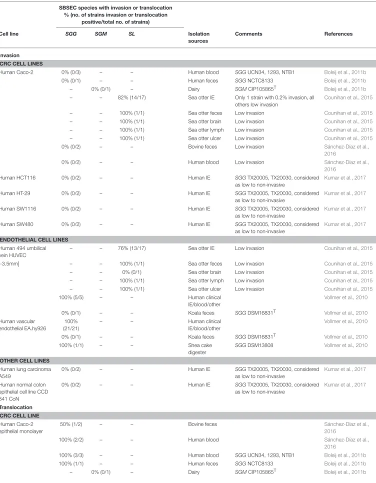
In the colon, adhesion of SBSEC members was assessed using epithelial CRC cell lines CMT-93, Caco-2 and HT-29 (Table 2). Adhesion to these CRC cell lines was observed for several SBSEC species includingSL,SGGandSGM. Animal-derivedSLstrains (sea otter IE, feces, brain and lymph node isolates) adhered highly variable to CMT-93 and Caco-2. Significant adhesion (>0.2–0.3%
of the inoculum) to CMT-93 and Caco-2 was rare within this strain pool and at slightly lower levels than theS.Typhimurium reference strain (Counihan et al., 2015). Adhesion to CMT-93 was generally lower than and without correlation to that of Caco-2 even forS.Typhimurium (Counihan et al., 2015) suggesting low suitability of CMT-93 cells for SBSEC adhesion assessment
T A B L E 2 | A d h e si o n o f S B S E C sp e c ie s to c e ll lin e s. S B S E C s p e c ie s w it h a d h e s io n % (n o . o f s tr a in s a d h e s io n p o s it iv e /t o ta l n o . o f s tr a in s ) C e ll li n e S G G S G M S G P S L S B Is o la ti o n s o u rc e s C o mme n ts R e fe re n c e s E P IT H E L IA L C E L L L IN E S B o vi n e ru m e n e p ith e lia lc e ll lin e n o t d iff e re n tia te d – – – – 1 0 0 % (2 /2 ) B o vi n e ru m e n K e ra tin iz a tio n p o si tiv e ly in flu e n c e d a d h e si o n . A d h e si o n st ro n g e r to b o vi n e th a n o vi n e c e lls S e m jé n a n d G á lfi, 1 9 9 0 B o vi n e ru m e n e p ith e lia lc e ll lin e n o n -k e ra tin iz e d – – – – 0 % (0 /4 ) B o vi n e ru m e n S ty ria k e t a l., 1 9 9 2 – – – – 0 % (0 /3 ) B o vi n e ru m e n B io ty p e II S ty ria k e t a l., 1 9 9 4 B o vi n e ru m e n e p ith e lia lc e ll lin e ke ra tin iz e d – – – – 1 0 0 % (4 /4 ) B o vi n e ru m e n S ty ria k e t a l., 1 9 9 2 – – – – 1 0 0 % (3 /3 ) B o vi n e ru m e n B io ty p e II S ty ria k e t a l., 1 9 9 4 O vi n e ru m e n e p ith e lia lc e ll lin e – – – – 1 0 0 % (2 /2 ) B o vi n e ru m e n K e ra tin iz a tio n p o si tiv e ly in flu e n c e d a d h e si o n . A d h e si o n st ro n g e r to b o vi n e th a n o vi n e c e lls . S e m jé n a n d G á lfi, 1 9 9 0 O vi n e ru m e n e p ith e lia lc e ll lin e n o n -k e ra tin iz e d – – – – 2 0 % (1 /5 ) B o vi n e ru m e n S ty ria k e t a l., 1 9 9 1 – – – – 0 % (0 /4 ) B o vi n e ru m e n S ty ria k e t a l., 1 9 9 2 O vi n e ru m e n e p ith e lia lc e ll lin e ke ra tin iz e d – – – – 1 0 0 % (5 /5 ) B o vi n e ru m e n S ty ria k e t a l., 1 9 9 1 – – – – 1 0 0 % (4 /4 ) B o vi n e ru m e n S ty ria k e t a l., 1 9 9 2 O ra l/p rim a ry b u c c a le p ith e lia lc e lls 1 0 0 % (3 /3 ) – – – 1 0 0 % (2 /2 ) H u m a n IE V o n H u n o ls te in e t a l., 1 9 9 3 – – – – 1 0 0 % (1 /1 ) B o vi n e fe c e s S B N C T C 8 1 7 7 , a d h e si o n si g n ific a n tly w e a ke r th a n h u m a n IE st ra in s V o n H u n o ls te in e t a l., 1 9 9 3 C R C C E L L L IN E S H u m a n C a c o -2 1 0 0 % (2 /2 ) – – – – B o vi n e fe c e s S á n c h e z-D ía z e t a l., 2 0 1 6 1 0 0 % (2 /2 ) – – – – H u m a n b lo o d S á n c h e z-D ía z e t a l., 2 0 1 6 1 0 0 % (1 /1 ) – – – – H u m a n fe c e s S G G N C T C 8 1 3 3 , d o se -d e p e n d e n t b in d in g E llm e ric h e t a l., 2 0 0 0 a – – 1 0 0 % (1 /1 ) – – H u m a n b lo o d S G P A T C C 4 3 1 4 4 , d o se -d e p e n d e n t b in d in g E llm e ric h e t a l., 2 0 0 0 a 1 0 0 % (3 /3 ) – – – – H u m a n b lo o d S G G U C N 3 4 , 1 2 9 3 , N T B 1 B o le ij e t a l., 2 0 1 1 b 1 0 0 % (1 /1 ) – – – – H u m a n fe c e s S G G N C T C 8 1 3 3 B o le ij e t a l., 2 0 1 1 b – 1 0 0 % (1 /1 ) – – – D a iry S G M C IP 1 0 5 8 6 5 T B o le ij e t a l., 2 0 1 1 b – – – 1 8 % (3 /1 7 ) – S e a o tt e r IE C o u n ih a n e t a l., 2 0 1 5 – – – 0 % (0 /1 ) – S e a o tt e r fe c e s C o u n ih a n e t a l., 2 0 1 5 – – – 0 % (0 /1 ) – S e a o tt e r b ra in C o u n ih a n e t a l., 2 0 1 5 – – – 0 % (0 /1 ) – S e a o tt e r ly m p h C o u n ih a n e t a l., 2 0 1 5 – – – 1 0 0 % (1 /1 ) – S e a o tt e r u lc e r C o u n ih a n e t a l., 2 0 1 5 (C o n ti n u e d )
T A B L E 2 | C o n tin u e d S B S E C s p e c ie s w it h a d h e s io n % (n o . o f s tr a in s a d h e s io n p o s it iv e /t o ta l n o . o f s tr a in s ) C e ll li n e S G G S G M S G P S L S B Is o la ti o n s o u rc e s C o mme n ts R e fe re n c e s H u m a n e p ith e lio id c a rc in o m a c e ll lin e K B E C A C C 8 6 1 0 3 0 0 4 1 0 0 % (1 /1 ) – – – – H u m a n fe c e s S G G N C T C 8 1 3 3 , d o se -d e p e n d e n t b in d in g , d is c o n tin u e d c e ll lin e d u e to H e L a d e riv a tio n E llm e ric h e t a l., 2 0 0 0 a – – 1 0 0 % (1 /1 ) – – H u m a n b lo o d S G P A T C C 4 3 1 4 4 , d o se -d e p e n d e n t b in d in g E llm e ric h e t a l., 2 0 0 0 a H u m a n H C T 1 1 6 1 0 0 % (2 /2 ) – – – – H u m a n IE S G G T X 2 0 0 0 5 , T X 2 0 0 3 0 K u m a r e t a l., 2 0 1 7 H u m a n H T-2 9 1 0 0 % (3 /3 ) – – – – H u m a n b lo o d S G G U C N 3 4 , 1 2 9 3 , N T B 1 B o le ij e t a l., 2 0 1 1 b 1 0 0 % (1 /1 ) – – – – H u m a n fe c e s S G G N C T C 8 1 3 3 B o le ij e t a l., 2 0 1 1 b – 1 0 0 % (1 /1 ) – – – D a iry S G M C IP 1 0 5 8 6 5 T B o le ij e t a l., 2 0 1 1 b 1 0 0 % (1 /1 ) – – – – H u m a n IE S G G U C N 3 4 M a rt in s e t a l., 2 0 1 5 1 0 0 % (5 /5 ) – – – – H u m a n IE S G G T X 2 0 0 0 5 , T X 2 0 0 3 0 , T X 2 0 0 3 1 (p ro m o tin g c e ll p ro lif e ra tio n ); S G G T X 2 0 0 0 8 , T X 2 0 0 3 4 , A T C C 4 3 1 4 3 (n o n -p ro m o tin g c e ll p ro lif e ra tio n ). A d h e si o n lo w e r fo r n o n -p ro m o tin g st ra in s K u m a r e t a l., 2 0 1 8 1 0 0 % (2 /2 ) – – – – H u m a n IE S G G T X 2 0 0 0 5 , T X 2 0 0 3 0 K u m a r e t a l., 2 0 1 7 H u m a n H T-2 9 M T X 1 0 0 % (1 /1 ) – – – – H u m a n IE h ig h e r a d h e si o n to H T-2 9 M T X vs . H T-2 9 M a rt in s e t a l., 2 0 1 5 M o u se re c tu m c a rc in o m a C M T-9 3 – – – 6 % (1 /1 7 ) – S e a o tt e r IE C o u n ih a n e t a l., 2 0 1 5 – – – 0 % (0 /1 ) – S e a o tt e r fe c e s C o u n ih a n e t a l., 2 0 1 5 – – – 0 % (0 /1 ) – S e a o tt e r b ra in C o u n ih a n e t a l., 2 0 1 5 – – – 0 % (0 /1 ) – S e a o tt e r ly m p h C o u n ih a n e t a l., 2 0 1 5 – – – 0 % (0 /1 ) – S e a o tt e r u lc e r C o u n ih a n e t a l., 2 0 1 5 H u m a n S W 1 1 1 6 1 0 0 % (2 /2 ) – – – – H u m a n IE S G G T X 2 0 0 0 5 , T X 2 0 0 3 0 K u m a r e t a l., 2 0 1 7 H u m a n S W 4 8 0 1 0 0 % (2 /2 ) – – – – H u m a n IE S G G T X 2 0 0 0 5 , T X 2 0 0 3 0 K u m a r e t a l., 2 0 1 7 E N D O T H E L IA L C E L L L IN E S H u m a n 4 9 4 u m b ili c a lv e in H U V E C – – – 4 1 % (7 /1 7 ) – S e a o tt e r IE C o u n ih a n e t a l., 2 0 1 5 – – – 0 % (0 /1 ) – S e a o tt e r fe c e s C o u n ih a n e t a l., 2 0 1 5 – – – 0 % (0 /1 ) – S e a o tt e r b ra in C o u n ih a n e t a l., 2 0 1 5 – – – 0 % (0 /1 ) – S e a o tt e r ly m p h C o u n ih a n e t a l., 2 0 1 5 – – – 0 % (0 /1 ) – S e a o tt e r u lc e r C o u n ih a n e t a l., 2 0 1 5 M o u se e n d o th e lia lt u m o r E O M A – – – 4 7 % (8 /1 7 ) – S e a o tt e r IE C o u n ih a n e t a l., 2 0 1 5 – – – 0 % (0 /1 ) – S e a o tt e r fe c e s C o u n ih a n e t a l., 2 0 1 5 – – – 1 0 0 % (1 /1 ) – S e a o tt e r b ra in C o u n ih a n e t a l., 2 0 1 5 – – – 0 % (0 /1 ) – S e a o tt e r ly m p h C o u n ih a n e t a l., 2 0 1 5 – – – 1 0 0 % (1 /1 ) – S e a o tt e r u lc e r C o u n ih a n e t a l., 2 0 1 5 H u m a n u n n a m e d sa p h e n o u s ve in 1 0 0 % (1 /1 ) – – – – H u m a n fe c e s S G G N C T C 8 1 3 3 , d o se -d e p e n d e n t b in d in g E llm e ric h e t a l., 2 0 0 0 a – – 1 0 0 % (1 /1 ) – – H u m a n b lo o d S G P A T C C 4 3 1 4 4 , d o se -d e p e n d e n t b in d in g E llm e ric h e t a l., 2 0 0 0 a (C o n ti n u e d )
T A B L E 2 | C o n tin u e d S B S E C s p e c ie s w it h a d h e s io n % (n o . o f s tr a in s a d h e s io n p o s it iv e /t o ta l n o . o f s tr a in s ) C e ll li n e S G G S G M S G P S L S B Is o la ti o n s o u rc e s C o mme n ts R e fe re n c e s H u m a n va sc u la r e n d o th e lia lE A .h y9 2 6 1 0 0 % (2 1 /2 1 ) – – – – H u m a n c lin ic a l IE /b lo o d /o th e r V o llm e r e t a l., 2 0 1 0 1 0 0 % (1 /1 ) – – – – K o a la fe c e s S G G D S M 1 6 8 3 1 T V o llm e r e t a l., 2 0 1 0 1 0 0 % (1 /1 ) – – – – S h e a c a ke d ig e st e r S G G D S M 1 3 8 0 8 V o llm e r e t a l., 2 0 1 0 O T H E R C E L L L IN E S H u m a n lu n g c a rc in o m a A 5 4 9 1 0 0 % (2 /2 ) – – – – H u m a n IE S G G T X 2 0 0 0 5 , T X 2 0 0 3 0 K u m a r e t a l., 2 0 1 7 H u m a n n o rm a lc o lo n e p ith e lia lc e ll lin e C C D 8 4 1 C o N 1 0 0 % (2 /2 ) – – – – H u m a n IE S G G T X 2 0 0 0 5 , T X 2 0 0 3 0 , a d h e re n c e p o si tiv e b u t si g n ific a n t lo w e r th a n to c a n c e r lin e s K u m a r e t a l., 2 0 1 7 T H P − 1 h u m a n m o n o c yt e s 1 0 0 % (1 /1 ) – – – – H u m a n fe c e s S G G N C T C 8 1 3 3 , d o se -d e p e n d e n t b in d in g E llm e ric h e t a l., 2 0 0 0 a – – 1 0 0 % (1 /1 ) – – H u m a n b lo o d S G P A T C C 4 3 1 4 4 , d o se -d e p e n d e n t b in d in g E llm e ric h e t a l., 2 0 0 0 a
and possibly the need to evaluate sea otter-derivedSLfor host specificity.
Adhesion abilities toward Caco-2 and HT-29 of human-derived SGG and dairy SGM was low (<15% of inoculum) to intermediate (20–50% of inoculum) forSGG and SGM/SE, respectively (Table 2). Low adhesion was comparable with that of S.Typhimurium whereas intermediate adhesion was comparable to that of E. coli and Lb. plantarum reference strains, but significantly lower than the 80–98% observed for E. faecalis. Differences were particularly evident forSGG NCTC8133 that more efficiently adhered to Caco-2 than HT-29 (Boleij et al., 2011b). A comparative assessment of blood-derived SGG of IE patients indicated significantly enhanced adhesion abilities to HT-29 cells, particularly among strains able to promote tumor cell proliferation (Kumar et al., 2018). This differentiation between proliferation-promoting and non-promotingSGGwas paralleled by the ability to colonize mice. Interestingly, mice colonization was increased in A/J type mice compared to C57BL/6 mice, which might be related to different host factors required for colonization (Kumar et al., 2018). Therefore, certain tumor-promoting strains might possess enhanced adhesion capabilities and thus a selective advantage particularly in a tumor environment presenting favorable factors. General adhesion of SBSEC members to CRC cell lines however seems limited in comparison to other gut pathogens.
Adhesion to Endothelial Cell Lines
Similar to epithelial cell lines, dose-dependent adhesion behavior was also observed forSGGNCTC8133 (human fecal isolate) and SGP ATCC43144 (human blood isolate) to human saphenous vein endothelial cell lines (Table 2). Especially at low inoculums, binding to endothelial cells was higher than to epithelial cells suggesting a preference toward endothelium (Ellmerich et al., 2000a). This was also observed among primarily human IESGG isolates adhering to EA.hy926 cells but not with animal feces-derived SGG DSM16831T (Vollmer et al., 2010). Mechanical stress on HUVEC cells had no influence on adherence suggesting thatSGGactively colonizes endothelial tissues (Vollmer et al., 2010). Interestingly, only blood isolates ofSGGexpress the blood-group antigen sialyl lewis-X (sLex) on their cell surface. sLex is normally expressed on the cell surface of leukocytes enabling rolling of leukocytes on the endothelium. This might increase SGGadhesion to endothelial cells (Hirota et al., 1996).
Significant adhesion to endothelial HUVEC-C cells was observed in multiple IE and septicemiaSLstrains isolated from sea otters (Counihan et al., 2015). The ability to adhere to cell lines other than HUVEC-C was minimal; only one strain showed adhesion to human epithelial Caco-2, human endothelial HUVEC-C and mouse endothelial tumor EOMA cells (Table 2). The other strains showed minimal adhesion to mouse/human epithelial or endothelial cells, suggesting key differences in adhesion mechanisms to different cell types and possibly host origin. In general, isolates from heart or blood adhered better to intact endothelial cells and support possible IE establishment without previous history of heart disease (Counihan et al., 2015). However, general adhesion of SL strains was in the range of 0.05–0.2% of the inoculum and thus significantly lower than the
2% of human clinical Staphylococcus aureus ATCC25923 used as reference (Counihan et al., 2015). The biological impact to trigger IE despite this significantly lower adhesion ability ofSL in contrast toS. aureuswill require further evaluation in relation to host specificity and mechanisms responsible for endothelial tissue colonization by SBSEC membersin vitroandin vivo.
Binding to Extracellular Matrix Proteins
Extracellular matrix proteins (ECM) are an important component to facilitate bacterial binding to epithelial and endothelial cell surfaces and thus niche colonization in humans and animals. Collagen thereby plays an important role. Collagen type I is present in organ capsules and scar tissue, such as on damaged heart valves. Collagen type IV is the main constituent of basement membranes and can become exposed at tumor sites (Tanjore and Kalluri, 2006; Boleij and Tjalsma, 2013).Adhesion to collagen type I and IV is a key feature of SGG (Table 3). MostSGG strains derived from human blood cultures of IE or bacteremia patients, pigeons suffering from streptococcosis (SGGandSGP) andSLstrains derived from sea otters with IE, but also fecal and dairy SBSEC isolates displayed binding to collagen type IV. In contrast, binding to collagen type I was a feature mostly associated with human blood-derivedSGG, SGP, SII and SL isolates and partially also withSGM isolates (Vanrobaeys et al., 2000a; Sillanpää et al., 2008; Vollmer et al., 2010; Boleij et al., 2011b; Counihan et al., 2015; Grimm et al., 2018). Among a panel of human and food-derivedSII and SL as well as dairy SGM, adhesion to collagen type I and IV was particularly present in human blood isolates (Boleij et al., 2011b; Jans et al., 2016). In contrast, SGG considered as commensals and isolated from human feces, pigeons and ruminants including the SGG type strain rarely bound to collagen type I, III and IV (Table 3). Similar differences were also observed between SGG from infected vs. SGG from healthy humans featuring high vs. low adhesion, respectively (Grimm et al., 2018).SGG NCTC8133 andSGPstrain ATCC43144 were only shown to bind collagen type IV whereasSGPDSM15351Tstrains displayed no adhesion to collagen type I and IV. Other collagen types bound by human IE-derivedSGGare collagen type II (96%) and to a lesser extent collagen type V (40%) (Table 3) (Ellmerich et al., 2000a; Sillanpää et al., 2008; Vollmer et al., 2010). Differences in collagen adhesion patterns therefore seem to exist between animal and human-derived strains even within the same species. Whether these different adhesion patterns amongSGGand other SBSEC members also translate into different abilities to cause disease remains to be investigated.
Connective tissue and the tumor-microenvironment contain an extensive network of ECM including collagen, laminins, fibronectin, proteoglycans, and hyaluronans (Peddareddigari et al., 2010).SLstrains from sea otters adhered to fibronectin, laminin, and hyaluronic acid in all cases (Table 3) (Counihan et al., 2015). Also human blood-derivedSIIandSLfeatured high adhesion abilities to fibronectin (Jans et al., 2016). Interestingly, theSLgenome features relevant hits to adhesion factors such as pneumococcal cell surface adherence protein A PavA involved in fibronectin-binding and the laminin-binding protein Lmb (Jin et al., 2013). Binding to fibronectin is also observed in the
SGGtype strain and the majority of pigeon-derivedSGGstrains (Table 3). Interestingly, fibronectin-binding is less prevalent in human-derived SGG and SGP with the exception of SGG NCTC8133 and human blood strainSGPATCC43144 (Ellmerich et al., 2000a; Vanrobaeys et al., 2000a; Sillanpää et al., 2008; Vollmer et al., 2010; Jans et al., 2016). Furthermore, rumen-derivedSB strains showed low or no binding to both human and porcine fibronectin (Styriak et al., 1999). Similar, theSIItype strain,SIIdairy andSLhuman commensal strains showed with a few exceptions only minor adhesion abilities to fibronectin, collagen type I and IV, mucin and fibrinogen (Table 3) (Jans et al., 2016).
Fibrinogen-binding is in contrast to fibronectin-binding a common feature also among human IE SGG strains, dairy SGM, human SII and SL blood isolates (Table 3) (Ellmerich et al., 2000a; Sillanpää et al., 2008; Vollmer et al., 2010; Jans et al., 2016). Human IE-derived SGGalso showed interactions with tenascin, laminin and vitronectin (Ellmerich et al., 2000a; Sillanpää et al., 2008; Vollmer et al., 2010). In contrast, rumen SBstrains showed mostly moderate or weak adhesion to bovine lactoferrin, vitronectin, heparin, and BSA. None of the SB strains bound to human serum albumin (Table 3) (Styriak et al., 1999). These differences in ECM adhesion patterns between animal and human strains might therefore be important for their colonization abilities of different body sites. These patterns furthermore suggest different adhesion mechanisms inSL,SGG and other SBSEC members (Lin et al., 2011b; Papadimitriou et al., 2014). Particularly human and animal blood isolates seem to have the ability to bind fibrinogen, while fibronectin-binding is variable, which implies different adhesion abilities regarding fibronectin in the tumor-microenvironment and fibrinogen at damaged sites requiring blood clotting.
Biofilm Formation, Exopolysaccharides,
Dextran Production and Capsular
Polysaccharides
Adhesion and biofilm-forming abilities are linked to colonization and persistence in the GIT. SB produce at least two types of polysaccharides: (1) water-soluble glucans, often dextrans, comprised ofα-1:6 linked glucose units (Bailey, 1959); and (2) capsular polysaccharide (Bailey and Oxford, 1958). GtfA inSGG was found to produce water-insoluble α-1,3-linked glucosidic polymers whereas GtfB encoded forα-1,3-linked water-insoluble and α-1,6-linked glucosidic water-soluble polymers (Lin et al., 2011b). Both types of polysaccharides have specific roles in adhesion, colonization and host immune evasion (Nobbs et al., 2009; Isenring et al., 2018). The ability to form biofilms is however not directly correlated to virulence and needs to be carefully distinguished (Vollmer et al., 2010).
Biofilm formation was observed with SBSEC strains from GIT, blood and food origin (Vollmer et al., 2010; Boleij et al., 2011b; Jans et al., 2016).SII and other SBSEC members were also observed to form biofilms on human teeth featuring various degrees of auto- and co-aggregation with other oral microbes (Shen et al., 2005; Arul and Palanivelu, 2014). Even outside a host, biofilm formation to uncoated plastic and stainless steel surfaces
T A B L E 3 | A d h e si o n o f S B S E C sp e c ie s to d iff e re n t e xt ra c e llu la r m a tr ix p ro te in (E C M ) ty p e s. S B S E C s p e c ie s w it h a d h e s io n % (n o . o f s tr a in s a d h e s io n p o s it iv e /t o ta l n o . o f s tr a in s ) E C M (s o u rc e ) S G G S G M S G P S II S L S B Is o la ti o n s o u rc e R e fe re n c e s C O L L A G E N Ty p e I( c a lf sk in ) 0 % (0 /9 ) – 0 % (0 /5 ) – – – P ig e o n st re p to c o c c o si s V a n ro b a e ys e t a l., 2 0 0 0 a Ty p e I( h u m a n ) 2 5 % (6 /2 4 ) – – – – – a n im a l G rim m e t a l., 2 0 1 8 0 % (0 /1 ) – – – – – F o o d G rim m e t a l., 2 0 1 8 7 8 % (3 5 /4 5 ) – – – – – H u m a n b lo o d G rim m e t a l., 2 0 1 8 0 % (0 /2 ) – – – – – H u m a n fe c e s G rim m e t a l., 2 0 1 8 0 % (0 /2 ) – – – – – U n kn o w n G rim m e t a l., 2 0 1 8 Ty p e I( ra t ta il) 1 0 0 % (1 /1 ) – – – 0 % (0 /1 ) – A n im a l Ja n s e t a l., 2 0 1 6 1 0 0 % (2 /2 ) – – – – – B o vi n e fe c e s S á n c h e z-D ía z e t a l., 2 0 1 6 – 6 0 % (3 /5 ) – 1 0 % (3 /2 9 ) – – D a iry Ja n s e t a l., 2 0 1 6 – – – 0 % (0 /1 ) – – F o o d c o n ta m in a tio n Ja n s e t a l., 2 0 1 6 – – – 7 5 % (3 /4 ) 1 0 0 % (1 /1 ) – H u m a n b lo o d Ja n s e t a l., 2 0 1 6 1 0 0 % (2 /2 ) – – – – – H u m a n b lo o d S á n c h e z-D ía z e t a l., 2 0 1 6 – – 0 % (0 /1 ) – – – H u m a n c e re b ro sp in a lflu id Ja n s e t a l., 2 0 1 6 1 0 0 % (1 /1 ) – – – 0 % (0 /4 ) – H u m a n fe c e s Ja n s e t a l., 2 0 1 6 7 3 % (1 1 /1 5 ) – 1 0 0 % (1 /1 ) – 0 % (0 /1 ) – H u m a n IE S ill a n p ä ä e t a l., 2 0 0 8 – – – 0 % (0 /5 ) – – H u m a n u n kn o w n Ja n s e t a l., 2 0 1 6 Ty p e I( so u rc e n /a ) 1 0 0 % (2 3 /2 3 ) – – – – – H u m a n IE a n d o th e rs * V o llm e r e t a l., 2 0 1 0 1 0 0 % (3 /3 ) – – – – – H u m a n IE B o le ij e t a l., 2 0 1 1 b 1 0 0 % (1 /1 ) – – – – – A n im a lf e c e s B o le ij e t a l., 2 0 1 1 b – 1 0 0 % (1 /1 ) – – – – D a iry B o le ij e t a l., 2 0 1 1 b Ty p e II (s o u rc e n /a ) 9 6 % (2 2 /2 3 ) – – – – – H u m a n IE a n d o th e rs * V o llm e r e t a l., 2 0 1 0 Ty p e III (c a lf sk in ) 2 2 % (2 /9 ) – 0 % (0 /5 ) – – – P ig e o n st re p to c o c c o si s V a n ro b a e ys e t a l., 2 0 0 0 a Ty p e IV (h u m a n ) 1 0 0 % (1 /1 ) – – – 0 % (0 /1 ) – a n im a l Ja n s e t a l., 2 0 1 6 1 0 0 % (2 /2 ) – – – – – B o vi n e fe c e s S á n c h e z-D ía z e t a l., 2 0 1 6 – 2 0 % (1 /5 ) – 1 4 % (4 /2 9 ) – – D a iry Ja n s e t a l., 2 0 1 6 1 0 0 % (1 /1 ) – – – – – F o o d G rim m e t a l., 2 0 1 8 – – – 0 % (0 /1 ) – – F o o d c o n ta m in a tio n Ja n s e t a l., 2 0 1 6 – – – 7 5 % (3 /4 ) 1 0 0 % (1 /1 ) – H u m a n b lo o d Ja n s e t a l., 2 0 1 6 – – 0 % (0 /1 ) – – – H u m a n c e re b ro sp in a lflu id Ja n s e t a l., 2 0 1 6 0 % (0 /1 ) – – – 0 % (0 /4 ) – H u m a n fe c e s Ja n s e t a l., 2 0 1 6 6 0 % (9 /1 5 ) – 0 % (0 /1 ) – 0 % (0 /1 ) – H u m a n IE S ill a n p ä ä e t a l., 2 0 0 8 – – – 0 % (0 /5 ) – – H u m a n u n kn o w n Ja n s e t a l., 2 0 1 6 (C o n ti n u e d )
T A B L E 3 | C o n tin u e d S B S E C s p e c ie s w it h a d h e s io n % (n o . o f s tr a in s a d h e s io n p o s it iv e /t o ta l n o . o f s tr a in s ) E C M (s o u rc e ) S G G S G M S G P S II S L S B Is o la ti o n s o u rc e R e fe re n c e s 3 8 % (9 /2 4 ) – – – – – A n im a l G rim m e t a l., 2 0 1 8 8 2 % (3 7 /4 5 ) – – – – – H u m a n b lo o d G rim m e t a l., 2 0 1 8 1 0 0 % (2 /2 ) – – – – – H u m a n b lo o d S á n c h e z-D ía z e t a l., 2 0 1 6 0 % (0 /2 ) – – – – – H u m a n fe c e s G rim m e t a l., 2 0 1 8 0 % (0 /2 ) – – – – – U n kn o w n G rim m e t a l., 2 0 1 8 Ty p e IV (m o u se sa rc o m a ) 8 9 % (8 /9 ) – 1 0 0 % (5 /5 ) – – – P ig e o n st re p to c o c c o si s V a n ro b a e ys e t a l., 2 0 0 0 a Ty p e IV (m o u se tu m o r) – – – – 1 0 0 % (1 0 /1 0 ) – S e a o tt e r b ra in §, fe c e s §, IE , u lc e r § C o u n ih a n e t a l., 2 0 1 5 Ty p e IV (s o u rc e n /a ) – – 1 0 0 % (1 /1 ) – – – H u m a n b lo o d E llm e ric h e t a l., 2 0 0 0 a 1 0 0 % (1 /1 ) – – – – – H u m a n fe c e s E llm e ric h e t a l., 2 0 0 0 a 9 6 % (2 2 /2 3 ) – – – – – H u m a n IE a n d o th e rs * V o llm e r e t a l., 2 0 1 0 1 0 0 % (3 /3 ) – – – – – H u m a n IE B o le ij e t a l., 2 0 1 1 b 1 0 0 % (1 /1 ) – – – – – A n im a lf e c e s B o le ij e t a l., 2 0 1 1 b – 1 0 0 % (1 /1 ) – – – – D a iry B o le ij e t a l., 2 0 1 1 b Ty p e V (h u m a n ) 4 0 % (6 /1 5 ) – 0 % (0 /1 ) – 0 % (0 /1 ) – H u m a n IE S ill a n p ä ä e t a l., 2 0 0 8 O T H E R E C M S O U R C E S F ib rin o g e n (h u m a n ) 1 0 0 % (1 /1 ) – – – 0 % (0 /1 ) – A n im a l Ja n s e t a l., 2 0 1 6 – 4 0 % (2 /5 ) – 7 % (2 /2 9 ) – – D a iry Ja n s e t a l., 2 0 1 6 – – – 0 % (0 /1 ) – – F o o d c o n ta m in a tio n Ja n s e t a l., 2 0 1 6 – – – 7 5 % (3 /4 ) 1 0 0 % (1 /1 ) – H u m a n b lo o d Ja n s e t a l., 2 0 1 6 – – 0 % (0 /1 ) – – – H u m a n c e re b ro sp in a lflu id Ja n s e t a l., 2 0 1 6 1 0 0 % (1 /1 ) – – – 0 % (0 /4 ) – H u m a n fe c e s Ja n s e t a l., 2 0 1 6 4 7 % (7 /1 5 ) – 1 0 0 % (1 /1 ) – 0 % (0 /1 ) – H u m a n IE S ill a n p ä ä e t a l., 2 0 0 8 7 8 % (1 8 /2 3 ) – – – – – H u m a n IE a n d o th e rs * V o llm e r e t a l., 2 0 1 0 – – – 0 % (0 /5 ) – – H u m a n u n kn o w n Ja n s e t a l., 2 0 1 6 F ib ro n e c tin (h u m a n ) 1 0 0 % (1 /1 ) – – – 0 % (0 /1 ) – A n im a l Ja n s e t a l., 2 0 1 6 – – – – – 2 0 % (2 /1 0 ) B o vi n e ru m e n S ty ria k e t a l., 1 9 9 9 – 6 0 % (3 /5 ) – 7 % (2 /2 9 ) – – D a iry Ja n s e t a l., 2 0 1 6 – – – 0 % (0 /1 ) – – F o o d c o n ta m in a tio n Ja n s e t a l., 2 0 1 6 – – – 7 5 % (3 /4 ) 1 0 0 % (1 /1 ) – H u m a n b lo o d Ja n s e t a l., 2 0 1 6 – – 0 % (0 /1 ) – – – H u m a n c e re b ro sp in a lflu id Ja n s e t a l., 2 0 1 6 0 % (0 /1 ) – – – 0 % (0 /4 ) – H u m a n fe c e s Ja n s e t a l., 2 0 1 6 3 3 % (5 /1 5 ) – 1 0 0 % (1 /1 ) – 0 % (0 /1 ) – H u m a n IE S ill a n p ä ä e t a l., 2 0 0 8 – – – 0 % (0 /5 ) – – H u m a n u n kn o w n Ja n s e t a l., 2 0 1 6 6 7 % (6 /9 ) – 6 0 % (3 /5 ) – – – P ig e o n st re p to c o c c o si s V a n ro b a e ys e t a l., 2 0 0 0 a (C o n ti n u e d )
T A B L E 3 | C o n tin u e d S B S E C s p e c ie s w it h a d h e s io n % (n o . o f s tr a in s a d h e s io n p o s it iv e /t o ta l n o . o f s tr a in s ) E C M (s o u rc e ) S G G S G M S G P S II S L S B Is o la ti o n s o u rc e R e fe re n c e s – – – – 1 0 0 % (1 0 /1 0 ) – S e a o tt e r b ra in §, fe c e s §, IE , u lc e r § C o u n ih a n e t a l., 2 0 1 5 F ib ro n e c tin (p o rc in e ) – – – – – 0 % (0 /1 0 ) B o vi n e ru m e n S ty ria k e t a l., 1 9 9 9 F ib ro n e c tin (s o u rc e n /a ) – – 1 0 0 % (1 /1 ) – – – H u m a n b lo o d E llm e ric h e t a l., 2 0 0 0 a 1 0 0 % (1 /1 ) – – – – – H u m a n fe c e s E llm e ric h e t a l., 2 0 0 0 a 3 0 % (7 /2 3 ) – – – – – H u m a n IE a n d o th e rs * V o llm e r e t a l., 2 0 1 0 H e p a rin – – – – – 0 % (0 /1 0 ) B o vi n e ru m e n S ty ria k e t a l., 1 9 9 9 L a c to fe rr in (b o vi n e ) – – – – – 2 0 % (2 /1 0 ) B o vi n e ru m e n S ty ria k e t a l., 1 9 9 9 L a m in in (m o u se tu m o r) – – – – 1 0 0 % (1 0 /1 0 ) – S e a o tt e r b ra in §, fe c e s §, IE , u lc e r § C o u n ih a n e t a l., 2 0 1 5 L a m in in (s o u rc e n /a ) – – 1 0 0 % (1 /1 ) – – – H u m a n b lo o d E llm e ric h e t a l., 2 0 0 0 a 1 0 0 % (1 /1 ) – – – – – H u m a n fe c e s E llm e ric h e t a l., 2 0 0 0 a 7 0 % (1 6 /2 3 ) – – – – – H u m a n IE a n d o th e rs * V o llm e r e t a l., 2 0 1 0 M u c in ty p e II (p o rc in e st o m a c h ) 1 0 0 % (1 /1 ) – – – 1 0 0 % (1 /1 ) – A n im a l Ja n s e t a l., 2 0 1 6 – 4 0 % (2 /5 ) – 1 7 % (5 /2 9 ) – – D a iry Ja n s e t a l., 2 0 1 6 – – – 0 % (0 /1 ) – – F o o d c o n ta m in a tio n Ja n s e t a l., 2 0 1 6 – – – 1 0 0 % (4 /4 ) 1 0 0 % (1 /1 ) – H u m a n b lo o d Ja n s e t a l., 2 0 1 6 – – 0 % (0 /1 ) – – – H u m a n c e re b ro sp in a lflu id Ja n s e t a l., 2 0 1 6 0 % (0 /1 ) – – – 0 % (0 /4 ) – H u m a n fe c e s Ja n s e t a l., 2 0 1 6 – – – 0 % (0 /5 ) – – H u m a n u n kn o w n Ja n s e t a l., 2 0 1 6 Te n a sc in 7 0 % (1 6 /2 3 ) – – – – – H u m a n IE a n d o th e rs * V o llm e r e t a l., 2 0 1 0 V itr o n e c tin (h u m a n ) – – – – – 2 0 % (2 /1 0 ) B o vi n e ru m e n S ty ria k e t a l., 1 9 9 9 V itr o n e c tin (s o u rc e n /a ) 2 2 % (5 /2 3 ) – – – – – H u m a n IE a n d o th e rs * V o llm e r e t a l., 2 0 1 0 B o vi n e se ru m a lb u m in (B S A ) 0 % (0 /1 ) – – – 0 % (0 /1 ) – A n im a l Ja n s e t a l., 2 0 1 6 – – – – – 0 % (0 /1 0 ) B o vi n e ru m e n S ty ria k e t a l., 1 9 9 9 – 4 0 % (2 /5 ) – 1 0 % (3 /2 9 ) – – D a iry Ja n s e t a l., 2 0 1 6 – – – 0 % (0 /1 ) – – F o o d c o n ta m in a tio n Ja n s e t a l., 2 0 1 6 – – – 7 5 % (3 /4 ) 1 0 0 % (1 /1 ) – H u m a n b lo o d Ja n s e t a l., 2 0 1 6 – – 0 % (0 /1 ) – – – H u m a n c e re b ro sp in a lflu id Ja n s e t a l., 2 0 1 6 0 % (0 /1 ) – – – 0 % (0 /4 ) – H u m a n fe c e s Ja n s e t a l., 2 0 1 6 0 % (0 /1 5 ) – 0 % (0 /1 ) – 0 % (0 /1 ) – H u m a n IE S ill a n p ä ä e t a l., 2 0 0 8 – – – 0 % (0 /5 ) – – H u m a n u n kn o w n Ja n s e t a l., 2 0 1 6 – – – – 1 0 0 % (1 0 /1 0 ) – S e a o tt e r b ra in §, fe c e s §, IE , u lc e r § C o u n ih a n e t a l., 2 0 1 5 H u m a n se ru m a lb u m in (H S A ) – – – – – 0 % (0 /1 0 ) B o vi n e ru m e n S ty ria k e t a l., 1 9 9 9 * 2 3 s tr a in s tota lo f w h ic h 2 o ri g in a te d fr o m fe c a ls a m pl e s of a koa la be a r (ty pe s tr a in ) a n d on e a n a e robi c s h e a c a ke di ge s te r is ol a te , bu t da ta w a s n ot e xtr a c ta bl e by s tr a in . §da ta de ri ve d fr o m s in g le s tr a in s .
was observed for all SBSEC species of blood, animal and dairy origin (Flint et al., 1997, 1999; Jans et al., 2016).
Polysaccharides are major constituents of biofilms (Christensen, 1989; Nobbs et al., 2009). SGG, SL and many SBSEC members are known to produce extracellular glucan encoded by glycosyl-transferases similar to GtfA, GtfB, and GtfC of S. mutans but lacking in SGP ATCC43144 (Rusniok et al., 2010; Lin et al., 2011b). Instead,SGP harbored a strain-specific exopolysaccharide biosynthesis gene cluster featuring sequence identity highest with those of Bacillus cereus and
Clostridium thermocellum (Lin et al., 2011b). Generally, this
suggests that from a common SBSEC ancestor,SGGlikely kept most biofilm-related loci while the respective loci were either absent or comprised of pseudogenes in SGP, SII and SGM potentially reducing or abrogating biofilm formation capabilities in comparison to SGG (De Vuyst and Tsakalidou, 2008; Lin et al., 2011b; Papadimitriou et al., 2014). Biofilm production might therefore be SBSEC-species dependent but older data are inconclusive in this respect.
Dextran production from rumen SB biotype II isolates of sheep, calve and cow is particularly dependent on available sugar compounds and a CO2 source. In contrast to capsular polysaccharides, the production of dextran is limited and directly correlated with the available sucrose concentration (Bailey and Oxford, 1958; Barnes et al., 1961; Cheng et al., 1976). The CO2 source can include HCO−3, which is readily available in the rumen (Bailey and Oxford, 1958; Barnes et al., 1961). For dextran production bySB, three different growth requirements are suggested: (i) biotin and ammonium chloride as sole vitamin and N-source, respectively, (ii) calcium, pantothenate, adenine, biotin, thiamine and arginine or glutamic acid, or (iii) xanthine and additional amino acids (Barnes et al., 1961). Prototroph SGG in contrast to SBbiotype II likely possess the metabolic capabilities to produce biofilm even in niches not meeting these growth requirements (Rusniok et al., 2010).
Dextran production furthermore seems to play a role in ruminal acidosis (Humer et al., 2018). It is hypothesized that the higher sucrose content of grain feed boosts dextran production in ruminants to form a slime in the rumen (Cheng et al., 1976; Kulp and Ponte, 2000; Humer et al., 2018). This slime, comprised of proteins and polysaccharides of other bacteria, increases viscosity and produces a froth foam eventually leading to bloat (Cheng et al., 1976). A key role in the slime production process is attributed toSBvia acidification and dextran production via its rumen-adapted dextran sucrase (Bailey and Oxford, 1958; Min et al., 2006).
Virulence Factors of SBSEC Members
Related to Adhesion and Colonization
General Aspects of the Cell Surface in Relation to Adhesion
The bacterial cell surface has important roles in the interaction with the environment, the host and for pathogenesis (Nobbs et al., 2009; Isenring et al., 2018). Lipoproteins featuring a serine-rich motif following a cysteine residue are frequently present on the surface of SGG UCN34, possibly linked to specific interactions with polysaccharides from the environment
(Rusniok et al., 2010). Wall-extracted antigens from the cell surface of SBSEC members bound equally well to epithelial and endothelial cell lines as whole SBSEC cells, supporting a role for cell surface factors in adhesion (Ellmerich et al., 2000a). Among surface proteins,SBsurface protein Sbs6, Sbs10, Sbs13, and Sbs16 as well as the histone-like protein HlpA, autolysin AtlA and the cell surface protein antigen C PaC are currently characterized SBSEC virulence factors besides pili. While HlpA is present in most SBSEC members, Sbs13, Sbs16, AtlA and PaC are mainly limited to SGG whereas Sbs6 and Sbs10 are also regularly observed inSE. AmongSGG, only blood-derivedSGG usually feature all seven surface proteins in contrast to rumen or fecal isolates (Table 4and Supplementary Data 1). This suggests reduced or different virulence characteristics of the other SBSEC members in comparison to blood-derivedSGG.
Specific studies were performed on enolase and HlpA. Enolase is a conserved anchorless surface protein involved in cross-linking of SGG UCN34 and human epithelial cells (Boleij et al., 2011a). The main interaction partner was identified as cytokeratin 8. Cytokeratin 8 is constantly expressed by epithelial cells, but at increased levels by CRC and could therefore play a role in the association ofSGGwith CRC (Boleij et al., 2011a).
HlpA is highly prevalent among SBSEC members possibly involved in adhesion (Table 4) (Boleij et al., 2009a; Lin et al., 2011b; Papadimitriou et al., 2014). HlpA is an anchorless bacterial surface protein that binds to lipoteichoic acid at the Gram-positive cell wall. Lipoteichoic acid was previously suggested to be involved in adhesion in cooperation with surface proteins (Von Hunolstein et al., 1993; Styriak et al., 1994). Binding to colon tumor cells is then further established via heparan sulfate proteoglycans (Boleij et al., 2009a). However, heparin (a heparan sulfate proteoglycan) and lipoteichoic acid compete for the same binding sites in HlpA which cannot efficiently bind simultaneously to both structures (Boleij et al., 2009a) supporting earlier observations that heparin treatment of rumenSBisolates inhibited lactoferrin-binding (Styriak et al., 1999). ARH-77syn myeloma cells overexpressing syndecan-1 (the predominant heparan sulfate proteoglycan on epithelial cells) displayed increased adherence of SB, other streptococci and staphylococci in contrast to ARH77 cells without syndecan-1 as well as E. faecalis or E. coli and other Gram-negative bacteria (Henry-Stanley et al., 2005). Therefore, heparan sulfate proteoglycans might play a significant role in epithelial interactions for staphylococci and streptococci to modulate interactions with tumor epithelial cells (Henry-Stanley et al., 2005; Boleij et al., 2009a).
The Capsule
The capsular polysaccharide of SBSEC members primarily consists of galactose, rhamnose and uronic acid. It is produced from glucose or other carbohydrates and in contrast to exopolysaccharides does not need CO2 for production (Bailey
and Oxford, 1958). Capsule properties are however strain dependent. Highly virulent strains, in this case onlySGGstrains, possessed a significantly thicker capsule whereas truncated genes in dairy isolate of SII or SGM might inhibit capsule production (Vanrobaeys et al., 1999; Boleij et al., 2011b; Papadimitriou et al., 2014). Genome data suggests a high diversity