Copyright © 1989, American Society for Microbiology
Interaction of the Lymphocyte-Derived Epstein-Barr Virus Nuclear
Antigen EBNA-1 with Its DNA-Binding Sites
C. HAL JONES,1 S. DIANE HAYWARD,2* ANDDAN R. RAWLINS'
Department ofMicrobiology and Immunology, Emory University School ofMedicine, Atlanta, Georgia30322,1 and the
Virology Laboratories, Department ofPharmacology and Molecular Sciences,Johns Hopkins University Schoolof Medicine, Baltimore, Maryland 212052
Received 18 July 1988/Accepted28September1988
TheEpstein-Barrvirus (EBV) nuclear antigen EBNA-1 playsanintegral role in the maintenance of latency
inEBV-infectedBlymphocytes. EBNA-1 bindstosequenceswithin the plasmid origin of replication (oriP). It is essential forthereplication of the latent episomal form of EBV DNA andmayalso regulate the expression
of the EBNA groupof latencygeneproducts. We have usedsequence-specific DNA-bindingassaystopurify
EBNA-1 awayfrom nonspecific DNA-bindingproteins inaB-lymphocyte cellextract.Theavailability of this
eucaryoticprotein has allowedanexamination ofthe interaction ofEBNA-1withits specific DNA-binding sites and anevaluation of possiblerolesforthedifferentbinding loci within the EBVgenome.DNAfilterbinding assaysand DNaseIfootprinting experimentsshowed that theintact RajiEBNA-1 protein recognized thetwo binding site loci in oriP and theBamHI-Q locus andno other sites in the EBV genome. Competitionfilter binding experiments withmonomerand multimerregion Iconsensusbinding sites indicated that cooperative
interactionsbetweenbinding siteshave relativelylittle impactonEBNA-1bindingtoregionI.An analysisof
the binding parameters ofthe Raji EBNA-1 to the three naturally occurring binding loci revealed that the
affinityof EBNA-1for the three loci differed. Theaffinity for the sites in regionIoforiPwasgreaterthan the
affinityfor thedyadsymmetrysites(regionII)of oriP, whilethephysically distantregionIIIlocus showed the
lowest affinity. Thisarrangementmayprovideamechanism whereby EBNA-1canmediate differing regulatory
functionsthrough differentialbindingtoitsrecognition sequence.
Epstein-Barr virus (EBV) infection of B-lymphoid cells
results in theestablishment ofalatentinfection in which the EBV genomes are maintained as nucleosome-covered epi-somes(15)andviralgeneexpression isrestrictedtoa small subset of thenuclear andmembrane-associated proteins (7, 8, 14, 21, 23). EBV infectionalso leadstoimmortalization of
B cells andtopolyclonal B-cell proliferation. These altered
B-cellgrowth properties, incombination withotherfactors,
e.g., chromosomal translocations, may lead to malignant
B-cell disease such as Burkitt's lymphoma or B-cell lym-phomainimmunosuppressed patients (24). EBV-carrying B
cellscanreadilybe established inculture, and these celllines provideanaccessiblesystemfor the analysis of mechanisms controllingthe establishment and maintenance of the latent state of EBV.
Some of the key requirements for the episomal form of
EBV DNA replication have been identified. Plasmids
con-tainingaDNAelement(oriP)from EBVaremaintained inan unintegrated state in cells if the viral protein EBNA-1 is
concomitantly expressedinthecells,suggestingthat EBNA-1istheonlyviralproduct requiredforplasmidmaintenance
(36). oriP, theminimalcis-actingsequence necessaryfor the maintenance of multicopy plasmids, is located within an 1,800-base-pair (bp) segment (bases 7,334 to 9,519 of the EBVgenome) and consists of two essential domains sepa-rated by approximately 1,000 bp (33, 35). Therequirement
for EBNA-1 proved to be mediated via sequence-specific
DNA binding to the two domains of oriP (25). DNase I protection studies showed thatregionIoforiPcontained 20
tandemly repeated bindingsites forEBNA-1,whileregionII (the dyadsymmetryregion)contained4overlapping binding
sites. Anadditional binding sitelocus (region III) wasalso
*Correspondingauthor.
identified in a separate region of the genome in BamHI-Q (25).
Inrecombinantplasmid constructionstheregionIlocus of oriP acts as an EBNA-1-dependent enhancer (28) and as such may modulate the expression from the two latency promoters that direct theexpression ofthe EBNAfamilyof genes. EBNA-1, thus, appearstobefunctionally pleotropic and,inadditiontotheinitiation of episomalDNAsynthesis,
may be involved in controlling the copy number of EBV plasmids, segregation oftheEBVgenomesafterreplication,
and autoregulation of its own transcription. The original
demonstration that EBNA-1 was a DNA-binding protein utilized a bacterially synthesized 28,000-molecular-weight fusionprotein,28KEBNA, containingthecarboxy-terminal
one-third of EBNA-1. Subsequently, it was reported (27) thatRajiEBNA-1boundnotonlytothethree loci identified
by28K EBNA, butalso to additional sites within the EBV
genome. Since interactions between EBNA-1 and the dif-ferentDNA-bindingloci formanintegralpartof the regula-tory mechanisms controlling latency, it was important to clearlydefine thebinding properties oflymphoblastoid cell-derived EBNA-1. In this study we report the isolation of EBNA-1 from Raji lymphoblastoid cells and characteriza-tion of the sequence-specific binding interaction of this native EBNA-1protein with elements of oriP and with the third EBNA-1bindinglocus inBamHI-Q.
MATERIALS AND METHODS
Cellsand media. Thelymphoblastoidcell lines Ramos(RA 1), CA-46, P3HR-1, CCRF-SB, B95-8, and Raji were ob-tained from the American Type Culture Collection, Rock-ville, Md., and were maintained at 37°C with 5% CO2 in RPMI 1640 medium supplemented with 10% fetal bovine serum. Before preparation of the protein extracts, larger
101
on November 10, 2019 by guest
http://jvi.asm.org/
A
SalI-A
BamH I-C
a
v _ = x
* ffi*
E a
X110 X
I I IL
I I
IR
I OU ° 0
1 ~z z I
RegionI RegionII
BamH1-0
I-4
_ s-z
_ E E
* * ffi
a 0
1% 11 I
0 11
V J(
,0 v
( )I
I I
Region II e
g.- pDH145
oriP
B
Consensus EBNA-1 Binding Site Plassid
5 GATCCAGATTAGGATAGCATATGCTACCCA pGH66 1 Copy
GTCTAATCCTATCGTATACGATGGGTCTAG5. pH65...2 Copies
pBS4...4 Copies
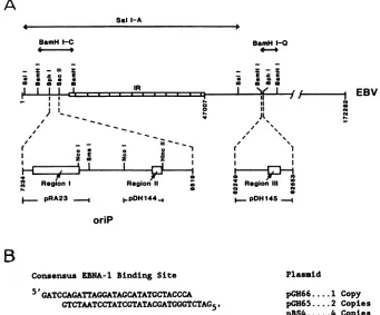
FIG. 1. (A) Location of the EBNA-1 binding sites in the EBVgenome. The EBVsequencescontained withinthe recombinantplasmids used in this study and the three distinct loci (regions I, II, and III) which contain EBNA-1 binding sites (25) are indicated. Base pair coordinatesarefrom Baeretal.(1). (B) Sequence oftheEBNA-1consensusbindingsiteoligonucleotideused in thisstudy.Thecopynumber
inthedifferentplasmids is also shown.
volumes(3to8 liters) of the cellsweregrowntodensitiesof 8 x
i0'
to 10 x 105 cells per ml at 37°C in sealed Spinnerculture bottles. The L-EBNA cells, a DNA-transfected mouse cell line constitutively expressing an integrated EBNA-1gene (19), were grown in Eagle minimal essential
mediumat37°Cin5% CO2 with 10%fetalbovineserum.For preparativepurposesthe L-EBNA cellswerepropagatedto confluencein 900-cm2 rollerbottles at37°C.
Plasmids and DNA. Plasmids pRA23 and pDH144 (Fig. 1A)weresubclones of theEBV(B95-8)oriPclone, pHEBO-1,which was agift from B. Sugden (33). Plasmid pRA23,a giftfromR. Ambinder,isareligation of the SmaIAfragment frompHEBO-1 and contains bases 7334to8187 of the B95-8
genome (1). Plasmid pDH144 was made by ligation ofthe
511-bp NcoI-HincII fragment (EBV bases 8622to9133) into pKP55,a1,967-bp derivative of pBR322 kindlyprovided by K. Peden. The cosmid clone cmSal-A contains the Sall A fragment (EBV bases 62259 to 62553) from EBV(M-ABA)
andwasobtained fromG. Bornkamm (22). Plasmid pDH145 wasconstructed by isolatinga497-bp SphIfragment froma
BamHI-Q-containing plasmid (pSL17) and ligating this into
theSphI site of pGH56,apUC18 derivative.
Synthetic oligonucleotides were made on an Applied Biosystems 380B Synthesizer by the Emory University MicrochemicalFacilityorby the Johns Hopkins Nucleotide
Synthesis Service. Complementarystrandsof theconsensus
EBNA-1 binding site (Fig. 1B) were produced with
over-hanging 5'-GATC ends, which facilitated the cloningof the
duplexed strands and conserved the spacing of the tandem repeats of this sequence observed in region I of oriP.
Complementary strands of the 49-base sequence TAAAA
GAAGTGAGAACGCGAAGCGTTCGCACTTCGTCCCA
ATATATATAwerealsosynthesizedand servedas acontrol for the experiments in which the oligonucleotide probes were used.
Synthetic copies of the consensus EBNA-1 binding site were inserted into the plasmid vector pGH59, a 1,967-bp pBR322 derivative that had been originally designated pKP54 (from K. Peden) andwas subsequently modified by
theaddition ofaBglII linker in themultiple cloning region.
Plasmids containing one (pGH66), two (pGH65), and four
(pBS4)tandemcopies ofthe bindingsitewereconstructed. Plasmid and cosmid DNAswere isolated and purified by
standard procedures, including two bandings on cesium chloride-ethidium bromidedensity gradients.
Preparationofproteinextracts. All extractionprocedures were carried out at4°C. Harvested cells werewashed with hypotonic bufferplus sucrose (25 mM N-2-hydroxyethylpi-perazine-N'-2-ethanesulfonicacid[HEPES] [pH 7.5],5 mM
KCl, 1 mM dithiothreitol [DTT], 1 mM phenylmethylsulfo-nyl fluoride [PMSF], 1 mM EDTA, 10 mM Na2S205, 10%
sucrose) andthenwere allowedto swell inhypotonicbuffer
(no sucrose)for 30min. The swollencellswerebrokenwith 15 strokes in a tight-fitting Dounce homogenizer, and the nucleiwereisolatedby centrifugation for 10minat1,500 x g.Thepelletednucleiwerelysed bytheaddition ofanequal volume ofhigh-salt extraction buffer (50 mM HEPES [pH 7.5], 2 mM DTT, 1 mMPMSF, 0.1 mM EDTA, 0.5 ,ug of
aprotinin per ml, 0.5 ,ug ofpepstatin A per ml, 0.5 ,ug of
leupeptin per ml, 0.2 mM N-tosyl-L-phenylalanine
chloro-methyl ketone [TPCK], 10% sucrose, 3.6 M KCI), and the lysate was incubated for 30 min. Extraction ofthe nuclear
IA EBV
C-|- pRA23_ .pDH144_4
on November 10, 2019 by guest
http://jvi.asm.org/
[image:2.612.142.483.81.364.2]lysate was continued essentially as described by Parker and Topol (20) and modified by Heberlein et al. (10) except that thefinal dialysis was against heparin-agarose chromatogra-phy buffer (HAG buffer) consisting of 25 mM HEPES (pH 7.5), 1 mM DTT, 0.1 mM EDTA, 5 mMNa2S205, 0.5 mM PMSF, 0.05
jig
of leupeptin per ml, 0.01% Nonidet P-40, and 20% glycerol with either 100 mM KCI or 125 mM NaCl. Crude extracts prepared in this manner were then fraction-ated by chromatography through heparin-agarose (Bio-Rad Laboratories, Richmond, Calif.). Nuclear extracts (1 to 2 mg of protein per ml of heparin-agarose) were applied to the columns, and the bound proteins were subsequently eluted with either discontinuous or continuous linear gradients of NaCl or ofKCl
in HAG buffer (0.35, 0.65, and 1.0 M [discontinuous] or 0.35 to 0.75 M [continuous]). Fractions containing sequence-specific DNA-binding activity were identified by a nitrocellulose filter binding assay.Crude extracts were also prepared from Escherichia coli DH1
cells
induced to express the 28K EBNA polypeptide from plasmid pNAK28 as described previously (19). The 28K EBNA protein was purified by chromatography on heparin-agarose as described above.Nitrocellulose
filter
binding assay. The protein-mediated retention of specific radioactively labeled DNA fragments onnitrocellulose
filters was used to detect and characterizeEBNA-like
DNA-binding activity in extracts. Specific and nonspecific DNA probes were prepared from plasmids or fromchemically
synthesized oligonucleotides. DNAprobes were obtained from plasmidIDNAs
by digesting the plasmids with the appropriate restriction enzymes, labeling by filling in the ends of the fragments with the polymerase I Klenow fragment anda-32P-labeled
deoxynucleoside triphosphates, and purification of the appropriate fragments by electroelu-tion from polyacrylamide gels. Synthetic oligonucleotide probes were prepared by annealing the complementary strands of the synthesized DNAs (described above), isolat-ing the duplex DNA molecules by chromatography over hydroxylapatite (Bio-Rad Laboratories), and labeling with T4 polynucleotide kinase and [a-32P]ATP. Standard binding reactions were carried out for 30min at22°C
and contained 25 mM HEPES (pH 7.5), 1 mM DTT, 5mM MgCl2, 1 mgof bovine serum albumin per ml, 250 mM NaCl or 125 mMKCl, 3 to5 fmol of32P-labeled DNA probe, and various amounts of either unlabeled sonicated salmon sperm DNA orpoly(dI-dC)
poly(dI-dC) (Pharmacia, Inc., Piscataway, N.J.) in a total volume of 0.05 ml. The reactions were then filtered in a Minifold apparatus (Schleicher & Schuell, Inc., Keene, N.H.) through nitrocellulose that had been treated as de-scribed by McEntee et al. (16), and each sample was washed three times with 0.3 ml of wash buffer (25 mM HEPES [pH7.5],
1 mM DTT, 5mM MgCl2, 250 mM NaCl or 125 mMKCl).
Thefilters
were then dried and the bound32P-DNA
was determined by liquid scintillation counting. For the competition assays unlabeled, uncut plasmid DNAs were added to the binding reactions.
Western immunoblot assay. Protein fractions were re-solved by electrophoresis on a discontinuous 10% sodium dodecyl sulfate
(SDS)-polyacrylamide
gel (13) and then electrophoretically transferred to nitrocellulose in 25 mM Tris hydrochloride (pH8.3)-12
mM glycine-0.1% SDS-10% methanol at 70 V and 300 mA for 3 h, using a Mighty Small Transphor apparatus (Hoeffer Scientific Instruments, San Francisco, Calif.). The filter was blocked for 16 h with3% gelatin in TBS (20 mM Tris hydrochloride [pH 7.5], 0.5 M NaCl, 10 U of heparin sulfate per ml) and then washed in TTBS (TBS with 0.3% Tween 20) for 15 min with threebufferchanges. The filter was incubatedfor 2 h with
naso-pharyngealcarcinomaserum no. 000506(kindlyprovidedby SusanSpring, National Cancer Institute, Bethesda, Md.)at a 1:200 dilution inTTBS-1% gelatin and then washed with TTBS (three times, 5
min
each time). The filter was devel-oped for16 hwithgold-conjugatedgoatanti-humanantibody
(Bio-Rad Laboratories)
in TBS with 0.2% bovine serum albumin-0.05% Tween20-0.02%
NaN3-0.4% gelatin. The stained membrane was washed and enhanced by silver staining, as suggested bythe manufacturer (Bio-Rad Labo-ratories). Prestained molecular weight standards(notshown) wereused to establish the approximate molecular weightsof anyimmunoreactive proteins.DNase Ifootprintingassay. DNAfragments containingtwo
tandem EBNA-1 consensusbinding sites were isolatedfrom
pGH65
(Fig.1B)
and labeled at one end by using the large fragment of polymerase I anda-32P-deoxynucleoside
tri-phosphates. The fragments were then used for DNase Ifootprint analysis, performed
as described previously(25),
andthe products ofthe assay were analyzed by autoradiog-raphy after electrophoresis on a denaturing polyacrylamide
gel.
Immunoprecipitation of DNA-protein complexes.
DNA-protein complexes
wereimmunoprecipitated
andanalyzed
essentially as described by McKay and DiMaio (17). Cosmid cmSal-A, which contains the entire oriP region (Fig. 1A), was restricted with BamHI and NcoI, and the fragments were32p labeled with the Klenow fragment of polymerase I. Standard binding reactions with 250 mM NaCl (described above)werecarriedoutusing the labeledcmSal-Afragments andproteinextractsfromE. coliexpressing the 28K EBNA fragment or from Raji cells. The volumes of the binding reactions were then increased to 0.2 ml by the addition of 0.15 ml of immunoprecipitation buffer (25 mM HEPES [pH 7.5], 125 mM NaCl, 0.05% Nonidet P-40, 0.1 mM PMSF, 4
,ug
ofpoly(dI-dC)
perml, 8,ug
oftRNA perml), and 1,ulof rabbit anti-28K EBNA or 1,ul
of preimmunization serum fromthe sameanimalwasadded andincubation was contin-ued at4°C
for 16 h. Immune complexes wereprecipitated by the addition of 25,ul
of protein A-Sepharose (Pharmacia, Inc.) equilibrated in immunoprecipitation buffer. The mix-ture was incubated for 2 h at0'C, and then the precipitate was washed five times with 0.5 ml ofimmunoprecipitation buffer. The washed beads were suspended in 50RI
of electrophoresis buffer (0.2% SDS, 10% glycerol, 0.025% bromphenol blue), heated for 1 min at 65°C, and then electrophoresedinan0.8%agarosegelin 40 mM Trisacetate(pH
8.3)-2
mM EDTA-0.1% SDS. The gel was dried and autoradiographed.RESULTS
Detection of EBNA-1 consensus site binding
activity
in extracts from mammalian cells. Nuclei from representative celllines that had previously been shown byimmunological methods to be either positive or negative for the EBNA-1 antigen were lysed by the addition of high molar salt solu-tions and the extracted proteins were precipitated with ammonium sulfate. The high-salt extracts were then frac-tionated by chromatography over heparin-agarose. Proteins bound to the heparin-agarose were eluted in a stepwise fashion with increasing concentrations of NaCl (0.35, 0.65, and 1.0 M), and each step was assayedfor the presence of a DNA-bindingactivitycapable ofinteracting specificallywith the EBNA-1 consensus binding sequences. Nitrocellulose filter binding assays demonstrated that the specific bindingon November 10, 2019 by guest
http://jvi.asm.org/
1.4
1.2
1.0*
O8
0.6-0.4
Q2 0 z
:3 0
m0 1.4
z
a L2. LO
Q8.
Q4*
Q2-A
. RAJIB.
RAMSlI
0
-0
*
4
t
C.'
CA-46bD
L-EBNA-. -EBNA-. ~~~~.~.-0 L
2&& ib 2b 4 2&
l
lb 2b 4b-PROTEIN (PS)4
2
.0 8
06
0.4
G2
14
.2 1.0
0.8
6
.4
.02
FIG. 2. Detectionofspecific bindingtothe consensus oligonu-cleotide in nuclear extracts ofEBV-positive cells. Nitrocellulose filter binding assays were performed using identically prepared extractsfromRaji (A), Ramos (B), CA-46 (C), andL-EBNA(D) cell lines. Nuclearextracts werepreparedasdescribed in Materials and Methods. The 0.65Mfractionfromaheparin-agarose columnwas
assayed for binding activity against a 32P-labeled double-stranded oligonucleotide (30 bp)representingthe EBNA-1 consensusbinding site
(4)
ora(49-bp) control DNAoligonucleotide(0).Thereactions contained25 ,ugof unlabeledsonicatedsalmon sperm DNA per ml.activity, when present, could be found in the 0.65 MNaCl
heparin-agarose fraction (data not shown). Figure 2 shows thedose-responsecurvesgenerated by fourof the cell types. Specific bindingwasreadily detectedinallEBNA-1-positive cell linestested, includingtheRajiand L-EBNAcells shown here. Raji is an
EBV-positive
Burkitt'slymphoma-derived
human lymphoblastoid cell line which expresses EBV la-tencygeneproducts,while theonly viralprotein synthesized by theL-EBNAcellline is EBNA-1. ThismouseL-cell line wasestablished by transfection with SV2-neo and a plasmid,
pGD5, containing
theEBV(P3HR-1)
BamHI K DNAfrag-mentwhichencodes EBNA-1. Thedemonstrationofbinding activitydirectedspecifically againstthe EBNA-1 consensus sitein extractspreparedfrom these latter cells suggests that theprotein-DNAinteractionsweremediated by the EBNA-1 gene product. Furthermore, the observation that L-EBNA cellscontained higher levels ofspecificbinding activity than didRajicells (Fig. 2) is consistent with immunological data indicatingahigher expression of the EBNA-1 protein in the converted mouse cell line (19). Othercell lines with detect-able binding activity included P3HR-1, a Burkitt's lym-phoma-derived human line; CCRF-SB, a human B-lympho-blastoidline obtained from an individual with acute lympho-blasticleukemia;and B95-8, a productively infected
marmo-set cell line (not shown). With the filter binding assay, detectablelevels of consensus site-specific binding were not found in the EBV-negative Burkitt's lymphoma line Ramos
or CA-46(Fig. 2) orin HeLa or Vero cells (not shown). Preparation of an EBNA-1 extract from Raji cells. To
FRACTION
R
u 32 44 51 55 59 68 ?6 84 C
-80K
FIG. 3. Fractionation ofRajinuclearextractbyheparin-agarose
chromatography. Raji nuclear extract prepared as described in Materialsand Methodswasloadedonto aheparin-agarosecolumnin HAG buffercontaining 350mMNaCl. Theproteinsretainedonthe columnwereelutedwithalineargradientbetween 350and 750mM NaCl. (A) Detection of sequence-specific DNA-binding activity.
DNA-bindingactivitywasdetectedbyperformingparallel nitrocel-lulose filter bindingassaysontheelutedfractions,using 32P-labeled DNAfragments containing eithertwoEBNA-1 consensusbinding
sites from pGH65
(0;
specificprobe, 93bp)orsequencesderived from plasmid vectorpGH59 (O; nonspecific probe, 121 bp). The reactions also contained0.8 ,ugofpoly(dI-dC) poly(dI-dC)perml. (B)Immunological detection ofEBNA-1.Representative fractions fromtheheparin-agarose gradientwereanalyzedby Western immu-noblotanalysis forthe presenceofproteinsthatwerereactivewith humanNPCserumcontainingantibodies directedagainstEBNA-1. The numbers above the lanes indicate the gradientfraction from which the sample was taken. Lane C contained a sample ofanunfractionatedproteinextract.
facilitate further studies of the sequence-specific
binding
activity detected in nuclear extracts prepared from Raji cells, the extracts were further purified by elution from heparin-agarosewith linear saltgradients ofeither KCI(0.25
to0.50M)orNaCl(0.35 to0.65M). The elution ofspecific binding activity was followed by performing parallel filter binding assayson theeluted fractions. In Fig. 3A atypical activity profile for the NaCl eluant from the column is presented. When the columns were developed in KCI, the peak of binding activityappearedinfractionscontaining0.30
to0.40M KCI. Theactivitywasfound in the 0.45to0.55M
fractions whenNaCl was applied asthe counterion.
Immu-nologically detectable EBNA was shown to copurify with the specific DNA-binding activity by Western immunoblot-ting (Fig. 3B). Representative fractions from the linear salt
on November 10, 2019 by guest
http://jvi.asm.org/
[image:4.612.64.302.79.332.2] [image:4.612.317.559.79.387.2]j-ori
P
t
.
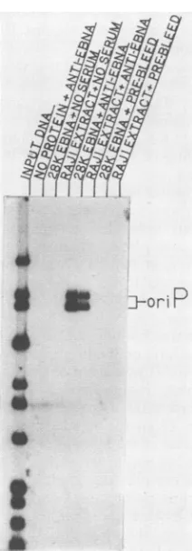
FIG. 4. Identification of DNA-binding activities by
immunoas-say. DNA-binding reactions were immunoprecipitated with rabbit antiserum directed against the bacterially synthesized 28K EBNA
fragmentorwith preimmunizationserumfromthesameanimal. The
probe used in thebindingreactions was a32P-labeledBamHI-NcoI
digest of cmSal-A (Fig. 1A). The fragments containing oriP are
indicated.
gradient (Fig. 3A) were electrophoresed on discontinuous SDS-polyacrylamide gels, transferred to nitrocellulose, and probed with anti-EBNA-positive human serum. The domi-nant immunoreactive protein appeared as an 80K
polypep-tide (compared withprestained molecular weight standards [not shown], which is slightly larger than the size usually reported for EBNA-1 extracted from Rajicells (9, 11, 32).
Although it seemed highly unlikely that the Raji cell binding activity would not be EBNA-1, the fractionation procedures applied to the extracts thus far do not yield a
homogeneous protein preparation. To further characterize the activity, we immunoprecipitated the products of the
DNA-binding reactions with rabbit antiserum directed against the 28K EBNA fragment (19). Binding reactions were prepared that included the bacterial or mammalian
protein extracts and a 32P-labeled BamHI-NcoI digest of cmSal-A whichcontainsregions I and II oforiP(Fig. 1). The
binding reactions were immunoprecipitated by the addition ofanti-EBNA-1 rabbit serum and removal of the immune
complexes with protein A-Sepharose. The immunoprecipi-tated products were resolved on a 0.75% agarose gel
con-taining0.1% SDS. TheoriP-specific bindingactivity can be
precipitated from the reactions containing the bacterial or
theRajiproteinswith theanti-EBNA-1 antibody(Fig. 4). No DNA-bindingactivity wasevident in thecontrols, including
those employing preimmunization serum.
DetectionofEBNA-1bindinglociintheEBVgenome. Ithad
been reported that EBV-specific DNA-binding interactions
distinct from those observed with 28K EBNA could be mediated by RajiEBNA-1 (27). Toaddress this question, an EBV DNA cosmid library containing overlapping DNA fragments representing the EBV genome (22) was assayed for the presence of Raji EBNA-1 interactive loci, using a
pool of the EBNA-1-positive fractions generated by heparin-agarosechromatography. End-labeledDNAfragments from eachcosmid were used as DNA probes in standard binding assays, and the binding reactions were filtered through nitrocellulose. The DNA fragments retained on the filters were then eluted from the nitrocellulose and analyzed by agarose gel electrophoresis (25).
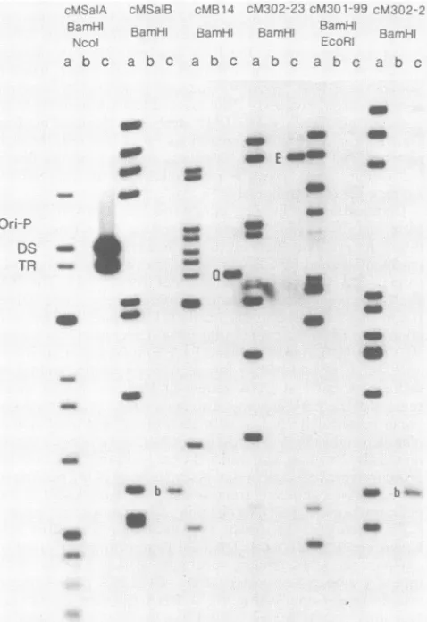
Under standard binding conditions five DNA fragments from the EBV genome were retained on the nitrocellulose filters in the presence of the Raji extract (Fig. 5). The fragmentsbound by EBNA-1 were the two DNA fragments from cmSal-A and the single fragment from cmB14. The 28K EBNA protein also selectively bound these three DNA fragments from the EBV cosmid library (25). The two additionalDNA fragments that were detected in this assay, EBV BamHI-E(cm302-23) and EBV BamHI-b (cmSal-B and cm302-21), resulted from the sequence-specific binding of cellular proteins to DNA sequence elements within those regions of the EBVgenome. The twocellular proteins do not copurify with EBNA-1 or with each other, and their molec-ular size and sequence binding specificities havebeen deter-mined (D. Rawlins, unpublished observations). The cellular proteins weredetected in this assay because of thepooling of theEBNA-1-containing fractions and are not present in the peak fractions of EBNA-1 used insubsequent experiments. Comparison of the specific DNA binding mediated by 28K EBNA and
Raji
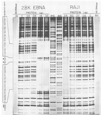
EBNA-1. DNase I footprinting was used to compare theDNA-binding specificity of Raji EBNA-1 with that of a similarly preparedextract of the 28K EBNAfusion protein from bacteria (Fig. 6). A DNA fragment containing two tandemconsensusEBNA-1 binding sites was labeled at a single 3' terminus with the Klenow fragment of DNA polymerase I and served as the probe in the protection assays. In Fig. 6, protection of the tandem binding sites by the bacterially synthesized EBNA fragment and by the Raji extract is clearly evident. As indicated by the bracketed sequences listed to the left ofthe footprint, the protection patterns exhibited by the extracts were remarkably similar despite the expected biochemical and physical differences between the 28K EBNA and the approximately 72- to80-kilodalton native viral protein. Inthisinvestigationand in our previous studies (25, 26), binding of the 28K EBNA to
tandem binding sites led to the appearance of DNase I-hypersensitive sites bothbetweenbinding sites andatthe 3' edge. However, these hypersensitive sites were not pro-duced by the protein extracts from Raji cells, even though the extent ofoverall protection from DNase cleavage was
equivalent.
Interaction of Raji EBNA-1 with the three binding loci from the EBVgenome. As described above, asurvey ofan EBV DNAcosmid libraryidentified threespecific binding locifor the Raji EBNA-1 protein. The three loci included the two
cis-acting elements of oriP (regions I and II) and alocus in the EBV BamHI Q fragment (region III) (Fig. 1A). Since EBNA-1 interactions at the different DNA loci may form part of the regulatory mechanisms involved in the
plasmid
maintenance functions of EBV and possibly also in the regulation of nuclear latency gene expression,the
binding
of Raji EBNA-1 to regionsI, II, and III was investigated.In Fig. 7thebindingprofiles obtainedbytheinteraction of increasing amounts of Raji EBNA-1 with DNA
fragments
on November 10, 2019 by guest
http://jvi.asm.org/
[image:5.612.126.232.75.378.2]a b C
-g
I-MP:s
T R
:3i
S
-s
-S_ Q
*M
N as'
go-40
N
In
a
FIG. 5. Survey ofanEBVcosmid 1 EBNA-1 binding sites by a DNA fil fraction of heparin-agarose-separated with radiolabeled clonedfragments of passing the entire EBV genome, a described previously (25). The cosrI enzyme usedtogenerate the DNAfra setoflanes.Lanescontainasample or retained onthe nitrocellulosefilter int
and the DNAretainedin the
presenco
The three EBNA-1 binding loci, tanc
metry (DS), and region III (Q), are i fragments indicated byEand bwereI notbyEBNA-1(seeResults).
containing
eitherregion
I,
regi
presented.
Theregion
Iprobe,
region
IIprobe, pDH144 (540 bp
clones of
pHEBO-1 (Fig.
1A)
ancbinding
sites(region I)
or 4 par sites(region
II).
Theregion
III prc derived froma subclone of theBand containstwo
potential
bindin of theprobes
should not have sbinding
since underthe condition exhibits noaffinity
fornonspecifi
molar amounts of each 35S-labe incubatedin the
binding
reactions DNAboundwas determined aftercomplexes through
nitrocelluloseof the inputDNAs,
approximately
twofold and fourfold asmuch
protein
wasrequired
forregions
II and III,respec-tively, as for region
I.
The relativebinding strengthsof the natural andsynthetic
EBNA-1bindingsiteswerealso examined in acompetition filterbindingassay
(Fig. 8). Increasing
amountsof unlabeled_ supercoiledrecombinantplasmidDNAswereadded to
bind-ing
reactionscontaining
a constant amount of EBNA-1 to_ns
- - compete forbinding
of EBNA-1 to a 32P-labeled EBNA-1 - E 04 consensusbinding
site. The salt concentration in these reactions wasadjusted
to 0.3 MNaCl,
whicheffectively
eliminated any
nonspecific protein-DNA
interactionsevenin _ the absence of any unlabeledcompetitor
DNA. Plasmids sopGH66,
pGH65,
andpBS4
contained one, two, and fourcopies,
respectively,
of the EBNA-1consensusbinding
site inaplasmid
vector,pGH59. pDH144
andpDH145
contained *> 5regions
II andIII, respectively,
inpGH59.
The region Icompetitor
wasrepresented
bypRA23,
which contains the _ 20naturally occurring binding
sites ofregion
I(Fig. 1).
_s
Thegeneral
pattern ofbinding
observed with the filter _ obinding
assays(Fig. 7)
wasagain
observed with the compe-titionassays;namely, region
Iwas abettercompetitor
than_s
region II,
andregion
III was the weakestcompetitor
for EBNA-1binding.
From the resultspresented
inFig.
8,
aquantitative
estimate of theamountofcompetitor
DNA that m it takestoreducebinding
of theprobe
tothe 50% levelcanbe determined. Inour
previous
studieswedemonstratedby
DNase I
footprinting
thatregion
I contains20tandem sites4 b for
binding by
EBNA-1 and that region II consists of 4partially
overlapping
binding
sites(25).
If theprotein-DNA
UI interactionsatthe individualsiteswere
quantitatively
equiv-alent,
a5-to10-folddifference in thecompeting strengths
ofregions
IandIIwould bepredicted. However,
toachieve the 50% levelofbinding,
the concentrations ofregions
I,II,
and IIIin the assays were0.013, 0.600,
and >3.50nM,
respec-tively.
Inotherwords,
inmolaramounts,45-fold lessregion
presenceofRaji IDNAwas
required
thanregion
II DNAtocompeteagainst
Iter binding assay. A pooled the consensus binding site for binding to EBNA-1. These
i Raji extract was incubated results
suggest
either that the individualbindingsitesarenot FEBV(M-ABA)DNA encom- equivalent or that binding of EBNA-1 to the cis-actingLnd binding was assayed as elements of oriP and to the
BamHI-Q
locus involves moreiid name and the restriction than
simple
independent
additions ofprotein
molecules to igmentsare listedaboveeach binding sites.ftheinputDNA(a),the DNA An indication that the primary DNA sequence of the
he absence ofRajiextract(b), binding site makes an important contribution to the relative
eof
Raji
EBNA-1extract(c).affinity
for EBNA-1 isprovided by
theobservation
that iem repeats (TR), dyad sym-pGH66,
which
contains
asingle region
I consensusbinding
oundbycellular proteinsand site, was as effective as region II and much stronger than
region
III in thecompetition
studies(Fig. 8).
Cooperativity
between
binding
sites was notreadily
apparent whenplas-mids
containing
two(pGH65)
andfour(pBS4)
copies
ofthe onII,
orregion
III are consensusbinding
site were used ascompeting
DNAs. pRA23 (796bp),
and theCompared
withpGH66,
theircompeting strengths
were 2.8 ), were derived from sub- and4.8,
respectively.
Thebinding
sites in theseplasmids
I containeither 20 tandem have thesame
spatial
arrangementsasthosefoundinregion
tially overlapping binding
I.Region
I (pRA23),containing
the 20naturally
occurring
)be, pDH145,
(320bp)
wasbinding sites, competed
37 times as well aspGH66
for amHl Qfragment
of EBV EBNA-1binding.
When this value iscompared
with 2.8 Ig sites. The different sizes times for thesynthetic
dimerbinding site,
itagain
suggests,ignificantly
influenced the thatcooperative
interactions betweenbinding
sites have a sof thisassay,theprotein
relatively
smallimpact
onEBNA-1binding.
ic DNA sequences. Equi-led DNA
fragment
were,and the percentage ofthe
rfilteringtheprotein-DNA
. To achieve 50% binding
DISCUSSION
One
approach
totheanalysis
ofthe molecularmechanismscontrolling latency
is toidentify
and characterizeon November 10, 2019 by guest
http://jvi.asm.org/
[image:6.612.65.304.83.431.2]A
T
T A
OA
F
5
A :
L c
*AC* iA:D%
z
28K EBNA
RAJ
zZ
OEN7-E PROTEIN t _
16 C -.4 8 i.6 3.2 APC AG 2-4 SO 23 : 80
-~~~~§!
t O
=
s~ -G J
_-_
-a =
__.0 _ _mwe
__-._~ __
_t
-
-im.. -.
_
g3~~~~~~~~MO
_
. -_
- -_
-n MM
FIG. 6. DNase I footprinting analysis: protection of a dimer of the EBNA-1 consensus binding site by the bacterially synthesized 28K EBNAfragment or by aheparin-agarose gradient fraction from Raji cells. Protected regions of the DNA are indicated by the brackets around the sequence listed to the left of thefootprint. DNaseI-hypersensitive bases are marked by the closed circles. The DNA sequence ladders (lanes A>C and A+G) were generated by the modified chemical procedure (2).
DNA interactions that take place at DNA sequences known
tohavearegulatory role in gene expression or to be essential for thereplicationprocess. Wehave adopted this approach for studying the interaction of EBNA-1 with the plasmid maintenance system (oriP) thatis integral to the
establish-mentoflatentinfectionsin B lymphocytes infected by EBV. oriP-specificDNAbinding by EBNA-1 was initially demon-strated by using a 28K fragment of EBNA-1 synthesized in bacteria. In this investigation we have characterized the interactions of the EBV EBNA-1 protein with oriP by using the entire EBNA-1 protein isolated from a latently infected humancell line.
Earlierattempts topurifyEBNA-1 werehampered by the relatively small amounts of antigenically reactive protein present in latently infected cells (30) and by the lack of a sensitive assay with which to monitor the purification
pro-cess. Our initial experiments were designed to determine whetherbinding to a consensus repeat unit of oriP could be utilizedto unambiguously detect EBNA-1 in crude cellular
extracts. We foundthata nitrocellulose filter binding assay incorporatingunlabelednonspecific DNA and relatively high salt concentrations could readily detect EBNA-like binding activity in extracts from all EBV-carrying cells tested and
also from transfected cells expressing the EBNA-1 gene. Theease of detection ofthebinding activity and the sensi-tivity ofthe assay mostlikely reflect the high specific binding affinity ofthe protein forits target DNA. By following the elution of thisspecificDNA-binding activity, a greater than 200-fold purification of EBNA-1 was achieved by repeated chromatographic separationof the nuclear extracts on hepa-rin-agarose. Although EBNA-1 was not purified to homoge-neity,noother DNA-binding activities were detected in the
extractunderstringent conditions andexperimentsdirectly confirmedthat the activity beingstudied was indeed medi-atedbythe EBNA-1protein. The sequence-specific binding copurified with an appropriately sized polypeptide which reacted with EBNA-positive human NPC serum, and the
oriPbinding activity was immunoprecipitated by monospe-cific antiserum raisedagainst purified28K EBNA.
Aqualitative comparisonof the DNase Ifootprints gener-atedbythe28K and RajiEBNAs revealedboth similarities and differences in the protection patterns. Each of the proteins appeared to protect exactly the samebases of the
consensussite,andonthe DNA strandtested,theprotection
was directionally asymmetricalrelative to the centerofthe
corepalindrome foundwithin thebinding site,with 10 bases
- - a-
-..Wb.awm& , - .,.
- .. . - -- ....
on November 10, 2019 by guest
http://jvi.asm.org/
[image:7.612.143.469.82.450.2]a~~~~~~~~~~~~~a a: z
40-w
a.
20-0.6 0.32 0.63 .3 2,5 50 1Z5
PROTEIN
(q)
FIG. 7. Binding of Rajiextract toregions IandIIoforiPandto the BamHI-Q locus (region III). Recombinant plasmids containing oriPregionI(pRA23),region II (pDH144), andregionIII(pDH145) were restricted with a combination ofSalI-NcoI(pRA23orpDH144) or BamHI-BgIII (pDH145). The digested fragments were 3' end labeled by filling in the ends with 35S-deoxynucleoside triphos-phates, using the Klenow fragment of polymerase I. The labeled fragments containing regionsI, II,andIII wereisolated and used in nitrocellulose filter binding assays. Five femtomoles ofthelabeled DNAfragmentswasincludedin eachparallel assay, alongwith the indicatedamounts ofprotein fromaconcentratedheparin-agarose gradient fraction from Rajicells.Symbols: 0,region 1 (796 bp); *, region 11 (540 bp); A,regionIII(320 bp).Theassays alsoincluded 25,g of unlabeled sonicated salmon spermDNAperml.
being protected 5' to the AT center of symmetry and 14 basesbeingprotected on the 3' side. Asymmetricalbinding is not uncommon under theseconditions and may reflect the directionalorientation oftheindividual EBNAbinding sites. Although the orientation of the binding sites in the tandem repeat region appears to vary in oriP isolates from different strains ofEBV(25), we do not know whetherthis has any functional significance in vivo. The presence of DNase I-hypersensitive sites3' to theregions protected by the 28K EBNArepresented the major difference noted between the footprints produced bythe bacterially synthesized and Raji EBNA-1. Although the difference in the sizes of the two polypeptides affords a reasonable explanation for the steri-cally limited access of the nuclease to the Raji EBNA-1-protected template, it does not explain the dramatic hyper-sensitivity found in the 28K EBNA-1 footprint. Again, this may indicate a characteristic of the DNA-protein interac-tion, in this case a change in the conformation of the bound DNA that is modified when the structure of the protein is intact. Consequently, for studies of conformation changes induced in the target sequences by EBNA-1 binding, use of theintactEBNA-1protein may be essential. Recently, it was reported from electron microscopic studies that EBNA-1 from Raji cells bound specifically to regions of EBV other
1.0-a
z D
.75.
0* In
z
a 0 .50
w
I-w cr
.25-0\
[image:8.612.81.290.78.349.2]0O067 a014 0.035 0.070 0.14 0.35 0.70 .4 3.5 COMPETITOR DNA (nM)
FIG. 8. Competition filter binding assaycomparingthe relative EBNA-1 binding affinity of single- and multiple-copy EBNA-1 consensusbinding siteswith thoseofplasmidscontaining region I, II, or III. The assay measured the amountof32P-labeled double-strandedoligonucleotide containingthe EBNA-1 consensusbinding
siteretained in the presence ofaconstant amountofEBNA-1and increasing amounts of unlabeled competitor DNA. The plasmid DNAs used were the following: vector (pGH59), one consensus EBNA-1binding site(lxEBS, pGH66),twobinding sites(2xEBS,
pGH65), four binding sites (4xEBS, pBS4), region I (pRA23), regionII(pDH144), andregionIII(pDH145). Thebinding reactions (25,ul) contained 2.5 fmol of32P-labeledprobe and4 p.gofprotein
from a concentrated heparin-agarose gradient fraction from Raji
cells.
thanregions I, II, and III(27). However,evenwhenapool of fractions from the heparin-agarose column was tested
under assay conditions which allowed binding of cellular factors, the Raji EBNA-1 still bound only to the oriP and BamHI-Q loci ofthe EBV genome. Earlier studies demon-strated thatEBNA-1wouldbindinanonspecificmanner(18, 30), and the significance of the low-stringency binding de-tected in the electron microscopic studies is unclear.
Inboth directbinding studiesandcompetitionassays we
haveobservedahierarchy of bindingtoregions I,II, and III; that is, region I has a higher relative binding affinity for EBNA-1 than doesregion II, and regionIII has the lowest affinity. The increased
affinity
ofregion I overregion
II is greaterthan would bepredicted fromthe numberof individ-ualbindingsites in each locus. In thecompetitionassaysthe single synthetic binding site, whichrepresents a consensusofthe region I sites, competed more effectively than the regionIIlocus,suggestingthatthedifferences inaffinity may reflect sequence variations found within theindividual bind-ing sites. Differences in the spatialarrangementofthe sites may also contribute. The region I sitesare centered 30 bp apart, whileregionII andregionIII sites are centeredonly 24and 21bpapart,respectively.Cooperativity between sites was not readily apparent when competition experiments wereperformed with plasmids containing one, two, and four copiesof the concensusbindingsite.
The establishment and maintenance of latency by EBV obviously require a carefully regulated schedule of DNA metabolism andgenomic segregationinvivo.Previously,we
proposeda"sink andorigin"model in which theinitiation of plasmid replication wouldbe controlled by the appropriate
on November 10, 2019 by guest
http://jvi.asm.org/
[image:8.612.323.560.82.286.2]interaction of EBNA-1 at the region II binding locus of oriP (25). Thus, the initiation of replication would depend upon the availability of active EBNA-1, a condition that could be modulated by sequestering the protein at the region I binding sites (the "sink") in a manner similar to that described for RepA protein-controlled replication of plasmid P1 (6). As shown here, the hierarchy of interaction ofEBNA-1 with the three specific binding elements is fully compatible with this model. However, the sink andoriginal model only addresses the regulation ofreplication and copy number of the latent EBV genome. The high-affinity binding of EBNA-1 to the region I binding sites may also provide a mechanism for chromosomal association and consequently segregation.
EBNA-1 appears to be functionally pleiotropic and, in addition to its role in the initiation of episomal DNA synthe-sis, may be involved in regulating the expression of the
nuclear class of latency proteins. An analysis of cDNA clones has shown that EBNA-1 is transcribed as a highly spliced mRNA from latency promoters at position 11305 in BamHI-C and position 14352 inBamHI-W (5, 29, 31). The open reading frame encodingEBNA-1 lies some 90 kilobases away in the BamHI Kfragment (12, 34). The transcripts for EBNA-2, -3A, -3B, -3C, and -5 are similarly highly spliced and appear to be controlled from the same latency promoters as EBNA-1 (3, 4, 29, 31). Reisman and Sugden (28) have shown that inrecombinant plasmid constructions, the region I locus of oriP acts as an EBNA-1-dependent enhancer. Although region I lies approximately 3 and 6 kilobases distant from the EBNA promoters, it would be within the operational range of an enhancer. Thus, in addition to the initiation of episomal DNA synthesis, EBNA-1 may function asanenhancer binding protein to positively regulate its own expression along with the expression of EBNA-2, -3A, -3B, and -3C and leaderprotein (also known as EBNA-4 or -5). Sinceoverproduction of EBNA-1 could potentially result in anincreased initiation of episomal DNA replication and loss of synchrony with celldivision, it seems likely that EBNA-1 synthesis is strictly controlled. Binding of EBNA-1 to the region III sites inBamHI-Q may provide a mechanism for an additional level of regulation. Our experiments show that EBNA-1 has a lower affinity for region III than for the loci in oriP. Thus, an EBNA-1 levels increase beyond that neces-sary to saturate oriP, binding would occur at region III. Since the primary EBNA-1 transcript passes through BamHI-Q, one could postulate that an EBNA-1-region III complex might impede or block the progression of RNA polymerase, thus reducing levels of EBNA-1 mRNA. Inthis
scenario, EBNA-1 would function as an enhancer binding protein to provide positive autoregulation through region I and as a transcriptional terminator to introduce negative autoregulation via region III. Although the exact function of the BamHI-Q binding site remains to be determined, the availability of the EBNA-1 affinity data now provides a basis fortestable experimental models.
ACKNOWLEDGMENTS
We thank C. Beckman, D. Ciufo, and M. Chiu for excellent technical assistance.
This work was supported by Public Health Service grants CA42245 and A122881 from the National Cancer Institute and the NationalInstitute of Allergy and Infectious Diseases, respectively.
LITERATURE CITED
1. Baer, R., A. T. Bankier, M. D. Biggin, P. L. Deininger, P.J. Farrell, T. J. Gibson, G. Hatful, G. S. Hudson, S. C.Satchwell, C. Seguin, P. S. Tufnell, and B. G. Barrell. 1984. Organization
of the B95-8Epstein-Barr virusgenome. Nature(London) 310: 207-211.
2. Bencini, D. A., G. A. O'Donovan, and J. R. Wild. 1984. Rapid chemicaldegradationsequencing. Biotechniques2:4-5. 3. Bodescot, M., 0. Bison, and M. Perricaudet. 1986. An
Epstein-Barrvirustranscriptionunitis atleast 84 kb long. Nucleic Acids Res. 14:2611-2620.
4. Bodescot, M., B. Chambraud, P. Farrell, and M. Perricaudet. 1984. Spliced RNA from the IR1-U2 region ofEpstein-Barr
virus: presence of an open reading frame for a repetitive polypeptide. EMBOJ. 3:1913-1917.
5. Bodescot, M., M. Perricaudet, and P. J. Farrell. 1987. A
pro-moterfor the highlysplicedEBNAfamilyofRNAsof Epstein-BarrVirus.J. Virol. 61:3424-3430.
6. Chattoraj, D., K. Cordes, and A. Abeles. 1984. Plasmid P1 replication: negativecontrolby repeated DNAsequences.Proc. Natl. Acad. Sci. USA 81:6456-6460.
7. Dambaugh, T., K. Hennessy, S. Fennewald, and E. Kieff. 1986. The virus genome and its expression in latent infection, p. 13-45. In M. A.Epstein andB. G. Achong (ed.), The Epstein-Barr virus: recent advances. J. Wiley & Sons, Inc., New York. 8. Dillner,J., B. Kallin, H. Alexander, I. Ernberg, M. Uno, Y.Ono,
G. Klein, and R. Lerner. 1986. An Epstein-Barrvirus (EBV)-determined nuclear antigen (EBNA-5) partly encoded by the transformation-associated Bam-WYH region of EBV DNA: preferential expression in lymphoblastoid cell lines. Proc. Natl. Acad. Sci. USA 83:6641-6645.
9. Fischer, D. K., M. R. Robert, D. Shedd, W. P. Summers, J. E. Robinson, J. Wolak, J. E. Stefano, and G.Miller. 1984. Identi-fication ofEpstein-Barr nuclear antigen polypeptide in mouse
andmonkey cells after gene transfer with a cloned 2.9-kilobase-pair subfragmentofthe genome. Proc. Natl. Acad. Sci. USA 81: 43-47.
10. Heberlein, U., B. England, and R. Tjian. 1985.Characterization of Drosphila transcription factors that activate the tandem
promoters of the alcohol dehydrogenase gene. Cell 41:965-977. 11. Hennessy, K., M. Heller, V. van Santen, and E. Kieff. 1983. Simplerepeat array in Epstein-Barr virus DNA encodes part of theEpstein-Barr nuclear antigen. Science220:1396-1398. 12. Hennessy, K., and E. Kieff. 1983. Oneof two Epstein-Barr virus
nuclear antigens contains aglycine-alanine copolymer domain. Proc. Natl. Acad. Sci. USA 80:5665-5669.
13. Laemmli, U. K. 1970.Cleavage of structuralproteins during the assembly of the head of bacteriophage T4. Nature (London) 227:680-685.
14. Laux, G., M. Perricaudet, and P. J. Farrell. 1988. A spliced Epstein-Barr virus gene expressed in immortalized lympo-hocytesis createdbycircularizationof thelinear viral genome. EMBO J. 7:769-774.
15. Lindahl, T., A. Adams, G. Bjursell, G. W. Bornkamm, C. Kaschka-Dierich, and U.Jehn. 1976. Covalentlyclosed circular duplex ofEBV in ahumanlymphoid cellline.J.Mol. Biol. 102: 511-530.
16. McEntee, K., G. M. Weinstock, andI. R. Lehman. 1980. RecA protein-catalyzed strand assimilation: stimulation by Esche-richia coli single-stranded DNA-binding protein. Proc. Natl. Acad. Sci. USA 77:857-861.
17. McKay, R., and D. DiMaio. 1981. Binding of an SV40 T antigen-relatedprotein to the DNAofSV40regulatorymutants. Nature(London) 289:810-813.
18. Milman, G., and E. S.Hwang. 1987.Epstein-Barr virus nuclear antigen forms a complex that binds with high concentration dependence to asingleDNA-bindingsite. J. Virol. 61:465-471. 19. Milman, G., A. L. Scott, M. S. Cho, S. C.Hartman,D. K.Ades, G. S. Hayward, P. G. Ki, J. T. August, and S. D. Hayward. 1985. Carboxyl-terminal domain of the Epstein-Barr virus
nu-clear antigen is highly immunogenic in man. Proc. Natl. Acad. Sci. USA 82:6300-6304.
20. Parker, C. S., and J. Topol. 1984. A DrosophilaRNA polymer-ase II transcription factorcontains a promoter-region-specific DNA-bindingactivity. Cell 36:357-369.
21. Petti, L., J. Sample, F. Wang, and E. Kieff. 1988. A fifth Epstein-Barr virus nuclear protein (EBNA3C) is expressed in
on November 10, 2019 by guest
http://jvi.asm.org/
latentlyinfectedgrowth-transformed lymphocytes.J.Virol. 62: 1330-1338.
22. Polack, A., G. Hartl, U. Zimber, U.-K. Freese, G. Laux, K. Takaki, B. Hohn, L. Gissman, and G. H. Bornkamm. 1984. A complete set of overlapping cosmid clones of M-ABA virus derived from nasopharyngeal carcinoma and its similarity to other Epstein-Barr virus isolates. Gene 27:279-288.
23. Powell, A. L. T., W. King, and E. Kieff. 1979. Epstein-Barr virus-specificRNA. III. Mapping of DNAencodingviralRNA in restringent infection. J.Virol.29:261-274.
24. Purtilo, D. T. 1987. Opportunistic cancers in patients with immunodeficiency syndromes. Arch. Pathol. Lab. Med. 111: 1123-1129.
25. Rawlins, D. R., G.Milman, S. D. Hayward, and G. S. Hayward. 1985.Sequence-specific DNAbindingof the Epstein-Barr virus nuclear antigen (EBNA-1) to clustered sites in the plasmid maintenance region. Cell42:659-668.
26. Rawlins, D. R., P. J. Rosenfeld, T. J. Kelly, G. R. Milman, K.-T. Jeang, S. D. Hayward, and G. S. Hayward. 1986. Sequence-specific interactions ofcellularnuclear factorIandEpstein-Barr virus nuclear antigen with herpesvirus DNAs, p. 525-542. In M. Botchan, T. Grodzicker, and P. A. Sharp (ed.), Cancer cells, vol. 4. DNA tumor viruses. Cold Spring Harbor Laboratory, ColdSpring Harbor, NY.
27. Reischig, J., D. Bartsch, A. Polack, V. Vonka, and I. Hirsch. 1987. Electron microscopy of binding of Epstein-Barr virus (EBV)nuclearantigen(EBNA-1) toEBV DNA. Virology 160: 498-501.
28. Reisman, D., and B. Sugden. 1986. trans activation of an Epstein-Barr viral transcriptional enhancer by theEpstein-Barr viral nuclearantigen 1.Mol. Cell.Biol. 6:3838-3846.
29. Sample, J., M. Hummel, D. Braun, M. Birkenbach, and E. Kieff. 1986. Nucleotide sequencesofmessenger RNAsencoding Ep-stein-Barr Virus nuclear proteins: a probable transcriptional initiation site.Proc. Natl. Acad.Sci. USA83:5096-5100. 30. Sculley, T. B., T. Kreofsky, G. R. Pearson, and T. C. Spelsberg.
1983. Partial purification of the Epstein-Barr virus nuclear antigen(s).J. Biol. Chem.258:3974-3982.
31. Speck, S., and J. Strominger. 1985. Analysis of the transcript encoding the latent Epstein-Barr virus nuclear antigen I: a potentiallypolycistronicmessagegenerated by longrange splic-ing of severalexons.Proc.Natl. Acad.Sci. USA 82:8305-8309. 32. Strnad, B. D., T. C. Schuster, R. F. Hopkins, III, R. H. Neubauer, and H. Rabin.1981.Identification ofanEpstein-Barr virus nuclearantigenbyfluoroimmunoelectrophoresisand rad-ioimmunoelectrophoresis. J.Virol.38:996-1004.
33. Sugden, B., K. Marsh, and J. Yates. 1985. A vector that replicates as a plasmid and can be efficiently selected in B-lymphoblasts transformed by Epstein-Barr virus. Mol. Cell. Biol. 5:410-413.
34. Summers, W. P., E. A.Grogan, D. Shedd, M. Robert,C.R.Liu, andG. Miller.1982.Stableexpression inmousecellsof nuclear neoantigen after transfer ofa3.4megadalton cloned fragment of Epstein-Barr virusDNA.Proc.Natl. Acad.Sci. USA 79:5688-5692.
35. Yates, J. L., N. Warren, D. Reisman, and B. Sugden. 1984. A cis-acting element from the Esptein-Barr viral genome that permits stable replicationof recombinant plasmids in latently infected cells.Proc. Natl. Acad. Sci. USA 81:3806-3810. 36. Yates, J. L., N.Warren, and B.Sugden. 1985. Stablereplication
ofplasmids derived from Epstein-Barr virus in various mamma-lian cells. Nature(London) 313:812-815.