Thesis submitted to the
TamilNaduDr.M.G.R.MedicalUniversity,
Chennai
For the degree of
Doctorate of Medicine (DM)
In
Clinical Haematology
By:
Dr.AnupamChakrapani, MD
For the year: August 2011
Department of Clinical Haematology
Christian Medical College, Vellore.
CERTIFICATE
This is to certify that this thesis titled “Diffuse Large B cell
Lymphoma:a single centre study”, is a bonafide work of the
candidate, Dr.AnupamChakrapani during the period from
August 2009 to August 2011 in partial fulfilment, towards the
award of degree of Doctorate of Medicine (higher specialty)
in Clinical Haematology for the examinations to be conducted
by the Dr.M.G.R Medical University in August 2011.
Dr. Alok Srivastava, MD, FRACP, FRCPA, FRCP
(Thesis Guide)
ACKNOWLEDGEMENT
(In the name of God, Most Gracious; Most Merciful)
I am heartily thankful to my guide and Professor, Dr.AlokSrivastava, whose encouragement,
guidance and support from the initial to the final level made this work possible. I take this
opportunity to express my gratitude to my teachers,Dr.Vikram Mathews, Dr Biju
George,Dr.AuroViswabandya, Dr Aby Abraham and Dr Rayaz Ahmad for their expert
opinions and guidance. I would like to gratefully thankProfessor and Head of Department
Pathology Dr.Sheila Nair and my colleague Dr Daborah in providing immunohistochemistry
data.Iam indebted to all my colleagues and friends in Clinical Haematology for their constant
support and encouragement. My deepest and sincere thanks to my parents, my wife Dr
Archana and daughter Aastha, who has been my constant source of support and
encouragement during this period.Last but not least Ioffer my regards and blessings to all the
CONTENTS
Sl. Number
Topic
Page number
1
Abstract 1-2
2
Introduction
3
3
Review of literature
4-12
4
Aims & Objectives
13
5
Patients &Methods
14-23
6
Results
24-42
7
Discussion
43-48
8
Conclusions 49-50
9 Proforma
i-ii
10
Bibliography
iii-vii
ABSTRACT
Background:
Diffuse large B cell lymphoma (DLBCL) is a heterogeneous group of B-cell lymphoma with variation in patient survival. Information regarding clinical presentation, staging, prognostic determinant (biological [GCB, non-GCB] and clinical[IPI]), and response to chemotherapy (CHOP and Rituximab CHOP) in exclusively nodal cases of DLBCL is limited.Aims and Objectives of the study:
To analyse the response to chemotherapy (CHOP and Rituximab CHOP) and to access the prognostic significance of IPI and biological sub-grouping of nodal DLBCL cases in our institution.Methodology:
All patients with nodal DLBCL cases who underwent treatment with minimum six months follow up in the Department of Haematology between January 2006 and April 2010 and whose slides and blocks could be retrieved from Department of Pathology were included in the study.Results:
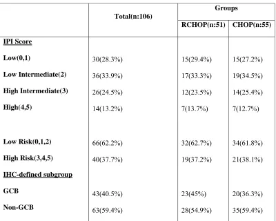
Of the 106 patients, 71(67%) male and 78(73.6%) patients were <60 years of age. 72(67.9%) presented with B-symptoms, 62(58.5%) had stage III/IV, and 80(75.5%) had high LDH at diagnosis. 22(20.8%) had one or more extra-nodal disease and 21(19.8%) had bulk disease. Out of 106 patients 66(62.2%) were in low IPI risk (0,1,2) and 40(37.7%) were in high IPI risk(3,4,5). Based on immune-histochemistry(Hanset.al) we classified 43(40.5%)patients as GCB DLBCL and 63(59.4%) as non-GCB DLBCL. The clinical characteristics of patients in sub groups were similar.The CR+CRu was 88% in Rituximab vs 70.9%% in non-Rituximab treated patients at the end of six cycles of chemotherapy (p=0.082). After a median follow up of 36 months (range:6-44months in RCHOP and 6-42 months in CHOP),the three year cumulative relapse free survival(RFS) and overall survival(OS) was 56.4% and 74.5% respectively in those who received CHOP chemotherapy. The addition of Rituximab improved the cumulative RFS and OS to 86.3% and 76.5% respectively, though the difference was not significant.Addition of Rituximab in high IPI risk group patients, improve EFS and OS at 24 months to 74.9% and 83.3% vs 19.8% and 41.5% in CHOP group(p=0.002). Rituximab treated patients in either GCB or non-GCB subgroup had similar EFS and cumulative RFS at median follow up of 24 months in comparison to non-Rituximab patients. In GCB group of patients the Rituximab significantly improves the OS (89.1%vs50.3%) at median follow up of 24 months (p=0.02). Neutropenia with or without fever was the most common chemotherapy related complication and was significantly more in RCHOP patients 68.6% vs 47.3% (p=0.032).
Conclusion:
This is the largest series of patients with DLBCL comprehensively evaluated and analyzed for outcome after treatment with CHOP and RCHOP. Patients were classified into low and high IPI risk group as well GCB and non-GCB origin of their disease. Addition of Rituximab has significant advantage in GCB and high IPI risk subgroups. In non-GCB and IPI low risk sub group, Rituximab increases relapse free survival but not the overall survival. Further analysis needs to be done with more number of patients and longer follow-up to truly understand the trend observed in this study for patients in India.Introduction
Diffuse Large B cell Lymphoma (DLBCL) is a neoplasm of B cells of hematopoietic system. Approximately one-third of all adult lymphomas are DLBCL, the most commonly occurring form of non-Hodgkin lymphoma (NHL) in the western world and India1,2,3.DLBCL is associated with an aggressive natural history, with median survival of less than one year in untreated patients. The cyclophosphamide, doxorubicin, vincristine and prednisolone (CHOP) chemotherapy has been the mainstay of therapy for several decades (six years overall survival 33%), since more intensive chemotherapy were more toxic and failed to demonstrate additional benefits27,28.
In largely separate efforts, remarkable progress has been made during the past decade in understanding the biological heterogeneity of DLBCL and improving survival for DLBCL patients with combination of CHOP and immunotherapy29.The integration of anti-lymphoma antibodies, notably rituximab®,into combination therapies for DLBCL have markedly improved patients outcomes across all subtypes32,33,34. Microarray analysis, gene expression
profiling (GEP) has uncovered distinct molecular signatures for DLBCL subtypes that have distinct clinical behaviours and prognoses16,17,23. Various immune-histochemical algorithms
have been developed to predict the almost similar results as GEP21,22. Most recently,
molecular signatures identified through GEP not only contributed prognostic information, but also have aided the new therapeutic targets. There is very minimal data from India on DLBCL looking into the cell of origin based on immunohistochemical algorithm and comparing the response of therapy in different sub-type.
Review of literatures
Introduction
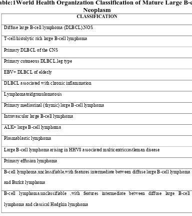
[image:14.612.112.496.294.725.2]DLBCL is a neoplasm of large B lymphoid cells with nuclear size equal to or exceeding normal macrophage nuclei or more than twice the size of a normal lymphocyte that has a diffuse growth pattern. The WHO system modified DLBCL classification to recognize multiple morphologic variants based on improved understanding of the variety of molecular abnormalities associated with DLBCL8 (Table:1).
Table:1World Health Organization Classification of Mature Large B-cell
Neoplasm
CLASSIFICATION
Diffuse large B-cell lymphoma (DLBCL),NOS
T-cell/histolytic rich large B-cell lymphoma
Primary DLBCL of the CNS
Primary cutaneous DLBCL,leg type
EBV+ DLBCL of elderly
DLBCL associated with chronic inflammation
Lymphomatoidgranulomatosis
Primary mediastinal (thymic) large B-cell lymphoma
Intravascular large B-cell lymphoma
ALK+ large B-cell lymphoma
Plasmablastic lymphoma
Large B-cell lymphoma arising in HHV8 associated multicentriccastleman disease
Primary effusion lymphoma
B-cell lymphoma,unclassifiable,with features intermediate between diffuse large B-cell lymphoma
and Burkit lymphoma
B-cell lymphoma,unclassifiable ,with features intermediate between diffuse large B-cell
Epidemiology
Lymphomas are the fifth most common systemic cancer, with the most common subtype being diffuse large B-cell lymphoma followed by follicular lymphoma and Hodgkin lymphoma. DLBCL represents approximately 30% of all lymphomas and is the most common subtype throughout the world1,4. In two large epidemiological study the reported incidence in India is 34%2 and 59.3%3 respectively. It is more common in the elderly .The median age is in the 7th decade but it may also occur in children and young adults. It is slightly more common in males than in females1,2,3.The incidence of NHL increased dramatically from the 1970s until the middle of the 1990s with an estimated 65,540 new cases expected in the united states in 2010.Several factors have contributed to this increased incidence including : more sensitive methods for identifying diagnostic cases, improvement in cancers reporting for haematological malignancies, changes in the classification systems used for lymphoid malignancies, and the epidemic of HIV infections occurring during this period with an associated increase in HIV-associated lymphomas5.For the majority of
patients, the aetiology of DLBCL remain unknown. Some factors that influence the risk of lymphoma include genetics, co-morbid diseases or their treatments (notably immunosuppressant), environmental factors such as ultraviolet,pesticide ,hair dyes, and diet. A subset of DLBCL, including immune-blastic and primary central nervous system (CNS) disease, is highly associated with Epstein-Barr virus although, unlike certain indolent histologies, the concept of antigen driven lymphoma genesis is less developed in DLBCL6.
Clinical Presentation
Most commonly patients present with a rapidly enlarging, painless lymph node in cervical, inguinal or axillary region. However in up to 40% of patients, the initially identified site is extra-nodal commonly involving the skin, gastrointestinal tract, central nervous system (CNS), lung, genitourinary tract or the bones7. Approximately 15% of the patients present with bone marrow involvement, about one-third have B-symptom (fever, night sweat, and weight loss),nearly one-half have Ann-Arbor system stage III/IV disease, and more than one half have an elevated serum lactate dehydrogenase (LDH)level5.
Clinical Prognostic Factors
Originally proposed in 1993,the international prognostic index(IPI) remains the primary clinical tool used to predict outcome for patients with DLBCL9.StageIII/IV disease, elevated LDH, age>60,Eastern Cooperative Oncology group(ECOG) performance status>2, and involvement of >1 extra-nodal site form the IPI score, with one point to each factor. The IPI scoring system nicely stratified patient into four groups with five years survival of 73%,51%,43% and 26% for 0-1,2,3,4-5 risk factors res with CHOP based regimen9.However
progression free survival(PFS):those with 1-2 risk factors had >80% expected PFS;and those with >3 risk factors had >50% PFS11.
Currently ,the original IPI remains as a prospectively designed and validated measure for assessing DLBCL risk12. In 2007,the revised International Working Group response criteria for malignant lymphoma strongly recommended the use of PET scan for patients with routinely FDG-avid, potentially curable lymphoma such as DLBCL13,14 .The PET is recommended 1)Before treatment to better delineate the extent of disease13,14,2) six to eight weeks after completion of therapy for assessment of complete response(CR) because CR is required for cure in DLBCL13,14,and3) in the context of clinical trial mid treatment to evaluate the prognostic ability of interval PET to predict the ultimate response to therapy and long term outcome15.
Biological Prognostic Factors
To segregate DLBCL into biological meaningful subgroups that might identify rational therapeutic targets, the Leukemia and Lymphoma Molecular Profiling Project began gene expression analyses of DLBCL biopsy sample by using DNA microarrays and identified biological distinct and prognostically meaningful molecular subgroups of DLBCL16,17.The
imaging, and modern therapies18,19,20.Despite its usefulness, gene expression profiling technology has not moved easily into community practices.
[image:18.612.101.510.165.351.2]As a result, immune-histochemical algorithm have been proposed and validated for classification of DLBCL into GCB and non-GCB(ABC)21,22. (Fig:1)
Fig:1 Immunohistochemistry based classification of DLBCL.
Treatment and outcomes
Newly diagnosed patient
Although DLBCL is associated with a median survival of less than 1 year in untreated patients1,this disease is commonly curable with conventional anthracycline-based chemotherapy. Advances in the management of DLBCL during the last decade , including the advent of monoclonal antibodies have led to excellent outcomes for many patients. Until recently, the CHOP regimen developed in 1970s28, remained the standard therapy for DLBCL27,28. The Southwest Oncology Group(SWOG) and the Eastern Cooperative Oncology group(ECOG) ,prospective randomized phase 3 trial that compared CHOP to three aggressive multi-agent regimen(m-BACOD,ProMACE/CytaBOM and MACOP-B) ,concluded that the standard CHOP regimen produced similar survival outcome with less toxicity ( 6 years OS for CHOP regimen was 33% as compared to 36%,34%,and 32% respectively for other three regimens)27.In 1997 ,rituximab became the first monoclonal
antibody approved for the use by US (FDA) for follicular lymphoma, and this immunotherapy was soon applied to DLBCL and other B-cell lymphomas29,30,31. Groupe d’
Etude des Lymphomes de l’Adulte (GELA) study32,33,34 in 2002 compared R-CHOP with
Relapsed patients
Novel Therapies for DLBCL
Although rituximab and R-chemotherapy regimens have greatly improved response rates and survival for patients with DLBCL, relapse remains a consistent clinical problem. Of particular concern are preliminary data from the CORAL trial indicating that although DLBCL is commonly cured with first-line R-CHOP, and many patients have been salvaged at relapse with ASCT in the past,41,42,43 current DLBCL patients are at higher risk when they relapse early following upfront R-CHOP chemotherapy and have a poor response to second-line rituximab-containing regimens even when theseregimens are consolidated with high-dose therapy and ASCT.42,43Novel approaches clearly are needed for DLBCL patients who relapse early after R-CHOP chemotherapy. These include other antibody therapies, lenalidomide51,52, SGN-40, bevacizumab, Syk inhibitors45 (fostamatinib disodium), enzastaurin50, histone deacetylase inhibitors, bortezomib47, antisurvivin agents, and mTOR
inhibitors.44A multicenter clinical trial that uses the Hans method to subtype DLBCL patients
and then randomizes non- GCB patients to bortezomib plus R-CHOP or RCHOP alone is now underway46,47,48. Whereas rituximab was the first monoclonal antibody approved for
cell NHL and clearly has revolutionized therapy for DLBCL, other antibodies targeting B-cell lymphomas are now available on an investigational basis, including AME-133, GA101, veltuzumab (all CD20), epratuzumab (CD22), dacetuzumab (CD40), galiximab (CD80), lexatumumab (TRAIL), as are other approaches to improve antibody therapy such as conjugation with radioisotopes or toxins53. Ultimately, understanding mechanisms by which malignant B-cells become resistant to rituximab and chemotherapy and determining means to address these mechanisms may provide pathways for approval of novel agents. Moreover, defining the biology of resistance and activity for various agents across DLBCL subtypes will
become increasingly important in the future as we attempt to select among regimens for newly diagnosed and relapsed patients.
AIMS &
AIMS AND OBJECTIVES
1. To sub classify the Diffuse large B cell lymphoma (DLBCL) in two group Germinal center(GCB) and non Germinal center(non-GCB) on the basis of expression of the three immunomarkersCD10, multiple myeloma oncogene 1 (MUM1), and polyclonal B-cell lymphoma 6 (BCL6)] .
2. To ascertain biological and clinical presentation, staging and international prognostic index (IPI).
3. To assess the response of chemotherapy (CHOP and Rituximab CHOP) in DLBCL as whole and in two subgroup(GC, non GC).
HYPOTHESIS
1. Diffuse large B cell lymphoma sub-classification based on Immuno-histochemical (IHC) stain algorithm on using formalin-fixed, paraffin-embedded tissues will correlates with historical data on gene expression profile.
2. Cell based origin and sub-classification will have prognostic impact on overall survival and event free survival.
3. The prognostic value of the DLBCL subgroup is statistically independent of the features included in the International Prognostic Indicator (IPI).
PATIENTS AND METHODS
This study protocol was approved by our Institutional Review Board (IRB).
Duration of the Scheme
: January 2006 to April 2010.
Settings of the study
: Department of Clinical Haematology, Department of Pathology
Diagnostic criteria
:
Morphology:
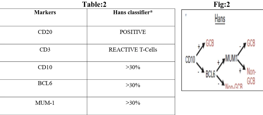
The involved tissue should fulfil the morphological description as per WHO 2008(World Health Organization Classification of Mature Large B-cell Neoplasm): Infiltration of the tissue by large B lymphoid cells with nuclear size equal to or exceeding normal macrophage nuclei or more than twice the size of a normal lymphocyte that has a diffuse growth pattern. [image:27.612.85.547.439.641.2]Immunomarkers:
For Germinal center(GCB), nonGerminal centre(nonGCB) sub-grouping.Table:2 Fig:2
Markers Hans classifier*
CD20 POSITIVE
CD3 REACTIVE T-Cells
CD10 >30%
BCL6 >30%
MUM-1 >30%
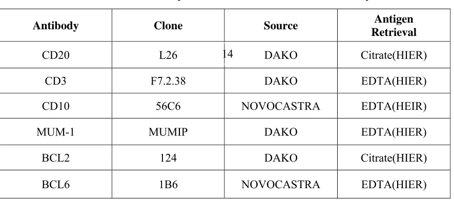
Table:3 Antibody used for Immunohistochemistry
Antibody Clone Source Antigen
Retrieval
CD20 L26 DAKO Citrate(HIER)
CD3 F7.2.38 DAKO EDTA(HIER)
CD10 56C6 NOVOCASTRA EDTA(HEIR)
MUM-1 MUMIP DAKO EDTA(HIER)
BCL2 124 DAKO Citrate(HIER)
BCL6 1B6 NOVOCASTRA EDTA(HIER)
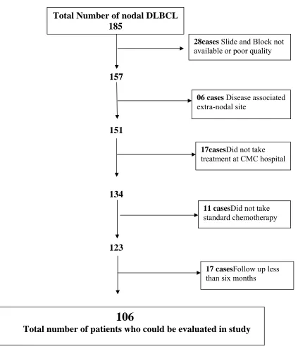
Table:4 Number of DLBCL patients studied
Total Number of nodal DLBCL
185
157
151
134
123
28cases Slide and Block not
available or poor quality
06 cases Disease associated
extra-nodal site
17casesDid not take
treatment at CMC hospital
11 casesDid not take
standard chemotherapy
17 casesFollow up less
than six months
106
Total number of patients who could be evaluated in study
Total Number of nodal DLBCL
PATIENTS
Inclusion criteria:
1. All the patients of primary nodal DLBCL disease diagnosed at CMCH will be taken as cases.
2. Cases of primary nodal DLBCL who have completed 6 cycles of CHOP/R-CHOP chemotherapy and have a minimum of 6 months follow-up.
3. Availability of hematoxylin and eosin stained and Immunohistochemistry slides from archival.
4.All cases should be CD20 positive.
Exclusion criteria:
1. Transformed DLBCL, 2. Primaryextra-nodal
3. Followup less than six months
METHODS
Data collection
Chemotherapy Protocols
RCHOP Chemotherapy:
Inj Rituximab 375mg/m
2Day 1
Inj Cyclophosphamide 800mg Day 1
Inj Adriamycin50 mg Day1
InjVincristine1.4mg Day 1
Tab Prednisolone 60mg Day 1-5
CHOPChemotherapy:
Inj Cyclophosphamide 800mg Day 1
Inj Adriamycin50 mg Day1
InjVincristine1.4mg Day 1
Tab Prednisolone 60mg Day 1-5
Total six cycles of chemotherapy repeated every 21 days .
Intrathecal Chemotherapy:
Inj Methotrexate 12.5 mg with each cycle.
Radiotherapy:
As per the indication , dose decision by Deparment of
Radiotherapy
Response criteria:
Complete response (CR):
Complete resolution of all clinically and radiologically detectable disease, all lymphoma related symptom, and all lymphoma related biochemical abnormalities (like elevated LDH). Lymph nodes and nodal masses must regress to normal size (defined as <1.5cm for lymph node initially >1.5cm). Lymph nodes measuring 1.1 to 1.5cm must regress to <1cm in greatest transverse diameter, or by more than 75% of the sum of the perpendicular diameter (SPD).
Complete Response unconfirmed(CRU):
The patient who fulfill criteria for CR with following exceptions, ie;Residual lymph node mass more than 1.5cm in maximum transverse diameter which have regressed more than 75% of the SPD. Individual node which were previously confluent must regress by more than 75% of the SPD compared with the size of the original mass.
Partial response (PR):
More than 50% decrease in SPD or no increase in size of the other lymph node, liver or spleen. Spleneic or liver nodule must regress by at least 50% in SPD and or appearance of new lesion.
Progressive Disease(PD):
More than or equal to 50% increase from the nadir in the SPD or any previously identified abnormal node 25% in longest diameter or appearance of new Lesion.IPI (International Prognostic Index)
1. Age: more than 60 years
2. Performance status(ECOG): of 2 or higher
3. Serum Lactate Dehydrogenase (LDH): level 1X normal 4. Extra-nodal sites: of 2 or more
5. Stage: III or IV
Staging: The Ann ArborStaging,modifiedCostwald
Stage 1: NHL is limited to one lymph node group (e.g., neck, underarm, groin, etc.) above
orbelow the diaphragm, or NHL is in an organ or site other than the lymph nodes (extra-nodal) but has not spread to other organs or lymph nodes.
Stage 2: NHL is limited to two lymph node groups on the same side of the diaphragm, or
NHL is limited to one extra-nodal organ and has spread to one or more lymph node groups on the same side of the diaphragm.
Stage 3: NHL is in two lymph node groups, with/without partial involvement of an
extranodalorgan or site above and below the diaphragm.
Stage 4: NHL is extensive disease,bone marrow involvement Additional Designations
A - absent (no) symptoms.
B - Presence of any of the following B symptoms: fever (greater than 101.5°), drenching
nightsweats, unexplained weight loss of 10% or more within the last 6 months, severe itching .
E -involvement of a single extranodal(other than the lymph nodes) site that directly adjoins
X - Presence of "bulky" disease, that is, a nodal mass whose greatest dimension is more than
10 centimeters in size, and/ora widening of the mediastinum (middle chest) by more than one-third.
Performance score: ECOG
0. Asymptomatic (Fully active, able to carry on all predisease activities without restriction)
1. Symptomatic but completely ambulatory (Restricted in physically strenuous activity but ambulatory and able to carry out work of a light or sedentary nature. For example, light housework, office work)
2. Symptomatic, <50% in bed during the day (Ambulatory and capable of all self care but unable to carry out any work activities. Up and about more than 50% of waking hours)
3. Symptomatic, >50% in bed, but not bedbound (Capable of only limited self-care, confined to bed or chair 50% or more of waking hours)
4. Bedbound (Completely disabled. Cannot carry on any self-care. Totally confined to bed or chair)
5. Death
Data analysis
Statistical analyses were performed with SPSS (windows 11.01 version, SPSS inc, Chicago), for all variables. Descriptive statistics was calculated for all variables. Theχ2 test/ Fishers exact test or t-test / Mann Whitney U test was used as appropriate to compare the differences
between groups for response to therapy. Overall survival (OS) was defined as the time from initiation of treatment to death or lost follow up. Event free survival (EFS) was defined as the time from initiation of treatment till first event or lost follow up. The event can be loss of response or death. The probability of OS and EFS was estimated using Kaplan-Meier method. For all tests, a two-sided p-value of 0.05 or less was considered statistically
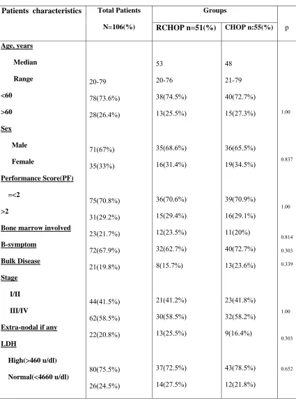
Table:5 Patients Characteristics
Patients characteristics Total Patients N=106(%)
Groups
p
RCHOP n=51(%) CHOP n:55(%) Age, years Median Range <60 >60 Sex Male Female Performance Score(PF) =<2 >2
Bone marrow involved
B-symptom
Bulk Disease
Stage
I/II
III/IV
Extra-nodal if any
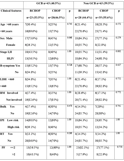
Table:7 Clinical characteristics in GCB and Non GCB subgroup
GCB n=43 (40.5%) Non GCB n=63 (59.5%)
Clinical features RCHOP
n=23 (53.5%) CHOP n=20(46.5%) p RCHOP n=28 (44.4%) CHOP n=35 (55.6%) p
Age >60 years
<60 years
7(30.4%)
16(69.6%)
5(25%)
15(75%)
0.745 6(21.4%)
22(78.6%)
10(28.5%)
25(71.4%)
0.572
Sex Male
Female
17(73.9%)
6(26.1%)
9(45%)
11(55%)
0.068 18(64.3%)
10(35.7%) 27(77.1%) 8(22.8%) 0.279 Stage I,II III,IV 10(43.5%) 13(56.5%) 8(40%) 12(60%)
1.00 10(35.7%)
18(64.3%) 11(31.4%) 24(68.5%) 0.802 B-symptom Yes No 15(65.2%) 8(34.8%) 15(75%) 5(25%)
0.526 17(60.7%)
11(39.3%)
20(57.1%)
15(42.8%)
0.427
LDH <460
>460
8(34.8%)
15(65.2%)
7(35%)
13(65%)
1.00 6(21.4%)
22(78.6%)
6(17.1%)
29(82.8%)
0.752
BM Involved
Not involved
4(17.4%)
19(82.6%)
3(15%)
17(85%)
1.00 8(28.6%)
20(71.4%)
6(17.1%)
29(82.8%)
0.772
Bulk Yes
No
4(17.4%)
19(82.6%)
6(30%)
14(70%)
0.473 4(14.3%)
24(85.7%)
7(20%)
28(80%)
0.741
IPI Low risk
High risk
14(60.8%)
9(39.1%)
12(60%)
8(40%)
1.00 18(64.3%)
10(35.7%)
23(65.7%)
12(34.2%)
1.00
RT Yes
No
3(13.1%)
20(86.9%)
6(30%)
14(70%)
0.263 4(14.3%)
24(85.7%)
5(14.2%)
30(85.7%)
1.00
PF =<2
>2
13(56.5%)
10(43.5%)
12(60%)
8(40%)
1.00 23(82.1%)
5(17.9%)
27(77.1%)
8(22.9%)
0.758
Table:8 Clinical characteristics in IPI low risk and high risk subgroup
IPI Low Risk n=66,(62.3%) IPI High Risk n=40,(37.7%)
Clinical features RCHOP
n=32,(48.4%) CHOP n=34,(51.6%) p RCHOP n=19,(47.5%) CHOP n=21,(52.5%) p
Age >60 years
<60 years
5(15.6%)
27(84.3%)
8(23.5%)
26(76.5%)
0.540 8(42.1%)
11(57.9%)
7(33.4%)
14(66.6%)
0.745
Sex Male
Female
21(65.6%)
11(34.4%)
22(64.7%)
12(35.3%)
1.00 14(73.6%)
5(26.4%)
14(66.6%)
7(33.4%)
0.736
Stage I,II
III,IV
20(62.5%)
12(37.5%)
21(61.7%)
13(38.3%)
1.00 00(00%)
19(100%) 2(9.5%) 19(90.5%) 1.00 B-symptom Yes No 18(56.25%) 14(43.75%) 23(67.6%) 11(32.4%)
0.447 14(73.6%)
5(26.4%)
17(80.9%)
4(19.1%)
0.712
LDH <460
>460
12(37.5%)
20(62.5%)
12(35.2%)
22(64.8%)
0.797 17(89.4%)
2(10.6%)
20(95.2%)
01(4.8%)
0.596
BM Involved
Not involved
5(15.6%)
27(84.4%)
3(8.8%)
31(91.2%)
0.469 07(36.8%)
12(63.2%)
8(38.0%)
13(62.0%)
1.00
Bulk Yes
No
2(6.2%)
30(93.8%)
7(20.5%)
27(79.5%)
0.151 6(31.5%)
13(68.5%) 6(28.5%) 15(71.5%) 1.00 GCB Non-GCB 14(43.7%) 18(56.3%) 12(35.3%) 22(64.7%)
0.615 9(47.3%)
10(52.6%)
8(38.0%)
13(62.0%)
0.750
RT Yes
No
3(9.3%)
29(90.7%)
5(14.7%)
29985.3%)
0.710 4(21.0%)
15(79.0%) 6(28.5%) 15(71.5%) 0.721 PF=<2 >2 27(84.4%) 5(15.6%) 27(79.4%) 7(20.6%)
0.752 9(47.4%)
10(52.6%)
12(57.1%)
9(42.9%)
0.752
RESULTS
Patients clinical and biological characteristics (Table:5,6,7,8)
In RCHOP group low risk(0,1,2),high risk(3,4,5) were 32(62.7%),19(37.2%) and CHOP group 34(61.8%),21(38.1%) respectively. The patients characteristics were almost similar in RCHOP and CHOP groups and none of the p value were significant( significant p<0.05).The clinical characteristics of the patients were further analysed in two subgroups; IPI based low risk and high risk groups and immune-marker based Germinal center B-cell like and Non-germinal center B-cell like groups. The female patients 11(55%) were more compared to male patients 9(45%) in germinal center group patients who got CHOP chemotherapy (p=0.06).Other clinical characteristics distribution were similar in two groups of patients ( non of the p value was significant).
TABLE: 9 Treatment
Total Group p RCHOP CHOP Chemotherapy Radiotherapy(consolidate) CNS therapy(intra-thecal)* 106 18(17.0%) 16(15.1%)** 51 7(13.7%) 5(9.8%)** 55 11(20%) 11(20%)** 0.179 0.445*At diagnosis:1(CHOP) and 0(RCHOP) had CNS disease **Prophylactic intrathecal therapy
TABLE:10 Post chemotherapy status RCHOP(n:51), CHOP(n:55)
Response After 3 cycle After 6 cycle
RCHOP CHOP p RCHOP CHOP p
Complete response(CR+CRU) Parital response(PR) Progressive disease(PD) 32(62.7%) 19(37.3%) 0(0%) 24(43%) 29(%) 2(3.6%)
0.079 45(88.2%)
1(1.9%) 2(3.92%) 3(5.88%) 39(70.9%) 6(10.9%) 7(12.7%) 3(5.4%) 0.082
TABLE:11 Status at last follow up (Mean follow-up 36 months)
Response Status at last follow up
RCHOP(n:51) CHOP(n:55) p
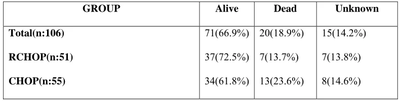
[image:44.612.103.506.266.650.2]Table:12 Status at the time of analysis
GROUP Alive Dead Unknown
Total(n:106)
RCHOP(n:51)
CHOP(n:55)
71(66.9%)
37(72.5%)
34(61.8%)
20(18.9%)
7(13.7%)
13(23.6%)
15(14.2%)
7(13.8%)
8(14.6%)
Table:13Unknown patient status at last follow up
GROUP Remission Relapse Months
(since last follow)
Total(n:15)
RCHOP(n:7)
CHOP(n:8)
8(53.3%)
7(100%)
1(12.5%)
7(46.6%)
0(00%)
7(87.5%)
(12-32months)
(17-40 months)
Chemotherapy and response (Table:9,10,11,12,13)
Survival analysis in different subgroups
Outcome comparison of CHOP and RCHOP
Table:14 Overall survival (OS)
Fig: 3A Overall survival in 106 patients
treated with CHOP and RCHOP
Table:15 Event Free Survival(EFS)
Fig: 3BEvent free survival in 106 Patients
treated with CHOP and RCHOP
Table:16 Cummulative RFS
Fig: 3C Cumulative RFS in 106 patients
Treated with CHOP and RCHOP
Group 12 months 24 months 36 months p RCHOP(n=51) 97.7% 78.4% 78.4% 0.190 CHOP(n=55) 86.3% 69.1% 69.1%
Group 12 months 24 months 36 months p RCHOP(n=51) 83.1% 73.4% 68.5% 0.190 CHOP(n=55) 76.2% 55.2% 47.9%
Group 12 months 24 months 36 months p RCHOP(n=51) 80.5% 73.4% 68.5% 0.13 CHOP(n=55) 77.7% 55.7% 51.1%
32
Analysis of response of CHOP and RCHOP chemotherapy:(Table:14,15,16
Fig:3 A,B,C)
Table:17 Outcome comparison in clinical IPI risk groups(median follow up 24 months)
IPI Risk group OS P EFS P RFS P
Low Risk(0,1,2) (n=66) RCHOP(n=32) 75.3% 0.26 75.1% 0.80 75.1% 0.29
CHOP(n=34) 86.7% 72.5% 76.8%
High Risk(3,4,5)(n=40) RCHOP(n=19) 83.3% 0.004 74.9% 0.002 74.9% 0.002
CHOP(n=21) 41.5% 19.8% 21.1%
Fig 4A:Overall survival in 66 IPI low risk patients with
CHOP and RCHOP Fig 4B: Event Free survival in 66 IPI low risk patients with CHOP and RCHOP
Fig 4C: Overall survival in 40 IPI high risk patients with CHOP and RCHOP
Fig 4D: Event Free Survival in 40 IPI high risk patients with CHOP and RCHOP
Analysis of response of chemotherapy in IPI risk subgroups : (Table:17
Fig:4 AB,C,D)
Outcome comparison GCB(n=43) and Non-GCB(n=63) groups(hans et.al)
Table:18
GCB treated with CHOP and RCHOP(median follow up 24 months)Treatment GP OS EFS RFS
RCHOP(n:23) 89.1% 80.2% 80.2%
CHOP(n:20) 50.3% 37.3% 46.3%
p 0.06 0.02 0.16
Fig 5A: Overall survival in 43GCB patients treated with CHOP and RCHOP
Fig 5B: Event free survival in 43GCB patients treated with CHOP and RCHOP
Fig 5C: Cumulative RFS in 43GCB patients treated with CHOP and RCHOP
Fig 5(A,B,C): Addition of rituximab to CHOP chemotherapy improved OS,EFS and Cumulative RFS
Table:19
Non GCB treated with CHOP and RCHOP(median follow up of 24 months)Treatment GP OS EFS RFS
RCHOP(n:28) 69% 66.9% 66.9%
CHOP(n:35) 79.2% 60.6% 60.6%
p 0.8 0.4 0.4
Fig 6A: Overall survival in 63 Non-GCB patients treated with CHOP and RCHOP
Fig 6B: Event free survival in 63Non-GCB patients treated with CHOP and RCHOP
Fig 6C: Cumulative RFS in 63Non-GCB patients treated with CHOP and RCHOP
Fig 6(A,B,C): Addition of rituximab to CHOP chemotherapy improved OS,EFS and Cumulative RFS
[image:52.612.84.525.116.630.2]Table:20
RCHOP in GCB and Non GCB(median follow up 24 months)Treatment GP OS EFS RFS
GCB(n:23) 89.1% 80.2% 80.2%
Non GCB(n:28) 69% 66.9% 66.9%
p 0.3 0.8 0.8
Fig 7A: Overall survival in 51 RCHOP treated patients in GCB and Non-GCB subgroup.
Fig 7B: Event free survival in 51 RCHOP treated patients in GCB and Non GCB subgroup.
Fig 7C: Cumulative RFS in 51 RCHOP treated patients in GCB and Non GCB
Fig 7(A,B,C): Addition of rituximab to CHOP chemotherapy improved OS,EFS and Cumulative RFS
in Germinal center B-cell and Non-Germinal center B-cell like subgroup of nodal DLBCL patients.
[image:53.612.83.522.77.628.2]Response analysis of chemotherapy in GCB and Non-GCB subgroup
(Table:18,19,20 Fig:5,6,7 A,B,C)
Table: 21 Chemotherapy related complication
Complication Total no(%) RCHOP CHOP p
Neutropenia
Hyperglycemia
Neuropathy
SIADH
DVT
61(57.5%)
26(24.5%)
3(2.8%)
6(5.7%)
3(2.8%)
35(68.6%)
14(27.5%)
3(5.9%)
5(9.8%)
2(4.1%)
26(47.3%)
12(21.8%)
0
1(1.8%)
1(1.8%)
0.032
0.652
0.108
0.103
40
Chemotherapy complications(Table:21)
Table:22 Incidence comparison of GCBvs non GCB with
Litrature
Studies Total number GCB Non-GCB
*Present 106 43(40.5%) 63(59.5%)
**Hans,Blood 2004 152 64(42%) 88(58%)
**Nyman,Blood2007 194 97(50%) 97(50%)
**Kaifu JCO 2008 243 121(49.7%) 122(50.3%)
**Shiozawa,Leuk.R2007 248 71(29%) 177(71%)
*Denovo DLBCL nodal site only **De novo DLBCL all sites
Table:23 Outcome comparison with Published literatures
Studies No of patients EFS* OS*
RCHOP CHOP RCHOP CHOP RCHOP CHOP
Present study
*at 3 years
51 55 68.5% 47.5% 78.5% 69%
Coiffer.BNEJM2002 *at 2 years
202 197 61% 43% 76% 63%
GELA JCO2005 *at 5 years
197 202 47% 29% 58% 45%
B Columbia JCO2005
*at 2 years 292 292 69% 51% 78% 42%
US intergpJCO2006 *at 5 years
279 267 52% 35% 67% 58%
MINT trial Lancet2006 *at 3 years
[image:58.612.81.540.445.691.2]Discussion
Clinical and biological characteristics
A total of 106 patients of primary nodal diffuse large b cell lymphoma with age greater than 15 years were included in the study. This comprises 71(67.0%) males and 35(33.0%) females with ratio approximately 2:12,3,4 . The median age of the patients in our study was 53 years
(range:20-76 years) in RCHOP group and 48 years(range:21-79 years) in CHOP group of patients. The median age reported in major western studies is in seventh decade. The median age of the group in SWOG 8516 trial ranged from 54-57 years which is comparable to our study1.The B-symptom was present in 72(67.9%) and the bone marrow was involved in 23(21.7%) of patients at diagnosis. 22(20.8%) cases has one or more extra-nodal involvement other than the primary and bulk disease i.e greater than 10 cm on presentation in 21(19.8%) of patients. The published studies have quoted an incidence of 40% bulky disease and 40% initially confined extra-nodal disease 2,29. Our study is on primary nodal disease so the above finding does not correlate with literature. In our study 80(75.5%) of cases has high LDH level (>460u/dl) comprises of 37(72.5%) patients in RCHOP group and 43(78.5%) patients in CHOP group. 22(20.8%) patients presented with anemia at diagnosis (Hb<10gm%) in . The LDH>ULN at diagnosis ranges from 30%-57% in MInT and RECOVER 60 trial.
In RCHOP group out of 51 patients, 23(45.1%) were GCB and 28(54.9%) patients were Non-GCB and in CHOP group out of 55 patients 20(36.3%) patients were GCB and 35(59.4%) patients were in Non-GCB group. In three major published western literature the frequency of distribution of GCB and Non-GCB in denovo DLBCL in all tissue is almost 50% in each group21,23,24(Table:22)and in asian population the published literature shows 30% and 70% respectively(schiozawa,leukR2007). Our study is only on nodal cases with frequency of distribution of GCB and Non-GCB is 40% and 60 respectively correlates nearest to hans.etal needs gene expression profiling for confirmation21.
Chemotherapy and response
Patients received either six cycles of CHOP chemotherapy;55(51.8%) patients or Rituximab with CHOP chemotherapy;51(48.2%) patients. 7(13.7%) patients in RCHOP and 11(20%) patients in CHOP group received consolidative radiotherapy either due to bulk disease or residual disease at end of six cycle of chemotherapy. Total of 16(15.1%) patients received prophylactic intrathecal methotrexate (5;9.8% patients in RCHOP and 11;20% patients in CHOP group) though only one patient had documented CNS disease in CHOP group.
After three cycles of chemotherapy the CR+Cru in CHOP was inferior 24(43%0 vs 32(62.7%) in RCHOP group (p=0.7) though p value not significant. After six cycle of chemotherapy in CHOP group, CR+Cru, PR, PD and SD were 39(70.9%), 6(10.9%), 7(12.7%)and 3(5.4%) respectively. It was better (p=0.08,) in RCHOP treatment group as the CR+Cru, PR, PD and SD were 45(88.2%),1(1.9%),2(5.88%) and 3(3.92%) respectively though the p value is not significant. At the mean follow up of 36 months the patients treated with RCHOP had CR+CRu rate better 37(75.2%) and relapse was less 10(19.6%) compared to CHOP chemotherapy(p=0.09).
The analysis shows trend (p value not significant) towards better CR rates after addition of Rituximab to CHOP chemotherapy and is comparable with the literature32,33 At the median follow up of 36 months the cumulative RFS was 73.4% in RCHOP and 55.7% in CHOP group (p=0.139). The EFS was 73.4% vs 55.2% (p=0.07) and OS 78.4% vs 69.1% (p=0.19) respectively in RCHOP and CHOP group. The advantage of 21% in event free survival (EFS) and 10% in overall survival (OS) with addition of Rituximab to CHOP chemotherapy is comparable with the literature (Table:23).
In low risk IPI group( 0,1,2) the OS, EFS and cumulative RFS at median follow-up of 24 months ,in RCHOP group of patients was 75.3%,75.1% and 75.1% ; and in CHOP group of patients was 86.7%,72.5% and 76.8% respectively( p value not significant).The OS is better in CHOP group most likely due poor follow up in relapse group of patients. In high IPI risk group( 3,4,5) the OS, EFS and cumulative RFS at median follow-up of 24 months ,in RCHOP grofollow-up of patients was 83.3%,74.9% and 74.9% ; and in CHOP group of patients was 41.5%,19.8% and 21.1%% respectively(high IPI risk group p=0.006 for OS, p=0.002 for EFS and p=0.002 for RFS) . Patients who were treated with only CHOP chemotherapy did bad in high IPI risk group with OS, EFS 41.5%,19.8% compared to low risk 86.7%,72.5% (p=0.001). Addition of Rituximab improves the OS, EFS 83.3%,74.9% in high risk comparable with the low risk 75.3%,75.1% (p=0.28).The advantage of Rituximab in high risk IPI nodal DLBCL correlates with published literature 23,29,36,37.
The Overall survival(OS), event free survival(EFS) and Cumulative relapse free survival(RFS) at median follow up of 24 months in GCB group were 89.1%,80.2% and 80.2% respectively in RCHOP chemotherapy patients vs 50.3%,37.3% and 46.3% respectively in CHOP chemotherapy patients,( OS p=0.06,for EFS p=0.02 and for RFS p=0.16).The survival correlates with Kai Fu JCO232008 paper which showed addition of Rituximab in GCB improves survival significantly.
median follow up of 24 months was 89.1 %,80.2% and 80.2% in GCB and 69%%,66.9%% and 66.9% respectively in Non-GCB patients( p=0.3). Addition of Rituximab improves survival more in GCB than non-GCB, though p value not significant correlates with Kai Fu JCO232008 paper, but not with Nyman Blood24 2008 which showed addition of Rituximab negates the survival advantage of GCB over Non-GCB.
Conclusion
1) The frequency of distribution of Germinal centre(GCB) and non-Germinal centre(non-GCB) in nodal DLBCL cases in our population is 40% and 60% respectively(Table:3).
2) The frequency of distribution of low IPI risk (0,1,2) and high IPI risk(3,4,5) in nodal DLBCL cases in our population is 62% and 37.7%% respectively.
3) Addition of Rituximab to CHOP chemotherapy improves event free survival by 20% and overall survival by 10% in our patients comparable to published literature (Table:14).
4) Addition of Rituximab to CHOP chemotherapy significantly improves overall survival in GCB and high IPI risk group comparable to published litratures.
5) In non-GCB and IPI low risk groups the Rituximab improves the event free survival and relapse free survival but not the overall survival.
Limitation of study
1) It is a retrospective study.
2) Selection bias; only nodal cases with minimum six months follow up patients selected.
3) DLBCL Classification based on cell of origin (GCB and non GCB) by three immunomarkers has its own limitations.
4) Short follow up and follow status of the few patients at the time of analysis not known.
PROFORMA:
Title:Diffuse Large B cell Lymphoma Single centre study
Sl.No:
Name: Hospital No: Sex: Age:
Date of diagnosis:
At diagnosis: Haemoglobin: Total leucocyte count: Platelet count: LDH: Viral serology (HIV, HbSAg, HCV):
Nodal(Site): Extranodal(Site):
B symptoms: Immunomarkers
Bone marrow aspirate and biopsy Ann Arbor staging:
Performance score (ECOG):
International prognostic index (score): Radiology: At diagnosis
After three cycles of chemotherapy After six cycles of chemotherapy At follow-ups
CSF examination:
Markers CD10 BCL6 MUM1 CD20 Impression
Chemotherapy Protocol:
CHOP Chemotherapy
Rituximab CHOP chemotherapy
Chemotherapy cycles date:
First cycle
Sixth cycle
Radiotherapy:
Intrathecal CNS therapy: Response:
After three cycles
After six cycles
At last follow up
Relapse date and site
Complication during treatment:
Neutropenia
Hyperglycemia
Neuropathy
SIADH
Others
Bibliography
1.Jaffe ES .The 2008 WHO classification of lymphoma:implications for clinical practice and translational research. Hematol Am SocEduc Program.2009:523-531
2.Distribution of various subtypes of non-Hodgkin's lymphoma in India:A study of 2773 lymphomas using R.E.A.L. and WHO Classifications K. N. NareshAnnals of Oncology 11(Suppl I):S63-S67 2000
3.Diffuse large Bcell Lymphoma :experience from tertiary care center in north india . KherR ,L.AIMMSDept of Medical oncologyMed Oncology 2009 April 7
4. Fisher RI, Miller TP, O’Connor OA. Diffuse aggressive lymphoma. Hematology (Am SocHematolEduc Program). 2004:221-236.
5.Friedberg JW, Fisher RI. Diffuse large Bcell lymphoma. HematolOncolClinNorthAm. 2008;22:941-952, ix.
6.Fisher SG .The emerging concept of antigen drivenlymphomas:epidemiology and treatment implications.CurrentopinOncol 2006:18(5):4117-24.
7.Christopher R. Improving outcome for patients with diffuse large B cell lymphoma CA Cancer J Clin 2010;60;3939-408.
8. Morton LM, Wang SS, Cozen W, et al. Etiologic heterogeneity among non- Hodgkin lymphoma subtypes. Blood. 2008;112:5150-5160.
9. A predictive model for aggressive non-Hodgkin lymphoma. The Int. Non-Hodgkin Lymphoma Prognostic Factors Project. N Engl J Med. 1993;329:987-994.
10.Sehn LH, Berry B, Chhanabhai M, et al. The Revised International Prognostic Index (R-IPI) is a better predictor of outcome than the Standard IPI.for patients with DLBCL treated with R-CHOP. Blood. 2007;109:1857-1861.
11.Sehn LH, The revised International Prognostic Index(R-IPI) is a better predictor of outcome than the standard IPI for patients with diffuse large B-cell lymphoma treated with RCHOP .Blood. 2007;109: 1857-1861.
12.Maritaziepert; Standard International Prognostic Index Remains a Valid predictor of outcome for patients with aggressive CD20+B-cell Lymphoma in Rituximab Era. JCO;May,volume28,2010 page;2373-2380.
14. Spaepen K, Stroobants S, Dupont P, et al. Early restaging positron emission tomography with (18)F-fluorodeoxyglucosepredictsoutcome in patients with aggressive non-Hodgkin lymphoma. Ann Oncol. 2002;13:1356-1363.
15.49. Itti E, Lin C, Dupuis J, et al. Prognostic value of interim 18F-FDG PET in patients with diffuse large B-Cell lymphoma: SUV based assessment at 4 cycles of chemotherapy. J Nucl Med. 2009;50:527-533.
16. RosenwaldAet al. The use of molecular profiling to predict survival after chemotherapy for diffuse large-B-cell lymphoma.NEJMed.2002;346:1937-1947.
17. Rosenwald A, Wright G, Leroy K, et al. Molecular diagnosis of primary mediastinal B cell lymphoma identifies a clinically favorable subgroup of diffuse large B cell lymphoma related to Hodgkin lymphoma. J Exp Med. 2003;198:851-862.
18. Iqbal J, Greiner TC, Patel K, et al. Distinctive patterns of BCL6 molecular alterations and their functional consequences in different subgroups of diffuse large B-cell lymphoma. Leukemia. 2007;21:2332-2343.
19. Iqbal J, Neppalli VT, Wright G, et al.BCL2 expression is a prognostic marker for the activated B-cell-like type of diffuse large B-cell lymphoma. J ClinOncol. 2006;24:961-968. 20. Iqbal J, Sanger WG, Horsman DE, et al.BCL2 translocation defines a unique tumor subset within the germinal center B-cell-like diffuse large B-cell lymphoma. Am JPathol. 2004;165:159-166.
21. Hans CP, Weisenburger DD, Greiner TC,et al. Confirmation of the molecular classification of diffuse large B-cell lymphoma by immune-histochemistry using a tissue microarray. Blood. 2004;103:275-282.
22. Choi WW, Weisenburger DD, Greiner TC,et al. A new immunostain algorithm classifies diffuse large B-cell lymphoma into molecular subtypes with high accuracy. Clin Cancer Res. 2009;15:5494-5502.
23. Addition of Rituximab to Standard Chemotherapy Improves the Survival of Both the Germinal Center B-Cell–Like and Non–Germinal Center B-Cell–Like Subtypes of Diffuse Large B-Cell Lymphoma Kai Fu, Dennis D. Weisenburger J ClinOncol 26:4587-4594. © 2008
24.HeidiNyman,Sirpa;Prognostic impact of immunohistochemically defined germinal center phenotype in diffuse large B-cell lymphoma patients treated with immune-chemotherapy. Blood,1,June2007.Vol109.
25.Mounier N, Briere J, Gisselbrecht C, et al: Rituximab plus CHOP (R-CHOP)overcomes bcl-2 associated resistance to chemotherapy in elderly patients with diffuse large B-cell lymphoma.Blood 2003;101;4279
26. Winter JN, Weller EA, Horning SJ, et al: Prognostic significance of bcl-6 protein expression in DLBCL treated with CHOP or R-CHOP:A prospective correlative stud y Blood 2006; 107:4207.
27.Fisher RI, Gaynor ER, Dahlberg S, et al. Comparison of a standard regimen (CHOP) with three intensive chemotherapy regimens for advanced non-Hodgkin lymphoma. N Engl J Med. 1993;328:1002-1006.69.
28.McKelvey EM, Gottlieb JA, Wilson HE,et al. Hydroxyldaunomycin (doxorubicin (Adriamycin)) combination chemotherapy in malignant lymphoma. Cancer. 1976;38:1484-1493.
29. Coiffier B. Diffuse large cell lymphoma.CurrOpinOncol. 2001;13:325-334.
30. Coiffier B, Haioun C, Ketterer N, et al. Rituximab (anti-CD20 monoclonal antibody)for the treatment of patients with relapsing or refractory aggressive lymphoma: a multicenter phase 2 study. Blood. 1998;92:1927-1932.
31. Coiffier B, Lepage E, Herbrecht R, etal.Mabthera (Rituximab) plus CHOP is superior to CHOP alone in elderly patients with diffuse large-B-cell lymphoma (DLCL): Interim results of a randomized GELA trial (abstract). Blood. 2000;96:223A. Abstract 950.
32. Coiffier B, Lepage E, Briere J, et al. CHOP chemotherapy plus rituximab compared with CHOP alone in elderly patients with diffuse large-B-cell lymphoma. N Engl J Med. 2002;346:235-242.
33. Feugier P, Van Hoof A, Sebban C, et al. Long-term results of the R-CHOP study in the treatment of elderly patients with diffuse large B-cell lymphoma: a study by the Grouped’Etude des Lymphomesdel’Adulte. J ClinOncol. 2005;23:4117-4126.
34. Coiffier B, Feugier P, Mounier N, et al. Long-term results of the GELA study comparing R-CHOP and CHOP chemotherapy in older patients with diffuse large B-cell lymphoma show good survival in poor-risk patients the GELA. J ClinOncolASCOAnnual Meeting Proceedings Part I. 2007; 25(18s). Abstract 8009.
35.Coiffier B. Long-term results of patients in the LNH-98.5 trial,the first randomized study comparing rituximab –CHOP to standard CHOP chemotherapy in DLBCL patients: a study by the Grouped’Etude des Lymphomesdel’Adulte. Blood 2010 116:2040-2045
36.Habermann TM, Weller EA, Morrison VA, et al. Rituximab-CHOP versus CHOP alone or with maintenance rituximab in older patients with diffuse large B-cell lymphoma. J ClinOncol. 2006;24:3121-3127.
37.Pfreundschuh M, Trumper L, Osterborg A, et al. CHOP-like chemotherapy plus rituximab versus CHOP-like chemotherapy alone in young patients with good-prognosis diffuse large-B-cell lymphoma: a randomized controlled trial by the Mab-TheraInternationalTrial (MInT) Group. Lancet Oncol.2006; 7:379-391.
38.Zelenetz AD, Abramson JS, AdvaniRH,et al. NCCN clinical practice guidelines in oncology: non-hodgkin lymphoma. J NatlComprCancNetw. 2010;8:288-334.
39. Philip T, Guglielmi C, Hagenbeek A, et al. Autologous bone marrow transplantation as compared with salvage chemotherapy in relapses of chemotherapy-sensitive non-Hodgkin lymphoma. N Engl J Med. 1995; 333:1540-1545.
40. Vose JM, Zhang MJ, Rowlings PA, et al. Autologous transplantation for diffuse aggressive non-Hodgkin lymphoma inpatients never achieving remission: a report from the Autologous Blood and Marrow Transplant Registry. J ClinOncol. 2001;19:406-413.
41. Oliansky DM, Czuczman M, Fisher RI,et al. The role of cytotoxic therapy with hematopoietic stem cell transplantation in the treatment of diffuse large B-cell lymphoma: update of the 2001 evidence-based review. Biol Blood Marrow Transplant.[Publishedonline ahead of print Jul 22 2010. PMID:20656046]
42. Gisselbrecht C, Glass B, Mounier N, et al. Salvage regimens with autologous transplantation for relapsed large B-cell lymphoma in the rituximab era. J ClinOncol.[Published online ahead of print Jul 262010. PMID: 20660832].
43. Gisselbrecht C, Glass B, Mounier N, et al. R-ICE versus R-DHAP in relapsed patients with CD20 diffuse large B-cell lymphoma(DLBCL) followed by autologous stem cell transplantation: CORAL study(abstract). J ClinOncol. 2009;27(suppl 15).Abstract 8509. 44. Leonard JP, Martin P, BarrientosJ,Elstrom R. Targeted treatment and new
agentsin diffuse large B-cell lymph.SeminHematol. 2008;45(suppl 2):S11-S16.
45. Friedberg JW, Sharman J, SweetenhamJ,et al. Inhibition of Syk with fostamatinibdi sodium has significant clinical activity in non-Hodgkin lymphoma and chronic lymphocytic leukemia. Blood. 2010;115:2578-2585.
46. Fisher RI, Bernstein SH, Kahl BS, et al. Multicenter phase 2 study of bortezomib inpatients with relapsed or refractory mantle cell lymphoma. J ClinOncol. 2006;24:4867-4874.
47. Dunleavy K, Pittaluga S, CzuczmanMS,et al. Differential efficacy of bortezomib plus chemotherapy within molecular subtypes of diffuse large B-cell lymphoma Blood. 2009;113: 6069-6076.
48. Furman RR, Martin P, Ruan J, et al. Phase1 trial of bortezomib plus R-CHOP in previously untreated patients with aggressive non-Hodgkin lymphoma. Cancer. [Publishedonline ahead of print Jul 27 2010.PMID: 20665890]
49. Leonard JP, Furman RR, Cheung Y-KK,et al. CHOP-R þ bortezomib as initial therapy for diffuse large B-cell lymphoma(DLBCL) (abstract). J ClinOncol 2007ASCO Annual Meeting Proceedings Part I.2007;25(18S):8031.
50. Robertson MJ, Kahl BS, Vose JM, etal.Phase 2 study of enzastaurin, a protein kinase C beta inhibitor, in patients with relapsed or refractory diffuse large B-cell lymphoma. J ClinOncol. 2007;25:1741-1746.
51.Wiernik PH, Lossos IS, Tuscano JM, et al.Lenalidomidemonotherapy in relapsed or refractory aggressive non-Hodgkin lymphoma.JClinOncol. 2008;26:4952-4957.
52. Nowakowski GS, LaPlant B, HabermannT,et al. A phase I/II Trial of lenalidomideandrchop (r2chop) in patients with newly diagnosed diffuse large B -Cell (DLBCL) and follicular grade 3 lymphoma (abstract). Blood.(ASH). 2009;114.Abstract 1669. 53. Micallef IN, Kahl BS, Maurer MJ, et al. Apilot study of epratuzumab and rituximabin combination with cyclophosphamide, doxorubicin, vincristine, and prednisone chemotherapy in patients with previouslyuntreated, diffuse large B-cell lymphoma.
54.Addition of Rituximab to Standard ChemotherapyImproves the Survival of Both the Germinal CenterB-Cell–Like and Non–Germinal Center B-Cell–LikeSubtypes of Diffuse Large B-Cell LymphomaKai Fu, Dennis D. Weisenburger J ClinOncol 26:4587-4594. ©
Coding for the master sheet
IPI RISK GROUP Low Risk(0,1):1 Low Inter(2):2 High Inter(3):3 High Risk(4,5):4 Low Risk(0,1,2):1 High Risk(3,4,5):2 Hans GCB:1 Non-GCB:2 LDH <460U/dl:1 >460U/dl:2 Sex Male:1 Female:2 Age <60 years:1 >=60 years:2 B-symptom Yes:1 No:1 CNS Involved:1 Not involved:2 Not done:3 Stage I,II:1 III,IV:2 Extra-nodal Yes:1 No:2 Performance score =<2:1 >:2 Bulk disease Yes:1 N:2 Bone-marrow Involved:IN Not involved:NI
S.NO Age Sex Date of Dx Site
involved Bulk Extranodal B symp
Perform
ance HB CNS Bone Marrow LDH Stage IPI HANS First Cycle Mid Course
1 31 M 1/4/2006 Cervical 2 2 1 1 13.7 2 NI 719 IIB 1 2 1/9/2006 1 2 27 M 3/6/2006 Cervical 2 1 2 1 14.2 3 NI 978 IIIBE 3 2 3/14/2006 1 3 30 F 4/29/2006 Cervical 1 2 1 1 7.6 3 NI 571 IIIBXS 3 1 5/4/2006 2 4 44 M 11/24/2008 Cervical 2 2 1 1 9.9 2 NI 491 IIIB 2 2 11/28/2008 2 5 44 M 5/31/2006 Cervical 2 1 1 3 10.6 3 NI 911 IVBE 3 1 6/17/2006 4 6 79 M 4/28/2006 cervical 2 2 1 3 11.4 3 NI 441 IIIB 3 2 5/9/2006 1 7 46 M 5/19/2006 Abdomen 2 2 1 2 11.8 3 NI 906 IVBS 3 2 6/9/2006 2 8 32 M 7/4/2006 Abdomen 1 2 2 1 6.7 3 IN 898 IVBX 4 2 7/7/2006 2 9 21 M 8/31/2005 Abdomen 1 2 1 2 8.4 3 IN 1646 IVBX 4 2 9/29/2005 2 10 63 M 1/31/2007 Axilla 2 2 2 1 14.1 3 NI 566 IIIA 3 2 2/21/2007 1 11 64 M 3/26/2007 inguinal 2 1 1 2 8.1 2 IN 939 IVBE 5 2 4/12/2007 2 12 35 F 4/25/2007 Abdomen 2 2 1 1 6.8 2 NI 994 IIIBS 2 2 5/1/2007 1 13 56 M 5/2/2007 Abdomen 2 1 1 3 12.6 2 NI 463 IIBE 3 1 5/4/2007 2 14 61 F 7/7/2007 Abdomen 1 2 1 2 10.4 3 NI 636 IIBX 3 1 8/28/2007 2 15 39 F 8/3/2007 Inguninal 2 2 1 1 11.7 2 NI 610 IIB 2 2 8/22/2007 1 16 47 M 10/29/2007 Cervical 1 2 1 1 11.6 2 NI 935 IIIBX 2 2 11/3/2007 2 17 40 M 12/20/2007 cervical 2 2 2 0 15.5 3 NI 531 IIA 1 1 1/3/2008 1 18 48 F 15/1/2008 Cervical 2 2 1 2 5.3 3 IN 1804 IVBS 4 1 2/29/2008 2 19 40 F 5/1/2008 Cervical 2 2 1 1 10.6 3 NI 614 IIIB 3 2 5/6/2008 1 20 54 F 6/5/2008 Cervical 2 2 1 0 11.8 3 NI 509 IIB 1 1 7/1/2008 2 21 52 M 6/19/2008 Cervical 1 2 1 1 12 3 NI 686 IIIBXS 2 1 7/1/2008 1 22 66 M 6/9/2008 Inguinal 2 2 2 2 11.7 3 NI 710 IIA 2 2 6/11/2008 2 23 62 M 9/24/2008 Cervical 1 2 1 1 7.4 3 NI 939 IIIBX 3 1 9/30/2008 2 24 40 M 10/6/2008 Cervical 1 2 1 1 9.9 3 NI 1570 IIIBX 3 2 10/18/2008 2 25 32 F 11/22/2008 inguinal 2 2 1 1 9.1 3 NI 2260 IVB 3 2 12/5/2008 2 26 63 M 1/22/2009 Axilla 1 2 1 1 12.8 3 NI 664 IIBX 2 2 2/3/2009 2 27 51 M 4/17/2009 Cervical 2 2 2 0 13.6 2 NI 556 IIA 1 2 4/29/2009 1
S.NO Date of
Compleltion CNStherapy Radiotherapy
Status post six
cycle Date LFU Mon
ths
Status
LFU Relapse Site Month
CR Alive Lost
follow Neutrope
Hype rgly Neu ro SIA DH Other s
1 4/25/2006 2 2 1 2/22/2010 49.6 1 No No 49.6 1 2 2 2 2 2 2 6/26/2006 2 2 1 5/2/2009 37.9 5 8/1/2008 Node 28.9 2 2 2 2 2 2 3 8/29/2006 2 1 1 7/27/2007 14.9 5 4/27/2007 Node 11.9 2 2 2 2 2 2 4 3/13/2009 2 2 3 12/17/2010 24.7 3 No No 24.7 1 2 2 2 2 2 5 9/8/2006 2 2 4 8/14/2007 14.5 4 10/24/2006 Marrow 4.8 2 2 1 2 2 2 6 9/8/2006 2 2 1 12/7/2007 19.3 5 3/26/2007 Node 10.9 2 2 1 1 2 2 7 2/16/2007 2 2 1 11/22/2010 54.1 5 3/20/2007 Node 10.0 1 2 2 2 2 2 8 11/10/2006 2 2 4 1/10/2007 6.2 4 10/11/2006 Node 3.3 2 2 1 2 2 2 9 1/6/2006 2 1 2 10/23/2006 13.7 5 9/21/2006 Node 12.7 2 2 1 2 2 2 10 6/6/2007 2 2 1 1/27/2009 23.9 5 8/14/2008 Node 18.4 2 2 2 2 2 2 11 7/26/2007 2 2 2 2/12/2008 10.6 5 1/31/2008 Skin 10.2 2 2 1 1 2 2 12 8/21/2007 2 2 1 1/11/2008 8.6 5 1/10/2008 Node 8.6 2 2 2 2 2 2 13 11/6/2007 2 1 3 9/17/2008 16.6 4 9/17/2008 Spine 16.6 2 2 2 2 2 2 14 12/26/2007 2 2 1 6/9/2008 11.1 5 6/9/2008 Node 11.1 2 2 1 2 2 2 15 12/14/2007 2 2 1 9/17/2010 37.5 1 No No 37.5 1 2 2 2 2 2 16 2/19/2008 2 2 1 8/3/2010 33.1 1 No No 33.1 1 2 2 2 2 2 17 4/19/2008 2 2 1 12/15/2010 35.8 1 No No 35.8 1 2 2 2 2 2 18 5/13/2008 1 1 4 7/10/2008 6.2 4 6/25/2008 Node 5.2 2 2 1 2 2 2 19 8/22/2008 2 2 1 3/24/2010 23 1 No No 23.0 1 2 1 2 2 2 20 12/1/2008 2 2 2 12/9/2009 18.1 5 2/25/2009 Node 8.7 1 1 2 2 2 2 21 10/14/2008 2 1 1 10/5/2010 24.5 1 No No 24.5 1 2 2 2 2 2 22 10/1/2008 2 2 2 1/30/2009 7.7 4 30/1/2009 No 7.7 1 1 2 2 2 2 23 2/13/2009 2 2 1 4/13/2009 6.6 1 No No 5.6 1 1 1 2 2 2 T B 24 2/3/2009 2 2 1 8/10/2010 22.1 1 No No 22.1 1 2 1 2 2 2 25 3/20/2009 2 2 4 5/29/2009 6.2 5 5/29/2009 Node 6.2 1 1 2 2 2 2 26 7/2/2009 2 2 1 2/2/2010 12.4 1 No No 12.4 1 2 1 2 2 2 27 9/1/2009 2 1 1 8/6/2010 15.6 1 No No 15.6 1 2 2 2 2 2
S.NO Age Sex Date of Dx Site
involved Bulk Extranodal B symp Perform
ance HB CNS Bone Marrow LDH Stage IPI HANS First Cycle Mid Course
28 60 M 6/1/2009 Cervical 2 2 1 0 10.5 3 IN 1144 IVB 4 2 6/7/2009 1 29 45 F 7/20/2009 Cervical 2 2 2 1 11.9 3 NI 636 IIIA 2 2 7/31/2009 2 30 34 F 11/21/2009 Cervical 2 1 1 0 11.2 3 NI 940 IIBE 2 2 12/4/2009 1 31 68 F 12/23/2009 Cervical 2 1 1 1 12.8 3 NI 1017 IIIBE 2 1 1/11/2010 1 32 36 M 3/31/2010 Inguinal 2 2 1 1 11.7 3 IN 980 IVB 3 2 4/2/2010 2 33 59 M 9/14/2009 Cervical 2 2 1 1 12.4 3 IN 483 IVB 4 2 9/16/2010 2 34 69 F 4/23/2007 Cervical 2 2 2 1 8.7 3 IN 567 IVB 4 1 5/3/2007 4 35 33 F 3/10/2007 Abdomen 1 2 1 2 6.7 3 NI 2335 IIIBX 2 1 4/6/2007 2 36 49 F 4/14/2007 Ingunal 1 1 1 3 6.7 3 NI 391 IIIBX 2 1 4/23/2007 2 37 41 M 9/11/2008 Nasophyx 2 2 2 1 14.5 2 NI 490 IIA 1 2 9/26/2008 1 38 58 M 9/4/2009 Abd flank 1 1 2 3 15.1 3 NI 2410 IIBE 2 2 9/22/2009 2 39 63 M 3/4/2006 Ingunal 2 2 1 1 13.1 3 IN 395 IVB 2 2 3/15/2006 2 40 61 M 1/3/2006 Nasophyx 2 2 1 1 15.5 2 NI 245 IIB 1 2 1/16/2006 2 41 53 M 9/28/2009 Cervical 2 2 1 1 14.3 2 IN 1102 IVB 2 2 9/29/2009 2 42 47 F 5/28/2009 Cervical 2 1 1 2 12.3 2 NI 681 IIIBE 3 1 5/29/2009 2 43 46 M 9/15/2009 Cervical 2 1 2 2 16 2 NI 340 IIIB 2 1 9/17/2009 2 44 22 F 10/27/2008 Tonsil 2 1 1 0 10.7 2 NI 327 IBE 0 2 11/8/2008 1 45 39 M 11/3/2008 Tonsil 2 1 1 1 15.6 2 NI 305 IIBE 1 1 11/24/2008 1 46 40 M 8/25/2009 Tonsil 2 1 1 1 13.7 2 NI 388 IBE 0 2 8/27/2009 1 47 55 F 10/15/2008 Tonsil 2 1 2 1 11.5 2 NI 460 IAE 0 1 11/3/2008 1 48 37 M 4/22/2006 Tonsil 1 1 1 2 13.8 2 NI 962 IIBX 1 2 5/1/2006 2 49 39 F 12/4/009 Tonsil 2 1 2 0 13.7 2 NI 463 IAE 0 1 12/9/2009 1 50 67 M 9/18/2009 Tonsil 2 1 1 1 12.8 2 NI 441 IAE 1 1 9/23/2009 1 51 60 M 4/18/2006 Tonsil 2 1 2 1 11.5 2 NI 551 IAE 2 2 4/29/2006 1 52 57 M 8/26/2008 Axilla 2 2 1 1 12 3 NI 553 IIIB 2 2 9/2/2008 2 53 54 M 4/30/2007 Nasophyx 2 2 1 1 15.3 2 IN 362 IVB 1 1 5/8/2007 1 54 53 F 1/19/2008 Nasophyx 2 2 2 1 13.1 3 NI 335 IIA 1 2 2/22/2008 1 55 69 M 1/27/2009 Nasophyx 2 2 1 2 12.9 2 NI 971 IIB 2 2 2/28/2009 1
S.NO Date of
Compleltion CNStherapy Radiotherapy
Status post six
cycle Date LFU Mon
ths
Status
LFU Relapse Site Month
CR Alive
Lost follow Neutro pe Hype rgly Neu ro SIA DH Other s
28 9/22/2009 2 2 1 11/11/2010 17.3 1 No No 17.3 1 2 2 2 2 2 29 11/27/2009 2 2 4 3/2/2010 7.4 4 12/29/2009 Node 2.3 1 2 2 2 2 2 30 3/23/2010 2 2 1 10/12/2010 10.7 1 No No 10.7 1 2 2 2 2 2 31 5/10/2010 2 2 1 11/10/2010 10.6 1 No No 10.6 1 2 1 2 2 2 32 7/30/2010 2 1 1 12/3/2010 8.1 1 No No 8.1 1 2 2 2 2 2 33 1/7/2010 2 1 2 5/13/2010 7.9 4 2/7/2010 Node 4.8 1 2 1 2 2 2 34 6/10/2007 2 2 4 11/13/2007 6.7 4 11/13/2007 Node 2.7 2 2 1 2 2 2 35 7/27/2007 1 2 1 11/26/2010 44.6 1 No No 44.6 1 2 2 1 2 2 36 9/21/2007 2 1 1 9/24/2010 41.1 1 No No 41.1 1 2 2 1 2 2 37 3/23/2009 1 2 1 12/23/2010 27.4 1 No No 27.4 1 2 1 2 2 2 38 1/7/2010 2 2 1 9/7/2010 12.1 1 No No 12.1 1 2 1 1 2 2 39 7/3/2006 2 2 1 11/16/2009 44.5 1 No No 44.5 1 2 1 1 2 1 40 10/6/2006 1 2 1 6/9/2007 17.1 1 3/16/2007 Node 14.4 2 2 1 1 2 2 41 1/16/2010 2 2 4 3/29/2010 6 4 1/21/2010 Node 3.8 1 2 1 2 2 2 42 9/7/2009 2 2 1 8/3/2010 14.2 5 I/27/2010 Node 8.0 1 2 1 1 2 2 43 1/19/2010 1 2 2 3/23/2010 6.2 4 3/23/2010 Node 6.2 1 2 1 1 2 2 44 3/7/2009 1 1 1 1/16/2010 14.7 1 No No 14.7 1 2 2 2 2 2 45 3/5/2009 1 2 1 11/17/2010 24.4 1 No No 24.4 1 2 2 2 2 2 46 1/8/2010 1 2 1 12/24/2010 16 1 No No 16.0 1 2 2 2 2 2 47 3/27/2009 2 2 1 9/28/2010 23.4 1 No No 23.4 1 2 2 2 2 2 48 9/11/2006 2 2 1 4/1/2009 35.3 1 No No 35.3 1 1 1 2 2 2 49 4/17/2010 1 1 1 10/7/2010 10.1 1 No No 10.1 1 2 2 2 2 2 50 2/12/2010 1 2 1 10/21/2010 13.1 1 No No 13.1 1 2 1 1 2 2 51 8/22/2006 2 2 1 9/9/2010 52.7 1 No No 52.7 1 2 1 1 2 2 52 1/7/2009 2 2 1 8/25/2010 24 1 No No 24.0 1 2 2 2 2 2 53 8/22/2007 2 2 1 11/8/2008 18.3 1 No No 18.3 1 1 2 2 2 2 54 5/22/2008 2 2 1 11/2/2010 33.4 5 11/2/2010 Node 33.4 1 2 1 2 2 2 55 6/29/2009 1 2 1 9/15/2010 19.6 1 No No 19.6 1 2 1 1 2 2
S.No Age Sex Date of Dx Site involved Bul k Extr anod al B symp Perform
ance HB CNS Bone
Marrow LDH Stage HANS IPI First Cycle Mid Course
1 61 M 5/12/2006 Cervical 1 2 1 1 13.1 3 NI 440 IIIAX 1 3 5/19/2006 2 2 59 M 6/19/2006 cervical 2 1 1 1 11.7 3 I