JOURNAL OFVIROLOGY, Feb. 1995, p. 801–808 Vol. 69, No. 2 0022-538X/95/$04.0010
Copyrightq1995, American Society for Microbiology
Human Foamy Virus Bel1 Transactivator Contains a Bipartite
Nuclear Localization Determinant Which Is Sensitive to
Protein Context and Triple Multimerization Domains
JUN CHANG,1KI JEONG LEE,1KYUNG LIB JANG,1† EUN KYEONG LEE,1‡ GWAN HYUK BAEK,1
ANDYOUNG CHUL SUNG1,2*
Department of Life Science1and Center for Biofunctional Molecules,2Pohang University of
Science and Technology, Pohang 790-784, Republic of Korea
Received 26 July 1994/Accepted 25 October 1994
The Bel1 protein of human foamy virus is a 300-amino-acid nuclear regulatory protein which transactivates the gene expression directed by the homologous long terminal repeat and the human immunodeficiency virus type 1 long terminal repeat. While previous reports suggested that the single basic domain of Bel1 from residues 211 to 222 and/or 209 to 226 is necessary and sufficient for efficient nuclear localization (L. K. Venkatesh, C. Yang, P. A. Theodorakis, and G. Chinnandurai, J. Virol. 67:161–169, 1993; F. He, J. D. Sun, E. D. Garrett, and B. R. Cullen, J. Virol. 67:1896–1904, 1993), our recent data showed that another basic domain, from amino acid residues 199 to 200, is also required for nuclear localization of Bel1 (C. W. Lee, C. Jun, K. J.
Lee, and Y. C. Sung, J. Virol. 68:2708–2719, 1994). To clarify this discrepancy, we constructed variousbel1-lacZ
chimeric constructs and several linker insertion mutants and determined their subcellular localization. When
the region of Bel1 containing basic domains was placed at an internal site of the lacZ gene, the nuclear
localization signal (NLS) of Bel1 consisted of two discontinuous basic regions separated by an intervening sequence. Moreover, insertion of specific amino acids between two basic regions disrupted the activity of the
Bel1 NLS. On the other hand, Bel1 residues 199 and 200 were not required to direct the Bel1–b-galactosidase
chimeric protein to the nucleus when the Bel1 NLS was appended to the amino terminus ofb-galactosidase.
These results indicate that the function of the Bel1 NLS is sensitive to the protein context within which the sequence is present. In addition, we demonstrated that the Bel1 protein forms a multimeric complex in the nuclei of mammalian cells by using a sensitive in vivo protein-protein interaction assay. Mutational analyses revealed that the regions which mediate multimer formation map to three domains of Bel1, i.e., residues 1 to 31, 42 to 82, and 82 to 111. Furthermore, our results show that the region of Bel1 from residues 202 to 226 prevents Bel1 from forming a multimeric complex.
The bel1 gene of human foamy virus (HFV) encodes a 300-amino-acid regulatory protein termed Bel1, which is a potent transcriptional activator required for transcription from the homologous and human immunodeficiency virus type 1 long terminal repeat promoters (18a, 18b, 25, 27, 35). The Bel1 protein has been shown to be essential for virus replication in vitro (28) and is localized to the nuclei of cells (18b, 28). Previous studies predict that the Bel1 protein is composed of discrete, function-specific modules (13, 26, 39a, 39b). The car-boxy terminus of Bel1 represents the domain responsible for the autonomous 30-amino-acid transcriptional activation do-main (26, 39b) and the augmenting dodo-main for HFV long terminal repeat-directed transactivation (39b). The central re-gion of Bel1 contains the promoter-binding (13) and/or regu-latory domain which controls the transcriptional activation do-main (26).
Most nuclear proteins contain specific sequences that facil-itate their transport into the nucleus through the nuclear mem-brane. The nuclear localization signals (NLSs) can be classified into three categories: prototypic NLSs consisting of short stretches of basic amino acids, such as the simian virus 40 large
T antigen (12, 17a, 17b, 23, 24, 38); relatively rare motifs with few basic residues, such as the influenza virus nucleoprotein NLS (5); and bipartite NLSs consisting of two clusters of basic residues separated by 10 to 12 amino acids, commonly includ-ing proline residues, such as those of Xenopus laevis nucleo-plasmin (7, 8, 36) and N1 (20). Many NLSs show little homol-ogy to other sequences except in that they possess many basic residues (6). On the basis of the simian virus 40 large T antigen paradigm, two basic clusters within Bel1 located at positions 193 to 200 and 214 to 223 were suggested as a putative NLS (11). Our recent mutational analyses of the Bel1 protein have revealed that missense mutations altering either one of the two basic segments (amino acids 199 to 200 and 213 to 223) result in predominantly cytoplasmic accumulation of Bel1 (26), sug-gesting that the Bel1 NLS consists of two basic regions sepa-rated by a 12-amino-acid spacer (a bipartite NLS). However, other mutational studies have reported that the Bel1 NLS is localized to the region defined by amino acid residues 209 to 226 (13) and/or 211 to 225 (39b) as a single basic cluster.
To identify and characterize the NLS sequence, most studies have used chimeric constructs consisting of various portions of a nuclear protein and a cytosolic protein. In this study, to define the precise region required for nuclear targeting of Bel1, various portions of Bel1 were placed at an internal region
or at the amino terminus ofb-galactosidase (b-gal) and tested
for the ability to locate a large cytosolicb-gal polypeptide to
the nucleus. Our results show that the Bel1 NLS consists of two essential basic amino acid domains which function in an
inter-* Corresponding author. Phone: 2294. Fax: 82-562-279-2199.
† Present address: Department of Microbiology, Pusan National University, Pusan, Republic of Korea.
‡ Present address: Department of Molecular and Cell Biology, Uni-versity of Connecticut, Storrs, CT 06269.
801
on November 9, 2019 by guest
http://jvi.asm.org/
dependent manner, as does that of Xenopus nucleoplasmin. However, the carboxy-terminal single basic domain of Bel1 is sufficient to confer the nucleus localization property on the
cytosolicb-gal protein only when the basic domain is attached
to the amino terminus ofb-gal, indicating that the ability of the
Bel1 NLS is location dependent.
Many eukaryotic transcription factors take the form of dimers or higher-order multimers as an absolute requirement for biological activity (2a, 4, 15, 22, 29, 40–42). Structural domains or motifs mediating specific interactions have been characterized, such as the leucine zipper, helix-loop-helix, and helix-span-helix proteins (16, 21, 22). In addition, several pro-teins, such as Myc, E12, and AP4, possess more than one dimerization domain (19, 30). To investigate whether the Bel1 protein functions as a monomer or an oligomer, we used an in vivo assay of protein-protein interaction (10). Our results show that the Bel1 protein forms a multimeric complex in the nuclei of mammalian cells, and domains for this specific multimer-ization were mapped to three regions i.e., residues 1 to 31, 42 to 82, and 82 to 111. Furthermore, we found that the region of Bel1 from residues 202 to 226 inhibits multimer formation.
MATERIALS AND METHODS
Construction of various Bel1–b-gal chimeric plasmids and linker insertion mutants.Plasmid pRc/CMVBel1, containing the entire bel1 open reading frame, was described previously (26). Plasmid pCH110 (Pharmacia LKB) contains the full-length prokaryotic lacZ gene under control of the simian virus 40 early
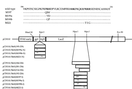
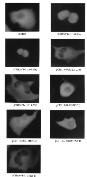
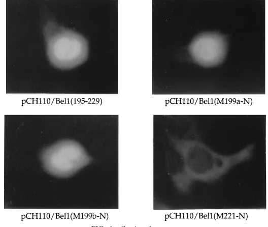
promoter. To construct pCH110/Bel1(184-236), the A nucleotide at position 9988 (all sequence positions refer to the full-length proviral HFV DNA; 28) of the bel1 gene in pRc/CMVBel1 was changed to C by site-directed mutagenesis to create an HpaI site. The plasmid DNA was then digested with HpaI and NcoI and the end was made flush with T4 DNA polymerase. The 150-bp DNA fragment was isolated by electroelution from a 4% polyacrylamide gel and was replaced with the HpaI fragment (624 bp) of vector pCH110 (Fig. 1A). pCH110/ Bel1(201-236) and pCH110/Bel1(214-236) were constructed by the same proce-dure, except that the inserts were the HincII-NcoI fragment (105 bp) of pRc/ CMVBel1-M199a (26) and the HincII-NcoI fragment (66 bp) of pRc/CMVBel1-M213 (26), respectively. To construct pCH110/Bel1(192-226), the Bel1 sequences from amino acids 192 to 226 were amplified with two primers (59-TTCTAC CAGTTGCCTCGAGGGTCCA-39and 59-AAGCACATGACTCGAGATCGA TGGATC-39; [the XhoI recognition sequence is underlined]) and then digested with XhoI. The resulting DNA fragment was cloned in frame into the HpaI site of pCH110 (Fig. 1A). Several derivatives of pCH110/Bel1(192-226), except pCH110/Bel1(M197-I), were also generated by the same procedure from the pRc/CMVBel1 derivatives (26; Fig. 1A). pCH110/Bel1(M197-I) was constructed by the same method following site-directed mutagenesis to change residues P-197, R-198, and P-199 to Q, I, and W, respectively, with primer 59-GACAG GATCGTGCCCAAGATCTGGTTTTGGACCTT-39. Suitable constructs were identified by both DNA sequencing and Western blotting (immunoblotting) with a monoclonal anti-b-gal antibody (Boehringer Mannheim Biochemicals). To construct amino-terminal chimeric plasmids, the region of Bel1 from residues 195 to 229 and its missense mutant derivatives (26) were amplified by PCR with primers 59-AGTTGCTCAGAGGTACCAAAACCAAGACCT-39 and 59-TGT CACTACTGGGTACCCATGACTCATTATC-39 (the KpnI recognition se-quence is underlined) and then digested with KpnI. The resulting fragments were cloned into the unique KpnI site of pCH110 (Fig. 1A). A synthetic oligonucle-otide (59-TGATGCTTTGTTAACATGTCACAG-39[the HpaI recognition se-quence is underlined]) complementary to nucleotides 10048 to 10071 of the bel1 gene was used to create a novel HpaI site at nucleotide 10057 to generate pRc/CMVBel1-M, resulting in the conversion of F-209 and D-210 to L and T, respectively. The 12-mer phosphorylated linkers (SalI, EcoRI, HindIII, and XbaI FIG. 1. (A) Schematic diagram of chimeric Bel1–b-gal expression vectors. The simian virus 40 early promoter of pCH110 directs high-level expression of a chimeric protein composed of 40 residues of Escherichia coli gpt and 20 residues derived from trpS fused to the lacZ-encodedb-gal protein. A series of internal Bel1–b-gal fusion vectors were constructed by replacing various regions of the Bel1 and mutant derivatives with the 624-bp HpaI-HpaI DNA fragment of pCH110. The amino-terminal chimera plasmids were constructed by inserting PCR-amplified DNA fragments of Bel1 into the KpnI site of pCH110. The amino acid sequences of Bel1 and mutant proteins encoded by these chimeric expression vectors are presented at the top in the single-letter code, and residues important for Bel1 nuclear targeting, identified by point mutational analysis, are in boldface. Amino acid residues of Bel1 in the chimeric constructs are in parentheses. HpaI recognition sites were removed by insertion of bel1 DNA fragments. (B) Immunofluorescence images of wild-typeb-gal and internal Bel1–b-gal fusion derivatives. (C) Immunofluorescence images of amino-terminal Bel1–b-gal chimeras.
802 CHANG ET AL. J. VIROL.
on November 9, 2019 by guest
http://jvi.asm.org/
[image:2.612.104.529.75.362.2]linkers; New England Biolabs) were inserted into the HpaI site of pRc/CMV Bel1-M to generate pRc/CMVBel1-M-SalI, pRc/CMVBel1-M-HindIII, pRc/ CMVBel1-M-EcoRI, and pRc/CMVBel1-M-XbaI, respectively. The correct in-frame sequences of these plasmids were confirmed by DNA sequencing and immunoblotting with rabbit anti-Bel1 serum (26).
Construction of Gal4-Bel1 fusion plasmids.Gal4-Bel1 derivatives were
[image:3.612.171.455.65.639.2]gen-erated by inserting various bel1 DNA fragments obtained by appropriate restric-tion enzyme digesrestric-tions and PCR into the SmaI site of pSG424 (9). Gal4-Bel1(1-260), Gal4-Bel1(1-226), Gal4-Bel1(1-82), and Gal4-Bel1(82-150) were previously described (26). Bel1(1-201), Bel1(1-196), Bel1(1-180), Gal4-Bel1(1-173), Gal4-Bel1(1-150), Gal4-Bel1(1-123), Gal4-Bel1(1-73), Gal4-Bel1 (1-54), Gal4-Bel1(1-43), and Gal4-Bel1(1-31) were constructed by inserting the FIG. 1—Continued.
VOL. 69, 1995 NLS AND OLIGOMERIZATION DOMAINS OF HFV Bel1 803
on November 9, 2019 by guest
http://jvi.asm.org/
PCR-amplified DNA fragments into the SmaI site of pSG424. An SmaI site was generated in a 59primer to facilitate in-frame fusion of the construction (59-AG ATTGTATCCCGGGTTCCTACGAAAA-39[the SmaI recognition sequence is underlined]). Several 39 primers were designed for generation of various carboxy terminus deletion mutants (201, 59-ACAGCGAAGGACCATATGGT GCCTAGG-39; 196, 59-CAGGTTTTGGTTCATGATCCGTGCTAG-39; 180, 59-TTAACAAGAGATCTGTTTCTTTGAT-39; 173, 59-AATAAACACAGCA GATCCAAC-39; 150, 59-GGGTATTCTACCCGTCGACTTCAACCTTA-39; 123, 59-TTCCCAAGAAACTGTAAAAGG-39; 73, 59-AGGATGTTTGTCCT CAATTTC-39; 54, 59-TTTGGTATATCGTCTGGGGCG-39; 43, 59-TTCAGGT TCCTCAGCAATAGT-39; 31, 59-CCTCTAGAGCATTTTCAGGGCCAAC-39). Gal4-Bel1(1-18) was obtained by partial digestion of Gal4-Bel1(1-54) with PstI and then religation. Gal4-Bel1(42-82) and Gal4-Bel1(82-120) were constructed by inserting a KpnI-BamHI fragment and an ApaI-BamHI fragment from pRc/ CMVBel1-M40 and pRc/CMVBel1-M120 (26) into the SmaI site of pSG424, respectively. Gal4-Bel1(112-150) and Gal4-Bel1(150-260) were also generated by inserting the PCR-amplified DNA fragments into the SmaI site of pSG424 (112sense, 59-AAGGATCCCTTTTACAGTTTCTTG-39; 150sense, 59-CTGAA TTCGGAATTTGGGTAAAAA-39; 260antisense, 59-AAGTAGCCCTGATAG TAGCGGTCC-39). pSbel1-S and Bel1(1-260)VP16 were previously described (26). pMC1, encoding herpes simplex virus type 1 VP16 amino acids 1 to 490, was previously described (1).
Cell culture, transfection, and chloramphenicol acetyltransferase (CAT) as-says.COS-7 and BHK-21 cells were grown in Dulbecco’s modified Eagle’s medium containing 10% fetal calf serum. For indirect immunofluorescence, approximately 2 3105cells on a glass coverslip were transfected with the
indicated plasmid DNA by the DEAE-dextran method (34). Cells (106) were
plated on a 100-mm-diameter dish at 24 h before transfection and transfected with 2mg of each of the reporter and activator plasmids. The cells were harvested and assayed for CAT activity at 48 h after transfection. The difference in trans-fection efficiency was normalized by using a second reporter plasmid, pGL2 (Promega), containing the luciferase gene. Luciferase activity was measured with a luciferase assay kit (Promega) in accordance with the supplier’s recommenda-tion. CAT enzyme reactions were carried out for 2 h of incubarecommenda-tion. When the level of acetylation of chloramphenicol was more than 90%, the lysates were diluted for the reaction and the CAT values were corrected by the dilution factor. All of the CAT assay data reported are from points in the linear range of the assay.
Immunofluorescence.For indirect immunofluorescence, transfected cells were fixed and permeabilized at 48 h after transfection with 95% methanol in phos-phate-buffered saline at2208C for 10 min. Cells were then reacted with a 1:100 dilution of a mouse anti-b-gal monoclonal antibody (Boehringer Mannheim Biochemicals), followed by a 1:80 dilution of anti-mouse immunoglobulin–fluo-rescein (Boehringer Mannheim Biochemicals). When pRc/CMVBel1 derivatives were transfected, a 1:80 dilution of polyclonal anti-Bel1 rabbit serum (26) and a 1:80 dilution of anti-rabbit immunoglobulin G–fluorescein isothiocyanate (GIBCO BRL) were used as the primary and secondary antibodies, respectively. The cells were photographed on a Carl Zeiss microscope equipped for fluores-cent illumination at a magnification of3400 with Kodak Gold 400 film.
RESULTS
The Bel1 NLS is a bipartite motif and is sensitive to the
protein context.Previous reports have identified the Bel1 NLS
by several methods, such as deletion (39b), missense mutation
(13, 26), and Bel1–b-gal chimeric constructs (13). However,
there is a conspicuous discrepancy in the results. In one case, missense mutations introduced into Bel1 residues 199 and 200
(R-199 and H-2003VD or GP) abolished nuclear targeting of
Bel1 (26), while in another case, an M197 missense mutant
(R-197, P-198, and R-1993QIW) and a Bel1 deletion mutant
(D194-200) retained nuclear targeting activity (13, 39b). To
determine the precise peptide sequence within Bel1 that is sufficient to function independently as an NLS, expression
vec-tors containing several subregions of Bel1 fused tob-gal were
constructed and their ability to target a large cytoplasmicb-gal
protein to the nucleus was tested (Fig. 1A). We placed a
portion of Bel1 in an internal region ofb-gal since the Bel1
NLS, in its normal context, is located not at the amino- or carboxy-terminal end but in an internal region. The subcellular localization of these chimeric proteins was examined by indi-rect immunofluorescence staining of COS-7 cells transfected with each expression plasmid (Fig. 1B). pCH110 expressing
wild-typeb-gal was detected predominantly in the cytoplasm
with little nuclear accumulation (Fig. 1B). In contrast, chimeric proteins containing the region of Bel1 from either residues 184 to 236 or 192 to 226 were detected predominantly in the nu-cleus. However, chimeric proteins of pCH110/Bel1(201-236) and pCH110/Bel1(214-236) containing the single basic domain of Bel1 displayed a mostly cytoplasmic staining indistinguish-able from that of pCH110 (Fig. 1B). Correct expression of the fusion proteins was verified by Western blot analysis with an
anti-b-gal monoclonal antibody (data not shown). These
re-sults suggest that the region of Bel1 from residues 192 to 226 including two basic segments is required for a large cytoplas-mic protein to be localized to the nucleus.
[image:4.612.187.451.74.297.2]Since some deletion mutants could change the structure significantly enough to mask the NLS motif required for intra-or intermolecular interactions, we generated various substitu-tion mutant derivatives of pCH110/Bel1(192-226) (Fig. 1A).
FIG. 1—Continued.
804 CHANG ET AL. J. VIROL.
on November 9, 2019 by guest
http://jvi.asm.org/
pCH110/Bel1(M197-I), pCH110/Bel1(M199a-I), and pCH110/ Bel1(M199b-I), which have missense mutations in the first basic cluster of the Bel1 NLS, showed severely impaired ability to direct chimeric proteins to the nucleus (Fig. 1B). Interest-ingly, pCH110/Bel1(M197-I) has the same mutation as M197, which was previously reported to be localized in the nucleus of transfected COS cells (13). We think that the inconsistency resulted from the use of different constructs and conditions in
which the segment of Bel1 was fused to b-gal. As expected,
pCH110/Bel1(M221-I) was localized exclusively in the cyto-plasm (Fig. 1B). These results further confirmed that two dis-continuous basic regions of Bel1, from residues 199 to 223, are
absolutely required to direct cytoplasmicb-gal to the nucleus.
However, previous reports showed that the single basic amino acid cluster of Bel1 from residues 209 to 226 and/or 211 to 225 was both necessary and sufficient for nuclear localization of the Bel1 protein (13, 39b). Previous studies investigated the activity of the Bel1 NLS by placing the potential NLS regions
at the amino terminus of b-gal. To determine whether the
discrepancy between our results and other previous reports is due to the difference in the position of the NLS within the lacZ gene, we fused the region of Bel1 from residues 195 to 229 and
its derivatives to the amino terminus ofb-gal (Fig. 1A) and
then determined the subcellular localization of these Bel1–b
-gal chimeric proteins (Fig. 1C). As expected, pCH110/Bel1 (195-229), containing the wild-type Bel1 sequence, but not
pCH110/Bel1(M221-N), can directb-gal to the nuclei of
trans-fected cells. In contrast, pCH110/Bel1(M199a-N) and pCH110/ Bel1(M199b-N) are localized preferentially in the nuclei of transfected cells, which is consistent with earlier reports (13). These results suggest that the second basic cluster of the Bel1 NLS itself is sufficient to function as a minimal nuclear local-ization signal only when it is located at the amino terminus of
b-gal, indicating that the ability of the Bel1 NLS is dependent
on the protein context within which it is present.
[image:5.612.118.446.77.447.2]Effects of linker insertion between the two basic clusters. Sequence comparison of the Bel1 NLS with other bipartite NLSs suggests that the NLS of the Bel1 protein is composed of a bipartite basic amino acid motif (Table 1). It was reported that the correct positioning of the two basic amino acid motifs could be essential for efficient nuclear targeting, whereas the precise sequence of the spacer region might not be important (6, 32, 36). To investigate whether the activity of the Bel1 NLS is affected by alteration of the length of the spacer between the two basic clusters, we introduced an HpaI site between two basic domains by site-directed mutagenesis to construct pRc/
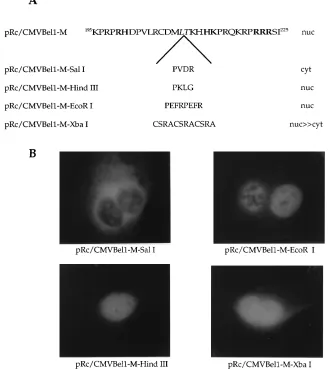
FIG. 2. Structure and immunofluorescence of mutant Bel1 proteins with linker insertions. (A) Sequences of Bel1 from residues 195 to 225 and pRc/CMVBel1-M linker insertion mutants. Phosphorylated synthetic linkers (12 bp) containing restriction endonuclease cleavage sites were inserted in frame into pRc/CMVBel1-M. The subcellular locations of the Bel1 proteins are given on the right. nuc, nucleus; cyt, cytoplasm. (B) Immunofluorescence images of the mutants indicated.
VOL. 69, 1995 NLS AND OLIGOMERIZATION DOMAINS OF HFV Bel1 805
on November 9, 2019 by guest
http://jvi.asm.org/
CMVBel1-M and then inserted several synthetic linkers into the HpaI site (Fig. 2A). As expected, pRc/CMVBel1-M was concentrated in the nuclei of transfected cells. Insertion of the
amino acid sequence PVDR (59-CCGGTCGACCGG-39; SalI
linker) caused cytoplasmic accumulation of Bel1 (Fig. 2B). In contrast, insertion of single or double repeats of other linkers (PKLG [pRc/CMVBel1-M-HindIII] and PEFRPEFR [pRc/ CMVBel1-M-EcoRI]) to give additional spacer lengths of four and eight amino acids had no effect on the nuclear targeting of Bel1. When the spacer length was increased by insertion of 12 amino acids (CSRACSRACSRA [pRc/CMVBel1-M-XbaI]), predominant nuclear staining with slight cytoplasmic accumu-lation was observed (Fig. 2B). These results indicate that the spacer region of the Bel1 NLS is moderately tolerant of inser-tion mutainser-tions, as is nucleoplasmin (36), but not when their separation is extended by a certain specific amino acid se-quence, such as PVDR.
The Bel1 protein forms multimeric complexes in the nuclei
of mammalian cells.Recently, it has been reported that several
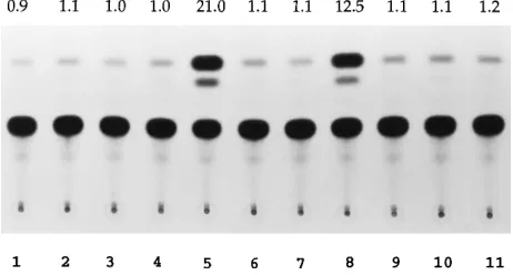
transactivators of human retroviruses, such as the Tat and Rev proteins of human immunodeficiency virus type 1 and the Rex protein of human T-cell leukemia virus type 1, form multimeric complexes in the nuclei of eukaryotic cells (2a, 2b, 42). To investigate whether the Bel1 protein functions as a monomer or an oligomer, like human immunodeficiency virus type 1 Tat and Rev, an in vivo protein-protein interaction assay was per-formed. We constructed a Gal4-Bel1(1-260) chimeric plasmid that contains the amino-terminal 147 residues of Gal4 to which Bel1 residues 1 to 260 were fused and fusion plasmid Bel1(1-260)VP16, which expresses a fusion protein consisting of the Bel1 sequence from residues 1 to 260 and the herpes simplex virus type 1 VP16 transcriptional activation domain (amino acids 423 to 490). The various expression vectors were trans-fected into BHK-21 cells, either alone or in combination but always in the presence of CAT reporter plasmid G5E1bCAT (3). Effective protein-protein interaction by Bel1 is predicted to spatially juxtapose the Gal4 DNA-binding domain and VP16 transcriptional activation domain encoded by the differ-ent plasmids, which in turn leads to transcriptional activation of the G5E1bCAT reporter gene, which contains Gal4-binding sites in the promoter. As shown in Fig. 3, expression of pSG424 containing a DNA-binding domain (amino acids 1 to 147) of Gal4, Gal4-Bel1(1-260), Bel1(1-260)VP16, pMC1, or pSbel1-S containing the entire bel1 open reading frame alone produced no significant activation of G5E1bCAT. However, coexpres-sion of Gal4-Bel1(1-260) with Bel1(1-260)VP16 or pSbel1-S resulted in stimulation of CAT gene expression by about
21-and 12-fold, respectively (Fig. 3, lanes 5 21-and 8), while cotrans-fection of pSG424 and Bel1(1-260)VP16 or pSbel1-S did not increase G5E1bCAT-derived gene expression (Fig. 3, lanes 4 and 7). The significant increases in CAT gene expression ap-pear to reflect a specific Bel1-Bel1 protein interaction in vivo. The higher level of CAT activity obtained with Bel1(1-260)VP16 than with pSbel1-S may be due to the difference in the strength of the transcriptional activation domain between VP16 and Bel1. To examine whether the Bel1-Bel1 interaction is specific, we cotransfected plasmid pMC1, expressing the entire domain of VP16, with Gal4-Bel1(1-260). Cotransfection of pMC1 and Gal4-Bel1(1-260) failed to produce significant stimulation of the G5E1b promoter (Fig. 3, lane 11), indicating that the Bel1 protein did not interact with VP16 in vivo. These results suggest that the Bel1 protein forms a multimeric com-plex in the nuclei of mammalian cells.
Multiple domains are involved in the oligomerization of
Bel1.To identify the sequences required for multimerization of
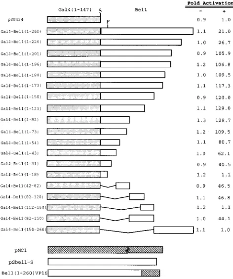
[image:6.612.57.300.93.200.2]Bel1, we generated a batch of Gal4-Bel1 chimeric proteins by introducing a series of deletions toward the amino-terminal side starting at residue 260 by using appropriate restriction enzymes or PCR and tested their ability to form multimers by assaying transient cotransfection into BHK-21 cells (Fig. 4). Deletion of residues 260 to 227 from Bel1 produced a slight increase in the efficiency of multimerization. Interestingly, fur-ther deletion of residues 226 to 202 from Bel1 produced an increase in CAT activity of about fourfold. This suggests that the region from residues 202 to 226 contains a negative regu-latory region which inhibits multimer formation with the Bel1 protein. This result is partially consistent with our recent find-ing that Bel1 contains a negative regulatory region from res-idues 153 to 226 which down-regulates the transactivation domain of Bel1 (26) if multimerization is an absolute require-ment of transcriptional activation. Further successive deletions from residue 201 to residues 197, 181, 174, 151, 124, and 83 did not show any significant effect on the multimerization of Bel1, indicating that the region of Bel1 from residues 1 to 82 still contains sufficient information to form a multimeric complex with Bel1. Deletion of Bel1 sequences from this point to res-idues 74, 55, 44, and 32 resulted in slightly progressive reduc-tions in the efficiency of multimerization which may have been
[image:6.612.320.551.100.224.2]FIG. 3. The Bel1 protein forms a multimeric complex in nuclei of mamma-lian cells. Plasmids pSG424, pSbel1-S, pMC1, and G5E1bCAT are described in the text. G5E1bCAT and various effector plasmids were cotransfected into BHK-21 cells, and CAT activity was measured at 48 h after transfection as described in Materials and Methods. Lanes: 1, pSG424; 2, Gal4-Bel1(1-260); 3, Bel1(1-260)VP16; 4, pSG424 and Bel1(1-260)VP16; 5, Gal4-Bel1(1-260) and Bel1(1-260)VP16; 6, pSbel1-S; 7, pSG424 and pSbel1-S; 8, Gal4-Bel1(1-260) and pSbel1-S; 9, pMC1; 10, pMC1 and pSG424; 11, pMC1 and Bel1(1-260)VP16. TABLE 1. Alignment of nuclear targeting sequences in Bel1 with
the bipartite motifs of other nuclear proteins
Protein Species of origin
Posi-tiona Sequence
Nucleoplasmin X 155 KRpaatKKagqaKKKKl
N1/N2 X 534 KRKteeesplKdKdaKK
c-FOS H, M, R RReRnKmaaaKcRnRRR
c-JUN H, M KRmRnRiaasKcRKRKl
C/EBP R 275 KKsvdKnsneyRvRReR
C/EBP R 288 RReRnniavRKsRdKaK
GCN4 Y 230 KRaRnteaaRRsRaRKl
Bel1 H 199 RHdpvlRcdmfeKhHKpRqKRpRRR
a
Each number refers to the position of the first amino acid shown in the primary sequence of the protein. The single-letter amino acid code is used, basic amino acids are in uppercase, and all other amino acids are in lowercase. X, xenopus; H, human; M, mouse; R, rat; Y, yeast (Saccharomyces cerevisiae).
806 CHANG ET AL. J. VIROL.
on November 9, 2019 by guest
http://jvi.asm.org/
due to removal of the region required for optimal multimer-ization. In contrast, an additional deletion of residues 31 to 19 from Bel1 resulted in complete loss of in vivo multimerization activity. Therefore, the serial carboxy-terminal deletion analy-sis demonstrates that multimer formation with Bel1 was me-diated by the region from residues 1 to 82 and down-regulated by the region from residues 202 to 226.
Further deletion analysis revealed that Gal4-Bel1(42-82) has CAT activity comparable to that of Gal4-Bel1(1-31). This sug-gests that the regions of Bel1 from residues 42 to 82 and 1 to 31 are required for effective multimerization. Surprisingly, Gal4-Bel1(82-120) and Gal4-Bel1(82-150), but not Gal4-Bel1 (112-150), produced an increase in CAT activity of about 45-fold, indicating that the region of Bel1 from residues 82 to 111 also can mediate the protein-protein interaction in vivo. In contrast, Gal4-Bel1(150-260) did not show any detectable CAT activity, suggesting that the region of Bel1 from residues 150 to 260 is not required for multimer formation. The expression levels of Gal4-Bel1 derivatives were confirmed by Western blot analysis with an anti-Gal4 rabbit antibody which reacts to the DNA-binding domain of Gal4. The Western blot analysis re-sult did not show any significant difference in the protein levels of Gal4-Bel1 derivatives (data not shown). Taken together, the available data show that the Bel1 protein contains three re-gions required for effective multimerization of Bel1 (residues 1 to 31, 42 to 82, and 82 to 111) and one region (residues 202 to 226) required for down-regulation of Bel1 multimerization.
DISCUSSION
In this report, we have demonstrated that the nuclear tar-geting signal of Bel1 is bipartite, comprising two interdepen-dent clusters of basic amino acids separated by an intervening spacer. In addition, our results showed that the second basic
cluster is sufficient to target the Bel1–b-gal chimeric
polypep-tide to the nucleus when it is located at the amino terminus of
lacZ but not when it is in an internal region. When the basic
cluster is located at the amino terminus of the chimeric pro-tein, it may be free enough to interact with factors involved in nuclear transport. Presumably, an amino acid sequence as ba-sic as the NLS would normally tend to be exposed at the hydrophilic surface of a protein. The second basic cluster thus
appears to be sufficient to direct Bel1–b-gal chimeric proteins
to the nucleus. These results indicate that the activity of the Bel1 NLS is dependent on the protein context within which it is present. It is likely that both basic clusters located in an internal region of a chimeric protein are required to act di-rectly in some steps involved in nuclear transport, or one of the two basic regions may be indirectly involved in providing the proper structural conformation for presenting the ‘‘true’’ sig-nal. It was reported that the capacity of the NLS to direct cytoplasmic proteins to the nucleus depends on the chimeric protein context (33, 37). Therefore, the conclusion that the Bel1 NLS is bipartite appears to be more justified since it is located in the internal region in the natural context of Bel1.
A sequence comparison identified similar compositions of a bipartite nucleoplasmin-like motif between Bel1 and a number of nuclear proteins (Table 1). The typical bipartite NLS con-sists of 2 basic residues followed by a spacer of 10 other resi-dues and then a second cluster in which 3 of 5 resiresi-dues are basic. Insertional mutagenesis of Bel1 showed that the spacer length can be increased to 14, 18, or 22 amino acids without significantly affecting nuclear targeting, indicating that there is no strict requirement for spacer length. However, we observed that the activity of the Bel1 NLS was abolished when a specific amino acid sequence, PVDR, was inserted into the spacer region. Similar results were reported in an earlier study in which the spacer length of the nucleoplasmin NLS was altered without disrupting the nuclear targeting activity, yet targeting was abolished by insertion of only one copy of the sequence QPWL (36). It is likely that inappropriate folding of the nu-clear targeting sequence by insertion of a specific foreign se-quence into the spacer region impairs the ability of the protein to interact with relevant cellular factors required for nuclear transport. Thus, efficient nuclear targeting presumably re-quires correct positioning of the two basic elements relative to each other.
[image:7.612.60.296.72.354.2]We have used an in vivo assay of protein-protein interaction (10) to assess the potential of the Bel1 protein to form specific multimers in the nuclei of mammalian cells. The results pre-sented in this report demonstrate that Bel1 can indeed form a multimeric complex, and the regions mediating the multimer-ization were mapped to three subdomains (residues 1 to 31, 42 to 82, and 82 to 111). Furthermore, the region from residues 202 to 226 negatively regulates multimer formation. It has been reported that several proteins, such as Myc, E12, and AP4, possess more than one dimerization domain and these multiple dimerization domains regulate dimer specificity (14, 21, 30). The presence of multiple domains in Bel1 for oligomerization could be expected to give specificity of multimer formation. In fact, the region of Bel1 from residues 1 to 88 is shared with Bet, a cytoplasmic protein expressed at a very high level in HFV-infected cells; Beo; and Bel3 (31). These facts indicate that the region of Bel1 from residues 82 to 111 may play a specific role
FIG. 4. Identification of regions affecting the multimerization of Bel1. A series of amino- and carboxy-terminal deletion mutant Bel1 proteins were gen-erated by using the appropriate restriction enzymes and PCR and then were fused in frame downstream of the Gal4 DNA-binding domain of pSG424. The ability of each Gal4-Bel1 fusion protein to multimerize with the Bel1-VP16 fusion protein expressed by the Bel1(1-260)VP16 plasmid was determined by assaying transient cotransfection into BHK-21 cells. pRc/CMV was used as a negative control (2) instead of Bel1(1-260)VP16 (1). The Gal4 DNA-binding domain, a part of Bel1, and part or all of VP16 are represented by the stippled, open, and hatched boxes, respectively. S, SmaI; P, PstI.
VOL. 69, 1995 NLS AND OLIGOMERIZATION DOMAINS OF HFV Bel1 807
on November 9, 2019 by guest
http://jvi.asm.org/
in multimeric complex formation with the Bel1 protein. This is supported by the fact that Gal4-Bel1(82-150), which contains both the positive regulatory domain and the multimerization subdomain of Bel1, effectively inhibits the native function of Bel1 as determined by an in vivo competition assay, whereas Gal4-Bel1(1-82) cannot (26). While the amino-terminal 55 res-idues were shown to be dispensable for activation (39a), the 82-to-120 region is included in the essential effector region (26, 39b). Although the data reported here demonstrate that HFV Bel1 can form specific multimers in the nuclei of mammalian cells, we do not know whether in vivo multimerization involves a bridging cellular factor. Also, it remains to be determined whether oligomerization of Bel1 is important for the transac-tivation function.
ACKNOWLEDGMENTS
We are grateful to Y. S. Lee for providing the microscope and technical advice.
This work was supported by grant BM-9411-23 from the Center for Biofunctional Molecules.
REFERENCES
1. Ace, C. I., M. A. Dalrymple, F. H. Ramsay, V. G. Preston, and C. M. Preston. 1988. Mutational analysis of the herpes simplex virus type 1 trans-inducing factor Vmw65. J. Gen. Virol. 69:2595–2605.
2. Bogerd, H. P., R. A. Fridell, W. S. Blair, and B. R. Cullen. 1993. Genetic evidence that the Tat proteins of human immunodeficiency virus types 1 and 2 can multimerize in the eukaryotic cell nucleus. J. Virol. 67:5030–5034. 2a.Bogerd, H. P., and W. C. Greene. 1993. Dominant negative mutants of
human T-cell leukemia virus type 1 Rex and human immunodeficiency virus type 1 Rev fail to multimerize in vivo. J. Virol. 67:2496–2502.
3. Cathy, Q. L., Y. Yun, J. P. Hoeffler, and J. F. Habener. 1990. Cyclic-AMP-responsive transcriptional activation of CREB-327 involves interdependent phosphorylated subdomains. EMBO J. 9:4455–4465.
4. Chen, J., C. Panagiotidis, and S. Silverstein. 1992. Multimerization of ICP0, a herpes simplex virus immediate-early protein. J. Virol. 66:5598–5602. 5. Davey, J., N. J. Dimmock, and A. Coleman. 1985. Identification of the
sequence responsible for nuclear accumulation of the influenza virus nucleo-protein in Xenopus oocytes. Cell 40:667–675.
6. Dingwall, C., and R. A. Laskey. 1991. Nuclear targeting sequences—a con-sensus? Trends Biochem. Sci. 16:478–481.
7. Dingwall, C., J. Robbins, and S. M. Dilworth. 1989. Characterization of the nuclear location sequence of Xenopus nucleoplasmin. J. Cell Sci. 11(Suppl.): 243–248.
8. Dingwall, C., J. Robbins, S. M. Dilworth, B. Roberts, and W. D. Richardson. 1988. The nucleoplasmin nuclear location sequence is larger and more com-plex than that of SV 40 large T antigen. J. Cell Biol. 107:841–849. 9. Fields, S., and S. K. Jang. 1990. Presence of a potent transcription activating
sequence in the p53 protein. Science 249:1046–1049.
10. Fields, S., and O. K. Song. 1989. A novel genetic system to detect protein-protein interaction. Nature (London) 340:245–246.
11. Flu¨gel, R. M.1991. Spumaviruses: a group of complex retroviruses. J. Ac-quired Immune Defic. Syndr. 4:739–750.
12. Goldfarb, D. S., J. Gariepy, G. Schoolnick, and R. D. Kornberg. 1986. Synthetic peptides as nuclear localization signals. Nature (London) 322:641– 644.
13. He, F., J. D. Sun, E. D. Garrett, and B. R. Cullen. 1993. Functional organi-zation of the Bel1 transactivator of human foamy virus. J. Virol. 67:1896– 1904.
14. Herkowitz, I. 1987. Functional inactivation of genes by dominant negative mutations. Nature (London) 329:219–222.
15. Hu, Y.-F., B. Lusher, A. Admon, N. Mermod, and R. Tjian. 1990. Transcrip-tion factor AP-4 dimerizaTranscrip-tion domains that regulate dimer specificity. Genes Dev. 4:1741–1752.
16. Jones, N. 1990. Transcriptional regulation by dimerization: two sides to an incestuous relationship. Cell 61:9–11.
17a.Kalderon, D., W. D. Richardson, A. T. Markham, and A. E. Smith. 1984. Sequence requirements for nuclear location of simian virus 40 large T anti-gen. Nature (London) 311:33–38.
17b.Kalderon, D., W. D. Richardson, A. T. Markham, and A. E. Smith. 1984. A short amino acid sequence able to specify nuclear location. Cell 39:499–509. 18a.Keller, A., E. D. Garrett, and B. R. Cullen. 1992. The Bel1 protein of human foamy virus activates human immunodeficiency virus type 1 gene expression via a novel DNA target site. J. Virol. 6:3946–3949.
18b.Keller, A., K. M. Partin, M. Lo¨chelt, H. Bannert, R. M. Flu¨gel, and B. R. Cullen.1991. Characterization of the transcriptional trans activator of hu-man foamy retrovirus. J. Virol. 65:2589–2594.
19. Kerppola, T. K., and T. Curran. 1991. Transcription factor interactions; basics on zippers. Curr. Opin. Struct. Biol. 1:71–79.
20. Kleinschmidt, J. A., and A. Seiter. 1988. Identification of domains involved in nuclear uptake and histone binding of protein N1 of Xenopus laevis. EMBO J. 7:1605–1614.
21. Lamb, P., and S. L. McKnight. 1991. Diversity and specificity in transcrip-tional regulation: the benefits of heterotypic dimerization. Trends Biochem. Sci. 16:417–426.
22. Landschulz, W. H., P. F. Johnson, and S. L. McKnight. 1988. The leucine zipper: a hypothetical structure common to a new class of DNA binding proteins. Science 240:1759–1764.
23. Lanford, R. E., and J. S. Butel. 1984. Construction and characterization of an SV 40 mutant defective in nuclear transport of T antigen. Cell 37:801–813. 24. Lanford, R. E., P. Kanda, and R. C. Kennedy. 1986. Induction of nuclear transport with a synthetic peptide homologous to the SV 40 T antigen transport signal. Cell 46:575–582.
25. Lee, A. H., K. J. Lee, S. Kim, and Y. C. Sung. 1992. Transactivation of human immunodeficiency virus type 1 long terminal repeat-directed gene expression by the human foamy virus bel1 protein requires a specific DNA sequence. J. Virol. 66:3236–3240.
26. Lee, C. W., C. Jun, K. J. Lee, and Y. C. Sung. 1994. The Bel1 protein of human foamy virus contains one positive and two negative control regions which regulate a distinct activation domain of 30 amino acids. J. Virol. 68:2708–2719.
27. Lee, K. J., A. H. Lee, and Y. C. Sung. 1993. Multiple positive and negative
cis-acting elements that mediate transactivation by Bel1 in the long terminal
repeat of human foamy virus. J. Virol. 67:2317–2326.
28. Lo¨chelt, M., H. Zentgraf, and R. M. Flu¨gel.1991. Construction of an infec-tious DNA clone of the full-length human spumaretrovirus genome and mutagenesis of the bel1 gene. Virology 184:43–54.
29. McIntyre, M. C., M. G. Frattini, S. R. Grossman, and L. A. Laimins. 1993. Human papillomavirus type 18 E7 protein requires intact Cys-X-X-Cys mo-tifs for zinc binding, dimerization, and transformation but not for Rb bind-ing. J. Virol. 67:3142–3150.
30. Mordacq, J. C., and D. I. H. Linzer. 1989. Co-localization of elements required for phorbol ester stimulation and glucocorticoid repression of pro-liferin gene expression. Genes Dev. 3:760–769.
31. Muranyi, W., and R. M. Flu¨gel.1991. Analysis of splicing patterns of human spumaretrovirus by polymerase chain reaction reveals complex RNA struc-tures. J. Virol. 65:727–735.
32. Nath, S. T., and D. P. Nayak. 1990. Function of two discrete regions is required for nuclear localization of polymerase basic protein 1 of A/WSN/33 influenza virus (H1N1). Mol. Cell. Biol. 10:4139–4145.
33. Nelson, M., and P. Silver. 1989. Context affects nuclear protein localization in Saccharomyces cerevisiae. Mol. Cell. Biol. 9:384–389.
34. Queen, C., and D. Baltimore. 1983. Immunoglobulin gene transcription is activated by downstream sequence elements. Cell 33:741–748.
35. Rethwilm, A., K. Mori, B. Maurer, and V. ter Meulen. 1990. Transacting transcriptional activation of human spumaretrovirus LTR in infected cells. Virology 175:568–571.
36. Robbins, J., S. M. Dilworth, R. A. Laskey, and C. Dingwall. 1991. Two interdependent basic domains in nucleoplasmin nuclear targeting sequence: identification of a class of bipartite nuclear targeting sequence. Cell 64:615– 623.
37. Roberts, B. L., W. D. Richardson, and A. E. Smith. 1987. The effect of protein context on nuclear location of signal function. Cell 50:465–475. 38. Smith, A. E., D. Kalderon, B. L. Roberts, W. D. Colledge, M. Edge, P. Gillett,
A. F. Markham, E. Paucha, and W. D. Richardson.1985. The nuclear localization signal. Proc. Trans. R. Soc. Lond. B 226:43–58.
39a.Venkatesh, L. K., and G. Chinnadurai. 1993. The carboxy-terminal tran-scription enhancement region of the human spumaretrovirus transactivator contains discrete determinants of the activator function. J. Virol. 67:3868– 3876.
39b.Venkatesh, L. K., C. Yang, P. A. Theodorakis, and G. Chinnandurai. 1993. Functional dissection of the human spumaretrovirus transactivator identifies distinct classes of dominant-negative mutants. J. Virol. 67:161–169. 40. Williams, T., and R. Tjian. 1991. Characterization of a dimerization motif in
AP-2 and its function in heterologous DNA-binding proteins. Science 251: 1067–1070.
41. Xia, Y.-P., and M. M. C. Lai. 1992. Oligomerization of hepatitis delta antigen is required for both the trans-activating and trans-dominant inhibitory activ-ities of the delta antigen. J. Virol. 66:6641–6648.
42. Zapp, M. L., T. J. Hope, T. G. Parslow, and M. R. Green. 1991. Oligomer-ization and RNA binding domains of the type 1 human immunodeficiency virus Rev protein: a dual function for an arginine-rich binding motif. Proc. Natl. Acad. Sci. USA 88:7734–7738.
808 CHANG ET AL. J. VIROL.