NATIONAL INSTITUTE OF SIDDHA
Chennai - 47AFFILIATED TO
THE TAMIL NADU DR. M.G.R. MEDICAL UNIVERSITY, CHENNAI - 600 032
A STUDY ON
AZHAL KEEL VAAYU
(DISSERTATION SUBJECT)
For the partial fulfillment of the
requirements to the Degree of
DOCTOR OF MEDICINE (SIDDHA)
ACKNOWLEDGEMENT
First of the author extremely grateful to Lord Almighty to empowered the author with his blessings and grace to complete this dissertation work successfully.
The author is extremely grateful to the almighty for the successful completion of this dissertation work,.
The author expresses here sincere thanks to the Vice – Chancellor, The Tamilnadu Dr. M.G.R. Medical University, Chennai-32..
The author extend her sincere thanks to Dr. V. Arunachalam M.D.(s), Director, National Institute of Siddha, Chennai, for granting permission to undertake a study in this dissertaion topic and also for providing all the basic facilities in order to carry out this work.
The author is very much grateful to Dr. G.Thiagarajan, M.D(s).,Associate Prof., and Head of the Department of Sirappu Maruthuvam, National Institute of Siddha, Chennai for his encouragement, suggestion and valuable guidance in this dissertation work.
The author is grateful to Dr.R.S.Ramaswamy, M.D(s), Associate Professor, Department of Sirappu Maruthuvam, National Institute of Siddha, Chennai for his valuable guidance and support in this dissertation work.
The author is grateful to Dr.T.R. Siddique Ali, M.D(s), Lecturer, department of Sirappu Maruthuvam, National Institute of Siddha, Chennai for his guidance and support in this dissertation work.
The author owes special thanks to Dr.k.v.Chandrasekaran M.B.B.S.,D.Ortho, M.S. Ortho Head of the Department of Orthopaedics, Dr.P.Thirunavukarasu M.B.B.S D.Ortho, Professor, Dr.M.Suresh babu M.B.B.S., M.S. Ortho, Tutor,
Dr.R.Sundarapandian, M.B.B.S., D.Ortho, Tutor of the Department of Orthopaedics, Chengalpattu Medical College Hospital.
The author exresses her thanks to Dr.D. Porselvi M.B.B.S., D.M.R.D. Department of Radiology, Chengalpattu Medical College Hospital.
The author expresses thanks to Dr.S.Venkatraman Ph.D., Director, C.L. Baid Metha College of Pharmacy, Thoraipakkam, Chennai – 4
I wish to thank Dr. S. Somasundaram, M.Sc, M.Phil, Ph.D., Asst. Professor, Department of Medicinal Botany for his guidance and help in identification of herbs in this dissertation work.
I also thank and Mr. Rathnam, and Mrs. M. Vimala, M.Lib, National Institute of Siddha,Chennai for their help in literature collection.
I wish to thank my friend Dr. M.Tamilselvi M.D (S) for her heartful help.
I also thank to my collegue and gratitude for their selfless help in this study.
INTRODUCTION
Man is a wonderful creature blessed by God. Imagination and laughter are milestones on the way that distinguishes mn from other animals. Of mere living, no man was ever proud; but the god life has always been his aim.
The aim of the History of Siddha Medicine is to present a faithful, clear and vivid picture of this system in all its manifestation and ramifications with all its inherent problems and relevancy to present age from its very beginning down the ages, as an integral component of the patterns of culture through which this system has passed in different ages and in different areas.
The unique nature of this system is its continuoes service to humanity for more than five thousand years in combating diseases and in maintaining its physical, mental, and moral health, while many of its contempraries had completed their courses long long age, since its origin, development and remifications have become obscure, any literary research on this subjective, to be scientific and useful, should commence with a comparative study of the medicines of those ancient civilizations, which will illuninate many of the dark cornersmof our systems.
Many principles on which very efficacious siddha medicines are prepared and treatments given, make us believe they had a detailed understanding of many of the fundamentals of biology, such as how enzymes work, how proteins perform mechanical, protective and catalytic function, and how proteins perform mechanical, protective and catlytic functions, and how glucose is metabolised for energy. The knowledge of the function of the enzymes Siddhas put into practice, is really marvellous when we consider the different ordinarilly inexplicable effective simple treatments of this system.
These medicines may be roughly divided into three classes as (1) Miracle medicines, (2) sophisticated medicines, and (3) Common medicines. Miracle medicines are becoming rare and should be learned direct from the masters who, having undergone all forms of initiation and hazards of apprenticeship, have reached perfection in all respects. Sophisticated medicines may be scientifically prepared and used by the well trained physicians without much risk. Common medicines are the most simple and very cheap things which were in wide use till the beginning of this century and are still in use in remote rural parts of our country.
Man is hale and healthy when his life moves along with nature when he violates against it he is oushed into mental stress, detriorates his physique and ultimately his sound mind.
In the legend of science the only system with rich Dravidian culture is the noble Siddha System of Medicine. It is holistic and treats the complete individual and not merely the disease. The treatment vries for different individuals based on their constitution (Prakriti) and dietary habits.
It is pround and pleasure for the author to be in this field and to take up the dissertaion on the disease that leads to many patients in despair.
AIM AND OBJECTIVES
Azhal keel Vaayu (Osteoarthritis) is a very common condition affecting the joints more frequently with aging. It’s a major cause of morbidity of the working force, throughout the world.
The extrapolation rate of osteoarthritis in our country is 78, 341,013 and the estimated population is 1, 065, 070, 607 (US census bureau, international Date Base – 2007)
So the author is much interested in choosing this disease as the topic for dissertation and treating the same with the help of Mirudharsingi Chunnam–65mg–twice a –day internally and poochu ennai externally.
The aim and objective of this dissertation, is stated as below.
1. Siddha system of medicine should reach the entire society of the world.
2. The unique aspect of Siddha principles namely the three dosham theories with respect of body constitution (yaakai), taste (suvai) and seasonal variation (Paruvakalam) are interpreted with the disease.
3. Relevant evidence from various Siddha literature and other system of medicine to be attached.
5. To know the correlation of aetiology, clinical features, signs and symptoms of Azhal keel vaayu in siddha aspect with osteoarthritis in modern aspect.
6. To have a detailed clinical investigations.
7. To have a clinical trial on Azhal keel Vaayu with Mirudharsingi Chunnam internally and poochu ennai externally.
8. To estimate the efficacy of Mirudharsingi Chunam and Poochu ennai
9. To evaluate the biochemical, pharmacological and toxicological reports of dissertation medicine.
SIDDHA ASPECTS
Siddhars, spiritual scientists explored and explained the reality of Nature and its relationship to man by their yogic awareness. According to Siddha philosophy, man is nothing but a miniature world containing the five basic elements.
Universe originally consisted of atoms which contributed to the five basic elements (Pancha boothas) namely, Earth, Water, Fire, Air and Ether which corresponds to the five sense of the human body and they were the fundamentals of all human body and all the corporal things.
The Earth (l{<*!gives shape to the body and release its energy, Bones, muscles, nerves represent it in the body.
The Water (fQI*!makes the earth supply and helps in the transmission of energy, serum, lymph, saliva, etc., represent it in the body.
The Fire (kQ*!makes the form of the body steady and gives vigour and stimulation. Digestion and circulation represent it in the body.
The Air (utq*!Ignites the fire and works as a life carrier and is the support of all contact and exchange. Respiratory and nervous system represent it in the body.
Man has gross physical body ( ^<K~zl<*!and subtle physical body (S,g<Gll<*/!!The life force which is different from material energy derived from food, pervades the gross physical through the subtle physical.
The food we eat has six tastes namely Sweet (-eqh<H*, Sour )Htqh<H*, Salt! )dh<H*,!
Bitter (jgh<H) , Pungent!)giIh<H*, Astringent (KuIh<H*/!
Each of them is a mixture of two basic elements.
-eqh<H!! .! l{<!!+ fQI Htqh<H!! .! l{<!+kQ dh<H! ! .! fQI!+ kQ
jgh<H!! .! gix<X!+!Ngibl< KuIh<H! .! l{<!+ Ngibl<
Panchboothas are the foundations for Three dosham Vaatham, Piththam, Kabam) which are the pillars that support our body structure
Vaayu Constitute Vaatham
Theyu Constitute Piththam
Appu Constitute Kabam
Any alterations in the level of mukkuttrams affects the normal functions of the body. This is obvious from the verses,
! lqgqEl<!GjxbqEl<!Ofib<osb<Bl<!F~OziI!
! ! utqLkzi!ou{<{qb!&e<X!
! The normal values of the mukkuttrams are in the ratio of Vaatham : Piththam : Kabam= 1 : ½ : ¼
“upr<gqb!uikl<!lik<kqjv!obie<xqz<! ! kpr<gqb!hqk<kf<!ke<eqzjv!uisq!
! npr<Gr<!ghf<kiemr<gqOb!giOzicz<!
! hqxr<gqb!sQuIg<Gh<!hqs!ogie<Xlqz<jzOb”
! ! ! ! .!G{uigml<!
AZHAL KEEL VAAYU
In Siddha literature Azhal keel Vaayu comes under the topic of Vatha disease. Keel Vaayu is the general term that includes all kinds of joint disease (locomotor system)
Description of the nomenclature
Azhal keel Vaayu = Azhal + keel + Vaayu
Azhal = Piththam
Keel = Joint
Vaayu = Vaatham
Initially the joint is affected by the vitiated vaatham, kabam and piththam is accompanied later. Also this is a disease of Pitha kaalam middle 1/3 of the life span.
CLASSIFICATION:
In Siddha maruthuvam, keel vaayu is classified into ten types. 1. Vali keel Vaayu
Ofib<!uVl<!upq
!)Aetiology):!giz!-bz<H!!.!Environmental Factors:
uikuIk<!ke!gizOlOki!oue<eqz<!
! ! lVUgqe<x!Neq!gx<gm!likl<!
! Nkjeh<!hsqObiM!giIk<kqjg!ke<eqz<!
! ! NmVOl!lx<x!likr<gt<!ke<eqz<!
! OhigOu!slqg<gqe<x!gizliGl<!
! ! ! ! .!B,gq!sqf<kil{q!
! The Vaatha disease will be precipiated in the months from Aani to karthigai (June to December)
hKlk<jkh<!h,g<g!jug<Gl<!hiElqgg<!giBl<!
! LKOueq!zqx<H!uqx<fQI!Lx<Xl<!.!gKole!
! ux<Xl<!ghlq0Gl<!uiBlqGl<!uip<lif<kIg<!
! Gx<x!fzqg<!Ogkqoke<!OxiK!
! ! ! ! ! .!sqk<k!lVk<Kuir<g!SVg<gl<!
! In Muthuvenil kaalm, the increased solar radiation increases the evaporation of water content in the world, on the same time these similar action on the body produces increases absorption of mucous for digestion and develop the vitality of Vatha disease. So, this disease occurs predominantly in muthuvennil kaalam.
d{Uujggt<!.!Diet:
! uik! Gx<xk<jk! lqjgh<hMk<kg<! %cb! gqpr<G! ujggt<,! GtqIs<sqjb! kvg<%cb!hkiIk<kr<gt<!lqGkqbig!d{<{z<,!GtqIf<k!gix<xqz<!-Vk<kz<,!ljpbqz<! fjekz<! heqg<gix<X! OlOz! uqPkz<! ljzh<hqvOksr<gtqz<! usqk<kz<! ntU! gmf<K!
dmZxU!ogit<tz<!lx<Xl<!hvl<hjvbqe<!giv{ligUl<!Okie<Xl</!
! okipqz<!ohX!jgh<Hg<giIk<kz<!KuIk<kz<!uqr<SgqER<OsiXl<!
! ! hjpbkil<!uvG!lx<jxh<!jhf<kqjebVf<kqeiZl<!
! wpqz<!ohxh<!hgZxr<gq!-vuqeqZxr<gikkiZl<!
! ! ljp!fqgi!GpzqeiOz!uikr<Ogi!hqg<Gr<giO{/!
! ! ! ! ! ! .!hvvis!Osgvl<!
! gsh<H,! KuIh<H! giIh<H! hkiIk<kr<gjt! lqGkqbig! d{<{z<! hjpb! OsiX! uvG! kqj{! d{<{z<! hgzqz<! K~r<gq! -vU! uqpqk<kqVk<kz<! Ngqb!
giv{r<gtqeiZl<!uikl<!lqGhMl</!
hpg<g!upg<gr<gt<!)
Habits)
oub<bqzqz<!fmg<jgbiZl<!lqgk<k{<{QI!Gcg<jgbiZl<!
! osb<bqjp!lgtqejvs<!OsIf<kEh!uqg<jgbiZl<!
! jhbOe!d{<jlbiZl<!higx<gib<!kqe<jgbiZl<!
! jkbOz!uikOvigl<!seqg<Gole<!xxqf<K!ogit<Ot/!
! ! ! ! ! ! ! .!OkjvbI!uigml<!
uikOfib<g<gie!-bz<H;!
)!Characteristic features of Vaatha)uikOl!gkqk<k!OhiK!uiBUolPl<Hr<!gi{<CI!
! ! uikOl!gkqk<k!OhiK!uiBuf<kqMR<!se<eq!Oki]l<!
! ! uikOl!gkqk<k!OhiK!uz<zMe<!olzqf<K!ogiz<Zl</!
! ! ! ! ! ! .!ngk<kqbI!sqgqs<si!vk<ei!kQhl</!
! uikl<! lqGl<OhiK! uiB! lqGl</! se<eqOkiml<! Ohie<x! hz! uqbikqgt<! uf<K!
OsVl<!dmz<!olzqBl</!
! ! uikuQX!ne<elqxr<giK!gMh<H{<mil<!u{<{L{<mil<!
! ! OliKgm<G!Ovigl<!SvL{<mi!lqVlZli!Lxr<gioke<Xl<!
! ! Ykiqb!uikleziG!fMg<gL{<mil<!ohiVt<gtbIf<k!
! ! kQokeOu!fvl<hqk<K!sf<Kgt<!OkiXr<gmg<Gf<!kqeLf<kiOe!
! ! ! ! ! ! .!OkjvbI!uigml<!
! uikl<! lqGl<OhiK! hsqbqe<jl,! dmz<! gMh<H,! Svl<,! -Vlz<,! dxg<glqe<jl,! dmz<! fMg<gl<! fvl<Hk<ktIs<sq! sf<Kgt<! OkiXl<! Gjmkz<! Ohie<x! GxqG{r<gt<!
Ofib<!uVl<!upq!.!Lg<Gx<x!hikqh<H!
ubK!d{U!
hpg<g!upg<gl<!
Sx<Xs<!S,pz<!
hqk<kl<!.!
sikgl<!
uqbiee<!.!
uikl<!.!
nhiee<!
ghl<!.!sf<kqgl<!
Ofib<!GxqG{r<gt<!)
Clinical Features)
hqk<k!gQz<uiB!ke<eix<!gQz<Lm<MuQr<gqs<!
! sqk<kI!osb<lVk<Kus<!sQIhmik!
! kg<gX!gib<s<sz<!g{<M!sizOu!kjekie!kf<Ok!
! olk<kX!sqgqs<js!ke<ekz<!ole<Olz<!fQg<Glh<hi!
! ! ! ! ! .!shihkq!jgObM!
! -K,!utqg<Gx<xl<!ke<eqjzbqz<!lqGf<Kt<tOhiK!npz<!Gx<xk<jkk<!K~{<mg<! %cb!d{U,!osb<jg!Lkzqbux<xiz<!hqxg<Gl<!Ofibil</!!-f<Ofibqz<,!Lm<cgtqz<! d{<miGl<! uQg<gl<! fiTg<Gfit<! ohVk<Kg<! ogi{<Om! uf<K,! lqGf<k! kQg<Gx<xk<kiz<! gQz<gtqeqjmObBt<t! hjs! ux{<M! hjsbx<Xg<! gQz<! njsBl<! Ohiokz<zil<!
fm<jmBjmkZl<! #gZg<#! #gZg<#! oge<x! YI! yzq! d{<miuKlib<! -Vg<Gl</!!
sqzOujtgtqz<!gQZg<Gg<!gQz<!%c!ym<cg<ogi{<M!yV!gpqOhiz!lmg<g!LcbilOz!
fqe<X!uqMuKl<!d{<M/!!-f<Ofib<g<G!sqXSvLl<!uVl</!
! It is characterized by swelling of joints associated with severe pain and pyrexia. Since it is not quickly responding to medicine the prolongs medical care is said to be essential. As piththa increases, kaba (mucuous) in the joint decrease and hence dryness occur. So, during flexion of the joint crepitation is produced.
Ofib<!g{qh<H!)
Diagnosis):
! !
Physicians ‘ Pori’ and Pulan’ are used as tools for examing the ‘pori pulan’ of the patient.
The above principles correspond to the methodology of 1. Inspection, 2. Interrogation and 3. Palpation in modern medicine, in arriving a clinical diagnosis of the disease.
1. Poriyalarithal (Inspection)
Pori is considered as the five senses of perception namely
1. Nose 2. Tongue 3. Eye 4. Skin 5. Ear
‘Poriyalarithal’ is examining the ‘pori’ of the patient by the physician for diagnosing.
2. Pulanalarithal (Palpation)
‘Pulan’ are five object of senses. They are,
1. Smell 2. Taste 3. Vision
4. Sensation to touch 5. Hearing
3. Vinaathal (Interrogation)
Vinathal is gathering the informations regarding the history of diseases, its clinical features etc, from the patient or his close relatives who are taking care of him, when the patient is not in a position to speak or of the patient is a child.
ntjugt<!
(Logics)
Alavaigal are used in clinical diagnose of a disease.
ntju!gi{<mz<!gVkz<!djv!nhiul<!ohiVt<!yh<hioxe<hI!
! ntju!OlZl<!yzqH{<jl!jbkqgk<!Okicbz<!ohe!fie<!
! gtju!gi{<hI!njubqx<xqe<!OlZl<!njxuI!njuobz<zil<!
! ntju!gi{<mz<!gVkz,<!djv!we<Xl<!&e<xqzmr<gqMOl/!
! ! ! ! ! ! .!squsqk<kqbiI!ntju!w{</!7!
! Alavai is divided in to ten types, they are
1. Observation - gi{<mz<! 6. Comparison - dhliel<!
2. Inference - gVkz<! 7. Inference by elimination
- hiiqOs]l<!
3. Authority, Literature
- djv! 8. Probability - sl<hul<!
4. Preception - nhiul<! 9. Tradition - JkQgl<!
5. Presumption - nVk<kh<hk<kq! 10. Natural Inference - -bz<H!
1. Kaandal (Inspection by Siddha Method)
Through ‘ kaandal’ the physician can directly see the patient hear the patients all the complaints and at length concludes a diagnosis.
2. Karuthal (Through Siddha Investigation)
Through Envagai thervu, Neerkuri and Neikuri, we can diagnose a disease by Karuthal.
3. Urai (Literature evidence of Siddha)
Comparative study of the signs and symptos of the patient with the reference books and come to a diagnosis.
Ennvagai thervugal (Eight diagnostic tools)
Siddhars have developed a unqiue method of diagnosing the disease by “Enn vagai thervugal’
“fic!^<hiqsl<!fi!fqxl<!olipq!uqpq! ! lzl<!&k<kvlqju!lVk<KuviBkl<”
.!Ofib<!fimz<!Ofib<!Lkz<!fimz!)Lkz<!higl<*!
! olb<g<Gxq!fqxl<!okieq!uqpq!fi!-Vlzl<!jgg<Gxq!
Hence the diagnosis is made by the following.
1. Naadi (Pulse) 5. Mozhi (Voice)
2. Sparisam (Sensation to Touh) 6. Vizhi (Eyes)
3. Naa (Tongue) 7. Malam (Faeces)
4. Niram Colour 8. Moothiram (Urine)
The speciality of eight tools of diagnosis is mentioned in the following verses also.
“fQcb!uqpqbqeiZl<!fqe<x!fig<Gxqh<hqeiZl<! ! uicb!OleqbqeiZl<!lzoliv!fQiqeiZl<!
! S,cb!uqbikqke<jes<!Sgl<!ohx!nxqf<K!osiz<Oz”/! !
! “kv{qBt<t!uqbikq!ke<je!bm<mir<gk<kiz<! ! kiexqb!Ou{<MuK!Obki!oue<eqz<!
! kqv{qbOkiI!fic!g{gt<!sk<kk<OkiM,
! Okgk<kqeK!hiqsl<!uV{l<!fig<G,
! Bqv{!lz!&k<kqvli!lqjug!otm<Ml<!
! bqkl<hmOukie<!hiIk<Kg<!Gxqh<Hr<!g{<M!
! hv{Vtiz<!ohiqObiIgt<!hikl<!Ohix<xqh<!
! h{<H!kuxilz<!h{<kR<!osb<uQOv”
! “okiGg<gZx<X!nm<muqjkh<!hiQm<js!ke<jek<! ! Kzg<gLxl<!h{<ckOv!oktquigh<!
! hGg<giqb!ficjb!fQ!hck<Kh<!hiVh<!
! hgIgqe<x!uiIk<jkjbh<!hiI!fijuh<!hiV,
! uGg<giqb!Okgolek<!okim<Mh<!hiV,
! utlie!siQvk<kqe<!fqxk<jkh<!hiV,
! sgqg<giqb!lzk<jkh<!hiI!szk<jkh<!hiV!
! siIf<k!uqpqkjeh<!hiIk<K!oktquib<g<!giO{/”
! ! ! ! ! .!!ngk<kqbI!juk<kqb!uz<zikq!.!711!
Azhal Keel Vaayu in relation with Ennvagai thervugal
1. Naadi (Pulse)
dmzqz<! dbqI! kiqk<kqVh<hkx<Gg<! giv{lie! sk<kq! wKOui! nKOu! kiK!
nz<zK!fic!weh<hMl</!
! “fic!we<xiz<!ficbz<z,!fvl<hqz<!kiOe! ! fzligk<!Kcg<gqe<x!KckiElz<z!
! fic!we<xiz<!uik!hqk<k!sqOzx<heLlz<z!
! fic!wPhk<kQvibqvf<!kiElz<z!
! fic!we<xiz<!n{<m!Ohv{<molz<zil<!
! fic!wPujgk<!Okix<xk<Kt<tib<!fqe<x!
! ficbKbi!vib<f<K!hiIk<kivieiz<!
! ficBXl<!ohiVt<!okiqf<K!fiM!uiOv”/!
Naadi is responsible for the existence of life and can be felt one inch proximal to the wrist on the radial side by means of palpation with the timps of index, middle and ring fingers corresponding vaatham, piththam and kabam respectively.
The three humours vaatham, piththam and kabam exists in the ratio 1: ½ : ¼ normally. Derangement in these ratios leads to various disease entities.
The three “Uyir thathukal” are formed by the combination of three nadigal with three vaayu.
a) Edakalai + Abaanan = Vaatham b) Pinkalai + Piranan = Piththam c) Suzhumunai + Samanan = Kabam
In Azhal Keel Vaayu the following types of naadi can be seen commonly. They are,
a) Vaatha piththam b) Vaatha kabam c) Pithatha vaatham d) Pithatha kabam e) Kaba vaatham
II. Sparism
In case of Azhal Keel Vaayu mild warmth noticed over the affected joint.
IV. Niram
In case of Azhal Keel Vaayu no abnormality is seen in Niram
V. Mozhi
In case of Azhal Keel Vaayu no abnormality was ruled out.
VI. Vizhi
In case of Azhal Keel Vaayu no abnormality is seen in vizhi.
VII. Malam
In case of Azhal Keel Vaayu constipation was reported in some cases.
VIII. Moothiram
Collection of urine for the determination of Neerkuri and Neikuri, a special diagnotic method.
Neerkuri and Neikuri
“nVf<K!lixqvkLl<!nuqOvilkib<!
! n0gz!nzuIkz<!ngizU,e<!kuqIf<kpx<!
! Gx<xt!uVf<kq!dxr<gq!jugjx!
! Ncg<!gzsk<!kiuqOb!giKohb<!
! OkiV!L%Ik<kg<!gjvGm<!hMfQIqe<!
! fqxg<Gxq!ofb<g<Gxq!fqVlqk<kz<!gmOe”/!
Prior to the day of urnine examination the patient is instructed to take a balanced diet and quantities of food must be proportionate to his routine in take. The patient could have no disturbed sleep. After wake up in the morning, the first urine voided is collected in a clear wide mouthed glass dish or Hina Clay container and is subjected to analysis of “neerkuri and neikuri” with in one and a half an hour. Then, neerkuri is to be found out by Neerkuri.
“uf<k!fQIg<giq!jbjm!l{l<!Fjv!wR<soze<! ! jxf<kqb!Ztju!bjxGK!LjxOb”
! ! ! ! ! .!sqk<k!lVk<Kuir<g!SVg<gl<!
Voided urine has the following characters 1. Niram - Colouration 2. Edai - Specific gravity 3. Manam - Smell
4. Nurai - Frothy nature
5. Enjal - Quantity of urine voided
Apart from these, the frequency of urination, abonormal constituents, such as sugar, protein, presence of blood, pus, renal calculus crystals also be to found out.
In Azhal Keel Vaayu patient straw or hay coloured urine is noticed.
Neikuri:
The speciality of neikuri is stated in the following verse.
! olb<g<Gxq!fqxf<okieq!uqpq!fiuqVlzl<!
! jgg<Gxq!LpuK~!dr<gx<xiI!kl<lqEl<!
! ohib<g<Gxq!olb<g<Gxq!HgZol!uIg<Gl<!
! ofb<g<Gxq!bkje!bqf<fQ{qzk<K!jvh<hOhil”.<!
! ! ! ! .!sqk<k!lVk<Ku!Ofib<!fimz<!Ofib<!Lkeimz<!kqvm<M!
The Process of dropped gingely oil indication
! “fqg<Gxqg<!Gjvk<k!fqVli!{!fQiqx<!
! sqxg<g!ou{<o{b<!ObiI!sqXKtq!fMuqMk<!
! oke<xk<!kqxf<okizq!Obgikjlk<kkq!
! eqe<xkqujz!Ohil<!ofxquqpqbxqbUl<!
! ose<xK!HgZ<!osb<kqjb!B{Ov”
- sqk<k!lVk<Ku!Ofib<!fimz<!Ofib<!Lkeimz<!kqvm<M!
The collected specimen as said above is to be analysed by following method. The specimen is kept open in a glass dish or hina clay container. It is to be examined under direct sunlight, without any shaking of the vessel. Then add one drop of gingely oil by at a distance of ½” or ¾” height observe cleanly the direction it spreads with in few minutes, and conclude the diagnosis as follows.
! “nvoue!fQ{<ce!n0Ok!uikl<! ! Npq!Ohiz<!hvuqe<!n0Ok!hqk<kl<!
! Lk<okik<K!fqx<gqe<!olipQuoke<!ghOl!
! Nvuqz<!NpqBl<!Npqbqz<!nvUl<!
! nvuqz<!Lk<Kl<!Npqbqz<!Lk<Kl<”.
Paruvakaalm (Seasonal Variation):
Sl.No Kalam
Kuttram
State of Kuttram Suvai
1. Kaar Kaalam (Aavani – Puratasi) (Aug 16 – Oct 15)
Vaatham↑↑ Piththam↑ Vettrunilai Valarchi Thannilai Valarch Enippu Pulippu Uppu 2. Koothir kaalam
(Iypasi – Karthigai) (Oct 16 – Dec 15)
Vaatham (-) Piththam ↑↑
Thannilai Adaithal Vettrunilai Valarchi Enippu Kaippu Thuvarppu 3. Munpanikaalam
(Markazhi – Thai) (Dec 19 – Feb 15)
Piththam (-) Thannilai Adaithal Enippu Pulippu Uppu 4. Pinpanikaalam
(Masi – Panguni) (Feb 16 – Apr 15)
Kabam ↑ Thanniklai Valarchi Enippu Pulippu
Thuvarppu 5. Elevenil kaalam
(Chithirai – Vaikasi) (Apr 16 – Jun 15)
Kabam ↑↑ Vetrunilai Valarchi Kaippu Karppu Thuvarppu 6. Mudhuvenil kaalam
(Aani – Aadi) (Jun 16 – Aug 15)
Vaatham ↑
Kabam (-)
Thannilai Valarchi Thannilai Adaithal
Thinai (Geographical Distribution)
It is divided in to five types.
1. Kurinji : Mountain regions and surroundings 2. Mullai : Forest regions and surroundings 3. Marutham : Cultivating regions and surroundings 4. Neithal : Sea and coastal region
5. Palai : Desert land only
Geographical Distribution play a vital role in altering Mukkutrams. Vaatha disease is prominent in Mullai and Naithal thinai.
Udal Kattugal
Our body consists of seven udal kattugal. It gives strength and structure to our body.
Sl.No Udal kattugal
Functions
1. Saaram It gives strength to the body and mind
2. Senneer Saram after absorption is converted into senneer. It is
responsible for knowledge strength, boldness and healthy complexion.
3. Oon Gives structure and shape to the body and is responsible for
the movements of the body.
4. Kozhuppu Lubricates the organs and proceed on its own works.
5. Enbu Protects the vital organs and used for movements and
nominates body structure
6. Moolai Present inside the bones and it gives strength and maintains
the normal condition of the bone.
7. Sukkilam
(or)
Suronitham
Sl.No Udal Kattugal
Increased Conditions Decreased Conditions
1. Saaram Leads to disease identical to the
increase in kapha like loss of appetite, excessive salivation
Loss of weight, tiredness, dryness of skin, laziness, diminished activity of the sense organs
2. Senneer Boils and tumous in different
parts of the body, splenomegaly Colic pain, increased blood pressure, reddish eye and skin, jaundice, leprosy, haematuria etc.
Tiredness, Lassitude, anaemia
3. Oon Tumours or extra growth
around the neck, face,
abdomen, thigh, genitalia etc.
Muscle wasting
4. Kozhuppu Identical to that of increased
oon associated with dysponea and loss of activity
Pain
5. Enbu Strong bones and teeth Weak bones, teeth, nails and
hairs 6. Moolai Heaviness, swollen eyes,
swollen phalanges, oliguria and non – healing ulcers
Ostoporosis and shunken eyes
7. Sukkilam (or)
Suronitham
Increased sexual activity and
signs identical to urinary calculi Failure to reproduce, pain in genitalia etc.
In the case of Azhal Keel Vaayu out seven Udalkattugal Saaram, Kozhuppu, Moolai, Enbu are commonly affected.
Saaram : Weakness, Pain in Knee joints
Mukkutram
Human body is influenced by Mukkuttrams (ie) Vaatham, Piththam and Kabam. They are responsible for normal physiological condition of the body.
Vaatham
uikl<!
uikl<!uiPlqml<;!
nhiee<! lzl<! -mgjz!
df<kqbqe<!gQp<!&zl<! gilg<ogic!
-Mh<H! wZl<H! Okiz<!
fvl<Hg<!%m<ml<! gQz<gt<!
lbqIg<giz<! De<!
uikk<kqe<!-bx<jg!h{<H;!
2/ Dg<g!L{<mig<gz<! 3/ &s<S!uqmz<!uir<gz<!
4/ leolipq!olb<gTg<Gs<!osbjzk<kiz<!
5/ lzl<!Lkzqb!hkqeie<G!uqjvUgjt!outqh<hMk<kz<!
6/ sivl<!Lkzqb!WP!dmx<gm<Mgm<Gl<!yk<k!fqgp<s<sqjbk<!kvz<! 7/ Jl<ohixqgm<G!ue<jljbg<!ogiMk<kz</!
uikl<!.!dmzqz<!osb<okipqz<;!
2/ dmz<Ofikz<!
3/ Gk<kz<!
4/ hqtk<kz<!Ohix<gi{z<!
5/ fvl<H!Lkzqb!Ge<xz<!
6/ fMg<gz<!
7/ -Xg<glikz<!
8/ fQIh<hjsbqe<jl!
9/ njsk<kz<!
:/ -jtk<kz<!
21/ Gjms<sz<!
22/ kc!Lkzqbux<xiz<!nch<hm<mK!Ohie<x!Oukje!
23/ jg!nz<zK!giz<!-ml<!uqm<Mh<!ohbIkz<!)&m<M!fPuz<*!
24/ dXh<Hk<!ktIs<sq!
25/ dXh<Hgt<!okipqz<!Hiqbilz<!lvl<!Ohizg<gqmk<kz<!
26/ lzl<,!sqXfQI!Lkzqbe!kQb<kz<!nz<zK!njmhMkz<! 27/ fQIOum<jg!
28/ og{<jmg<giz<,!okijm!Lkzqbe!ofiXr<gqh<!OhiuK!Ohizk<Okie<xz</! 29/ wZl<Hg<Gt<!Kjth<hK!Ohie<x!d{Is<sq!
2:/ lbqIg<%s<!osxqkz<!
31/ jg!giz<gjt!lmg<gUl<!fQm<mUl<!-bzikhc!osb<kz<!
32/ ws<SjuBl<!KuIh<hib<!-Vk<kz<!nz<zK!KuIhig!uib<!fQVxz<!
uikk<kqe<!G{l<;!
uxm<sq!(dry)
GtqIs<sq!(Cold)
n[k<Kul<!(Subtle)
gcel<!(Rough)
njskz<!(Unstable)
-zG!(Light)
uikk<kqe<!ujggt<;!
2/ hqvi{e<;!
&s<S!uqMkZl<!uir<GkZl<!osb<Bl<!
3/ nhiee<;!
lzszk<jk!gQp<!Ofig<gq!kt<Tl<!
4/ uqbiee<;!
dmzqZt<t! njsBl<! ohiVt<! njsbih<! ohiVt<! we<El<! -v{<cZlqVf<K! dXh<Hgjt!fQm<mUl<!lmg<gUl<!osb<Bl</!
5/ dkiee<;!
uif<kqjb!wps<osb<Bl</!
Vaatham is a kinetic energy, which influences all movements.
Sl.No Name Locations Physiologic functions 1. Piranan Heart and Lower and
Upper Respiratory Tracts
Controls knowledge, mind and five objects of sense useful for breathing
2. Abanan Lower abdomen and
extremities Responsible for urination, expels faeces and foetus, dioscharge sperm and menstruation.
3. Viyanan Mainly at heart Responsible for movement of all parts of the body and sued to fell the sensation
4. Uthanan Chest Responsible for vomiting cough, hiccough, sneezing
5. Samanan Stomach Aids for proper digestion. It controls the activity of other vaayus
6. Naagan Eyes Responsible for opening and
closing of the eyes
7. Koorman Heart and Eyes Responsible for vision and yawning and controls lacrimation 8. Kirukaran Throat Responsible for salivation nasal
secretion and appetite
9. Thevathathan Eruvai & Karuvai For laziness, sleeping and anger defecation, reproduction
10. Thananjeyan Nose Responsible for bloating of the body after death. It escapes on the third day after death through the cranium when it bursts.
In the case of Azhal Keel Vaayu
1. Abanan - Habitual constipation
Piththam
Piththam is responsible for all the transformation. Piththam is located in urinary bladder, heart, head, umbilicus, abdomen, blood, sweat, skin and eye.
Piththam is classified into 5 types. They are,
1. Anala Piththam - Responsible for digestion of food 2. Ranjaga Piththam - Responsible for colour of blood
3. Sathagam - Located in heart and is responsible for normal activities of the body.
4. Alosagam - Responsible for normal vision
5. Prasagam - Responsible for the complexion of skin. In case of Azhal Keel Vaayu
1. Sathagam - Difficulty in walking, climbing upstairs, squatting, sitting cross legged (Daily Activities).
Kabam
Stabilizes, maintains and lubricates all movements.
Kabam is classified in to 5 types, they are
1. Avalambagam : Heart is the center for avalambagam. It controls all other forms of kabam
2. Kilethagam : Stomach is the center for kilethagam. It give moisture and softness to the ingested food and helps for digestion
3 Bothagam : Tongue is the center for Bothagam and it is responsible for the sense of taste
4. Dharpagam : Head is the center for Dharpagam. It gives cooling effect to eyes
5. Santhigam : It lies in the joints and is responsible for the locomotive action of movable bony joints.
In case of Azhal Keel Vaayu,
Humor Increase Decrease
Vaatham Distended abdomen,
Constipation, Weakness, Insomnia, Tremors Breathlessness, Blackish discoloration Body pain, Feeble Voice, Syncope,
Diminished capability of brain
Piththam Yellowish disoloration of eyes, skin, urine and motion
Polyphagia, Polydypsia,
Burning sensation all over the body,
Sleeplessness
Cold, Pallor,
Decreased appetite
Symptoms associated with growth of kabam
Kabam Loss of appetite,
Excessive salivation, Heaviness, Dyspnoea, Excessive sleeping, Whiteness, Diminished activity
Prominence of bone edges, Dry cough,
Lighness,
Relation between Suvai, Panjabootha and Mukkutram
Sl.No Suvai Panjabootha Mukkutram
1. Enippu (Sweet)
Piruthivi + Appu Kapha ↑
Vatha ↓ (-) Pitha ↓ (-) 2. Pulippu (Sour) Piruthivi + Theyu Kapha ↑
Pitha ↑
Vatha ↓ (-) 3. Uppu (Salty) Appu + Theyu Kapha ↑
Pitha ↑
Vatha ↓ (-) 4. Kaippu
(Bitter)
Vaayu + Space Vatha ↑
Kapha ↓ (-) Pitha ↓ (-) 5. Karppu
(Pungent) Vaayu + Theyu Vatha ↑ Pitha ↑
Kapha ↓ (-) 6. Thuvarppu
(Astringent) Piruthivi + Vaayu Vatha ↑ Kapha ↓ (-) Pitha ↓ (-)
↑ - Valarchi
Lg<Gx<x!OuXhiM!)
pathophysiology):
Vaatham is mainly responsible for proper locomotor functions. Bones and joints are considered be the vatha place.
In Azhal keel vaayu, vaatha is the first vitiated which has an impact over Vianan and Abaanan (among the types of Vaatham)
Deranged Vianan leads to pain and difficulty in movements while Abaanan leads to constipation.
Along with Vaatham, Kabam is altered from its normal proportion, Santhegam is affected and this leads to abnormality in joints movements.
Atlast piththam is altered with affection in Saathagam as it hinders the desire in locomotion.
giz!fqjzgtqz<!Lg<Gx<xl<!)Three doshams in seasonal variation)
! ke<eqjz!utIs<sq! Oux<Xfqjz!
utIs<sq!
ke<eqjz!
njmkz<!
uikl<! LKOueqz<! giIgizl<! %kqIgizl<!
hqk<kl<! giIgizl<! %kqIgizl<! Le<heq!
! Vaatham vitiates during muthuvenil ie., during Summer, the environment is hot it leads to dryness, similarly the body is affected by excessive heat and loses its energy through perspiration and the digestion is impaired.
So, in Azhal keel vaayu the disease shows its exacerbation during Muthuvenil kalam.
Udal Vanmai – Body Immunity
The Udal Vanmai is classified in to 3 types. They are, 1. Iyarkkai Vanmai
2. Seyarkai Vanmai 3. Kaala Vanmai
1. Iyarkkai Vanmai :
Natural immunity of the body itself by birth 2. Seyarkai Vanmai:
Improving the health by intake of nutritious food materials, activities and medicines.
3. Kaala Vanmai:
Development of immunity according to age and environment
Gnanenthiriyam.
Gnanenthiriyam are Mei, Vaai, Kan, Mooku and Sevi
1. Mei : Feels all types of sensation 2. Vai : For recognize taste 3. Kann : Meant for vision 4. Mooku : For recognize smell 5. Sevi : For hearing
In case of Azhal Keel Vaayu no abnormalities are seen
Kanmenthriyam
Kanmenthriyam are kai, kaal, vaai, eruvaai and karuvai
1. Kai : Majority of normal works done by hands 2. Kaal : For Walking
3. Vaai : For Speaking 4. Eruvaai : For defaecation 5. Karuvaai : For reproduction
In case of Azhal Keel Vaayu “Kaal” affected.
Ofib<!fqkiel<
!)Differential diagnosis):Azhal keel vaayu is differentiated from other types of keel vaayu as follows:
1. Vali Keel Vaayu:
dryness of mouth, pyrexia, headache, palpitation, constipation and sweating. In advanced cases it may affect the heart and produce “Thamaraga Vaayu”.
2. Iya Keel Vaayu:
It is characteruized by severe pain in the joints associated with emaciation of the body, anorexia, insomnia, cough, hiccough, vomiting, anamemia and dropsy. The common sites are spinal cord, hip joint and knee joint.
3. Vali Iya keel Vaayu:
Line of Treatment
In Siddha system, the main aim of the treatment is to cure Udarpini (due to Mukkuttram) and Manapini (due to changes in Mukkunam). Treatment is not only for perfect healing but also for the prevention and rejuvenation.
Thiruvalluvar says about physicians duty, study the disease, study the cause, seek subsiding ways and do what is proper and effective.
“Ofib<!fic!Ofib<!Lkz<!fic!nK!k{qg<Gl<! uib<!fic!uib<h<hs<!osbz<”/!
“dx<tietUl<!hq{qbtUr<!gizLl<! gx<xie<!gVkqs<!osbz<”
! ! ! ! ! ! ! .!kqVg<Gxt<!
! So it is essential to know the disease, the aetiology, the nature of the patient, severity of the illness, the seasons and the time of occurrence must be observed clearly.
Line of treatment is as follows: Kaappu (prevention) Neekkam (Treatment) Niraivu (Retoration)
Kaappu: (Prevention)
1. Teaching good moral habits. 2. Avoid Stress and strain.
3. Taking purgatives once in 6 months.
4. Always have good mental thoughts by doing meditation. 5. Yoga
! All the patients were also avised to follow Siddhars preventive measures which would give immortality of body and soul, quoted in Pathartha Guna Chinthamani as
follows.
“kq{<{!lqv{<Mt<ot!sqg<g!umg<gilx<!
! oh{<{qe<hi!ozie<jxh<!ohVg<gilz<!.!d{<[r<giz<!
! fQiSVg<gq!OliI!ohVg<gq,!ofb<BVg<gq!B{<huIkl<! ! OhVjvg<gqx<!OhiOl!hq{q”
!
! “hiZ{<Ohil<;!w{<o{b<ohxqe<!ouf<fQiqx<!Gtqh<Ohil< ;! ! hgx<H{Ovil<;!hgx<XbqOzil< ;!hObikvL!&k<k! ! WzR<OsI!GpzqbOvi!ctoubqZl<!uqVh<Ohil<!
! -v{<mmg<Ogil< ;!ye<jxuqOmil< ;!-mK!jgbqx<!hMh<Ohil<! ! &zR<OsI!gxq!FgOvil<!&k<k!kbqI!d{<Ohil< ;!
! Lkeitqx<!sjlk<kgxq!bLokeqE!lVf<Okil< ;! ! Rizf<kie<!uf<kqcEl<!hsqk<okipqb!U{<O{il<!
! “d{<hkqV!ohiPokipqb!&e<X!ohiPK{<O{il< ;! ! dxr<Gukq!viouipqbh<!hgZxg<gR<!osb<Ouil< ;! ! oh{<!gmjlk<!kqr<gTg<OgiI!gize<xq!lVOuil< ;! ! ohVf<kig!olMk<kqcEl<!ohbIk<K!.!fQvVf<Okil<”
! l{<!hvU!gqpr<Ggtqx<!gVj{bqe<xq!HsqObil;<! ! uijpbqtl<!hqR<osipqbg<!geqbf<kz<!osb<Ouil< ;
f{<Hohx!U{<mhqe<H!GxfjmBr<!ogit<Ouil<!
! fleiIg<gqr<!OgKgju!filqVg<Glqmk<Ok”/! !
! “NX!kqr<gm<!ogiV!kmju!ule!lVf<!kbqz<Ouil<! ! nmI!fie<G!lkqg<ogiVgix<!OhkqBjx!FgIOuil<!
! OkXlkq!obie<xjvg<OgiI!kvfsqbl<!ohXOuil<!
! kqr<gtjvg<!gqv{<MkvR<!sutuqVh<HXOuil<!
! uQXsKI!fim<ogiVgiz<!ofb<ogiVgi!zqMOuil<!
! fiXgf<kl<!Hm<lqju!fMfqsqbqe<!LgOvil<!
! fleiIg<gqr<!OgKgju!filqVg<G!lqmk<Ok!
!
! “hgk<okiPg<G!likvsr<!gvf<Kjmh<h!lqju!f<K~m<! ! hmofVr<Ogil< ;!kQh!jlf<kI!lvfqpzqz<!usqObil< ;! ! Sgh<H{Is<sq!bsehg!ek<kV{R<!osb<Obil< ;! ! KR<sZ{!uqVlzR<js!ObiglPg<!gijm! ! uGh<ohMg<gqx<!sqf<Kgs!lqju!lijz!uqVh<Ohil<! ! ux<szf<okb<!ul<hqKIsx<!Gvju!uqm!lim<Omil<! ! fgs<szL!Ljts<szLf<!okxqg<Glqm!l[Ogil<! ! fleiIg<gqr<!OgKgju!filqVg<G!lqmk<Ok”
Neekkam (Treatment)
The aim of treatment is based on
a) To bring the three Thodams in to normal equilibrium state. b) To treat the patient according to symptoms, by internal medicine
For normalizing three Thodams,
“uqOvsek<kiz<!uikl<!kiPl<! ulek<kiz<!hqk<kl<!kiPl<!
fsqb!nR<sek<kiz<!ghl<!kiPl</”
Here Azhal Keel Vaayu, Vaatha humor is deranged mainly. Administration of laxatives or purgatives to the patients brings the vitiate vatha into normal.
Treatment:
The treatment in Siddha system includes not only the removal of signs and symptoms of a disease but also in total uprootment of the diseases.
This is achieved by normalizing the vitiated Mukkuttrams there by retaining body’s natural health.
In Azhal keel vaayu, the deranged vaatham is brought to its normal state by purgation (uqOvsel<*/!
• 10 ml of Sithathi ennai was given with Sombu kudineer early morning with empty stomach - Firstday only
• Mirudharsingi chunnam : 65 mg twice- a –day given with ghee after taking food.
• Poochu Ennai : external application over the affected joint.
hk<kqbl<!)
Dietary restrictions)
! -s<sihk<kqbk<kqz<!fQg<Gl<!ohiVt<gt<!
! gMG!fx<xqzk<!ok{<o{b<!%p<hi{<mr<gt<!gmjz!
! uMu!kigqb!okr<Gli!uVg<jg!fx<gibl<!
! lcuq!zikout<Tt<tqogit<!Hjgbqjz!lKoh{<!
! -mX!higOzi!mgk<kq!fQg<gqmzqs<!sihk<kqbl<!
! ! .!sqk<k!lVk<Kuir<g!SVg<gl<!
! gMG,! wt<ofb<,! gz<bi{! h,seqg<gib<,! gt<,! gmjz,! Okr<gib<,! lir<gib< ,! hzi,!gibl< ,!dt<tqh<h,{<M,!ogit< ,!Hjgbqjz,!oh{<gt<!OsIg<jg,!higz<,!ngk<kq! -jugjt!-s<sihk<kqbk<kqz<!fQg<g!Ou{<Ml</!
! “HtqKuI!uqR<Sl<!gxqbiz<!H,iqg<Gl<!uikl<”
Htqh<H,!KuIh<H!SjuBt<t!d{U!ujggjt!fQg<g!Ou{<Ml</!
Implementation of special medicine methods to the Needy (Sirappu Maruthuva Parigara Muraigal – (Yogasanas and Thokkanam)
MODERN ASPECTS
Osteo Arthritis
Introduction
Osteoarthritis can be defined as a gradual loss of articular cartilage, combined with thickening of the subchondral bone; bony outgrowth (osteophytes) at joint margins; and mild, chronic nonspecific synovial inflammation.
Epidemiology:
Osteoarthritis is by far the most common joint disorder throughout the world, and is one of the leading causes of disability in the elderly.
Although the disease commonly affects the cervical and lumbar spine, most epidemiologic studies report that it has a predilection for weight – bearing joints in the leg and certain joints in the hand.
The prevalence of Osteoarthritis in all joints correlates strikingly with age. One – third of people aged 65 years and older have knee Osteoarthritis that is evident by
radiograph. Before the age of 50, men are more likely to have Osteoarthritis than women, but after age 50, it is women who are more likely to be affected.
Farming 1-9 years increases the risk of osteoarthritis 4 times, farming 10 or more
years increases the risk 9 times.
Worldwide estimates are that 10% of men and 18.0% of women aged over 60
years have symptomatic osteoarthritis.
80% of those with osteoarthritis will have limitations in movement, and 25%
cannot perform their major daily activities of life.
Local Factors:
Excess Weight :
Population – based studies of osteoarthritis consistenly have shown that overweight people are at greater risk of developing knee osteoarthritis than average – weight controls.
Obese women are four to five times more likely to have knee osteoarthritis than persons of average weight. Studies suggest that obese people with knee osteoarthritis are at greater risk than thinner people for disease progression. Weight reduction is likely to lesson the symptoms of knee Osteoarthritis.
Injury and Occupation:
Major acute knee injuries, including cruciate ligament and meniscal tears, are common causes of knee osteo arthritis. Osteoarthritic changes have been reported in up to 89% of people after meniscectomy. Most people who have experienced complete anterior curciate ligament rupture will develop knee osteo arthritis.
Osteo arthritis is associated with a variety of sport activities including running (hip osteoarthritis), Soccer playing (knee and hip osteoarthritis), and Football playing (knee osteoarthritis).
Pathogenesis
Normal Articular Cartilage
Normal cartilage has two main components. One is the extracellular matrix, which is rich in collagens (mainly types II, IX and XI) and proteoglycans 9 mainly aggrecan.
Aggrecan is a central core protein bearing numerous glycosaminoglycans chains of chondroitin sulfate and keratan sulfate, all capable of retaining water.
Passage of Normal Cartilage to Aging cartilage
Several structural and biochemical changes involving the non collagenous component of the matrix occur during aging. These changes alter biochemical properties of the cartilage that are essential for the distribution of forces in the weight-bearing zone.
Glycosaminoglycans are modified qualitatively; they become shorter as the cartilage ages. The concentration of type 6 keratan sulfate increases during aging, to the detriment of type 4 keratan sulfate.
These quantitative and qualitative change in proteoglycan reduce the capacity of the molecules to retain water. Thus, aging cartilage contains less water, which alters the biochemical properties of the cartilage. Fissures that develop aging are due mainly to stress fractures of the collangen network.
Osteoarthritic joints
Osteoarthritic joints have abnormal cartilage and bone, with synovial and capsular lesions.
Macroscopically, the most characteristic elements are
Reduced joints space.
Formation of osteophytes (protrusion of bone and cartilage) mostly at the margins
joints.
These changes are the result of several histologic phases
Phase 1: edema and microcracks
.
The first recognizable change osteoarthritis is edema of the extracellualr matrix, principally in the intermediate layer. The cartilage loses its smooth aspect, and
microcracks appear. There is a focal loss of chondrocyting with areas of chondrocute proliferation.
Phase 2: fissuring and pitting
.
The microcrack deepen perpendicularly in the direction of the forces of tangential cutting and along fibrils of collagen. Vertical form in the subchondral bone cartilage. Clusters of chondrocytes appear around these clefts and at the surface.
Phase 3: erosion
Fissures cause fragements of cartilage to detach and “fall” into the articular cavity osteocartilaginous loose bodies and uncovering the subchondral bone. Subchondral microcysts develop These fragement cause the mild synovial inflammation of OA.
The resulting inflammation often is more focal than inflammation that occurs during rheumatoid synovitis. Histologically, OA synovitis is characterized by mild, nonspecific lymphoplasmocytic and histiocytic infiltration.
AETIOLOGY
Aetiology
The causes of osteoarthritis are varied.
Endocrine:
People with diabetes may be prone to osteoarthritis. Other endocrine problems also may promote development, including acromegaly, hypothyroidism, hyperparathyroidism, and obesity.
Post traumatic:
Traumatic causes can be further divided into macrotrauma or microtrauma. An example of macrotrauma is an injury to the joint such as bone break causing he bones to line up improperly (mal alignment), lose stability, or damage cartilage. Microtrauma may occur over time (chronically). An example of this would be repetitive movements or the overuse noted in several occupations.
Inflammatory joint disease:
This category would include infected joints, chronic gouty arthritis, and rheumatoid disease.
Metabolic:
Congenital or developmental:
Abnormal anatomy such as unequal leg length may be a cause of osteoarthritis.
Genetic:
A genetic defect my promote breakdown of the protective architecture of cartilage. Examples include collagen disturbances such as Ehlers-Danlos syndrome.
Neuropathic:
Diseases such as diabetes can cause nerve problems. The loss of sensation may affect how the body knows the position and condition of the joints or limbs. In other words, the body can’t tell when it is injured.
Other:
Nutritional problems may cause osteoarthritis. Other disease such as hemophilia and sickle cell anaemia are further examples.
Secondary Causes of Osteoarthritis
¾ Calcium deposition.
¾ Congenital or development
¾ Endocrine
¾ Genetic defect
¾ Infectious
¾ Metabolic Neuropathic
Classification of Osteoarthritis
Osteoarthritis is a common cartilage disease and a major cause of pain and disability in older adults. Primary osteoarthritis results from changes caused by specific inflammatory or metabolic conditions, while secondary osteoarthritis is caused by other conditions that damage cartilage.
Primary Osteoarthritis Secondary Osteoarthritis
Usually limited to one or a small number of joints
May be limited to a small number of joints if injury – related, or may be in joints throughout body, if disease related No specific inflammatory or metabolic
condition known to be associated with arthritis is present
Conditions that cause damage to cartilage are present, such as;
¾ Inheritied diseases of iron, calcium or coper storage such as
hemochromatosis,
hyperparathyroidism, or Wilson’s diseases.
¾ Neurologi disorders that result in the loss of nerve function.
¾ Congenital diseases that cause an imbalance in the joints
Signs and symptoms
The most common signs and symptoms of osteoarthritis are
¾ Joint soreness after periods of overuse or inactivity.
¾ Stiffness after period of rest that goes away quickly when activity resumes.
¾ Morning stiffness, which usually lasts no more than 30 minutes.
¾ Pin caused by the weakening of muscles surrounding the joint due to inactivity.
¾ Joint pain is usually less in the morning and worse in the evening after a day’s activity
¾ Deterioration of coordination, posture and walking due to pain and stiffness.
Diagnosis
There is no single sign, symptom, or test result that allows a definitive diagnosis of osteoarthritis. Instead, the diagnosis is based on a consideration of several factors, including the presence of the characteristic signs and symptoms of osteoarthritis and the results of laboratory tests and x-rays.
Diagnostic criteria
Formal criteria and helpful for diagnosing osteoarthritis in knee joints. The criteria for osteoarthritis of the knee include the presence of knee pain plus at least three of the following characteristics:
¾ Age greater than 50 years
¾ Morning stiffness lasting less than 30 minutes
¾ Bony tenderness of the knee
¾ Bony enlargement of the knee
¾ No detectable warmth of the joint to the touch.
Complications list for Osteoarthritis:
The list of complications that have been mentioned in various sources for Osteoarthritis includes;
Life style effects include
¾ Depression
¾ Anxiety
¾ Feelings of helplessness
¾ Limits on daily activities
¾ Job limitations
¾ Loss of everyday family joys and responsibilities
¾ Rapid, complete breakdown of cartilage resulting in loose tissue
¾ Material in the joint (Chrondrolysis).
¾ Bone death (Osteonecrosis).
¾ Stress fractures (hairline crack in the bone that develops).
¾ Gradually in response to repeated injury or stress
¾ Bleeding inside the joint
¾ Infection in the joint
¾ Rupture of the tendons and ligaments in the joint.
ANATOMY OF THE KNEE JOINTS
This hinge joint is formed by the condyles of the femus, the condyles of the tibia and the posterior surface of the patella. The anterior part of the capsule consists of the tendon of the quardriceps femoris muscles which also supports the patella.
Intracapsular structures include two cruciace ligments which cross each other, extending from the intercondylar notch of the femus to the intercondylar eminence of the tibia. They help to stabilize the joint.
Semilunar cartilages of menisci are incomplete discs of white fibro cartilage lying on top of the articular condyles of the tibia. They are wedge –shaped, being thicker at their outer edges. They help to stablize the joint by preventing lateral displacement of the bones.
Bursae and pads of fat are numerous. They prevent friction between a bone and a ligament or tendon and between the skin and the patella. Synovial membrane covers the circiate ligaments and the pads of fat.
ARTICULAR CARTILAGE
The ends of the bones in a snovial joint are covered with a layer of articular cartilage. This is an avascular tissue that consists of cartilage cells 9chondrocytes) embedded in a thick matrix of proteoglycans, water, type II collagen and smaller amounts of other proteins.
Although there is no cell division in normal cartilage, chondrocytes are metabolically active cells that are responsible for synthesis and turnover of cartiage matrix throughout life. The matrix consists of meshwork of type II collagen fibrils that run through a hydrated ‘gel’ of proteoglycan molecules, the most important of which is aggrecan.
Aggrecan consists of a core protein, to which several glycosaminoglycan (GAG) side – chains are attached. GAGs consist of long chains of disaccharide repeats, in which the disaccharide consists of one ordinary sugar linked to an amino sugar.
The most important GAGs in aggrecan are chondroit in sulphate (glucoronic acid and sulphated acetygalactosamine) and keratan sulphate (galactose and sulphated N-acetlglucosamine). Cartilage also contains hyaluronan, a long GAG consisting of multiple glucoronic acid and N- acetylgalactosamine disaccharide repeats.
INTEGRITY OF THE KNEE JOINT
1. Lateral motion of the knee joint in extension is controlled by capsule, collateral ligaments and cruciate ligaments, in flexion, by the same structures minus the fibular collateral ligament.
2. Rotatory motion of the knee joint in extension is controlled by the capsule, collateral ligaments and cruclate ligaments, in flexion, by the same structures minus the fibular collateral ligament.
3. Forward gliding of the tibia on the femur is controlled by the anterior curciate ligament and the quadriceps.
4. Backward guiding of the tibia on the femur is controlled by the posterior cruciate ligament and the posterior capsule.
5. Lateral gliding of the tibia on the femur is controlled by the tibia, intercondylar spine and the femoral condyles with the aid of all the ligaments.
6. Hyperextension is controlled by both collateral ligaments, both cruciate ligaments, both menisci, the posterior aspect of the articular capsule, the oblique popliteal ligament and the architecture of the femoral condyles.
7. Hyperflexion is controlled by both cruciate ligaments, both menisci, the femoral attachment of the posteior aspect of the capsule, the femoral attachement of both heads of gastrocnemius muscle, and the bony structure of the condyles of the femur and the tibia.
tissue attachment between them. The tibial collateral ligament glides forward and backwards in extension and flexion.
Aggrecan has a strong negative charge because of the sulphate any hydroxyl groups in the GAG residues: as a consequence, it binds large numbers of water molecules to assume a shape that occupies the maximum possible volume available The expansive force of the charged and hydrated aggrecan, combined with the restrictive force of the collagen meshwork, gives articular cartilage excellent shock – absorbing properties.
With ageing, the amount of chondroitin sulphate in cartilage decreases, whereas that of keratan sulphate increases. The end result is a reduction in water content and impairment of cartilage’s shock absorbing properties. Age – related charges in cartilage differ from those found in osteoarthritis, where there is abnormal chondrocyte division, loss of proteoglycan from matrix and an increase in water content.
Cartilage matrix is constantly being turned over, and in health a perfect balance is maintained between synthsis and degradation of matrix components. Matrix degradation is throught to be mediated by proteolytic enzymes such as aggrecanse and matrix metalloproteinases that degrae the core protein of aggrecan and other matrix proteins. Other enzymes termed glycosidases degrade the GAG side – chains.
The mechanisms by which proteoglycan turnover is regulated in normal cartilage are poorly understood, but pro – inflammatory cytokines such as interleukin – 1 (IL –1) and tumour necrosis factor (TNF) which are involved in joint inflammation are known to upregulate production of aggrecanase, metalloproteinases and other enzymes that cause matrix degradation, thereby promoting cartilage damage.
This is offset by upregulation of inhibitors of proteinases in cartilage, called tissue inhibitors of metalloproteinases (TIMP), which oppose the effects of degrading enzymes and protect against matrix degradation.
Zones of articular cartilage:
1. Superficial Layer (tangential Zone)
Makes up 10% of cartilage
Consists of 2 sub – zones
i. Fibrilar Sheet / Lamina Splendens is the more superficial layer.
Clear film consisting of a sheet of small fibrils with little polysaccharide and
no cells.
ii. Cellular layer with flattened chondrocytes.
Thinnest layer, with highest content of collagen and the lowest concentration of proteoglycans.
Collagen (type IX) is arranged at right angles to adjacent bundles and parallel to the articular surface.
Subsequently has greatest ability to resist shear stresses and serves as a gliding surface for joint.
May also function to limit passage of large molecules between synovial fluid and cartilage.
Superficial zone is the first to show changes of osteoarthritis.
2. Transitional Layer
.- This zone is involves transition between the shearing forces of surface layer to compression forces in the cartilage layers.
- Composed almost entirely of proteoglycans - Spherical chondrocytes.
- Less strongly bound
3. Deep radial layer:
4. Calcified Cartilage Layer:
- Contains the tidemark layer
- Tidemark is pasophilic line which straddles the bound between calcified and uncalcified cartilage.
- Separes hyaline cartilae from subchlodral bone
PROTOCOL
1. BACKGROUND
Osteoarthritis is a non - inflammatory disorder of movable joints characterized by detorioration of articular cartilage and formation of new bone at the joint surfaces and margins. This disorder is also known as degenerative joint disease. Osteoarthritis at Knee joints is the leading cause of chronic disability in developed countries. The distribution of osteoarthritis in men & women is similar. Ages between 40-65Yrs commonly affected. In this age group, the prevalence is 68%.
In Siddha system, Azhal keel vaayu is the equivalent to osteoarthritis.
According to Siddha literature, Azal keel vaayu is one of the types of keel vaayu
diseases. Keel vaayu disease is one among the 80 Vaatha diseases that is called Santhuvaatham. It is also called as Mootuvali and Santhusoolai.
About the disease
“hqk<k!gQz<uiB!ke<eix<!gQz<Lm<MuQr<gqs<! ! sqk<kI!osb<lVk<Kus<!sQIhmik!ke<jlkigqk<!
! kk<kie!gib<s<sz<!g{<M!sizOu!kjekie!kf<Ok!
! olk<kX!sqgqs<js!ke<ekz<!ole<Olz<!fQg<Glh<hi”!
.!shihkq!jgObM!
! -K,!utqg<Gx<xl<!ke<eqjzbqz<!lqGf<Kt<tOhiK!npz<!Gx<xk<jkk<!K~{<mg<! %cb!d{U,!osb<jg!Lkzqbux<xiz<!hqxg<Gl<!Ofibil</!!-f<Ofibqz<,!Lm<cgtqz<! d{<miGl<! uQg<gl<! fiTg<Gfit<! ohVk<Kg<! ogi{<Om! uf<K,! lqGf<k! kQg<Gx<xk<kiz<! gQz<gtqeqjmObBt<t! hjs! ux{<M! hjsbx<Xg<! gQz<! njsBl<! Ohiokz<zil<!
fm<jmBjmkZl<! #gZg<#! #gZg<#! oge<x! YI! yzq! d{<miuKlib<! -Vg<Gl</!!
sqzOujtgtqz<!gQZg<Gg<!gQz<!%c!ym<cg<ogi{<M!yV!gpqOhiz!lmg<g!LcbilOz!
fqe<X!uqMuKl<!d{<M/!!-f<Ofib<g<G!sqXSvLl<!uVl</!
! It is characterized by swelling of joints associated with severe pain and pyrexia. Since it is not quickly responding to medicine the prolongs medical care is said to be essential. As piththa increases, kaba (mucuous) in the joint decrease and hence dryness occur. So, during flexion of the joint crepitation is produced.!
In Siddha text, Korakkar Santhraregai there is a preparation named Mirutharsingi Chunnam which is indicated for vaatham 80. In Ugimunivaithyakaaviam text, there is a
external drug called poochu ennai which is indicated for muzhangal piddipu.
So, I would like to estimate their efficacy for the treatment of Azhal keel vaayu.
2. AIMS
(a) Primary aim
To estimate the efficacy of Mirutharsingi Chunnan and Poochu ennai in the
treatment of Azhal keel vaayu
(b) Secondary aim
3. POPULATION & SAMPLE
The population consists of all patients with Azhal keel vaayu satisfying the
inclusion and exclusion criteria mentioned below. The sample consists of patients attending the IPD/OPD of the Ayothidoss Pandithar Hospital of the National Institute of Siddha, Chennai – 47.
4. SAMPLE SIZE
The trial size will be 50 patients
5. INCLUSION CRITERIA
1. Aged between 40 and 65yrs.
2. Willing to give blood specimen & willing to take X-ray for the investigation when required.
3. Willing to be in – patient for 48 days, or willing to attend OPD once in 8 days for 48 days.
6. EXCLUSION CRITERIA
1. Hypertension 2. Stomach carcinoma 3. Peptic Ulcers
7. WITHDRAWAL CRITERIA
1. Any drastic changes occurring in haematological parameters & in urine analysis.
2. Development of any gastro-intestinal disturbances.
1. Occurrence of any other serious illness.
8. TRIAL DRUG & DURATION
Purgation: Siththathi ennai - 10 ml with sombukudineer (at early morning)
– First day on admission
Mirutharsingi chunnam - 65 mg with Ghee – twice a day, after food
Poochu ennai - q.s. - External application
Trial treatment period - 48days.
9. TESTS & ASSESSMENTS
(a) Clinical assessment
Pain, swelling, warm, redness, tenderness, morning stiffness, crepitations, periarticular muscle atrophy, movements of the joints, measurements of the joints in both knee joints.
(b) Investigations
10. CONDUCT
Azhal Keel Vaayu patients satisfying the inclusion & exclusion criteria will be
admitted to the trial. Informed consent will be obtained from the patients.
A day before starting trial treatment, cleansing of Vaatha Kuttram by purgation
will be carried out. X-ray will be taken before treatment and at the end of the treatment. Lab investigations will be carried out before treatment, on 24th day and at the end of the treatment.
For IP patients, the trial drug will be administered by the doctor. For Op patients, the trial drugs will be issued for 8 days. They will be asked to come to Op with unconsumed medicines and return them. On the 8th day, the trial drug will be given to the patient for another 8 days. At each clinical visit, clinical assessment will be done.
11. FORMS
Form I - Selection proforma - Used before admission of the patients to the
trial.
Form II - Assessment proforma - Used once in 8 days during treatment.
12. ANALYSIS
RESULTS AND OBSERVATION
Table 1 Gender distribution
Gender Cases
No. Percentage
Male 28 46.7 Female 32 53.3
Total 60 100.0
Observation:
Among the selected 60 patients, the prevalence of the disease was found to be higher in females i.e. 53.3%.
Table 2 Age distribution
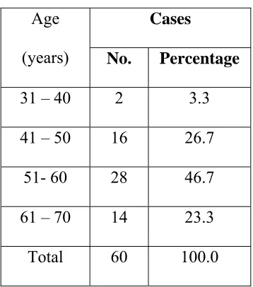
Age (years)
Cases
No. Percentage
31 – 40 2 3.3 41 – 50 16 26.7
51- 60 28 46.7 61 – 70 14 23.3
[image:70.612.183.362.477.680.2]Observation:
[image:71.612.153.454.219.394.2]The prevelance of the disease was found to be higher in the age group 51-60 years.
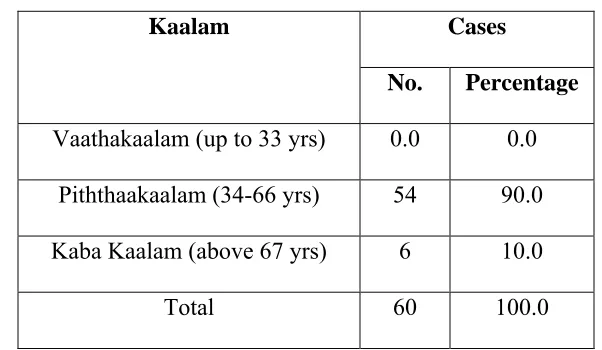
Table :3 Kaalam (Life span)
Kaalam Cases
No. Percentage
Vaathakaalam (up to 33 yrs) 0.0 0.0 Piththaakaalam (34-66 yrs) 54 90.0 Kaba Kaalam (above 67 yrs) 6 10.0
Total 60 100.0
Observation:
Out of 60 cases, 90% of the cases were found to be in Piththakaalam i.e. between 33 –66 years.
4. Paruvakaalam
Among 60 patients, 42 (70%) cases were admitted to the trial in Koothirkaalam and the remaining 18 (30%) cases were admitted in Munpanikaalam