Copyright © 2001, American Society for Microbiology. All Rights Reserved.
Effect on Polyomavirus T-Antigen Function of Mutations in a
Conserved Leucine-Rich Segment of the DnaJ Domain
HONGYUN LI, KARIN SO¨ DERBA¨RG, HAMID HOUSHMAND, ZHI-YONG YOU,ANDGO¨ RAN MAGNUSSON*
Department of Medical Biochemistry and Microbiology, Biomedical Center, Uppsala University, Uppsala, Sweden Received 15 July 1999/Accepted 17 November 2000
The N-terminal part of the mouse polyomavirus T antigens contains a highly conserved segment (-LLELLKL-), including amino acid residues 13 to 19. The sequence motif is predicted to form alpha helix I in the DnaJ domain of the T antigens. Four mutants with conservative substitutions of amino acid residues 13 and 14 were constructed. Of the four substitutions, L13M, L13I, L13V, and L14V, only L13V resulted in a phenotypic change. In transfected mouse cells, L13V large T antigen showed a more than 100-fold-reduced viral DNA synthesis. The viral replication could not be rescued by cotransfection of the cells with DNA expressing small t antigen or a large T antigen truncated at the C terminus that would compensate for a defect in host cell stimulation. In contrast to the effect on DNA replication, the L13V substitution in large T antigen did not prevent complex formation with Hsc70 and the Rb protein. Also, the activity of the protein in transactivation of transcription from the adenovirus E2 promoter was unimpaired, showing that the transcription factor E2F was released from pRb. The L13V substitution also caused a defect in small t antigen. However, this phenotypic change was due to protein instability. In contrast, middle T antigen with the L13V substitution remained stable and functional in cellular transformation. Together, the data show that the effect of the L13V substitution did not abrogate the Hsc70 interaction of the DnaJ domain. However, it is possible that the substitution of amino acid residue 13 affected specific DnaJ functions of large T antigen.
Polyomaviruses establish persistent infections in their hosts. The limited size of the viral genome makes the replication of viral DNA dependent on cellular enzymes. For this reason, viral early proteins, the T antigens, induce quiescent cells to enter the cell division cycle. Mouse polyomavirus encodes four early proteins: large, middle, small, and tiny T antigen (47, 51). These proteins are translated from mRNAs formed from a precursor by differential splicing. The four mRNAs have a common 5⬘-terminal sequence corresponding to 79 translated codons. Downstream of the splice points in mRNA, translation results in polypeptide segments unique to each T antigen.
The N-terminal common region of the T antigens (crt) is homologous to the conserved domain of the DnaJ family of molecular chaperones (5, 28). The J domain, consisting of approximately 70 amino acid residues that form four alpha helices, binds to and stimulates the ATPase activity of proteins belonging to the Hsp70/DnaK family (9). In a loop between helices II and III there is a conserved amino acid motif, the J box (-HPD-), which contacts Hsp70 in the formation of a binary complex (21). The DnaJ-homologous structure of poly-omavirus T antigen has DnaJ activity, since binding to Hsp70 protein and activation of its ATPase activity have been dem-onstrated (27, 47). Besides the -HPD- motif in the J domain, the T antigens of many polyomaviruses contain a second, highly conserved leucine-rich motif located at a position cor-responding to alpha helix I. However, this leucine-rich motif is not particularly conserved in other DnaJ proteins (see Fig. 1), suggesting that it might confer a T-antigen-specific function. In
mouse polyomavirus the motif at the putative alpha helix I has the sequence -LLELLKL-.
Large T antigen controls viral DNA synthesis by binding to the origin of replication and forming a homomultimeric com-plex that unwinds the two DNA strands (14). In this process, large T antigen also interacts with cellular replication proteins, directing them to the viral replicon. In simian virus 40 large T antigen, the J domain was shown to be involved in the initiation reaction, probably in the formation or dissociation of protein complexes (8). The J domain of large T antigen is also involved in the interaction with cellular proteins indirectly involved in replication. One such interaction is with the Rb family of proteins (48, 53). In mouse polyomavirus large T antigen, the segment -DLFCYE-, located on the C-terminal side of the J domain at amino acid residues 141 to 146, mediates binding to the pocket of these proteins (pRb, p107, and p130) (17, 31, 50). However, for dissociation of the complex with transcription factor E2, interaction of pRb with the J domain of large T antigen appears to be necessary, since mutant polypeptides with amino acid substitutions of the -HPD- motif are defective in this respect (48). The released transcription factors posi-tively regulate the expression of a set of genes whose products participate in replication, including the synthesis of viral DNA. Binding of pRb to large T antigen is also necessary for its activity in immortalization of rodent embryonic cells (1, 10, 24, 25, 46).
Middle T antigen is the main transforming protein of mouse polyomavirus. It has no known enzyme activity but acts by binding to cellular polypeptides involved in transduction of growth signals (reviewed in references 12 and 26). The contri-bution of the J domain to the function of middle T antigen has not been systematically studied. However, deletions affecting this segment of the polypeptide do not damage its transform-* Corresponding author. Mailing address: Box 582, SE-751 23
Upp-sala, Sweden. Phone: 46-18-4714560. Fax: 46-18-509876. E-mail: mago @bmc.uu.se.
2253
on November 9, 2019 by guest
http://jvi.asm.org/
ing activity or binding to protein phosphatase 2A (7, 19). Small t antigen is also able to bind to the A-subunit of protein phosphatase 2A (43). An apparently separate function of small t antigen is to stimulate the activity of large T antigen in viral DNA replication. Whether this effect of small t antigen is caused by direct activation of large T antigen or by influence on cellular replication factors is not known.
Functional studies of the J domain, using mutant proteins, have been focused on the highly conserved J box. Here we report on the effects of mutations altering alpha helix I of the J domain of polyomavirus T antigens.
MATERIALS AND METHODS
Cells, genomes, and transfection methods.NIH 3T3 and Swiss 3T6 mouse fibroblast cells were obtained from ATCC (Manassas, Va.), and Fischer rat FR3T3 cells were a gift from F. Cuzin (Nice University). Primary cell cultures of rat embryo fibroblasts (REF) were established from 15- to 16-day-old whole rat embryos (50). Cell cultures in 6-cm-diameter petri dishes were maintained in Dulbecco modified Eagle medium (DMEM) supplemented with serum as indi-cated. Transfection experiments were carried out with growing cells that had been plated 20 h earlier at a density of 3⫻105cells per petri dish. In various
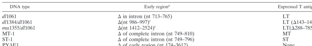
DNA transfection experiments we used DEAE-dextran-chloroquine (36), Lipo-fectamine as recommended by the manufacturer (Life Technologies), or copre-cipitation with calcium phosphate (56). Polyomavirus genomes were propagated as recombinants of plasmid pBR322 or pML (35), joined at the singleBamHI sites. Mutantsdl1061 (40), MT-1, and ST-1 (58) have deletions that restrict early viral gene expression to large, middle, or small T antigen, respectively. Mutant mu1355/dl1061 produces an N-terminal fragment of large T antigen which is inactive in viral DNA synthesis but retains the ability to bind the cellular pRb (50), and mutantdl1384/dl1061 has a deletion at nt 986 to 997, encoding a large T antigen that is deficient in pRb binding (50). The reporter gene constructs pPYLcat and pE2cat have been described elsewhere (34, 55). In these plasmids the chloramphenicol acetyltransferase gene is located downstream of the poly-omavirus late promoter or the adenovirus E2 promoter, respectively. The pre-viously published genotypes and expression properties of polyomavirus genomes are summarized in Table 1. For analysis of viral DNA replication a reporter plasmid, PyOrirep/pUC18, was used. It contained the polyomavirus origin of
DNA replication (nt 4634 to 5293 and 1 to 174) inserted into theBamHI site of pUC18 DNA.
Wild-type and mutant large T antigen were expressed using the pcDNA3 vector (InVitrogen). The large-T-antigen-coding sequence of polyomavirus mu-tant LT-1 DNA and its derivatives,crtL13V/E15D,crtL14V/E15D, andcrtP43S, were inserted into theEcoRV site of pcDNA3. Wild-type and mutantcrtL13V/ E15D small t antigen were expressed from the early coding sequences of ST-1 DNA inserted into pcDNA3. The Rb protein p105 was expressed from plasmid pSGRb (kindly provided by W. G. Kaelin), containing cDNA cloned in pSG5 (Stratagene).
Analysis of protein expression.For radioactive labeling of polypeptides, the cells were first incubated for 30 min in methionine-free DMEM buffered with 20 mM HEPES. [35S]methionine was added (150Ci per culture), and the
incuba-tion of the cells was continued for 4 h. The cell were lysed in a buffer consisting of 10 mM Tris-HCl (pH 8.0), 137 mM NaCl, 1.0 mM dithiothreitol, 1.0 mM MgCl2, 1.0 mM CaCl2, 1.0 mM EDTA, 10% (vol/vol) glycerol, 1.0% (vol/vol)
Nonidet P-40, 50g of phenylmethylsulfonyl fluoride, and 1.0g of aprotinin per
ml. After 30 min at 0°C, nuclei and cell debris were removed by centrifugation. For immunoprecipitation and immunoblot analysis of T antigen, the monoclonal antibodies (MAb) LT1 (13), F4, and F5 (42) were used. For immunoprecipita-tion of pRb and Hsc70 we used the MAb Ab1 (Oncogene Sciences) and MAb SP822 (Stressgene), respectively. In immunoprecipitation, antibodies were cap-tured using protein G-Sepharose (Pharmacia Biotech).
Analysis of viral DNA replication.Viral DNA was excised from the recombi-nant plasmids by digestion with BamHI, recircularized by treatment with T4 DNA ligase at a concentration of 5g of DNA per ml, and used for transfection of 3T6 cells. Low-molecular-weight DNA was selectively extracted from cells, and viral DNA was partially purified (40). It was cleaved withDpnI andBamHI, resolved by agarose gel electrophoresis, and transferred to a hybridization mem-brane (52). DNA on the memmem-brane was annealed with32P-labeled polyomavirus
DNA (15), and bound radioactivity was quantified using a Molecular Imager G-450 (Bio-Rad). In analyses of viral DNA replication with large T antigen expressed at a high level, the origin of viral DNA replication was present on a separate plasmid, PyOrirep/pUC18. Here, newly replicated DNA was isolated
after digestion withDpnI andPstI and was detected using a32P-labeledPstI
fragment excised from the same plasmid.
Analysis of transcriptional transactivation.REF cultures were transfected with a mixture of pPYLcat or pE2cat DNA and a second plasmid expressing large T antigen. Protein extracts were prepared at 40 h posttransfection by addition of 0.10 ml of 10 mM Tris-HCl (pH 7.9), 150 mM NaCl, 1.5 mM MgCl2,
and 0.5% (vol/vol) Nonidet P-40. Chloramphenicol acetyltransferase (CAT) ac-tivity in 0.04-ml portions was assayed by the method of Gorman et al. (20), as modified by Herbomel et al. (23). Incubations were done at 37°C for 3 h. The substrate and the products were separated by thin-layer chromatography. For quantitation of the reaction, the thin-layer chromatograms were analyzed in a Molecular Imager.
RESULTS
Mutant construction. The conserved amino acid motif
[image:2.612.52.559.84.159.2]-LLELLKL- in the common region of mouse polyomavirus T antigens (Fig. 1) is predicted to form the alpha helix I (5) of the J domain. To construct mutants with base-pair substitutions in the codons of this conserved motif, a uniqueBglII cleavage site was introduced by changing bp 219 of polyomavirus DNA from A-T to T-A. This transversion resulted in the conservative amino acid replacement E15D. The mutation did not detect-ably alter the activity of mouse polyomavirus large T antigen in the initiation of viral DNA replication (data not shown). In further mutagenesis we replaced the highly conserved residues L13 and L14 of polyomavirus T antigens. Mutations leading to the amino acid substitutions L13M, L13V, and L13I were in-troduced. In addition, a mutation leading to the P43S substi-tution was introduced to provide a reference for defective DnaJ activity (8, 49). The mutants were denotedcrt(common region T antigen). None of these amino acid substitutions at residue 13 led to disruption of the predicted alpha-helical structure. All these mutations were made in the genetic back-ground of dl1061, having a deletion that restricts early gene expression to large T antigen. To investigate the effect of the TABLE 1. Polyomavirus genomes and T-antigen expression
DNA type Early regiona Expressed T antigensb
dl1061 ⌬in intron (nt 713–765) LT
dl1384/dl1061 ⌬(nt 986–997)c LT (⌬143–146)
mu1355/dl1061 ⌬(nt 1412–2524)c LT(⌬288–785)
MT-1 ⌬of complete intron (nt 749–810) MT
ST-1 ⌬of complete intron (nt 749–796) ST
PY⌬E1 ⌬of early region (nt 174–3612) None
aNucleotide numbers are nominal and refer to the standard sequence with EMBL accession number J02288. Deleted nucleotides (nt) are indicated by⌬. bLT, MT, and ST designate large, middle, and small T antigen, respectively. Deleted amino acid residues are indicated by⌬.
cThe mutant also has thedl1061 deletion.
on November 9, 2019 by guest
http://jvi.asm.org/
crtmutations on middle and small t antigen, anAvaI restriction fragment (nucleotides 658 to 1018) was substituted for the homologous fragment of MT-1 and ST-1 DNA. In these viral genomes deletions corresponding to intervening sequences in RNA splicing restrict early gene expression to middle T anti-gen and small t antianti-gen, respectively.
Activity of large T antigens produced bycrtmutants in DNA
replication.To test the activity of the mutant large T antigens
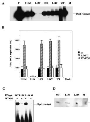
in viral DNA replication, mouse 3T6 cells were transfected with the mutant genomes prepared by excision from recombi-nant plasmids. At 40 h posttransfection, viral DNA was selec-tively extracted from the cells and partially purified. After digestion with the methylation-dependent restriction endo-nucleasesDpnI andBamHI that linearized the DNA mole-cules, they were resolved by agarose gel electrophoresis and blotted onto a hybridization membrane. Detection of polyoma-virus DNA was done by annealing with a 32P-labeled probe.
The autoradiogram is shown in Fig. 2A, and quantitative data from a similar experiment (Fig. 2B) showed that large T anti-gens with the L13I, L13M, and L14V substitutions supported viral DNA replication at 75 to 90% of the level reached with the wild-type protein. In contrast, the mutant expressing large T antigen with the L13V substitution replicated very poorly. The amount of viral DNA produced during 40 h was only 0.1% of that for thedl1061 control.
To rule out that the L13V substitution made large T antigen unstable, DNA replication was analyzed with large T antigen expressed at a high level, allowing parallel determination of protein. NIH 3T3 cells were transfected with pcDNA3/LT-wt, pcDNA3/LT-L13V/E15D, pcDNA3/LT-L14V/E15D or, as a negative control, pcDNA3 mixed with PyOrirep/pUC18.
Anal-ysis of newly replicated DNA isolated at 40 h posttransfection showed (Fig. 2C) that the L13V substitution in large T antigen severely decreased its activity in the initiation of viral DNA replication also in this experiment. The synthesis of a reporter plasmid, containing an origin of DNA replication, in cells transfected with pcDNA3/LT-L13V/E15D was only 1% of the amount obtained in cells expressing wild-type or L14V/E15D large T antigen. To determine the quantity of large T antigen in the cells, protein was extracted from parallel, transfected
cultures and was resolved by sodium dodecyl sulfate-polyacryl-amide gel electrophoresis (SDS-PAGE). Analysis of large T antigen by immunoblotting showed (Fig. 2D) that the amount of immunoreactive protein in cells expressing the L13V mutant protein was slightly lower than the corresponding amounts of wild-type and L14V mutant protein. However, this difference was too small to explain the large effect of the L13V substitu-tion on viral DNA replicasubstitu-tion. Immunofluorescence analysis using MAb LT1 showed that both L13V and L14V large T antigens were located in the nucleus (data not shown). Hence, large T antigen with the L13V substitution appeared to have lost most of its activity in the initiation of viral DNA synthesis in both 3T6 and NIH 3T3 cells.
Attempts to rescue the replication function of mutant large
T antigens.Besides controlling the initiation of each round of
viral DNA synthesis, large T antigen participates in the induc-tion of cellular DNA synthesis that is a prerequisite for the viral replication. If the substitution of amino acid residue 13 or 14 inhibited the binding of large T antigen to the origin of viral DNA replication, or any ensuing cis activity in viral DNA synthesis, then other T-antigen proteins would probably be unable to rescue that function. If, on the other hand, a mutant large T antigen was primarily deficient in the perturbation of cell cycle control, rescue of function might be possible.
Small t antigen enhances viral DNA replication in 3T6 mouse fibroblasts. The mechanism is unknown but may relate to site-specific phosphorylation of large T antigen required for its activity in viral DNA replication. To investigate the effect on the activity of the fourcrtmutant large T antigens, 3T6 cells were cotransfected with ST-1, expressing wild-type small t an-tigen, and each of the plasmids encoding wild-type orcrt mu-tant large T antigen. The amounts of viral DNA formed during 40 h following transfection are shown in Fig. 2B. Small t anti-gen increased viral DNA synthesis in all cases. The synthesis of crtL13I/E15D/dl1061, crtL13M/E15D/dl1061, and crtL14V/ E15D/dl1061 reached almost the same level as that ofdl1061 DNA. In contrast, mutantcrtL13V/E15D/dl1061 remained at 0.1% of the control level. However, the activity of this mutant large T antigen also was stimulated by small t antigen. We showed earlier (50) that mutant large-T-antigen polypeptides with substitutions in the pRb binding site (-DLFCYE-) at amino acid residues 141 to 146 were partially defective in viral DNA synthesis. In these cases cotransfection with a genome expressing a 287-amino-acid-residue N-terminal fragment of large T antigen but with a normal pRb binding site overcame the deficiency. Here we investigated whether this truncated large T antigen (mu1355/dl1061) had a stimulatory effect on the viral DNA synthesis of the fourcrtmutant forms of the protein. Mouse 3T6 cells were transfected with the two types of DNA, and the amount of newly synthesized viral DNA was determined. The result showed (Fig. 2B) that the activity in viral DNA replication of thecrtmutant large T antigens was not increased by the coexpression of themu1355 large T an-tigen. Hence the L13V substitution in large T antigen ap-peared to have a direct effect on the activity of the protein in viral DNA replication. However, this experiment does not ex-clude additional effects of the mutation on large-T-antigen activities.
Binding ofcrtmutant large T antigen to Hsc70 and pRb.To
[image:3.612.61.287.72.155.2]investigate whether the L13V substitution affected the DnaJ
FIG. 1. Conserved amino acid motif in the N-terminal part of poly-omavirus (POV) T antigens. Deduced amino acid sequence of indi-cated T-antigen segments encoded by mouse polyomavirus (EMBL accession no. J02288), hamster polyomavirus (accession no. M26281), Kilham mouse polyomavirus (accession no. M55904), lymphotropic polyomavirus (accession no. K02562), simian virus 40 (accession no. V01380), BK virus (accession no. V01108), and budgerigar fledgeling disease virus (accession no. M20775) are shown. The homologous segment of the HDJ-1 protein (accession no. X62421) and the con-sensus amino acid sequence of DnaJ proteins (Prosite accession no. PD000231) are included as a reference. The highly conserved leucine residues in T antigens are highlighted by shading.
on November 9, 2019 by guest
http://jvi.asm.org/
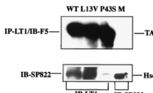
activity of large T antigen, binding to the Hsc70 protein was tested. NIH 3T3 cells that have a high constitutive expression of this polypeptide were used for the analysis. The cells were transfected with plasmids expressing wild-type or mutant crtL13V/E15D large T antigen. As a negative control, a plas-mid, pcDNA3/LT-P43S, was used that encoded a mutant large T antigen with a P43S substitution in the universally conserved -HPD motif. This mutant large T antigen does not form a stable complex with Hsc70 (8, 49). At 42 h posttransfection,
protein was extracted and the cleared lysates were immuno-precipitated with either MAb LT1 directed against large T antigen or MAb SP822 directed against Hsc70. After SDS-PAGE, protein was transferred to nitrocellulose membranes by blotting. The membranes were then developed with MAb F5 directed against large T antigen or MAb SP822. Chemilu-minograms show (Fig. 3) that cells transfected with plasmids encoding wild-type, mutantcrtL13V/E15D, andcrtP43S large T antigens expressed easily detectable amounts of the large-FIG. 2. Activity of wild-type (WT) and mutant large T antigen in viral DNA replication. (A and B) Cultures of mouse 3T6 cells (5⫻105cells)
grown in DMEM containing 10% horse serum were transfected with 0.1g of viral DNA using the DEAE-dextran method. The cells were transfected either with a viral genome expressing wild-type or mutant large T antigen (LT) alone (A) (black columns in panel B) or together with a second viral genome expressing small t antigen (ST-1) or truncated large T antigen (dl1355). Low-molecular-weight DNA was selectively extracted at 42 h posttransfection, partially purified, cleaved withDpnI andBamHI, and subjected to agarose gel electrophoresis. DNA was transferred from the gel to a hybridization membrane and was annealed with32P-labeled polyomavirus DNA. Radioactivity retained on the filters
was then determined by autoradiography (A) or quantified in a Molecular Imager. The columns represent the average values of determinations of samples from duplicate cultures, and the error bars show the variation. (C and D) Cultures of NIH 3T3 cells (5⫻105cells) grown in DMEM
containing 10% fetal bovine serum were transfected, using Lipofectamine, with 1.0 g of PyOrirep/pUC18 and 1.0 g of pcDNA3LT-wt,
-L13V/E15D, or -L14V/E15D, encoding large T antigen. One culture was transfected with only PyOrirep/pUC18 DNA (lanes M). (C) Newly
replicated DNA was analyzed as described above. (D) Cell extracts from parallel transfected cultures were analyzed by immunoblotting, using MAb F5. T Ag shows the position of large T antigen.
on November 9, 2019 by guest
http://jvi.asm.org/
[image:4.612.148.455.71.485.2]T-antigen protein. Moreover, there was no apparent difference in the association of wild-type and mutantcrtL13V/E15D large T antigen with Hsc70. In contrast, in extracts of cells trans-fected with pcDNA3/LT-P43S there was no detectable Hsc70 protein coimmunoprecipitated with large T antigen.
The -DLFCYE- segment of large T antigen (amino acid residues 142 to 146) binds to the Rb pocket family of proteins. However, for some functional interactions between large T antigen and the pRbs, a functional J domain of the former protein is also required (48, 49, 57). To test whether large T antigen with the L13V substitution bound to pRb, we cotrans-fected cells with one plasmid encoding pRb (pSGRb) and a second plasmid encoding wild-type or mutantcrtL13V/E15D large T antigen. As a negative control, the mu1384 large T antigen, with amino acid residues 143 to 146 deleted, was used (50). At 42 h posttransfection, [35S]methionine was added to
the cultures and protein was labeled for 4 h. After lysis of the cells, large T antigen was immunoprecipitated with MAb LT1, and pRb was immunoprecipitated with MAb Ab1 from the extract of cells transfected with the pRb-encoding plasmid alone. Polypeptides were resolved by SDS-PAGE and then analyzed by autoradiography (Fig. 4). Extracts of cells trans-fected with plasmids encoding wild-type or mutant large T antigen contained a 90-kDa polypeptide reacting with MAb LT1. Its electrophoretic mobility was somewhat heterogenous, consistent with the known modifications of large T antigen in mammalian cells (4, 22). A ca. 105-kDa polypeptide coimmu-noprecipitated with wild-type and mutantcrtL13V/E15D large T antigen but not with the mu1384 protein. A similar ca. 105-kDa polypeptide, reacting with MAb Ab1, was observed in cells transfected with only pSGRb, but was below the detection level in untransfected cells. Together, the data indicated that wild-type and mutantcrtL13V/E15D large T antigen but not mu1384 large T antigen coimmunoprecipitated with pRb. Thus, the L13V substitution did not impair the formation of large T antigen-pRb complexes.
Transcriptional transactivation bycrtmutant large T
anti-gens. Polyomavirus large T antigen is able to transactivate
transcription by more than one mechanism. The transcription factor E2F is activated by release from the Rb family of
pro-teins after binding of large T antigen (39). Mutant large T antigens with amino acid substitutions in the conserved loop of the J domain were shown to bind to pRb but were defective in the induction of E2F release (48, 57). A second, for the poly-omavirus protein as yet uncharacterized, transactivation mech-anism operates on various transcription units, including the viral late genes (6, 29, 34). This mechanism may involve bind-ing of the cellular p300 to the C-terminal part of large T antigen (33, 38). To test the ability of ourcrtmutant large T antigens in transcription transactivation, they were expressed together with reporter plasmids containing the adenovirus E2 (pE2cat) or polyomavirus late promoter (pPYLcat) upstream of thecatgene. To ensure that wild-type pRb was expressed, secondary REF cells were used in this experiment. They were transfected with a recombinant plasmid encoding wild-type or mutant large T antigen mixed with the plasmid carrying the reporter gene. As a negative control for large T antigen ex-pression, we used the plasmid pPY⌬E1, which has most of the T-antigen-coding sequences deleted but retains the regulatory elements of the viral genome. Under the conditions of the experiment, CAT expression from the polyomavirus late pro-moter (Fig. 5A) was relatively high even in the absence of large T antigen. Coexpression of wild-type large T antigen or any of the four mutant forms stimulated the activity of the viral late promoter two- to fourfold. The small differences in the activity of the wild-type and various mutant large T antigens are prob-ably not significant. The level of CAT expression from the adenovirus E2 promoter was low in REF cells (Fig. 5B). How-ever, in the presence of large T antigen it was stimulated four-to sevenfold. The wild-type and all four mutant forms of large T antigen had similar effects on the activity of the E2 promoter. Hence the substitutions of amino acid residues 13 and 14 of large T antigen did not impair the DnaJ function of the protein that is required for a functional interaction with pRb to release the transcription factor E2F (48).
Stability of mutantcrtL13V small and middle T antigen.An
analysis of the small-t-antigen activity on
large-T-antigen-de-FIG. 4. Complex formation of wild-type and mutant large T anti-gen with pRb. Cultures of NIH 3T3 cells were transfected, using Lipofectamine, with plasmid pSGRb (2.0g) alone or mixed with 1.0 g of pcDNA3/LT-wt (WT), -L13V/E15D (L13V), ordl1384 (1384), encoding large T antigen. As a control (M), cells were transfected with pcDNA3 without an insert. At 42 h posttransfection, cells were labeled for 4 h with [35S]methionine. Protein extracts were prepared, and
immunoprecipitation (IP) was done with MAb LT1 directed against large T antigen or with MAb Ab-1 directed against pRb. Immunopre-cipitated material was resolved by SDS-PAGE in an 8% gel, and radioactivity was detected by autoradiography. The positions in the gel of large T antigen (TAg at 90 kDa) and pRb (105 kDa) relative to markers with known size are indicated.
FIG. 3. Complex formation of mutant and wild-type large T anti-gen with Hsc70. Cultures of NIH 3T3 cells (5 ⫻ 105 cells) were
transfected, using Lipofectamine, with plasmid pcDNA3/LT-wt (WT), -L13V/E15D (L13V), or P43S encoding large T antigen. As a negative control, cells were transfected with pcDNA3 without an insert (M). At 42 h posttransfection, cell extracts were prepared and incubated with either MAb LT1 (anti-large T antigen) or MAb SP822 (anti-Hsc70). Immunoprecipitated (IP) material was resolved by SDS-PAGE fol-lowed by immunoblotting, using either MAb F5 (anti-LT) or MAb SP822. The positions in the gel of large T antigen (TAg) and Hsc70 relative to markers with known size are indicated.
on November 9, 2019 by guest
http://jvi.asm.org/
[image:5.612.96.250.71.164.2]pendent viral DNA synthesis showed that the L13M, L13I, and L14 substitutions did not impair this function (the activity of wild-type small t antigen is shown in Fig. 2B). However, the crtL13V/E15D small t antigen was completely inactive (data not shown). To test whether this result was due to instability of the mutant small t antigen, the expression plasmid pcDNA3/ ST-L13V/E15D was constructed. NIH 3T3 cells were trans-fected with this plasmid or pcDNA3/ST-wt. As a negative con-trol, pcDNA3 vector DNA without an insert was used. At 42 h posttransfection, cells were lysed and extracted protein was analyzed by immunoblotting using MAb F4. The result shows (Fig. 6A) that the cells did not contain detectable amounts of the mutantcrtL13V/E15D small t antigen, although the wild-type protein was easily detectable. Together with the activity determination of mutant crtL13V/E15D/ST-1, the data
indi-cated that a substitution at position 13 of leucine for valine, but not for isoleucine or methionine, made small t antigen labile. The lability of small t antigen with the L13V substitution raised the question of whether it had a similar destabilizing effect on middle T antigen, since the sequences of the N-terminal 192 amino acid residues of the two proteins are co-linear. The activity of middle T antigen in cellular transforma-tion reflects its overall activity. Therefore, the mutant derivatives of polyomavirus MT-1 DNA were used for trans-fection of FR3T3 cells. Two days after transtrans-fection, the cells were replated at a fivefold-lower density, and after another 10 days, foci of cells were isolated by trypsinization in glass cylin-ders. Clones of transformed cells were obtained at similar yields with wild-type and all fourcrtmutant DNAs. Protein was extracted from cells of individual clones and was analyzed by immunoblotting using MAb F5, which recognizes middle T antigen. The experiment showed (Fig. 6B) that all the clones of transformed cells, but not the negative control, contained im-munoreactive material with an electrophoretic mobility corre-sponding to 55 kDa. This result indicated that the L13V
[image:6.612.60.289.73.406.2]sub-FIG. 6. Stability of mutant small and middle T antigens. (A) Cul-tures of NIH 3T3 cells were transfected as described in the legend to Fig. 5. At 42 h posttransfection, cell extracts were prepared and sub-jected to SDS-PAGE in a 12% gel, followed by immunoblotting using MAb F4 reacting with small t antigen. Lane M, material from cells transfected with pcDNA3 without insert; control lane, small t antigen produced in insect cells. (B) FR3T3 cells were transfected, using the calcium phosphate coprecipitation method with MT-1 DNA, encoding wild-type protein or mutant protein with the indicated substitutions at residues 13 and 14, respectively. Clones of transformed cells were isolated, and extracts were prepared from cultures of these cloned cells. Proteins were resolved by SDS-PAGE in an 8% gel, and T antigen was identified by immunoblotting using MAb F5. Lane M, extract of untransformed FR3T3 cells. The positions in gels of middle T antigen (MT), small T antigen (ST), and of markers with known sizes are indicated.
FIG. 5. Effect of wild-type and mutant large T antigen on the poly-omavirus late promoter and the adenovirus E2 promoter. Growing REF cultures (5⫻105cells) were transfected, using Lipofectamine,
with 1.0g of pPYLcat (A) or pE2cat (B) DNA mixed with 1.0g of polyomavirus DNA cloned in plasmid pML. As a control to expression of wild-type and mutant large T antigen, the mutant PY⌬E1 with a deletion of the early region was used. Cell extracts were prepared at 40 h posttransfection, and CAT activity was assayed. The14C-labeled
chloramphenicol substrate and the acetylated products were separated by thin layer chromatography. The radioactive spots were identified by autoradiography and quantified in a phosphorimager. The relative enzyme activity in the absence of large-T-antigen expression was set to 1. Wild-type and mutant large T antigen (LT) are denoted by WT and the amino acid substitutions, respectively.
on November 9, 2019 by guest
http://jvi.asm.org/
[image:6.612.345.514.316.584.2]stitution did not have any negative effect on the stability or activity of middle T antigen.
DISCUSSION
Processing of mouse polyomavirus early RNA by differential splicing leads to four types of mRNA which share a 5⬘-terminal exon segment. Therefore, the four translation products, the T antigens, have identical N-terminal parts consisting of 79 amino acid residues. Most of this segment forms a structure homologous to the DnaJ domain (9, 28). Within this part of the T antigens, two short amino acid sequences are highly con-served in the polypeptides encoded by all known polyomavi-ruses. One is the -HPDKGG- motif containing the J box. The other is highly conserved in T antigens but not in other DnaJ proteins. Amino acid residues 11 to 20 of polyomavirus T antigens are predicted (30) to form an alpha helix, correspond-ing to helix I of the DnaJ protein. This putative helix is longer (residues 10 to 19) than the homologous structure in the hu-man Hsp40 protein HDJ-1 (residues 5 to 10). In the alpha helix I of HDJ-1, the predicted structure was confirmed by nuclear magnetic resonance analysis (45). In large T antigen, leucine residue 13, which is sensitive to substitution, would be located in alpha helix I or just outside this structure if it has the same size as in the HJD-1 protein. The high sequence conservation of this T-antigen segment suggests that it has a strongly se-lected function. However, this function is not necessarily re-lated to DnaJ activity. Interestingly, the conserved leucine-rich motif is related to the conserved region 1 of the adenovirus E1A protein [(E/D)X3LX(E/D)LX2(L/I)], which is known to
participate in binding to several cellular proteins, such as pRb, Cdk2, and p300, also called CREB-binding protein. (16, 54). To study the function of the leucine-rich segment in polyoma-virus T antigens that corresponds to alpha helix I, we intro-duced minimal amino acid changes that were unlikely to dis-rupt its potential helical structure. In mutagenesis of codons for amino acid residues 13 and 14 in the T antigens, we se-lected conservative shifts to residues which were not repre-sented in the corresponding polypeptides of any known poly-omavirus (Fig. 1). L13 was completely conserved in T antigens. Thus, we made thecrtmutants L13I, L13M, and L13V. Posi-tion L14 is less conserved, but a valine residue has not been observed at this position in any of the known T antigens. Therefore, we made the mutant L14V. All these mutants were constructed with DNA containing thecrtE15D mutation that results in a unique restriction endonuclease cleavage site. Since an aspartic acid residue is present at this position in the T antigens of the human and simian polyomaviruses, we did not anticipate any phenotypic effect from the replacement of the glutamic acid residue. This assumption was supported by sev-eral experiments that showed normal T-antigen functions of thecrtE15D mutant (data not shown). To test the effect of the other mutations on individual polyomavirus T antigens, thecrt mutations were combined with deletion mutants that restricted splicing of the viral early RNA. Effects on viral DNA synthesis transcriptional transactivation, binding to Hsc70 and pRb, and cellular immortalization were then analyzed in transfection experiments. We also analyzed the effect of the mutations on the function of middle T antigen. However, in keeping with earlier studies of deletion mutants (7, 19), the J domain of
middle T antigen appeared to have little influence on the known functions of the protein. Previous analyses of the DnaJ domain of T antigens have been focused on the interaction with DnaK homologs, such as Hsp70 and Hsc70, and the spec-ificity of interactions with cellular targets (8, 11). The J domain of T antigens can functionally substitute for DnaJ in Esche-richia colicells (27). It binds to Hsc70 in mammalian cells (8, 48) and interacts with this protein by stimulating its ATPase activity (47). The cooperation with Hsc70 or its homologs is required for functional interactions with other cellular polypeptides, such as pRb (48, 53, 57).
Mutation of the highly conserved J box (-HPD-) impairs the function of simian virus 40 large T antigen in initiation of viral DNA synthesis (8). A nonnuclear localization might explain this phenotype, since DnaK proteins have been reported to mediate nuclear translocation (41). However, Sheng et al. (48) showed that J-box-defective large T antigen had a nuclear localization. Large T antigens with substitutions of amino acid residue 13 or 14 also accumulated in the cell nucleus, as shown by immunofluorescence (data not shown). The defect of the protein with an alteration of the HPD box is probably in one or several of the numerous protein-protein interactions that are involved in the initiation process of DNA replication. Thecrt mutants analyzed in the present study produce T antigens with a normal J box. However, even one of the conservative amino acid substitutions in alpha helix I of the J domain, L13V, caused a distinctive phenotype in viral DNA synthesis. The impairment of viral DNA replication was traced to defects of both large (Fig. 2) and small (Fig. 6A) T antigen. In large T antigen the L13V substitution had a profound effect on viral DNA synthesis that was not rescuable by coexpression of a truncated form of the polypeptide with an intact J domain. This N-terminal fragment of large T antigen has been shown to stimulate cellular and viral DNA synthesis by advancing the cell cycle (50). The activity ofcrtL13V/E15D large T antigen was stimulated by coexpression of small t antigen (Fig. 2B). However, in relation to the activity of the wild-type protein, the defect ofcrtL13V/E15D large T antigen remained. Hence it is probable that large T antigen with the L13V substitution was impaired in its interaction with viral DNA or cellular replica-tion factors. However, purifiedcrtL13V/E15D large T antigen had normal DNA binding and unwinding activities (unpub-lished data). The polyomaviruscrtL13V large T antigen was recently reported to be unstable when expressed in both mouse and rat cells (32). In our investigation, the L13V substitution had little effect on the stability of large T antigen in NIH 3T3 cells (Fig. 2C and D). Moreover, analysis of stability in BHK cells, following a pulse-chase protocol, showed that both wild-type and L13V/E15D large T antigens had half-lives of approx-imately 8 h (data not shown). It is possible that the combina-tion of L13V and E15D substitucombina-tions resulted in a more stable protein than the isolated L13V substitution. Paradoxically, a C-terminal fragment of large T antigen, devoid of the DnaJ domain, is able to support viral DNA. However, it is probable that the full-length and truncated versions of the protein be-have differently in the assembly of DNA replication complexes, providing an explanation for the involvement of the DnaJ structure in viral DNA synthesis.
Small t antigen stimulates viral DNA synthesis, at least in some types of mouse cells (2, 37). The simian virus 40 small t
on November 9, 2019 by guest
http://jvi.asm.org/
antigen has been shown to transactivate the cyclin A gene, and mutation of the J box disturbs this effect (44). Of the four mutants producing T antigens with amino acid substitutions of 13L and 14L, only thecrtL13V/E15D small t antigen was de-fective in stimulating viral DNA synthesis. However, this phe-notype was linked to instability of the protein (Fig. 6A). Since neither large nor middle T antigen was destabilized by the L13V substitution, the unique part of the proteins probably influences the structure of the DnaJ domain and its stability.
The effect of the L13V amino acid substitution on the func-tion of large T antigen in viral DNA replicafunc-tion raised the question of whether the observed defect was in DnaJ activity on DnaK-like proteins. Since mutation of the J box results in loss of binding to Hsc70 and transcription factor E2F activa-tion, we investigated whether the L13V and P43S substitutions in large T antigen caused similar defects. The experiment was done with NIH 3T3 cells, which have a high constitutive level of Hsc70. In a coimmunoprecipitation experiment there was no difference in binding to Hsc70 of the wild-type large T antigen and the protein with the L13V substitution. In contrast, large T antigen with the P43S substitution in the J box did not form a stable complex with Hsc70 (Fig. 3). This result suggests that conservative substitutions in alpha helix I of large T antigen were not critical for the interaction with Hsc70.
Binding of the pRb to large T antigen does not occur unless the -DLFCYE- segment (residues 141 to 146) is present (17, 31, 50). However, for displacement from pRb of the transcrip-tion factor E2F, the DnaJ domain of large T antigen has to be functional. In contrast, transactivation by large T antigen of the polyomavirus late promoter is independent of pRb binding, since the -DLFCY- motif in the protein is not required for this function (50). The cellular pRb formed a complex with crtL13V/E15D large T antigen but not with the truncated dl1384 protein lacking residues 143 to 146 (Fig. 4). The large T antigen with the L13V substitution also induced activation of the transcription factor E2F, since all thecrtmutants (L13M, L13I, L13V, and L14V) were positive in transactivation of both the E2F-regulated E2 promoter from adenovirus and the poly-omavirus late promoter (Fig. 5). Analysis of cellular immor-talization corroborated the conclusion that E2F was activated, since lines of secondary REFs were established after transfec-tion with genomes expressing each of the four mutatransfec-tions (data not shown).
Our data show that the four tested mutants have a DnaJ function, as analyzed by interactions of large T antigen with Hsc70 and pRb. However, the L13V substitution in large T antigen caused a distinct defect of the protein in the initiation of viral DNA replication. Since the amino acid replacement mapped in the core of the DnaJ domain, the functional defect must by definition reflect a DnaJ activity. Thus, we propose that the DnaJ domain of polyomavirus T antigens has more than one specificity for the interaction with other proteins. Support for this idea is provided by the work of Pipas (5), which shows that the L-to-F substitution of conserved residue 19 in simian virus 40 large T antigen (Fig. 1), which corre-sponds to residue 19 in the polyomavirus protein, did not inhibit viral DNA synthesis but instead abolished the activity of the protein in cellular transformation and in assembly of virus particles.
After completion of this paper, a paper by Berjanskii et al.
was made public (3). These investigators demonstrated that residues 13 and 14 of polyomavirus T antigens form part of the J-domain alpha helix I. It was also shown that the L13V sub-stitution in an N-terminal fragment of T antigen inactivated its DnaJ function in a complementation assay performed withE. colicells, possibly due to disruption of alpha helix I. In the reported structure, the side chain of residue 13 is buried in the helix.
ACKNOWLEDGMENT
The experiments described in this report were supported financially by the Swedish Cancer Society.
REFERENCES
1.Asselin, C., and M. Bastin.1985. Sequences from polyomavirus and simian virus 40 large T genes capable of immortalizing primary rat embryo fibro-blasts. J. Virol.56:958–968.
2.Berger, H., and E. Wintersberger.1986. Polyomavirus small T antigen en-hances replication of viral genomes in 3T6 mouse fibroblasts. J. Virol.60: 768–770.
3.Berjanskii, M. V., M. I. Riley, A. Xie, V. Semenchenko, W. R. Folk, and S. R. Van Doren.2000. NMR structure of the N-terminal J domain of murine polyomavirus T antigens: implications for DnaJ-like domains and for muta-tions of T antigens. J. Biol. Chem.,275:36094–36103.
4.Bockus, B. J., and B. Schaffhausen.1987. Localization of the phosphoryla-tions of polyomavirus large T antigen. J. Virol.61:1155–1163.
5.Brodsky, J. L., and J. M. Pipas.1998. Polyomavirus T antigens: molecular chaperones for multiprotein complexes. J. Virol.72:5329–5334.
6.Cahill, K. B., A. J. Roome, and G. G. Carmichael.1990. Replication-depen-dent transactivation of the polyomavirus late promoter. J. Virol.64:992– 1001.
7.Campbell, K. S., K. R. Auger, B. A. Hemmings, T. M. Roberts, and D. C. Pallas.1995. Identification of regions in polyomavirus middle T and small t antigens important for association with protein phosphatase 2A. J. Virol. 69:3721–3728.
8.Campbell, K. S., K. P. Mullane, I. A. Aksoy, H. Stubdal, J. Zalvide, J. M. Pipas, P. A. Silver, T. M. Roberts, B. S. Schaffhausen, and J. A. DeCaprio. 1997. DnaJ/hsp40 chaperone domain of SV40 large T antigen promotes efficient viral DNA replication. Genes Dev.11:1098–1110.
9.Cheetham, M. E., and A. J. Caplan.1998. Structure, function and evolution of DnaJ: conservation and adaptation of chaperone function. Cell Stress Chaperones3:28–36.
10. Cowie, A., J. de Villiers, and R. Kamen.1986. Immortalization of rat embryo fibroblasts by mutant polyomavirus large T antigens deficient in DNA bind-ing. Mol. Cell. Biol.6:4344–4352.
11. DeCaprio, J. A.1999. The role of the J domain of SV40 large T in cellular transformation. Biologicals27:23–28.
12. Dilworth, S. M.1995. Polyoma virus middle T antigen: meddler or mimic? Trends Microbiol.3:31–35.
13. Dilworth, S. M., and B. E. Griffin.1982. Monoclonal antibodies against polyoma virus tumor antigens. Proc. Natl. Acad. Sci. USA79:1059–1063. 14. Fanning, E., and R. Knippers.1992. Structure and function of simian virus
40 large tumor antigen. Annu. Rev. Biochem.61:55–85.
15. Feinberg, A. P., and B. Vogelstein.1983. A technique for radiolabeling DNA restriction endonuclease fragments to high specific activity. Anal. Biochem. 132:6–13.
16. Flint, J., and T. Shenk.1997. Viral transactivating proteins. Annu. Rev. Genet.31:177–212.
17. Freund, R., P. H. Bauer, H. A. Crissman, E. M. Bradbury, and T. L. Ben-jamin.1994. Host range and cell cycle activation properties of polyomavirus large T-antigen mutants defective in pRB binding. J. Virol.68:7227–7234. 18. Gjørup, O. V., P. E. Rose, P. S. Holman, B. J. Bockus, and B. S.
Schaff-hausen.1994. Protein domains connect cell cycle stimulation directly to initiation of DNA replication. Proc. Natl. Acad. Sci. USA91:12125–12129. 19. Glenn, G. M., and W. Eckhart.1995. Amino-terminal regions of polyoma-virus middle T antigen are required for interactions with protein phospha-tase 2A. J. Virol.69:3729–3736.
20. Gorman, C. M., L. F. Moffat, and B. H. Howard.1982. Recombinant ge-nomes which express chloramphenicol acetyltransferase in mammalian cells. Mol. Cell. Biol.2:1044–1051.
21. Greene, M. K., K. Maskos, and S. J. Landry.1998. Role of the J-domain in the cooperation of Hsp40 with Hsp70. Proc. Natl. Acad. Sci. USA95:6108– 6113.
22. Hassauer, M., K. H. Scheidtmann, and G. Walter.1986. Mapping of phos-phorylation sites in polyomavirus large T antigen. J. Virol.58:805–816. 23. Herbomel, P., B. Bourachot, and M. Yaniv.1984. Two distinct enhancers
with different cell specificities coexist in the regulatory region of polyoma. Cell39:653–662.
on November 9, 2019 by guest
http://jvi.asm.org/
24.Jat, P. S., and P. A. Sharp.1989. Cell lines established by a temperature-sensitive simian virus 40 large-T-antigen gene are growth restricted at the nonpermissive temperature. Mol. Cell. Biol.9:1672–1681.
25. Jat, P. S., and P. A. Sharp.1986. Large T antigens of simian virus 40 and polyomavirus efficiently establish primary fibroblasts. J. Virol.59:746–750. 26.Kaplan, D. R., D. C. Pallas, W. Morgan, B. Schaffhausen, and T. M. Roberts.
1989. Mechanisms of transformation by polyoma virus middle T antigen. Biochim. Biophys. Acta948:345–364.
27. Kelley, W. L., and C. Georgopoulos.1997. The T/t common exon of simian virus 40, JC, and BK polyomavirus T antigens can functionally replace the J-domain of the Escherichia coli DnaJ molecular chaperone. Proc. Natl. Acad. Sci. USA94:3679–3684.
28. Kelley, W. L., and S. J. Landry.1994. Chaperone power in a virus? Trends Biochem. Sci.19:277–278.
29. Kern, F. G., S. Pellegrini, A. Cowie, and C. Basilico.1986. Regulation of polyomavirus late promoter activity by viral early proteins. J. Virol.60:275– 285.
30. Kneller, D. G., F. E. Cohen, and R. Langridge.1990. Improvements in protein secondary structure prediction by an enhanced neural network. J. Mol. Biol.214:171–182.
31. Larose, A., N. Dyson, M. Sullivan, E. Harlow, and M. Bastin.1991. Poly-omavirus large T mutants affected in retinoblastoma protein binding are defective in immortalization. J. Virol.65:2308–2313.
32. Lemieux, B., and M. Bastin.2000. Polyomavirus large T antigen mutants affected in viral DNA replication. Virology269:370–376.
33. Lill, N. L., M. J. Tevethia, R. Eckner, D. M. Livingston, and N. Modjtahedi. 1997. p300 family members associate with the carboxyl terminus of simian virus 40 large tumor antigen. J. Virol.71:129–137.
34. Linder, S., M. Nilsson, I. Martens, and G. Magnusson.1990. A viable mouse polyomavirus mutant without immortalizing or transforming activities. Vi-rology179:78–86.
35. Lusky, M., and M. Botchan.1981. Inhibition of SV40 replication in simian cells by specific pBR322 DNA sequences. Nature (London)293:79–81. 36. Luthman, H., and G. Magnusson.1983. High efficiency polyoma DNA
trans-fection of chloroquine treated cells. Nucleic Acids Res.11:1295–1308. 37. Martens, I., S. A. Nilsson, S. Linder, and G. Magnusson.1989. Mutational
analysis of polyomavirus small-T-antigen functions in productive infection and in transformation. J. Virol.63:2126–2133.
38. Nemethova, M., and E. Wintersberger.1999. Polyomavirus large T antigen binds the transcriptional coactivator protein p300. J. Virol.73:1734–1739. 39. Nevins, J. R.1992. E2F: a link between the Rb tumor suppressor protein and
viral oncoproteins. Science258:424–429.
40. Nilsson, S. V., and G. Magnusson.1983. T-antigen expression by polyoma mutants with modified RNA splicing. EMBO J.2:2095–2101.
41. Okuno, Y., N. Imamoto, and Y. Yoneda.1993. 70-kDa heat-shock cognate protein colocalizes with karyophilic proteins into the nucleus during their transport in vitro. Exp. Cell Res.206:134–142.
42. Pallas, D. C., C. Schley, M. Mahoney, E. Harlow, B. S. Schaffhausen, and T. M. Roberts.1986. Polyomavirus small t antigen: overproduction in bac-teria, purification, and utilization for monoclonal and polyclonal antibody production. J. Virol.60:1075–1084.
43. Pallas, D. C., L. K. Shahrik, B. L. Martin, S. Jaspers, T. B. Miller, D. L. Brautigan, and T. M. Roberts.1990. Polyoma small and middle T antigens
and SV40 small t antigen form stable complexes with protein phosphatase 2A. Cell60:167–176.
44. Porras, A., J. Bennett, A. Howe, K. Tokos, N. Bouck, B. Henglein, S. Sathya-mangalam, B. Thimmapaya, and K. Rundell.1996. A novel simian virus 40 early-region domain mediates transactivation of the cyclin A promoter by small-t antigen and is required for transformation in small-t antigen-depen-dent assays. J. Virol.70:6902–6908.
45. Qian, Y. Q., D. Patel, F. U. Hartl, and D. J. McColl.1996. Nuclear magnetic resonance solution structure of the human Hsp40 (HDJ-1) J-domain. J. Mol. Biol.260:224–235.
46. Rassoulzadegan, M., Z. Naghashfar, A. Cowie, A. Carr, M. Grisoni, R. Kamen, and F. Cuzin.1983. Expression of the large T protein of polyoma virus promotes the establishment in culture of “normal” rodent fibroblast cell lines. Proc. Natl. Acad. Sci. USA80:4354–4358.
47. Riley, M. I., W. Yoo, N. Y. Mda, and W. R. Folk.1997. Tiny T antigen: an autonomous polyomavirus T antigen amino-terminal domain. J. Virol.71: 6068–6074.
48. Sheng, Q., D. Denis, M. Ratnofsky, T. M. Roberts, J. A. DeCaprio, and B. Schaffhausen.1997. The DnaJ domain of polyomavirus large T antigen is required to regulate Rb family tumor suppressor function. J. Virol.71:9410– 9416.
49. Sheng, Q., T. M. Love, and B. Schaffhausen.2000. J domain-independent regulation of the Rb family by polyomavirus large T antigen. J. Virol.74: 5280–5290.
50. Soderbarg, K., and G. Magnusson.1993. Lytic functions of mutant polyoma-virus large T-antigen with deletion of retinoblastoma protein-binding motif. Virology193:281–288.
51. Soeda, E., J. R. Arrand, N. Smolar, J. E. Walsh, and B. E. Griffin.1980. Coding potential and regulatory signals of the polyoma virus genome. Nature (London)283:445–453.
52. Southern, E. M.1975. Detection of specific sequences among DNA frag-ments separated by gel electrophoresis. J. Mol. Biol.98:503–517. 53. Stubdal, H., J. Zalvide, K. S. Campbell, C. Schweitzer, T. M. Roberts, and
J. A. DeCaprio.1997. Inactivation of pRB-related proteins p130 and p107 mediated by the J domain of simian virus 40 large T antigen. Mol. Cell. Biol. 17:4979–4990.
54. Wang, H. G., Y. Rikitake, M. C. Carter, P. Yaciuk, S. E. Abraham, B. Zerler, and E. Moran.1993. Identification of specific adenovirus E1A N-terminal residues critical to the binding of cellular proteins and to the control of cell growth. J. Virol.67:476–488.
55. Weeks, D. L., and N. C. Jones.1983. E1A control of gene expression is mediated by sequences 5⬘to the transcriptional starts of the early viral genes. Mol. Cell. Biol.3:1222–1234.
56. Wigler, M., A. Pellicer, S. Silverstein, R. Axel, G. Urlaub, and L. Chasin. 1979. DNA-mediated transfer of the adenine phosphoribosyltransferase lo-cus into mammalian cells. Proc. Natl. Acad. Sci. USA76:1373–1376. 57. Zalvide, J., H. Stubdal, and J. A. DeCaprio.1998. The J domain of simian
virus 40 large T antigen is required to functionally inactivate RB family proteins. Mol. Cell. Biol.18:1408–1415.
58. Zhu, Z. Y., G. M. Veldman, A. Cowie, A. Carr, B. Schaffhausen, and R. Kamen.1984. Construction and functional characterization of polyomavirus genomes that separately encode the three early proteins. J. Virol.51:170–180.