Copyright © 2003, American Society for Microbiology. All Rights Reserved.
The Processing of eIF4GI by Human Rhinovirus Type 2 2A
pro
:
Relationship to Self-Cleavage and Role of Zinc
Walter Glaser, Andrea Triendl, and Tim Skern*
Institute of Medical Biochemistry, Division of Biochemistry, University of Vienna, Vienna Bio Center, A-1030 Vienna, Austria
Received 11 November 2002/Accepted 21 January 2003
The 2A proteinase (2Apro) of human rhinoviruses (HRVs) is a cysteine protease containing a structurally
important zinc ion. In the viral polyprotein, the enzyme cleaves between the C terminus of VP1 and its own N terminus. 2Aproalso processes the two isoforms of the cellular protein, eukaryotic initiation factor 4G (eIF4G).
We have shown that mature HRV2 2Apro, when translated in vitro in rabbit reticulocyte lysates, efficiently
cleaves eIF4GI, although the enzyme was not immediately active upon synthesis. Here, we examine the relationship between self-processing and eIF4GI cleavage. The onset of both reactions first occurred at least 10 min after initiation of protein synthesis. Furthermore, when self-processing was prevented by a specific mutation between VP1 and 2Apro, the VP1-2Aproprecursor was essentially unable to cleave eIF4GI, implying
that self-processing is a prerequisite for eIF4GI cleavage. 2Aprosynthesized in the presence of a potent zinc
chelator is inactive; however, upon addition of excess zinc, HRV2 2Aprorapidly gained activity. Finally, the
presence of the zinc chelator in the culture medium can protect HeLa cells from HRV infection.
A crucial step in the life cycle of many viruses is the proteo-lytic processing of viral and cellular proteins (6). This is espe-cially evident with certain picornaviruses which encode pro-teinases which not only process viral proteins but also carry out specific and controlled proteolysis of the eukaryotic initiation factor 4GI (eIF4GI) and its isoform 4GII (11, 20, 27). These proteins are components of the eIF4F complex, which is re-sponsible for recruitment of capped mRNA to the ribosome (19). Cleavage of the eIF4G isoforms prevents recruitment of capped mRNAs, thus disabling host cell protein synthesis. Pi-cornaviral mRNAs can, however, still be translated under these conditions, since protein synthesis initiates at a higher-ordered structure, termed the internal ribosome entry site,
within the 5⬘untranslated region (2).
Picornaviruses have evolved two different types of protein-ases for cleavage of the eIF4G isoforms (23). Foot-and-mouth disease virus (FMDV) encodes a papain-like cysteine
protein-ase, the leader proteinase (Lpro), at the very N terminus of the
polyprotein (10, 12). Lprofrees itself from the polyprotein by
cleavage between its own C terminus and the adjacent protein,
VP4 (26). Proteolysis of eIF4GI by Lpro has recently been
shown to be enhanced by an apparently direct interaction of
the C-terminal extension (CTE) of Lpro with the eIF4G
iso-forms (7).
In contrast, human rhinoviruses (HRVs) and enteroviruses (polioviruses and coxsackieviruses) encode a different cysteine
proteinase, namely the 2A proteinase (2Apro), which is
respon-sible for cleavage of the eIF4G isoforms (17, 23). This enzyme differs from the majority of cysteine proteinases by possessing a chymotrypsin fold as well as containing zinc (22, 25). The zinc
ion, located about 20 A˚ from the active site, appears not to play
a role in catalysis; instead, it has been proposed to play a structural role, possibly taking over the role of a disulfide bridge found at an equivalent position in extracellular
chymo-trypsin-like proteinases (22, 28). 2Apro is located toward the
center of the viral polyprotein and processes itself from the growing polypeptide chain by cleavage between the C terminus of the preceding protein, VP1, and its own N terminus.
Pro-cessing at the C terminus of 2Aprois carried out by the second
viral proteinase, 3Cpro.
Despite much investigation, the mechanism of cleavage of
eIF4G isoforms by human rhino- and enteroviral 2Aprois still
not completely understood. Evidence for direct cleavage of
eIF4GI by 2Apro from poliovirus, coxsackievirus B4, and
HRV2 has been shown (3, 15, 16, 29). In contrast, cleavage of eIF4GI during poliovirus infection appears to occur indirectly
via activation of cellular proteinases as well as by 2Apro(32,
33).
We have previously investigated in detail the relationship
between FMDV Lpro self-processing and its ability to cleave
eIF4GI. eIF4GI proteolysis was independent of whether the
Lpro was expressed as a mature protein or whether it was
expressed as a polyprotein with subsequent VP4/VP2
se-quences. Indeed, inhibition of Lproself-processing by mutation
of the cleavage site between Lpro and VP4 did not inhibit
eIF4GI processing (8). Finally, the onset of eIF4GI cleavage was rapid and occurred at low concentrations; proteolysis of
eIF4GI could be observed even before mature Lpro was
de-tectable by fluorography (8, 9).
HRV2 2Apro is not active immediately after synthesis. In
contrast to Lpro, when mature HRV2 2Apro was expressed in
rabbit reticulocyte lysates (RRLs), cleavage of eIF4GI did not
begin until at least 10 min after synthesis of 2Apro had been
initiated (9). To investigate whether self-processing also did not commence until this time, we constructed a plasmid
(pHRV2 VP1-2Apro) encoding the HRV2 VP1 and 2Apro
(HRV2 nucleotides 2318 to 3586 cloned downstream of the
* Corresponding author. Mailing address: Institute of Medical Bio-chemistry, Division of BioBio-chemistry, University of Vienna, Vienna Bio Center, Dr. Bohrgasse 9/3, A-1030 Vienna, Austria. Phone: 43 1 4277 61620. Fax: 43 1 4277 9616. E-mail: [email protected].
5021
on November 8, 2019 by guest
http://jvi.asm.org/
encephalomyocarditis virus internal ribosome entry site in the
plasmid pCITE), enabling us to examine HRV2 2Apro
self-processing between the C terminus of VP1 and its own N
terminus. Following linearization with BamHI, mRNA was
transcribed in vitro as described previously (9) from pHRV2
VP1-2Aproand translated in vitro in RRLs. In vitro translation
reactions (typical volume, 50 l) contained 70% RRL
(Pro-mega), 20Ci of [35S]methionine (1,000 Ci/mmol; Hartmann
Analytic), 0.8 U of RNasin/l, and unlabeled amino acids
except methionine at 20M. After preincubation for 2 min at
30°C, translation was started by addition of mRNA to a final
concentration of about 10 ng/l. Samples were taken at
differ-ent time points and placed on ice; unlabeled methionine and cysteine were then added to a 2 mM concentration (each) followed by Laemmli sample buffer. Proteins were separated by sodium dodecyl sulfate-polyacrylamide gel electrophoresis (PAGE) and examined as described previously (8, 9) either by fluorography to detect the radiolabeled translation products (Fig. 1A) or by immunoblotting with an antibody against the N terminus of eIF4GI to monitor the status of the endogenous eIF4GI in the RRL (Fig. 1B).
The unprocessed precursor protein VP1-2Aprohas a
molec-ular mass of 49 kDa, while those of the processed products
VP1 and 2Apro are 33 and 16 kDa, respectively. The 10-min
time point (Fig. 1A, lane 2) shows more than 90% unprocessed
VP1-2Apro, indicating that in contrast to FMDV Lpro, 2Aprois
not immediately active upon synthesis. Complete cleavage is achieved between 60 and 90 min. The amounts of VP1 de-crease over time, indicating nonspecific degradation of the
protein in the lysate; this has been observed for certain pro-teins expressed in RRLs (21). The band corresponding to
2Apro is less intense than VP1, since it contains only two
methionines, compared to the 9 methionines of VP1. To com-pare the effect of self-processing on eIF4GI cleavage, RNA
from a plasmid encoding mature 2Aprowithout VP1 was also
synthesized and translated (Fig. 1, lanes 12 to 14). The mature
2Aproin this construction is preceded by five additional amino
acids from pCITE (MetAlaThrThrMet); therefore, the 2Apro
has four methionines compared to the two present in 2Apro
after processing from VP1. Thus, the intensity of the respective bands is about double.
The time course cleavage of eIF4GI is shown in Fig. 1B. Intact eIF4GI runs as a series of bands around 220 kDa due to microheterogeneity at the N terminus, while the N-terminal
cleavage products of eIF4GI generated by 2Aprocleavage
mi-grate at around 140 kDa. At the 10-min time point (Fig. 1B, lane 2), essentially no eIF4GI cleavage has occurred; once initiated, cleavage is rapid, being almost complete after 30 min. Similar rates of eIF4GI cleavage were observed in the absence
of self-processing with 2Apro alone (lanes 12 to 14). Thus,
independent of whether self-processing of HRV2 2Aprotakes
place or not, and in contrast to FMDV Lpro, 2Aproappears not
to be immediately active upon synthesis but rather requires time to gain activity. In addition, the presence of the VP1
sequence appears to have no effect at the rate at which 2Apro
gains activity.
The VP1-2Aproprecursor cannot cleave eIF4GI.To further
investigate the relationship between the two cleavage
reac-FIG. 1. Kinetics of self-processing and eIF4GI cleavage by HRV2 2Aproand 2AproG1W. RRLs were incubated with mRNAs (⬃10 ng/l) encoding HRV2 VP1-2Apro(lanes 1 to 5), VP1-2AproG1W (lanes 6 to 9), or just 2Apro(lanes 12 to 14) or without RNA (lane 11). An mRNA encoding VP1-2AproC106A, an inactive 2Aprovariant in which the active site cysteine is replaced by an alanine, was also translated (lane 10). Samples were taken at the designated time points after mRNA addition, and protein synthesis was terminated by addition of Laemmli sample buffer. Aliquots were then analyzed by PAGE. (A) Fluorogram of 17.5% acrylamide PAGE showing protein synthesis. The positions of the uncleaved precursor VP1-2Aproand the cleavage products VP1 and 2Aproare marked. The fluorogram was exposed for 17 h. (B) Immunoblot of 6% acrylamide PAGE showing the status of eIF4GI. The positions of the uncleaved eIF4GI and the N-terminal cleavage product (cpN) detected by the anti-eIF4GI antibody are marked. Protein standards in kDa are indicated in both panels.
on November 8, 2019 by guest
http://jvi.asm.org/
[image:2.603.132.451.72.309.2]tions, we decided to inhibit self-processing by mutating the
VP1-2Apro cleavage site. We demonstrated previously in a
bacterial system (24) that replacement of the P1⬘ site amino
acid glycine by a tryptophan completely prevents self-process-ing. Therefore, using standard methods of PCR mutagenesis,
we constructed a variant of VP1-2Aproin which the first amino
acid of 2Apro, glycine, was replaced by a tryptophan (pHRV2
VP1-2AproG1W). mRNA from this plasmid was then
trans-lated in vitro in RRLs, and the effect on eIF4GI cleavage was monitored (Fig. 1, lanes 6 to 9). As shown in Fig. 1A, only the unprocessed precursor protein is observed, confirming that
2Aprocan no longer process itself from VP1. Interestingly, the
failure to process VP1 from 2Apro also results in a lack of
eIF4GI cleavage (Fig. 1B); even after 90 min of incubation, no
significant eIF4GI cleavage by VP1-2AproG1W was seen. To
rule out any effect of this mutation on the catalytic activity of
2Apro, we translated an mRNA encoding the 2AproG1W
pro-tein without VP1. This variant showed normal eIF4GI cleavage rates (data not shown). Thus, although it contains a potentially
catalytically active enzyme, the uncleaved VP1-2Aproprecursor
has essentially almost no ability to process eIF4GI. This is in
contrast to FMDV Lpro, which can still cleave eIF4GI even
when self-processing is inhibited by mutations introduced at the cleavage site (8).
What are the molecular mechanisms behind these
differ-ences between 2Apro and Lpro? Examination of the
three-dimensional structures of the two enzymes reveals a clear dif-ference between the lengths and conformations of the termini
at which processing occurs. Thus, Lprohas a C-terminal
exten-sion of 18 amino acids which projects well away from the main
globular domain and lies about 30 to 40 A˚ from the active site.
This suggests that the CTE can move away from the active site, leaving it available for other substrates. In contrast, the N
terminus of HRV2 2Apro is closer to the active site, and only
five amino acids of the N terminus project away from the globular domain; this suggests that the uncleaved VP1 protein blocks the enzyme’s active site, thus preventing access to other substrates.
TPEN, a zinc chelator, prevents acquisition of an active conformation by HRV2 2Apro. The delay in onset of 2Apro
cleavage of both self-processing and eIF4GI cleavage sug-gested that the enzyme was initially synthesized in an inactive
form. Since HRV 2Aprois known to contain zinc (25, 30), we
decided to investigate whether the incorporation of the zinc ion into the enzyme was rate limiting by removal or addition of zinc to the lysate. We used the highly specific zinc chelator
N,N,N⬘,N⬘-tetrakis-(2-pyridylmethyl)-ethylenediamine (TPEN)
(Sigma). TPEN has a very high specificity for Zn2⫹ (pK
a ⫽
15.4) but low affinities for Ca2⫹(pK
a⫽3) and Mg2⫹
(negli-gible) (4). The effects of zinc depletion on HRV2 2Aproactivity
were assayed by preincubating the RRL with different concen-trations of TPEN (dissolved in ethanol at a stock concentration of 25 mM and diluted with water to the appropriate
concen-tration immediately before use) before adding the VP1-2Apro
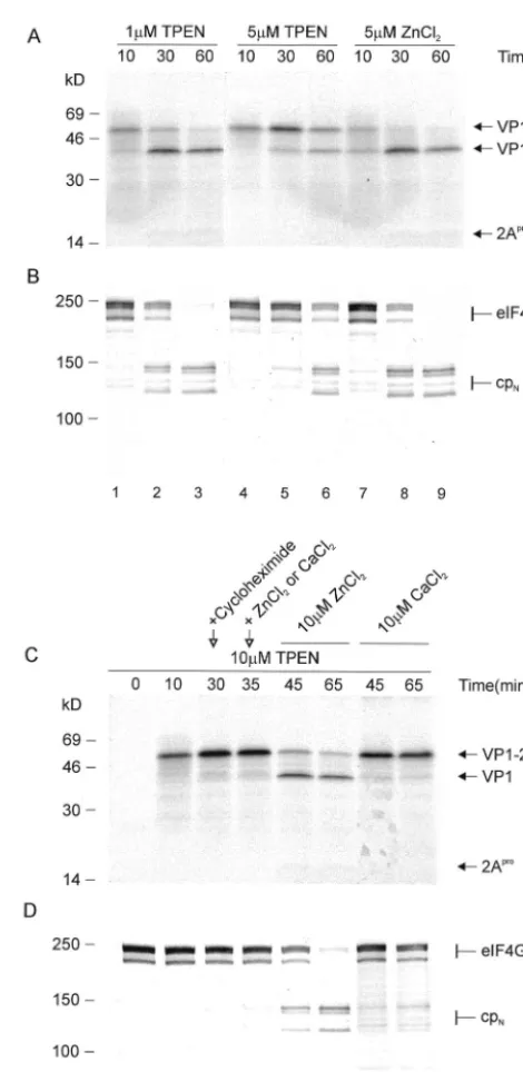
mRNA. Figure 2A and B show that increasing concentrations
of TPEN inhibit 2Aproself-processing and eIF4GI-processing
ability. At 5M TPEN, 50% self-processing and eIF4GI
cleav-age are observed at 60 min, whereas the control shows 50% cleavage at 30 min and almost complete cleavage at 60 min (compare Fig. 2, lanes 5 and 6 with Fig. 1, lanes 3 and 4). In
contrast, the addition of zinc in the absence of TPEN does not
influence 2Aproactivity in any way (Fig. 2, lanes 7 to 9). This
suggests that the endogenous zinc concentration in the RRL is
[image:3.603.303.538.65.547.2]sufficient for 2Apro activity, despite the presence of the very
FIG. 2. Effect of zinc concentration on HRV2 2Aproactivity. (A and B) The mRNA VP1-2Apro (⬃10 ng/l) was used to program RRLs; the indicated concentrations of the zinc chelator TPEN or ZnCl2were added to the translation mixture on ice prior to incubation. The HRV2 VP1-2ApromRNA (⬃10 ng/l) was translated in RRLs in the presence of 10M TPEN. After 30 min, cycloheximide was added to a final concentration of 10 ng/l to inhibit further protein synthesis. (C and D) After a further 5 min of incubation, ZnCl2or CaCl2was added to a final concentration of 10M, respectively. Aliquots were removed at the indicated time points, and protein synthesis (A and C) and eIF4GI state (B and D) were monitored as for Fig. 1.
on November 8, 2019 by guest
http://jvi.asm.org/
high amounts of EGTA used to chelate the calcium ions re-quired for micrococcal nuclease treatment. The translation machinery is not influenced by zinc depletion or higher zinc
amounts, since concentrations up to 200 M of TPEN and
zinc, respectively, were tested without observable effects on protein synthesis (data not shown).
Zinc-depleted HRV2 2Aprois rapidly activated by addition
of zinc without requiring denaturation and renaturation.
Pre-vious experiments (25, 30) have shown that HRV2 2Aprobinds
the zinc very tightly. The zinc cannot be extracted by 10 mM
EDTA; removal of the zinc from HRV2 2Apro was achieved
only after denaturation. The zinc-depleted denatured enzyme could be reactivated by renaturation in the presence of zinc. We were interested whether the de novo synthesized protease could be activated by addition of zinc without any special denaturation and renaturation steps. Therefore, it was
neces-sary to completely inhibit HRV2 2Apro; this was achieved by
translating the VP1-2Apro mRNA in the presence of 10 M
TPEN (Fig. 2C and D, lanes 11 and 12). After 30 min,
cyclo-heximide was added to a concentration of 10 ng/l to stop the
synthesis of new 2Apro(Fig. 2, lane 12). At 35 min, we added
either ZnCl2to a 10M concentration to activate the 2Aproor
CaCl2 as a control to the reaction mix and continued the
incubation (Fig. 2, lanes 13 to 17). The presence of 10 M
TPEN inhibits 2Apro almost completely, since after 65 min
without ZnCl2(Fig. 2, lane 17) essentially no self-processing or
eIF4GI cleavage is observed. In contrast, just 10 min after
addition of 10M ZnCl2(Fig. 2, lane 14), both VP1-2Aproand
eIF4GI cleavage had reached about 50%. The absence of the previously observed 10-min delay indicates that the
incorpora-tion of zinc into de novo-synthesized VP1-2Aprois a very rapid
step.
It is worth noting that neither the addition of TPEN nor that
of zinc (both compounds were tested at 5 M) affected Lpro
activity in any way, showing that the effect is specific to HRV2
2Apro(data not shown).
TPEN can protect HeLa cells from the viral cytopathic ef-fect.Since the presence of TPEN can prevent the activation of
de novo synthesized HRV2 2Apro, we investigated whether
TPEN can be used to block HRV replication in cell culture. TPEN is able to permeate membranes and thus ideally suited for this purpose. To examine any possible differences between minor and major group HRVs, both HRV2 and HRV14 were used. HeLa cells were incubated with different concentrations of TPEN, and the cells were infected with HRV2 or HRV14 or incubated with medium alone. The viability of the cells was
then assayed after 24 h using the Cell Titer 96 AQueous
non-radioactive cell proliferation assay (Promega). Briefly, 106cells
were seeded into a 96-well plate 1 day prior to infection. Cells were infected using HRV serotypes 2 and 14 at 20 50% tissue culture infective doses/cell in the presence or absence of the indicated TPEN concentrations. Each experiment was re-peated three times. Twenty-four hours after infection, cell vi-ability was determined by adding the tetrazolium compound followed by incubation for 2 h at 37°C and subsequent mea-surement of absorption at 492 nm in a Labsystems Multiscan RC plate reader.
Figure 3 shows that uninfected HeLa cells remain viable in the presence of TPEN up to concentrations between 1.25 and
1.8 M. Higher TPEN concentrations are increasingly
cyto-toxic. The cytotoxic effect of TPEN is not unexpected, since depletion of zinc in HeLa cells leads to activation of specific caspases which then induce apoptosis (5, 13, 18). The concen-trations of TPEN at which apoptosis was induced were very similar to those reported previously (18).
Despite the loss of cell viability due to the induction of apoptosis by TPEN, protection of cells from viral infection was
observed at TPEN concentrations between 1.8M and 2.5M
(Fig. 3). With HRV14 (Fig. 3B), the protection was more pronounced, with 60% of cells protected at a concentration of
1.25 M, a concentration below that required for inducing
apoptosis. Thus, the inhibition of HRV infection with a potent zinc chelator is feasible.
It has been known for many years that the addition of zinc itself inhibits HRV replication. The mechanism of this inhibi-tion remains unclear, but it appears to involve binding of zinc to the part of the polyprotein containing the capsid precursors (14). In contrast, we have shown that inhibition of replication of two serotypes could be achieved by the chelation of zinc, brought about by the presence of TPEN. Although chelation of zinc by TPEN induces apoptosis, a concentration range was found which protected cells to a significant extent against in-fection by both HRV2 and HRV14. Notably, the concentration
range of 1.5 to 2M is clearly lower than the 10M required
[image:4.603.306.544.77.361.2]for 2Aproinhibition in RRLs. This implies either that the zinc
FIG. 3. Protection of HeLa cells by TPEN from HRV infection. HeLa cells were incubated in 96-well plates with the indicated TPEN concentrations and incubated with 20 50% tissue culture infective doses (open bars) of HRV2 (A) or HRV14 (B) or medium (closed bars) as a control. Twenty-four hours after infection, cell viability was determined. The mean and error bars were calculated from three separate experiments.
on November 8, 2019 by guest
http://jvi.asm.org/
concentration is lower in HeLa cells than in RRLs or that TPEN is also affecting other cellular processes. Since TPEN has been shown to interfere with endocytosis (1), it seems possible that viral entry may also be affected.
The rapid induction of apoptosis by TPEN in HeLa cells would seem to preclude its use as a possible antiviral com-pound. However, other cell types, such as macrophages, have been shown to remain unaffected by TPEN concentrations as
high as 150M (1). Thus, future work will be directed towards
determining the TPEN sensitivities of cell lines capable of supporting HRV replication. An investigation of the useful-ness of TPEN and related zinc chelators as inhibitors of HRV and human enterovirus replication can then be made with the cell lines most resistant to TPEN. Furthermore, it will be of interest to examine whether TPEN can also inhibit the
repli-cation of polio- and coxsackieviruses. Although the 2Apros of
these viruses have not been directly shown to contain zinc, site-directed mutagenesis and amino acid alignments (31) strongly imply the ability of these proteinases to bind zinc and thus their sensitivity to TPEN during viral replication.
This work was supported by the Austrian Science Foundation (grants P-13367 and P-16189 to T.S.).
We thank D. Blaas, E. Gaudernak, J. Seipelt, and members of our laboratory for stimulating discussions.
REFERENCES
1. Aballay, A., M. N. Sarrouf, M. I. Colombo, P. D. Stahl, and L. S. Mayorga. 1995. Zn2⫹depletion blocks endosome fusion. Biochem J.312:919–923. 2. Belsham, G. J., and R. R. Jackson.2000. Translation initiation on
picorna-virus RNA, p. 869–900.InN. Sonenberg, J. W. B. Hershey, and M. B. Mathews (ed.), Translational control of gene expression, vol. 39. Cold Spring Harbor Laboratory Press, Cold Spring Harbor, N.Y.
3. Bovee, M. L., B. J. Lamphear, R. E. Rhoads, and R. E. Lloyd.1998. Direct cleavage of elF4G by poliovirus 2A protease is inefficient in vitro. Virology 245:241–249.
4. Cherny, R. A., J. T. Legg, C. A. McLean, D. P. Fairlie, X. Huang, C. S. Atwood, K. Beyreuther, R. E. Tanzi, C. L. Masters, and A. I. Bush.1999. Aqueous dissolution of Alzheimer’s disease Abeta amyloid deposits by bio-metal depletion. J. Biol. Chem.274:23223–23228.
5. Chimienti, F., M. Seve, S. Richard, J. Mathieu, and A. Favier.2001. Role of cellular zinc in programmed cell death: temporal relationship between zinc depletion, activation of caspases, and cleavage of Sp family transcription factors. Biochem. Pharmacol.62:51–62.
6. Dougherty, W. G., and B. L. Semler.1993. Expression of virus-encoded proteinases—functional and structural similarities with cellular enzymes. Microbiol. Rev.57:781–822.
7. Foeger, N., W. Glaser, and T. Skern.2002. Recognition of eIF4G isoforms by picornaviral proteinases. J. Biol. Chem.277:44300–44309.
8. Glaser, W., R. Cencic, and T. Skern.2001. Foot-and-mouth disease leader proteinase: involvement of C-terminal residues in self-processing and cleav-age of eIF4GI. J. Biol. Chem.276:35473–35481.
9. Glaser, W., and T. Skern.2000. Extremely efficient cleavage of eIF4G by picornaviral proteinases L and 2A in vitro. FEBS Lett.480:151–155. 10. Gorbalenya, A. E., E. V. Koonin, and M. M. Lai.1991. Putative
papain-related thiol proteases of positive-strand RNA viruses. Identification of rubi-and aphthovirus proteases rubi-and delineation of a novel conserved domain associated with proteases of rubi-, alpha- and coronaviruses. FEBS Lett. 288:201–205.
11. Gradi, A., Y. V. Svitkin, H. Imataka, and N. Sonenberg.1998. Proteolysis of human eukaryotic translation initiation factor eIF4GII, but not eIF4GI, coincides with the shutoff of host protein synthesis after poliovirus infection. Proc. Natl. Acad. Sci. USA95:11089–11094.
12. Guarne´, A., J. Tormo, K. Kirchweger, D. Pfistermueller, I. Fita, and T. Skern.1998. Structure of the foot-and-mouth disease virus leader protease: a papain-like fold adapted for self-processing and eIF4G recognition. EMBO J.17:7469–7479.
13. Jiang, S., S. C. Chow, M. J. McCabe, Jr., and S. Orrenius.1995. Lack of Ca2⫹involvement in thymocyte apoptosis induced by chelation of intracel-lular Zn2⫹. Lab. Investig.73:111–117.
14. Korant, B. D., and B. E. Butterworth.1976. Inhibition by zinc of rhinovirus protein cleavage: interaction of zinc with capsid polypeptides. J. Virol.18: 298–306.
15. Kuechler, E., J. Seipelt, H.-D. Liebig, and W. Sommergruber.2002. Picor-navirus proteinase-mediated shutoff of host cell translation: direct cleavage of a cellular initiation factor, p. 301–311.InB. L. Semler and E. Wimmer (ed.), Molecular biology of picornaviruses. ASM Press, Washington, D.C. 16. Liebig, H.-D., E. Ziegler, R. Yan, K. Hartmuth, H. Klump, H. Kowalski, D.
Blaas, W. Sommergruber, L. Frasel, B. Lamphear, R. Rhoads, E. Kuechler, and T. Skern.1993. Purification of two picornaviral 2A proteinases: inter-action with eIF-4G and influence on translation. Biochemistry32:7581–7588. 17. Lloyd, R. E., M. J. Grubman, and E. Ehrenfeld.1988. Relationship of p220 cleavage during picornavirus infection to 2A proteinase sequencing. J. Virol. 62:4216–4223.
18. McCabe, M. J., Jr., S. A. Jiang, and S. Orrenius.1993. Chelation of intra-cellular zinc triggers apoptosis in mature thymocytes. Lab. Investig.69:101– 110.
19. Merrick, W. C., and J. W. B. Hershey.2000. Pathway and mechanism of initiation of protein synthesis, p. 33–88.InN. Sonenberg, W. C. Merrick, and J. W. B. Hershey (ed.), Translational control of gene expression, vol. 39. Cold Spring Harbor Laboratory Press, Cold Spring Harbor, N.Y. 20. Morley, S. J., P. S. Curtis, and V. M. Pain.1997. eIF4G: translation’s mystery
factor begins to yield its secrets. RNA3:1085–1104.
21. Oberst, M. D., T. J. Gollan, M. Gupta, S. R. Peura, J. D. Zydlewski, P. Sudarsanan, and T. G. Lawson.1993. The encephalomyocarditis virus 3C protease is rapidly degraded by an ATP-dependent proteolytic system in reticulocyte lysate. Virology193:28–40.
22. Petersen, J. F., M. M. Cherney, H. D. Liebig, T. Skern, E. Kuechler, and M. N. James.1999. The structure of the 2A proteinase from a common cold virus: a proteinase responsible for the shut-off of host-cell protein synthesis. EMBO J.18:5463–5475.
23. Skern, T., B. Hampoelz, E. Bergmann, A. Guarne´, J. Petersen, I. Fita, and M. N. G. James.2002. Structure and function of picornavirus proteinases, p. 199–212.InB. L. Semler and E. Wimmer (ed.), Molecular biology of picor-naviruses. ASM Press, Washington, D.C.
24. Skern, T., W. Sommergruber, H. Auer, P. Volkmann, M. Zorn, H. D. Liebig, F. Fessl, D. Blaas, and E. Kuechler.1991. Substrate requirements of a human rhinoviral 2A proteinase. Virology181:46–54.
25. Sommergruber, W., G. Casari, F. Fessl, J. Seipelt, and T. Skern.1994. The 2A proteinase of human rhinovirus is a zinc containing enzyme. Virology 204:815–818.
26. Strebel, K., and E. Beck.1986. A second protease of foot-and mouth disease virus. J. Virol.58:893–899.
27. Svitkin, Y. V., A. Gradi, H. Imataka, S. Morino, and N. Sonenberg.1999. Eukaryotic initiation factor 4GII (eIF4GII), but not eIF4GI, cleavage cor-relates with inhibition of host cell protein synthesis after human rhinovirus infection. J. Virol.73:3467–3472.
28. Tsukada, H., and D. M. Blow.1985. Structure of alpha-chymotrypsin refined at 1.68 A˚ resolution. J. Mol. Biol.184:703–711.
29. Ventoso, I., S. E. MacMillan, J. W. Hershey, and L. Carrasco.1998. Polio-virus 2A proteinase cleaves directly the eIF-4G subunit of eIF-4F complex. FEBS Lett.435:79–83.
30. Voss, T., R. Meyer, and W. Sommergruber.1995. Spectroscopic character-ization of rhinoviral protease 2A: Zn is essential for the structural integrity. Protein Sci.4:2526–2531.
31. Yu, S. Y. F., and R. E. Lloyd.1992. Characterization of the roles of conserved cysteine and histidine residues in poliovirus 2A-protease. Virology186:725– 735.
32. Zamora, M., W. E. Marissen, and R. E. Lloyd.2002. Multiple eIF4GI-specific protease activities present in uninfected and poliovirus-infected cells. J. Virol.76:165–177.
33. Zamora, M., W. W. Marissen, and R. E. Lloyd.2002. Poliovirus-mediated shutoff of host translation: an indirect effect, p. 313–320.InB. L. Semler and E. Wimmer (ed.), Molecular biology of picornaviruses. ASM Press, Wash-ington, D.C.