Copyright © 2001, American Society for Microbiology. All Rights Reserved.
Interleukin-8 Stimulates Human Immunodeficiency Virus
Type 1 Replication and Is a Potential New Target
for Antiretroviral Therapy
BRIAN R. LANE,1,2KARIN LORE,3PAUL J. BOCK,1,2JAN ANDERSSON,4MICHAEL J. COFFEY,5 ROBERT M. STRIETER,5†ANDDAVID M. MARKOVITZ1,2*
Divisions of Infectious Diseases1and Pulmonary and Critical Care Medicine,5Department of Internal Medicine, and
Graduate Program in Cellular and Molecular Biology,2University of Michigan Medical Center, Ann Arbor,
Michigan 48109-0640, and Department of Microbiology, Pathology and Immunology3and
Department of Medicine, Center for Infectious Medicine,4Karolinska Institutet, Stockholm, Sweden
Received 25 January 2001/Accepted 21 May 2001
Production of the C-X-C chemokines interleukin-8 (IL-8) and growth-regulated oncogene alpha (GRO-␣) in
macrophages is stimulated by exposure to human immunodeficiency virus type 1 (HIV-1). We have
demon-strated previously that GRO-␣then stimulates HIV-1 replication in both T lymphocytes and macrophages.
Here we demonstrate that IL-8 also stimulates HIV-1 replication in macrophages and T lymphocytes. We further show that increased levels of IL-8 are present in the lymphoid tissue of patients with AIDS. In addition,
we demonstrate that compounds which inhibit the actions of IL-8 and GRO-␣via their receptors, CXCR1 and
CXCR2, also inhibit HIV-1 replication in both T lymphocytes and macrophages, indicating potential thera-peutic uses for these compounds in HIV-1 infection and AIDS.
Altered cytokine production by cells exposed to human im-munodeficiency virus type 1 (HIV-1) contributes to the sys-temic symptoms of AIDS (cachexia, anorexia, and malaise) (29), HIV-1-related brain disease (21), and recruitment of immune cells to infected tissue (42). The introduction of highly active antiretroviral therapy (HAART) has dramatically re-duced HIV-1 mortality in the United States since 1996 (38). Unfortunately, many patients cannot tolerate therapy, and in others, resistance to the drugs develops (19). Therefore, new viral and cellular targets have been sought for the treatment of HIV-1 infection, either alone or in combination with HAART (24). Cytokines and their receptors are one group of such potential targets for therapy of HIV-1 infections.
In the past few years, it has been shown that the C-C che-mokines RANTES, MIP-1␣and MIP-1suppress HIV-1 rep-lication (12, 13). The actions of these chemokines are believed to be related to the fact that they are ligands for CCR5, the principal coreceptor used by monocytotropic isolates of HIV (1, 11, 14, 17, 18). Similarly, SDF-1␣, the only known ligand for CXCR4, the principal coreceptor for T-tropic isolates of HIV, inhibits the replication of CXCR4-using isolates of HIV (X4 HIV) (8, 20, 36). RANTES, MIP-1␣, MIP-1, and SDF-1␣
inhibit HIV-1 replication both by competing with HIV for binding to CCR5 or CXCR4 and by causing internalization of their respective receptors (2, 3, 41). Interestingly, under some circumstances these same chemokines can actually enhance HIV-1 replication (16, 23, 25, 26, 33, 40, 43). The mechanisms by which RANTES and SDF-1␣ can act to augment HIV-1
replication include increasing viral attachment to, and entry into, target cells (16, 23, 26, 43), activating intracellular signal-ing pathways (23, 25), and augmentsignal-ing viral gene expression from the HIV-1 long terminal repeat (33).
The role that other chemokines, including two members of the C-X-C chemokine family, interleukin-8 (IL-8) and growth-regulated oncogene alpha (GRO-␣), may play in controlling HIV-1 replication and pathogenesis has not been well estab-lished. IL-8 has been demonstrated to attract neutrophils and T cells, stimulate monocyte adherence, and mediate angiogen-esis by interacting with the C-X-C chemokine receptors CXCR1 and CXCR2 (6, 22, 27, 31, 47). GRO-␣was identified initially as a melanoma growth factor and later as a neutrophil chemoattractant (6). GRO-␣shares 43% amino acid identity with IL-8 and functions similar to IL-8 by means of its ability to ligate CXCR2 (6). Previous investigations have found either a slight inhibitory effect or no effect of IL-8 on HIV-1 replication (10, 32, 35), and GRO-␣was not previously known to have any effect on viral replication. In addition, neither CXCR1 nor CXCR2 has been demonstrated to function as a coreceptor for HIV entry (19).
There is currently great interest in agents that block these same chemokines, or their cognate receptors, for the treatment of a number of illnesses, particularly inflammatory diseases (7). For example, an IL-8-specific monoclonal antibody is currently in use in clinical trials of patients with psoriasis (46). Other drug discovery efforts aimed at these pathways produced SB225002, the first reported potent and selective nonpeptide inhibitor of a chemokine receptor (45). This small molecule inhibitor acts as an antagonist of IL-8 binding to CXCR2 (50% inhibitory concentration⫽22 nM), and has⬎150-fold selec-tivity over CXCR1 and other chemokine receptors (45).
Several recent findings suggest that interfering with IL-8 and GRO-␣function would be an effective therapy for HIV-1 in-fection. First, elevated levels of both IL-8 and GRO-␣ are * Corresponding author. Mailing address: 5220 MSRB III, 1150 W.
Medical Center Dr., Ann Arbor, MI 48109-0640. Phone: (734) 647-1786. Fax: (734) 764-0101. E-mail: [email protected].
† Present address: Department of Medicine, Division of Pulmonary and Critical Care Medicine, UCLA School of Medicine, Los Angeles, CA 90095-1922.
8195
on November 9, 2019 by guest
http://jvi.asm.org/
present in the serum and lungs of HIV-1-infected individuals (15, 34, 44). We have recently demonstrated that exposure of MDM to HIV-1 leads to increased IL-8 production, an effect mediated by Tat and the inflammatory cytokine tumor necrosis factor alpha, as well as by gp120 (B. R. Lane et al., submitted for publication). In addition, we have described a novel auto-crine/paracrine loop in which HIV-1 gp120 ligation of CXCR4 on monocyte-derived macrophages (MDM) stimulates the production of GRO-␣, and GRO-␣further stimulates HIV-1 replication (30a).
We demonstrate here that IL-8 stimulates HIV-1 replication in MDM and T lymphocytes. We also show that increased levels of IL-8 are present in the lymphoid tissue of patients with AIDS. Antibodies that neutralize IL-8 activity, and anti-bodies that block binding to the receptors CXCR1 and CXCR2, can inhibit HIV-1 replication in macrophages and T cells. Blocking the actions of IL-8 and GRO-␣with the small-molecule inhibitor of CXCR2 SB225002 also markedly reduces HIV-1 replication. Thus, we have shown that the autocrine/ paracrine loop in which IL-8 and GRO-␣participate is a po-tential target for antiretroviral therapy. Therapeutic com-pounds currently under development for chemokine-mediated inflammatory disease therefore have the potential to be ex-ploited for the therapy of HIV infection and AIDS.
MATERIALS AND METHODS
Isolation and preparation of human AM, MDM, and peripheral blood lym-phocytes (PBL).Alveolar macrophages (AM) were collected by bronchoalveolar lavage of nonsmoking volunteers without lung disease or HIV-1 infection as described previously (30). The recovered bronchoalveolar lavage fluid was cen-trifuged to collect the cellular portion, and cells were resuspended in Dulbecco modified Eagle medium (DMEM) supplemented with 10% fetal bovine serum, 2 mM glutamine, 100 U of penicillin per ml, and 100g of streptomycin per ml (complete DMEM). AM were purified by plastic adherence and cultured 5 days before infection.
Peripheral blood mononuclear cells (PBMC) were collected by venipuncture of healthy volunteers as described previously (30). PBMC contained approxi-mately 20% CD14⫹monocytes as determined by flow cytometry. To separate the
PBMC into subpopulations composed mainly of monocytes or lymphocytes, PBMC were subjected to a plate adherence step for 2 h. Adherent cells were consistently⬎90% peripheral blood monocytes (PBM) as determined by Diff-Quik analysis and⬎85% CD14⫹as determined by flow cytometric staining with
a phycoerythrin-conjugated mouse anti-human monoclonal antibody to CD14 (M5E2; PharMingen), as well as ⬎99% viable as determined by trypan blue exclusion. PBM were differentiated into MDM by culture in complete DMEM for up to 2 weeks (3 days in most experiments) prior to infection.
Nonadherent PBMC following the plate adherence step were enriched for lymphocytes and contained less than 2% CD14⫹monocytes. These
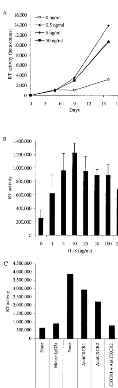
monocyte-FIG. 1. Exogenous IL-8 stimulates HIV-1 replication in AM and MDM. (A) AM were cultured for 5 days before infection with HIV-1BaLand then treated with IL-8 at either 0.5, 5, or 50 ng/ml. Superna-tants (25%) were collected twice weekly and replaced with fresh me-dium. Viral replication was assessed by quantifying the RT activity present in the supernatants at several time points after infection. (B) MDM were treated with IL-8 at the indicated concentrations every 3 days beginning 1 day before infection with HIV-1BaL. Supernatants were analyzed for RT activity 8 days after infection. The increase observed with IL-8 treatment in the range from 5 to 100 ng/ml was determined to be statistically significant relative to no treatment (P⬍ 0.05, Student’sttest). (C) Antibodies to CXCR1 and CXCR2 block the ability of IL-8 and GRO-␣(each at 25 ng/ml) to stimulate HIV-1BaLreplication in MDM. Supernatants were analyzed for RT activity 19 days after infection. Data shown are representative of five experi-ments.
on November 9, 2019 by guest
http://jvi.asm.org/
[image:2.612.77.270.76.708.2]depleted PBMC (PBL) were cultured at 1⫻106to 2⫻106/ml in RPMI 1640
supplemented with 10% fetal bovine serum, 2 mM glutamine, 100 U of penicillin per ml, and 100g of streptomycin per ml (complete RPMI). PBL were stim-ulated with 5g of phytohemagglutinin (PHA; Sigma, St. Louis, Mo.) per ml for 1 to 3 days and then maintained in IL-2 (40 U/ml; Hoffmann-La Roche, Nutley, N.J.). In some experiments, PBL were also depleted of CD8⫹cells with magnetic
Dynabeads M-450 CD8 as instructed by the manufacturer (Dynal, Lake Success, N.Y.).
Preparation of HIV-1 stocks.All of the HIV-1 isolates used in this study were originally obtained from the NIH AIDS Reagent Program. Stocks of HIV-1BaL
were prepared by infection of HOS-CD4-CCR5 cells and of HIV-1BRUby
infection of CEM-SS cells. For some experiments, viral stocks were prepared by infection of PHA-activated, CD8-depleted PBL; results were identical to those obtained using the cell line-derived viral isolates (data not shown). Other viral stocks were used directly as provided by the NIH AIDS Reagent Program.
HIV-1 infection of MDM and PBL.For each experiment, multiple wells of macrophages or PBL were infected with equal reverse transcriptase (RT) counts of HIV-1 (30⫻106to 300⫻106cpm of RT used per 105cells). This amount of
CPM of RT activity per cell corresponds to a multiplicity of infection of between 0.001 and 0.01 as determined by titration on HOS-CD4-CCR5 and CEM-SS cell lines. MDM were cultured for 3 days prior to infection, washed after an overnight incubation with the virus, and cultured in complete DMEM. PHA-activated PBL were washed and incubated in complete RPMI plus IL-2 (40 U/ml) and any other treatments overnight. PHA-activated PBL were then spin infected by incubation with HIV-1 for 4 h at 2,500 rpm, washed, and incubated in medium plus IL-2 and other treatments. A portion of the medium (25%) was removed from the MDM and PBL supernatants and replaced twice weekly.
Recombinant human IL-8 and GRO-␣, and monoclonal antibodies to CXCR1, CXCR2, IL-8, and GRO-␣, were obtained from R&D Systems (Min-neapolis, Minn.) and added as indicated in the figure legends. SB225002 was
obtained from Calbiochem (San Diego, Calif.) and reconstituted in dimethyl sulfoxide (DMSO). All other chemicals were obtained from Sigma.
RT assay.Quantification of the RT activity present in the supernatants using a poly (A)-oligo(dT) template primer was used as a measure of HIV-1 replica-tion as previously described (39). RT activity was assayed using32P-labeled dTTP
incorporated into DNA bound to DE81 paper (Whatman) and quantified using either the Series 400 PhosphorImager and ImageQuant software (Molecular Dynamics, Sunnyvale, Calif.) or a Betascope radioisotope imaging system. RT activity is reported as phosphorimager counts, except in Fig. 1A and Fig. 2A, where activity is reported as beta counts.
Patients and controls.Tissue biopsies from lymph nodes and tonsils were collected from 1-infected patients and snap-frozen. The biopsies from HIV-1-infected patients (n⫽4) with recent seroconversion were taken within 6 months after verified infection. These patients had a mean viral load of 46,795 HIV-1 RNA copies/ml of plasma and a mean CD4 count of 563/l. Untreated, asymptomatic, chronically HIV-1-infected patients (n⫽5) had a mean viral load of 12,000 copies/ml and a mean CD4 count of 510/l. Patients treated with ongoing HAART (n⫽2) had a mean viral load of⬍50 copies/ml and a mean CD4 count of 960/l. Patients with AIDS (n⫽3) had a mean viral load of 1,000,000 copies/ml and a mean CD4 count of 54/l. Control tissue was obtained from HIV-1 seronegative healthy adults (n⫽3) after elective tonsillectomy following institutional review board approval from Huddinge University Hospi-tal, Huddinge, Sweden.
Detection of IL-8 expression by immunohistochemistry.Cryopreserved biop-sies embedded in OCT compound (Tissue-TEK; Miles, Elkhart, Ind.) were cut in 8-m-thick sections, mounted on HTC glass slides (Novakemi, Stockholm, Swe-den), and fixed with 2% formaldehyde (Sigma) in phosphate-buffered saline for 15 min at room temperature. Slides were then washed with balanced salt solution (BSS) (Gibco Ltd., Paisley, United Kingdom) and stored at⫺20°C for future use. The staining procedure used to identify IL-8 expression in tissue sections at the single-cell level has previously been described (4, 5). Briefly, endogenous perox-idase was blocked by 1% H2O2in BSS supplemented with 0.01 M HEPES buffer
(Gibco) and 0.1% saponin (Riedel de Haen, AG Seelze, Germany). The IL-8-specific monoclonal antibody (NAP-1; 2g/ml; Immunokontakt, Bioggio, Swit-zerland) was diluted in BSS-saponin and applied overnight at room temperature. After washes in BSS, the sections were incubated with a biotin-labeled secondary antibody (goat anti-mouse immunoglobulin G [IgG]; Caltag Laboratories, South San Francisco, Calif.) for 30 min, followed by incubation with an avidin-biotin-horseradish peroxidase complex (Vectastain; Vector Laboratories). A color re-action was developed by addition of 3⬘-diaminobenzidine tetrahydrochloride (Vector Laboratories). The sections were counterstained with hematoxylin.
The tissue sections were examined in a Leica RXM microscope (Leica, Wetz-lar, Germany) equipped with a 3CCD color camera (DXC-950p; Sony Corpo-ration, Tokyo, Japan). IL-8-expressing cells were identified at the single-cell level due to a characteristic staining pattern of protein localized in the Golgi stacks (4). The specificity of immunofluorescent staining was determined by incubation in the presence of a 10-fold molar excess of recombinant IL-8, which abolished staining. In addition, PBMC stimulated with lipopolysaccharide were used as a positive control for staining. The number of positively stained cells in the total tissue (3⫻106to 5⫻106m2) was counted manually. As the evaluation of
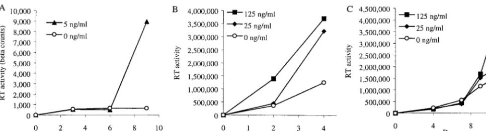
[image:3.612.62.545.72.202.2]separate positive cells can be difficult due to expected clusters of IL-8-expressing cells, the number of positive cells was sometimes presented as a range. FIG. 2. Exogenous IL-8 stimulates HIV-1 replication in PBL. (A) CD8-depleted PBL were treated with the indicated concentrations of IL-8 and infected with HIV-1BaL(A), HIV-1BRU(B), or HIV-2CBL23(C). Supernatants were collected twice weekly and replenished with fresh medium.
TABLE 1. IL-8-expressing cells in the lymphoid tissue of HIV-1-infected individualsa
Category
No. of cells expressing IL-8
in lymphoid tissue positive cellsMean no. of (Pvalueb)
1 2 3 4 5
AIDS 14–16 19–24 20–40 22.2
Seronegative controls 0 2–4 4 2.3 (0.026) Recent HIV-1
sero-converters 0 1 1 10 3.0 (0.034)
Chronic, asymptomatic
HIV-1 infection 1–2 1–2 1–4 2–5 2–6 2.6 (0.037) HAART-treated
HIV-1 infection 1 8 4.5 (0.012)
aThe number of cells that stained positively for IL-8 is indicated for patients
in each of several clinical categories (described in Materials and Methods).
bRelative to individuals with AIDS (Student’sttest). The differences between
the other groups were not found to be statistically significant.
on November 9, 2019 by guest
http://jvi.asm.org/
RESULTS
Exogenous IL-8 stimulates HIV-1 replication in MDM and PBL.Previous studies have found increased IL-8 in the context of HIV-1 infection, but none have implicated IL-8 in HIV-1 pathogenesis (10, 15, 28, 34, 37). We have recently demon-strated that exposure of MDM to HIV-1 leads to increased IL-8 production, an effect mediated by Tat and the inflamma-tory cytokine tumor necrosis factor alpha, as well as by gp 120 (Lane et al., submitted). As certain chemokines (e.g., RANTES, MIP-1␣, MIP-1, and SDF-1␣) have been demon-strated to play important roles in HIV-1 pathogenesis, mainly as inhibitors of viral entry (9, 12, 36), we studied the effect of IL-8, and signaling through its receptors CXCR1 and CXCR2, on HIV replication in macrophages and T lymphocytes. Rep-lication of the macrophage-tropic R5 isolate HIV-1BaLin AM was increased by treatment with IL-8 (Fig. 1A). HIV-1BaL replication in MDM was also increased significantly by the addition of IL-8 at doses of between 5 and 100 ng/ml (Fig. 1B). These amounts of IL-8 are most likely physiologic, since they are within the range produced by MDM following exposure to HIV-1 (Lane et al., submitted). We evaluated the addition of IL-8 before infection, after infection, or before and after in-fection in a number of experiments. Each of these three treat-ment regimens resulted in an increase in HIV-1 replication,
and we found no significant difference between the regimens in a number of experiments (data not shown). In experiments performed with MDM from 17 different donors, IL-8 increased HIV-1 replication in 15 of the 17 donors (mean, 6.2-fold; range, 0.5- to 50-fold). IL-8 does not appear to confer suscep-tibility to productive infection with X4 HIV on MDM, as viral replication was not detected when MDM from multiple donors were infected with HIV-1BRU in the presence or absence of exogenous IL-8 (data not shown).
We have recently demonstrated that a second chemokine that can signal through CXCR2, GRO-␣, stimulates HIV-1 replication at a range of doses similar to that of IL-8 (30a). We therefore hypothesized that the effect of these chemokines may be mediated by the receptor CXCR2 and possibly by CXCR1, which can also act as a receptor for IL-8. Indeed, the effects of IL-8 and GRO-␣ on HIV-1 replication were blocked by a combination of antibodies that prevent interaction with CXCR1 and CXCR2 (Fig. 1C). These data indicate that IL-8, which is produced in response to exposure to HIV-1, enhances HIV-1 replication in primary human MDM by acting through its receptors (CXCR1 and CXCR2).
We next investigated whether IL-8 had an effect on HIV-1 replication in CD4⫹T lymphocytes. Addition of IL-8 in doses exceeding 5 ng/ml stimulated viral replication in PHA-acti-FIG. 3. IL-8-expressing cells in lymphoid tissue from HIV-1-infected individuals. IL-8-expressing cells found by intracellular immunohisto-chemical staining of a lymph node section from an AIDS patient are shown at a magnification of⫻200. In the high-power magnification (⫻630; inset), the intracellular localization of IL-8 protein to the Golgi-endoplasmic reticulum complex can be seen.
on November 9, 2019 by guest
http://jvi.asm.org/
vated, CD8-depleted PBL (Fig. 2). Replication of the R5 iso-late HIV-1BaLin activated PBL was stimulated in 10 of the 11 donors tested (mean, 4.8-fold; range, 0.8- to 19-fold) by the addition of IL-8 (Fig. 2A). IL-8 also stimulated the replication of X4 isolates of HIV, including HIV-1BRU (Fig. 2B) and HIV-2CBL23 (Fig. 2C). As observed with infection of MDM, stimulation of HIV-1BaL replication in PBL is mediated through both CXCR1 and CXCR2, as anti-CXCR2 alone par-tially reduced, and CXCR2 in combination with anti-CXCR1 completely prevented, the stimulatory effect of exog-enous IL-8 (data not shown).
IL-8 is increased in the lymphoid tissue of AIDS patients.
Previous investigators have demonstrated that levels of IL-8 are increased in the sera and lungs of HIV-1-infected individ-uals (15, 34, 44). To determine whether IL-8 levels are also increased in the microenvironment in which much of HIV-1 replication occurs in vivo, we evaluated the expression of IL-8 in lymphoid tissue from individuals at various stages of clinical disease progression. Tissue was collected from individuals with AIDS, from those whose infections had been successfully treated with HAART, from those with chronic asymptomatic HIV-1 infection, from recent HIV-1 servoconverters, and from seronegative controls (Table 1). IL-8 was readily detected in the lymphoid tissue by immunohistochemical staining (Fig. 3). Examination of tissue at high power demonstrated that IL-8 is present in the Golgi-endoplasmic reticulum complex within lymphoid cells (Fig. 3). Preincubation of tissue sections with a 10-fold molar excess of IL-8 eliminated the signal, demonstrat-ing the specificity of the immunohistochemical staindemonstrat-ing (refer-ence 4 and data not shown). A clear increase in the number of IL-8-expressing cells was present in individuals with AIDS compared with individuals in all of the other groups (Table 1). Thus, significantly increased IL-8 expression is seen at a major site of HIV-1 replication, lymphoid tissue, in patients with high viral loads and disease progression.
Anti-IL-8, anti-CXCR1, and anti-CXCR2 antibodies and the small-molecule inhibitor SB225002 inhibit HIV-1 replication
in MDM and PBL. As increased levels of IL-8 are present
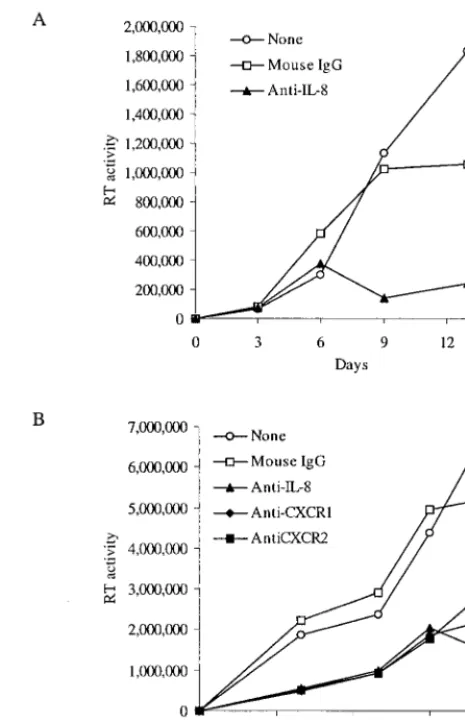
locally at the sites of HIV-1 infection in vivo, and as IL-8 stimulates HIV-1 replication, we hypothesized that inhibition of endogenous IL-8 might reduce HIV-1 replication. Our ex-periments using exogenous IL-8 indicated that small amounts of IL-8 (5 to 125 ng/ml) are sufficient to stimulate HIV-1 replication (Fig. 1 and 2). These amounts of IL-8 are in the same range as the amount induced by exposure of MDM to HIV-1 and produced constitutively by activated PBL (Lane et al., submitted). To test this hypothesis, we incubated HIV-1-infected cells with antibodies known to antagonize IL-8 func-tion. Addition of an antibody that neutralizes IL-8 activity markedly reduced HIV-1BaL replication in MDM compared with the addition of a mouse IgG control (Fig. 4A). Further-more, when the IL-8-specific antibody or antibodies that pre-vent interaction with the IL-8 receptors CXCR1 and CXCR2 were added to HIV-1BRU-infected PBL, viral replication was reduced considerably (Fig. 4B). Control mouse IgG antibody had no effect on HIV-1 replication in PBL.
As antibodies that neutralize the function of IL-8 (Fig. 4A) and GRO-␣(30a) and antibodies that prevent interaction with CXCR1 and CXCR2 (Fig. 4B) reduce HIV-1 replication, we hypothesized that inhibition of the pathways by which these
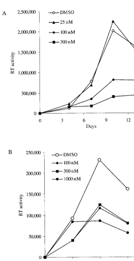
chemokines signal might serve as a target for anti-HIV drugs. We therefore tested the efficacy of a recently discovered in-hibitor of this pathway that is the type of compound potentially more suitable for clinical trials and use. The small-molecule inhibitor of CXCR2 SB225002 significantly inhibited replica-tion of HIV-1BaLin MDM (Fig. 5A) and of HIV-1BRUin PBL (Fig. 5B). The doses of SB225002 used in these experiments did not significantly affect cellular viability, proliferation, and activation of MDM or PBL, as determined by 3-(4,5-dimeth-ylthiazol-2-yl)-2,5-diphenyltetrazolium bromide (MTT) assay (see legend for Fig. 5). Replication of HIV-1 in MDM and PBL was also significantly diminished by a peptide inhibitor of IL-8 and GRO-␣function (data not shown). These data indi-cate that endogenous IL-8 production and signaling through CXCR1 and CXCR2 play stimulatory roles in HIV-1 replica-FIG. 4. Depletion of endogenous IL-8 inhibits HIV-1 replication in MDM and PBL. (A) MDM were treated with either mouse IgG1 or an antibody that neutralizes IL-8 activity (each at 20g/ml) and infected with HIV-1BaL. Medium was collected for the RT assay and replen-ished twice weekly. (B) Immunodepletion of IL-8 or blocking the IL-8 receptors CXCR1 and CXCR2 suppresses HIV-1 replication in CD8-depleted PBL. CD8-CD8-depleted PBL were treated with the indicated antibodies (20g/ml) and infected with HIV-1BRU. Medium was col-lected and replenished twice weekly. Supernatants were analyzed for RT activity on the indicated number of days after infection. Data shown are representative of four experiments.
on November 9, 2019 by guest
http://jvi.asm.org/
[image:5.612.313.546.74.436.2]tion in two major targets of infection, MDM and T lympho-cytes, and point to the existence of an autocrine/paracrine loop involving IL-8 and HIV-1 replication.
DISCUSSION
The presence of elevated levels of IL-8 in both the sera and lungs of individuals infected with HIV-1 has led several groups to suggest that IL-8 plays a role in HIV-1 pathogenesis, but little evidence has been presented to support these claims (15, 28, 34). Previous investigators found that inhibition of IL-8 had no effect on the activation of macrophages by HIV-1 Tat and that IL-8 had either no effect or a modest inhibitory effect on HIV-1 replication (10, 32, 35). However, we have recently demonstrated that IL-8 is produced by macrophages in re-sponse to HIV-1 and by endothelial cells in rere-sponse to Ka-posi’s sarcoma-associated herpesvirus and that IL-8 plays an important role in the early angiogenesis of Kaposi’s sarcoma (Lane et al., submitted). Here, we demonstrate that IL-8 stim-ulates HIV-1 replication in both MDM and activated T lym-phocytes, findings that are somewhat at odds with those of previous investigators. In contrast to these previous studies, we performed experiments with primary human macrophages and lymphocytes from a large number of individuals and found an effect with amounts of IL-8 that are similar to the amounts of IL-8 found in the sera and lungs of HIV-1-infected individuals. Previous investigators most likely did not detect an effect of IL-8 on HIV-1 replication because the doses used were well above the optimal dose for IL-8 (5 to 125 ng/ml). Our findings that IL-8 expression is increased in the lymphatic microenvi-ronment in AIDS patients and that inhibition of the activity of endogenous IL-8 markedly reduces HIV-1 replication suggest that the role of IL-8 in HIV-1 replication is likely to be bio-logically relevant. Of further note is the observation that IL-8 stimulates HIV-1 replication in two important target cells of infection, T cells and macrophages. Thus, HIV-1 infection leads to elevated production of IL-8 by MDM, and IL-8 then completes an autocrine/paracrine loop by, in turn, increasing HIV-1 replication in macrophages and lymphocytes. This loop presents an attractive target for antiretroviral therapy.
We have now demonstrated that blocking IL-8 and GRO-␣
[image:6.612.57.281.88.525.2]with either antibodies to IL-8, GRO-␣, CXCR1, or CXCR2 or the small-molecule inhibitor SB225002 inhibits HIV-1 replica-tion in two important target cells of infecreplica-tion, T lymphocytes and macrophages. SB225002 is a small-molecule inhibitor of chemokine receptor signaling that acts preferentially on CXCR2 (45). We have found that SB225002 is able to inhibit viral replication in both lymphocytes and macrophages, with-out negatively affecting cellular viability, at doses in the nano-molar range. Thus, inhibitors of CXCR2 signaling may break the vicious cycle in which HIV-1 infection leads to elevated production of IL-8 and GRO-␣, which then complete auto-crine/paracrine loops by in turn increasing HIV-1 replication. As one IL-8-specific monoclonal antibody (Abgenix) is cur-rently in clinical trials for psoriasis and additional compounds are being developed for the treatment of other inflammatory diseases, it appears attractive to test these agents for use as antiretroviral therapies. Our laboratory findings with SB225002 provide evidence that this or related compounds may be useful as antiretroviral agents as well. The fact that inhibitors of IL-8 FIG. 5. The small-molecule inhibitor SB225002 inhibits HIV-1
rep-lication in macrophages and T lymphocytes. (A) MDM were treated with 0.01% DMSO (the carrier control) or SB225002 at the doses indicated every 3 days beginning 1 day before infection with HIV-1BaL. The data shown are the mean of the RT activity present in triplicate wells measured on the days indicated. Cellular viability, proliferation, and activation were measured on day 13 by an MTT-based assay and determined to be 0.353 ⫾0.036, 0.382⫾0.040, 0.293⫾0.022, and 0.276⫾0.040 for treatment with 0, 25, 100, and 300 nM SB225002, respectively. (B) PHA-activated PBL were treated with DMSO (0.01%) or the indicated doses of SB225002 every 3 days beginning 1 day before infection with HIV-1BRU. HIV-1 replication was assayed by measuring the amount of RT activity in the supernatants on days 3, 6, and 9 after infection. Cellular viability, proliferation, and activation were measured on day 9 by an MTT-based assay and determined to be 0.052⫾0.017, 0.031⫾0.020, 0.023⫾0.017, and 0.046⫾0.018 for treatment with 0, 100, 300, and 1,000 nM SB225002, respectively. These experiments are representative of six (A) and three (B) inde-pendent experiments performed with cells from different donors.
on November 9, 2019 by guest
http://jvi.asm.org/
and CXCR2 function are already major targets for drug de-velopment, along with our findings, which indicate that IL-8 and GRO-␣ and their receptors may play a role in HIV-1 disease, calls for an exploration of the therapeutic potential of blocking this chemokine axis in HIV infection and AIDS.
ACKNOWLEDGMENTS
This work was supported by National Institutes of Health (NIH) grants AI36685 (D.M.M.) and HL57885 (M.J.C.) and by an NIH grant to the General Clinical Research Center at the University of Michigan (M01-RR00042). B.R.L. and P.J.B. were supported in part by the University of Michigan Medical Scientist Training Program (NIH grant NIGMS T32 GM07863) and the Graduate Program in Cellular and Molecular Biology (NIH grant GM07315). B.R.L. was additionally supported by the Molecular Mechanisms of Microbial Pathogenesis Training Program (NIH grant AI07528) and by funds from the Harvey Fellows Program.
HIV-1BRUwas provided by Steven King and Gary Nabel. HIV-1BAL
was obtained from Suzanne Gartner, Mikulas Popovic, and Robert Gallo through the AIDS Research and Reference Reagent Program, Division of AIDS, NIAID, NIH.
REFERENCES
1.Alkhatib, G., C. Combadiere, C. C. Broder, Y. Feng, P. E. Kennedy, P. M. Murphy, and E. A. Berger.1996. CC CKR5: a RANTES, MIP-1␣, MIP-1
receptor as a fusion cofactor for macrophage-tropic HIV-1. Science272:
1955–1958.
2.Alkhatib, G., M. Locati, P. E. Kennedy, P. M. Murphy, and E. A. Berger.
1997. HIV-1 coreceptor activity of CCR5 and its inhibition by chemokines: independence from G protein signaling and importance of coreceptor down-modulation. Virology234:340–348.
3.Amara, A., S. L. Gall, O. Schwartz, J. Salamero, M. Montes, P. Loetscher, M. Baggiolini, J. L. Virelizier, and F. Arenzana-Seisdedos.1997. HIV core-ceptor downregulation as antiviral principle: SDF-1␣-dependent internaliza-tion of the chemokine receptor CXCR4 contributes to inhibiinternaliza-tion of HIV replication. J. Exp. Med.186:139–146.
4.Andersson, J., J. Abrams, L. Bjork, K. Funa, M. Litton, K. Agren, and U. Andersson.1994. Concomitant in vivo production of 19 different cytokines in human tonsils. Immunology83:16–24.
5.Andersson, J., T. E. Fehniger, B. K. Patterson, J. Pottage, M. Agnoli, P. Jones, H. Behbahani, and A. Landay.1998. Early reduction of immune activation in lymphoid tissue following highly active HIV therapy. AIDS
12:F123–F129.
6.Baggiolini, M., B. Dewald, and B. Moser.1994. Interleukin-8 and related chemotactic cytokines-CXC and CC chemokines. Adv. Immunol.55:97–179. 7.Baggiolini, M., and B. Moser.1997. Blocking chemokine receptors. J. Exp.
Med.186:1189–1191.
8.Bleul, C. C., M. Farzan, H. Choe, C. Parolin, I. Clark-Lewis, J. Sodroski, and T. A. Springer.1996. The lymphocyte chemoattractant SDF-1 is a ligand for LESTR/fusin and blocks HIV-1 entry. Nature382:829–833.
9.Bleul, C. C., R. C. Fuhlbrigge, J. M. Casasnovas, A. Aiuti, and T. A. Springer.1996. A highly efficacious lymphocyte chemoattractant, stromal cell-derived factor 1 (SDF-1). J. Exp. Med.184:1101–1109.
10. Capobianchi, M. R., I. Abbate, G. Antonelli, O. Turriziani, A. Dolei, and F. Dianzani.1998. Inhibition of HIV type 1 BaL replication by MIP-1␣, MIP-1, and RANTES in macrophages. AIDS Res. Hum. Retroviruses14:233–240. 11. Choe, H., M. Farzan, Y. Sun, N. Sullivan, B. Rollins, P. D. Ponath, L. Wu, C. R. Mackay, G. LaRosa, W. Newman, N. Gerard, C. Gerard, and J. Sodroski.1996. The beta-chemokine receptors CCR3 and CCR5 facilitate infection by primary HIV-1 isolates. Cell85:1135–1148.
12. Cocchi, F., A. L. DeVico, A. Garzino-Demo, S. K. Arya, R. C. Gallo, and P. Lusso.1995. Identification of RANTES, MIP-1 alpha, and MIP-1 beta as the major HIV-suppressive factors produced by CD8⫹T cells. Science270:1811–
1815.
13. Coffey, M. J., C. Woffendin, S. M. Phare, R. M. Strieter, and D. M. Marko-vitz.1997. RANTES inhibits HIV-1 replication in human peripheral blood monocytes and alveolar macrophages. Am. J. Physiol.272:L1025–L1029. 14. Deng, H., R. Liu, W. Ellmeier, S. Choe, D. Unutmaz, M. Burkhart, P. Di
Marzio, S. Marmon, R. E. Sutton, C. M. Hill, C. B. Davis, S. C. Peiper, T. J. Schall, D. R. Littman, and N. R. Landau.1996. Identification of a major co-receptor for primary isolates of HIV-1. Nature381:661–666.
15. Denis, M., and E. Ghadirian.1994. Dysregulation of interleukin 8, interleu-kin 10, and interleuinterleu-kin 12 release by alveolar macrophages from HIV type 1-infected subjects. AIDS Res. Hum. Retroviruses10:1619–1627. 16. Dolei, A., A. Biolchini, C. Serra, S. Curreli, E. Gomes, and F. Dianzani.1998.
Increased replication of T-cell-tropic HIV strains and CXC-chemokine re-ceptor-4 induction in T cells treated with macrophage inflammatory protein
(MIP)-1␣, MIP-1and RANTES beta-chemokines. AIDS12:183–190. 17. Doranz, B. J., J. Rucker, Y. Yi, R. J. Smyth, M. Samson, S. C. Peiper, M.
Parmentier, R. G. Collman, and R. W. Doms.1996. A dual-tropic primary HIV-1 isolate that uses fusin and the beta-chemokine receptors CKR-5, CKR-3, and CKR-2b as fusion cofactors. Cell85:1149–1158.
18. Dragic, T., V. Litwin, G. P. Allaway, S. R. Martin, Y. Huang, K. A. Na-gashima, C. Cayanan, P. J. Maddon, R. A. Koup, J. P. Moore, and W. A. Paxton.1996. HIV-1 entry into CD4⫹cells is mediated by the chemokine
receptor CC-CKR-5. Nature381:667–673.
19.D’Souza, M. P., J. S. Cairns, and S. F. Plaeger.2000. Current evidence and future directions for targeting HIV entry: therapeutic and prophylactic strat-egies. JAMA284:215–222.
20.Feng, Y., C. C. Broder, P. E. Kennedy, and E. A. Berger.1996. HIV-1 entry cofactor: functional cDNA cloning of a seven-transmembrane, G protein-coupled receptor. Science272:872–877.
21. Gartner, S.2000. HIV infection and dementia. Science287:602–604. 22. Gerszten, R. E., E. A. Garcia-Zepeda, Y. C. Lim, M. Yoshida, H. A. Ding,
M. A. Gimbrone, Jr., A. D. Luster, F. W. Luscinskas, and A. Rosenzweig.
1999. MCP-1 and IL-8 trigger firm adhesion of monocytes to vascular en-dothelium under flow conditions. Nature398:718–723.
23. Gordon, C. J., M. A. Muesing, A. E. Proudfoot, C. A. Power, J. P. Moore, and A. Trkola.1999. Enhancement of human immunodeficiency virus type 1 infection by the CC-chemokine RANTES is independent of the mechanism of virus-cell fusion. J. Virol.73:684–694.
24. Haynes, B. F., G. Pantaleo, and A. S. Fauci.1996. Toward an understanding of the correlates of protective immunity to HIV infection. Science271:324– 328.
25. Kelly, M. D., H. M. Naif, S. L. Adams, A. L. Cunningham, and A. R. Lloyd.
1998. Dichotomous effects of beta-chemokines on HIV replication in mono-cytes and monocyte-derived macrophages. J. Immunol.160:3091–3095. 26. Kinter, A., A. Catanzaro, J. Monaco, M. Ruiz, J. Justement, S. Moir, J.
Arthos, A. Oliva, L. Ehler, S. Mizell, R. Jackson, M. Ostrowski, J. Hoxie, R. Offord, and A. S. Fauci.1998. CC-chemokines enhance the replication of T-tropic strains of HIV-1 in CD4⫹T cells: role of signal transduction. Proc.
Natl. Acad. Sci. USA95:11880–11885.
27.Koch, A. E., P. J. Polverini, S. L. Kunkel, L. A. Harlow, L. A. DiPietro, V. M. Elner, S. G. Elner, and R. M. Strieter.1992. Interleukin-8 as a macrophage-derived mediator of angiogenesis. Science258:1798–1801.
28.Lafrenie, R. M., L. M. Wahl, J. S. Epstein, K. M. Yamada, and S. Dhawan.
1997. Activation of monocytes by HIV-Tat treatment is mediated by cytokine expression. J. Immunol.159:4077–4083.
29. Lahdevirta, J., C. P. Maury, A. M. Teppo, and H. Repo.1988. Elevated levels of circulating cachectin/tumor necrosis factor in patients with acquired im-munodeficiency syndrome. Am. J. Med.85:289–291.
30. Lane, B. R., D. M. Markovitz, N. L. Woodford, R. Rochford, R. M. Strieter, and M. J. Coffey.1999. TNF-alpha inhibits HIV-1 replication in peripheral blood monocytes and alveolar macrophages by inducing the production of RANTES and decreasing C-C chemokine receptor 5 (CCR5) expression. J. Immunol.163:3653–3661.
30a.Lane, B. R., R. M. Strieter, M. J. Coffey, and D. M. Markovitz.2001. Human immunodeficiency virus type 1 (HIV-1)-induced GRO-␣production stimu-lates HIV-1 replication in macrophages and T lymphocytes. J. Virol.75:
5812–5822.
31. Larsen, C. G., A. O. Anderson, E. Appella, J. J. Oppenheim, and K. Mat-sushima.1989. The neutrophil-activating protein (NAP-1) is also chemotac-tic for T lymphocytes. Science243:1464–1464.
32. Mackewicz, C. E., H. Ortega, and J. A. Levy.1994. Effect of cytokines on HIV replication in CD4⫹lymphocytes: lack of identity with the CD8⫹cell
antiviral factor. Cell. Immunol.153:329–343.
33. Marechal, V., F. Arenzana-Seisdedos, J. M. Heard, and O. Schwartz.1999. Opposite effects of SDF-1 on human immunodeficiency virus type 1 repli-cation. J. Virol.73:3608–3615.
34. Matsumoto, T., T. Miike, R. P. Nelson, W. L. Trudeau, R. F. Lockey, and J. Yodoi.1993. Elevated serum levels of IL-8 in patients with HIV infection. Clin. Exp. Immunol.93:149–151.
35. Nagira, M., A. Sato, S. Miki, T. Imai, and O. Yoshie.1999. Enhanced HIV-1 replication by chemokines constitutively expressed in secondary lymphoid tissues. Virology264:422–426.
36. Oberlin, E., A. Amara, F. Bachelerie, C. Bessia, J. L. Virelizier, F. Arenzana-Seisdedos, O. Schwartz, J. M. Heard, I. Clark-Lewis, D. F. Legler, M. Loetscher, M. Baggiolini, and B. Moser.1996. The CXC chemokine SDF-1 is the ligand for LESTR/fusin and prevents infection by T-cell-line-adapted HIV-1. Nature382:833–835.
37. Ott, M., J. L. Lovett, L. Mueller, and E. Verdin.1998. Superinduction of IL-8 in T cells by HIV-1 Tat protein is mediated through NF-B factors. J. Immunol.160:2872–2880.
38. Palella, F. J., Jr., K. M. Delaney, A. C. Moorman, M. O. Loveless, J. Fuhrer, G. A. Satten, D. J. Aschman, and S. D. Holmberg.1998. Declining morbidity and mortality among patients with advanced human immunodeficiency virus infection. HIV Outpatient Study Investigators. N. Engl. J. Med.338:853–860. 39. Potts, B. J., W. Maury, and M. A. Martin.1990. Replication of HIV-1 in
primary monocyte cultures. Virology175:465–476.
on November 9, 2019 by guest
http://jvi.asm.org/
40.Schmidtmayerova, H., B. Sherry, and M. Bukrinsky.1996. Chemokines and HIV replication. Nature382:767.
41. Signoret, N., J. Oldridge, A. Pelchen-Matthews, P. J. Klasse, T. Tran, L. F. Brass, M. M. Rosenkilde, T. W. Schwartz, W. Holmes, W. Dallas, M. A. Luther, T. N. Wells, J. A. Hoxie, and M. Marsh.1997. Phorbol esters and SDF-1 induce rapid endocytosis and down modulation of the chemokine receptor CXCR4. J. Cell Biol.139:651–664.
42. Swingler, S., A. Mann, J. Jacque, B. Brichacek, V. G. Sasseville, K. Williams, A. A. Lackner, E. N. Janoff, R. Wang, D. Fisher, and M. Stevenson.1999. HIV-1 Nef mediates lymphocyte chemotaxis and activation by infected mac-rophages. Nat. Med.5:997–1103.
43. Trkola, A., C. Gordon, J. Matthews, E. Maxwell, T. Ketas, L. Czaplewski, A. E. Proudfoot, and J. P. Moore. 1999. The CC-chemokine RANTES increases the attachment of human immunodeficiency virus type 1 to target cells via glycosaminoglycans and also activates a signal transduction pathway that enhances viral infectivity. J. Virol.73:6370–6379.
44. Villard, J., F. Dayer-Pastore, J. Hamacher, J. D. Aubert, S. Schlegel-Haueter, and L. P. Nicod.1995. GRO alpha and interleukin-8 in Pneumo-cystis cariniior bacterial pneumonia and adult respiratory distress syndrome. Am. J. Respir. Crit. Care Med.152:1549–1554.
45. White, J. R., J. M. Lee, P. R. Young, R. P. Hertzberg, A. J. Jurewicz, M. A. Chaikin, K. Widdowson, J. J. Foley, L. D. Martin, D. E. Griswold, and H. M. Sarau.1998. Identification of a potent, selective non-peptide CXCR2 an-tagonist that inhibits interleukin-8-induced neutrophil migration. J. Biol. Chem.273:10095–10098.
46. Yang, X. D., J. R. Corvalan, P. Wang, C. M. Roy, and C. G. Davis.1999. Fully human anti-interleukin-8 monoclonal antibodies: potential therapeutics for the treatment of inflammatory disease states. J. Leukoc. Biol.66:401–410. 47. Yoshimura, T., K. Matsushima, S. Tanaka, E. A. Robinson, E. Appella, J. J.
Oppenheim, and E. J. Leonard.1987. Purification of a human monocyte-derived neutrophil chemotactic factor that has peptide sequence similarity to other host defense cytokines. Proc. Natl. Acad. Sci. USA84:9233–9237.