A Dissertation on
A STUDY ON
POST TONSILLECTOMY
IMMEDIATE AND DELAYED COMPLICATIONS
Submitted to the
THE TAMILNADU DR. M.G.R. MEDICAL UNIVERSITY
In partial fulfilment of the requirements
For the award of the degree of
M.S.BRANCH IV
(OTORHINOLARYNGOLOGY)
GOVERNMENT STANLEY MEDICAL
COLLEGE & HOSPITAL
THE TAMILNADU DR. M.G.R. MEDICAL UNIVERSITY,
CHENNAI, TAMILNADU
DECLARATION
I, Dr. S.GERALD PARISUTHAM, Solemnly declare that the
dissertation, titled “A STUDY ON POST TONSILLECTOMY
IMMEDIATE AND DELAYED COMPLICATIONS” is a bonafide work done by me during the period of AUG 2012 to SEP 2013 at Government
Stanley Medical College and Hospital, Chennai under the expert supervision
of PROF.DR.T.BALASUBRAMANIAN, M.S., D.L.O., Professor and Head, Department of Otorhinolaryngology, Government Stanley Medical
College and hospitals, Chennai.
This dissertation is submitted to The Tamil Nadu Dr. M.G.R.
Medical University in partial fulfilment of the rules and regulations for the
M.S. degree examinations in Otorhinolaryngology to be held in April 2014.
Chennai-1 DR.S.GERALD PARISUTHAM
CERTIFICATE
This is to certify that the dissertation presented “A STUDY ON POST
TONSILLECTOMY IMMEDIATE AND DELAYED COMPLICATION” by DR.S.GERALD PARISUTHAM, is an original work done in the Department of Otorhinolaryngology, Government Stanley
Medical College and Hospital, Chennai in partial fulfillment of the
regulations of the Tamilnadu Dr. M.G.R. Medical University for the award
of degree of M.S. (Otorhinolaryngology) Branch IV, under my supervision
during the academic period 2012-2014.
THE DEAN, PROF.DR.T.BALASUBRAMANIAN,
Govt. Stanley Medical College, PROFESSOR AND HEAD OF DEPT,
Chennai-1 Dept of Otorhinolaryngology,
Govt. Stanley Medical
College and Hospitals, Chennai-1 Place : Chennai
ACKNOWLEDGEMENTS
I wish to express my sincere thanks to Prof. Dr.
GEETHALAKSHMI, M.D., Phd, DEAN, Government Stanley Medical College and Hospital for having permitted me to utilize the facilities of the
hospital for the conduct of the study.
My heartfelt gratitude to Prof. Dr. T.BALASUBRAMANIAN, M.S.,
D.L.O., Professor and Head, Department of Otorhinolaryngology, Government Stanley Medical College and Hospital for his motivation,
valuable suggestions, expert supervision and for making all necessary
arrangements for conducting this study.
I owe my sincere thanks to Prof. Dr. N.SEETHALAKSHMI M.S.,
D.L.O., and PROF.DR.RAMANIRAJ M.S., D.L.O, for their support, guidance and encouragement during this study.
I wish to thank my Assistant professors DR.K.ATHIYAMN M.S,
I also thank Mrs.Radhakalaiselvan, Audiologist and speech pathologist of ENT Department, Government Stanley hospital for her expert
assistance.
My sincere thanks to all those post graduates who helped me during
this study period.
I thank the staff nurses and theatre personnel, Government Stanley
Hospital for their cooperation and assistance.
Last but not the least, my indebtedness and gratitude to all the patients
who are the cornerstone of my study, and who most willingly and selflessly
subjected themselves to this study for the sake of benefit to their
CONTENTS
S.NO TOPIC P.NO
01. INTRODUCTION
02. AIMS AND OBJECTIVES
03. REVIEW OF LITERATURE
04. MATERIALS AND METHODS
05. OBSERVATION AND RESULTS
06. DISCUSSION
07. SUMMARY
08. CONCLUSION
09. ANNEXURE
i BIBLIOGRAPHY
ii MASTERCHART
iii PROFOMA
iv ETHICAL COMMITTEE APPROVAL LETTER
v PATIENT INFORMATION SHEET
A STUDY ON POST TONSILLECTOMY IMMEDIATE
AND DELAYED COMPLICATIONS
ABSTRACT
This is an analytical study on evaluation of incidence of post
tonsillectomy immediate and delayed complications. Since
adenotonsillectomy is done by both conventional and coblation method, the
incidence of immediate and delayed complications were compared in both
technique and statistical significance is obtained. This study includes 150
patients of age group above 5 and below14 years who underwent
adenotonsillectomy, among them 100 patients undergone conventional
technique and 50 patients under gone coblation technique. The study
emphasis explaining the risk of postoperative haemorrhage to patient,
expertise training of Surgeons in securing haemostasis in both conventional
and modern technique, recording and analyzing the complications to
improve patient safety.
1. INTRODUCTION
Aggregated Mucosa Associated Lymphoid Tissue (MALT) in
the subepithelial pharyngeal layer at the entrance of the
aerodigestive tract are collectively called as the Waldeyer’s ring.1
WALDEYER’S RING consists of : 1. Adenoid or Lushka tonsil
2. Tubal tonsils
3. Nodules (Posterior pharyngeal wall)
4. Palatine tonsils
5. Lateral pharyngeal bands
6. Lingual tonsil
WALDEYER’S RING
Tonsils and adenoid act as host defense and sentinels2 at
the portal of aerodigestive tract. The T-l ymphocytes in the
parafollicular region of these lymphoid aggregates provide
cell mediated immunit y. The cr ypts in the tonsils increase the
surface area for contact with foreign substances.
B-l ymphoc ytes in the germinal centers of these l ymphoid
tissue produce IgA antibodies. The most commo n problems
affecting the tonsils and adenoid tissue are recurrent infections
(throat or ear), significant enlargement and obstruction that
causes swallowing and breathing problems. Abscesses around
the tonsils and chronic infections can also affect the tonsils and
adenoids, making them sore and swollen. Tumors are rare, but
can grow on the tonsils.
METHODS OF TONSILLECTOMY
COLD HOT
1. Dissection and snare method 1. Electrocautery
2. Guilletone method 2. Laser tonsillectomy
3. Intracapsular tonsillectomy 3. Coblation tonsillectomy
4. Hormonic scalpel 4. Radiofrequency
5. Plasma mediated ablation technique
6. Cryosurgical technique
Varying techniques for tonsillectomy have been described in cold and
hot Tonsillectomy. There remains a debate as to the optimal method with
the least patient morbidity.
2. AIM OF THE STUDY
1. To evaluate the incidence of immediate and delayed complications
following adenotonsillectomy.
2. To compare the postoperative morbidity in cold and hot
tonsillectomy.
3. REVIEW OF LITERATURE
HISTORY OF TONSILLECTOMY
Roman surgeon Aulus Cornelius Celsus and Paul of Aeigina3 in 30
AD were the first to describe early tonsillectomies by blunt removal
using fingernail or hook and to remove them with a knife.
Guillotine: Philip Syng Physick (1768-1867), of Philadelphia in 1828
modified an instrument which was earlier designed by Benjamin Bell for
uvulotomy and used it for tonsillectomy. It is the predecessor of all
guillotomes. Physick’s tonsillectomy had two plates with knife sliding
between them. Fahnestock introduced a guillotine with a prong or fork to
catch the tonsil, known as Malhieus guillotine. This was later modified by
Morel Mackenzie.
Greenfield Sluder of Louis after improvization of the instrument
demonstrated its safety. Hence guillotine tonsillectomy is also known as
Sluder tonsillectomy in his honour.
1909 - George Ernest Waugh of England removed entire tonsils by
dissection.
1917 - Samuel J. Crowe published his report on 1000 tonsillectomies
and popularized the use of Crowe-Davis mouth gag and sharp
dissection.
1930 - Fowler of Philadelphia performed modern tonsillectomy.
1970 - Cryosurgery of freezing the surface of tonsil at -195degree for 1-1.5 minutes revolutionized Tonsillectomies but the failure rate
were more due to regrowth of tonsil and excessive scarring. Brymill
Cryospray II liquid nitrogen unit with a vaccum probe is used.
1994 – Laser tonsillectomy described by Grud Meyer-Schwickerath
achieved the functional reduction in tonsil volume by Co2 and KTP-
532 laser without the morbidity of pain and bleeding.
2002 - Microdebrider and Coblation tonsillectomy evolved with
minimal intra operative bleeding. Now a daysCoblation has become
a more commonly practiced technique for tonsillectomy.
Newer tools such as the Coblator and Plasma Knife have gained
popularity with some surgeons, which make the practicing patterns even
ANATOMY
ADENOID EMBRYOLOGY
Adenoids begin forming in the 3rd month of fetal development.
Glandular primordia on posterior pharynx are infiltrated with lymphocytes. It is covered by pseudostratified ciliated epithelium. It is
fully formed by 7th month as a single pyramidal mass of tissue on the base
of posterior-superior nasopharyngeal surface folded without true crypts3.
BLOOD SUPPLY
1. Ascending palatine branch of the facial artery
2. Ascending pharyngeal artery
3. Pharyngeal branch of internal maxillary artery.
NERVE SUPPLY
Glossopharyngeal nerve and Vagus nerve are the two innervators.
LYMPHATIC DRAINAGE
HISTOLOGY
Histology of Adenoid
The adenoid is lined by a pseudostratified ciliated columnar
epithelium3 that is plicated to form numerous surface folds. The
nasopharyngeal epithelium lines a series of mucosal folds,
around which the lymphoid parenchyma is organized into
follicles and is subdivided into 3-4 lobes by connective tissue
TONSIL
EMBRYOLOGY
Palatine tonsils begin to develop in the 3r d month of fetal
development3. Tonsil develops from the ventral second
pharyngeal pouches as 8-12 buds of epithelium growing into the pharyngeal walls, forming 10-30 crypts. Branching of cr ypts
occur in the last trimester. Paired, sitting within tonsillar sinus
is limited anteriorl y b y palatoglossal arch, posteriorly b y
palatophar yngeal arch, laterall y b y superior pharyngeal
BLOOD SUPPLY
1. Tonsillar and ascending palatine branches of facial artery
2. Ascending pharyngeal artery
3. Dorsal lingual branch of the lingual artery
4. Palatine branch of maxillaryarter y.
NERVE SUPPLY
Innervation to the tonsil comes from the Sphenopalatine
ganglion via lesser palatine and glossopharyngeal nerves.
LYMPHATIC DRAINAGE
No afferent lymphatics, efferents drain into upper deep
cervical lymph nodes.4
HISTOLOGY
Histology of Tonsil
The tonsil consists of a mass of l ymphoid follicles
supported b y a connective tissue framework. The l ymphoc ytes
are dense in the center of each nodule, an area commonl y
referred to as the germinal center (because multiplication of
l ymphocytes takes place at this center). The tonsillar crypts
penetrate nearly the whole thickness of the tonsil and
distinguishes it histologically from other lymphoid organs. The luminal surface of the tonsil is lined with nonkeratinizing
stratified squamous epithelium, and it is continuous with that of
the remainder of the oropharynx.
PATHOPHYSIOLOGIC VARIANTS TONSIL
Tonsillar involution begins at puberty; by old age, only a
little tonsillar tissue remains. Tonsillar crypts contain
desquamated epithelial debris and cells. Usually, this debris is
cleared from the cr ypts. Rarely, debris ma y remain within the
ADENOID
The adenoid grows rapidly after birth and usually
undergoes a degree of involution and atrophy from the age of
7-10 years. It is rarely seen in adults.
IMMUNOLOGY AND FUNCTION
It is part of the secondary immune system with no afferent
lymphatics.
Immunologic structure is divided into 4 compartments:
I. Reticular crypt epithelium,
II. Extrafollicular area,
III. Mantle zone of the lymphoid follicle, and
IV. Germinal center of the lymphoid follicle.
PATHOPHYSIOLOGY
Microbiology of adenotonsillitis
Group A beta-hemolytic Streptococci5 is the most commonly
recognized pathogen. Many other organisms are also involved, few of
particular importance are beta-lactamase producing organisms like
In polymicrobial infections7 beta-lactamase producing organisms can
protect Group A Streptococci from eradication with Penicillins.
Any virus infection can initiate an attack of acute tonsillitis which
also predisposes to secondary bacterial infection. The viruses implicated in acute tonsillitis include adenovirus, Epstein barr virus and herpes simplex
virus. Anaerobes8 have also been found to be present in moderate amounts
in 30% of superficial swabs.
PATHOPHYSIOLOGY OF ADENOTONSILLAR HYPERTROPY:
Pathologic manifestations include recurrent adenoiditis, recurrent acute
tonsillitis, peritonsillar abscess, obstructive sleep apnea and corpulmonale9.
OSA
Adenotonsillar hypertrophy clearly plays a role in the pathogenesis of
childhood OSAS10. The vast majority of children with OSAS has large
tonsils and adenoids, which usually improves after adenotonsillectomy11.
Isono and colleagues studied children with OSAS during anesthesia and
skeletal muscle paralysis and determined that the site of upper airway
closure was at the level of the tonsils and adenoids, whereas in normal
ADENOTONSILLITIS
Chronic adenotonsillar hypertrophy 12—manifesting as various degrees
of airway obstruction in children—has become the most common indication
for adenotonsillectomy. Typically, the tonsils and adenoids are very small
at birth and progressively enlarge over the first five years of life as a result
of increased immunologic activity. Brodsky and colleagues reported that
the hypertrophied and chronically infected tonsils and adenoids had greater
loads of pathogenic bacteria, especially β-lactamase producers, than non
diseased tonsils and adenoids. These studies were based on core samples
that were believed to be more accurate than surface cultures of the tonsils
and adenoids. It is possible that equilibrium exists between the normal flora
of the adenotonsillar tissue and their local immunologic response and that
this equilibrium can become disrupted with recurrent acute viral or bacterial
infections.
CLASSIFICATION
Acute tonsillitis is classified as
1. Acute catarrhal or superficial tonsillitis
2. Acute follicular tonsillitis
3. Acute parenchymatous tonsillitis
1. Acute catarrhal or superficial tonsillitis
Tonsillitis as part of generalized pharyngitis.
2. Acute follicular tonsillitis
ACUTE FOLLICULAR TONSILLITIS
Infection spreads to the crypts which becomes filled with the purulent
material, which presents at the openings of the crypts as yellowish spots.
3. Acute parenchymatous tonsillitis
Tonsil is uniformly enlarged and red.
4. Acute membranous tonsillitis
Exudates from the crypts coalesces to form a membrane on the
CHRONIC TONSILLITIS - CLASSIFICATION
1. Chronic follicular tonsillitis
2. Chronic parenchymatous tonsillitis
3. Chronic fibrotic tonsillitis.
CHRONIC FOLLICULAR TONSILLITIS
SYMPTOMS
1. Sore throat13
2. Difficulty in swallowing
3. Odynophagia
SIGNS
1. Halitosis
2. Hyperaemia of pillars
3. Significantly enlarged and tender jugulodigastric node
COMPLICATIONS
1. Chronic Tonsillitis
2. Peritonsillar abscess14
3. Parapharyngeal / Cervical abscess
4. Acute otitis media
5. Rheumatic fever15
6. Acute glomerulonephritis
4. MATERIALS AND METHODS
STUDY METHOD
The analysis is based on patient attending the Department of
Otorhinolaryngology at Stanley medical college hospital.
An Analytical study of 150 patients of age group 5-14 years who had
undergone adenotonsillectomy between August 2012 to September 2013
are included.
MATERIALS AND METHODS
The study was conducted in 150 patients who had undergone
adenotonsillectomy in Government Stanley Medical College.
Ethical committee approval was obtained.
INCLUSION CRITERIA
1. Age group: 5 – 14 Years
2. Sex : Male and Female
i. Acute recurrent tonsillitis,
ii. Peritonsillitis,
iii.Streptococcal carriers,
iv.OSA,
v. Conductive hearing loss due to secretory otitis media16
vi.Diphtheria carriers.
EXCLUSION CRITERIA
1.Patients with age < 5 years and > 14 years,
2.Acute tonsillitis,
3.Blood dyscariasis,
4.Palatal abnormalities like submucus cleft palate,
5.Down’s syndrome17
CLINICAL ASSESSMENT - HISTORY TAKING
Accurate history taking is essential.
History of
i. throat pain,
ii. odynophagia,
iii. difficulty in swallowing,
v. snoring,
vi. ear block and
vii. recurrent upper respiratory tract obstruction should be taken.
CLINICAL EXAMINATION
1. Adenoid facies - Elongated face, short open upper lip, crowded teeth, high arched plate and hyponasal speech.
2. EXAMINATION OF THROAT: [Oral cavity and Oro
pharynx].
The oral cavity includes
1. Lips
2. Teeths
3. Gums
4. Tongue
5. Palate - both hard and soft
6. Floor of the mouth
The oropharynx include
1. Uvula
2. Soft palate
3. Anterior and posterior tonsillar pillars
4. Tonsils
5. Posterior pharyngeal wall.
GRADING OF TONSIL18
Grade 0 : Tonsils absent
Grade 1 : Hidden behind tonsillar pillars
Grade 2 : Extend to pillars
Grade 3 : Visible beyond pillars
POST NASAL EXAMINATION:
Postnasal examination can be done by using St.Clair Thompson’s
post nasal mirror. This examination can be augmented using a 2.7 mm
internal diameter 0° nasal endoscope. The size of the adenoid can be graded
using Clemens grading.
CLEMEN’S CLINICAL GRADING OF ADENOID ENLARGEMENT 20
GRADE I - Adenoids filling 1/3 of vertical portion of choana
GRADE-II - Adenoids filling1/3 to 2/3 of the choana
GRADE III - Adenoids filling 2/3 to complete choana
GRADE IV - Complete choanal obstruction
RADIOLOGICAL ASSESSMENT21
X ray Neck Soft tissues – Lateral view CT image of the Nasopharynx
ADENOID HYPERTROPHY
X ray neck lateral view of the soft tissues will usually reveal the
degree of adenoid hypertrophy.
X ray cervical spine in Down’s syndrome to check C1 C2
subluxation.
Following investigations are done and the patient is assessed prior to
surgery.
BLOOD: Haemoglobin % Total count
Differential count
Erythrocyte sedimentation rate
Bleeding time
Clotting time
Blood grouping and Rh typing
Prothrombin time
Absolute Partial thromboplastin time
URINE: Albumin, Sugar and deposits X ray chest PA view
Diagnostic Nasal Endoscopy
SURGICAL PROCEDURE AND METHODS ANAESTHESIA
General anesthesia is administered via cuffed endotracheal tube.
POSITION
SISTER ROSE POSITION
“ROSE POSITION” - patient lying in supine position with head and neck in
extension by keeping sand bag under the shoulder.
1. Toothed and non-toothed Waugh's forceps
2. Dennis Browne Tonsil holding forceps
3. Mollison Tonsil dissector and anterior pillar retractor
4. Luc's forceps
5. Scissor
6. Birkett Curved artery forceps
7. Negus second artery forceps
8. Eve Tonsillar snare
9. Boyle Davis mouth gag with three sizes of tongue blades
10. Jenning mouth gag
11. Kliner Mouth gag
12. Doughty tongue blade
13. Russell Davis tongue blade
14. Magauren plate
15. Draffin’s bipods
16. St.Clair Thompson adenoid curette with cage
17. Beckman adenoid curette without cage
18. Adenoid through cutting forceps
19. Laforce adenotome
20. Muck forceps
21. Colveler tonsillar forceps
23. Birkett straight artery forceps
24. Negus knot tier and ligature pusher
25. Irwin More pillar suturing needle
26. Ballenger’s guillotine
27. Doyen's mouth gag
28. Tonsil swab
29. Yankauer pharyngeal suction tube.
PROCEDURE
Boyle - Davis mouth gag is introduced, slided in and fixed in optimum
position using Draffin’s bipods.
SURGICAL STEPS OF ADENOIDECTOMY
1. CONVENTIONAL ADENOIDECTOMY23
A red rubber catheter is introduced through the anterior
nares and brought out through the oral cavity retracting the soft
palate. Nasopharynx is examined by digital palpation to confirm the
presence and size of adenoids.
St. Clair Thomson’s adenoid curette with guard is introduced into
nasopharynx till it touches the posterior border of nasal septum. Curette
with guard is passed to engage the adenoids. At this level head should be
slightly flexed to avoid injury to atlanto occipital joint during curettage.
With gentle sweeping movements adenoid is curetted in midline. No attempt
should be made to curette adenoid more laterally so as to avoid injury to the
Eustachian tubes. Lateral mass should be removed ideally using a currete
without cage to prevent injury to Eustachian tube orifice. Pack is kept in the
nasopharynx to obtain haemostasis.
2.ENDOSCOPIC ASSISTED COBLATION ADENOIDECTOMY
The nasal cavities and nasopharynx are examined with 0 or 30 degree
2.7mm Karl storz endoscope. Appropriate sized adenoid curette is selected
and is placed transorally into the nasopharynx. Under nasal endoscopic
guidance the adenoid tissue is curetted either by Conventional curettage or
by Coblation technique. Endoscopic assisted technique24 has the advantage of complete removal of adenoid without remnants.
SURGICAL STEPS OF TONSILLECTOMY
DISSECTION AND SNARE METHOD
INCISION OF TONSIL CAPSULE BY DISSECTION METHOD
1. Tonsil is grasped using Tonsil holding forceps at the superior pole
and drawn firmly in the inferomedial direction, thus exposing
mucosa lateral to the free edge of anterior faucial pillar. The
curvilinear incision is made half way between the superior and inferior pole of tonsil to the depth of the surgical ‘capsule’ of the
tonsil.
2. The plane of dissection is the loose areolar tissue plane which lies between the capsule and the pharyngeal superior constrictor muscle
and it is essential that the dissection is performed strictly in this
plane25.
4. Towards the lower pole of the tonsil there is a firm fibrous
triangular fold which tends to hold the dissection at this point. A
cold –wire snare25 is threaded over the tonsil which is finally removed by closing the snare at the level of tonsillolingual sulcus.
This ensures that the lingual ‘tongue’ of lymphoid tissue is
removed with the tonsil proper.
REMOVAL OF TONSIL BY SNARE
R
5. Minor bleeding usually stops spontaneously if a gauze swab is
placed gently and firmly in the tonsillar fossa for 2 to 3 minutes.
Minor persistent bleeding may be controlled quickly and effectively
with insulated diathermy forceps. The fossa should be absolutely
dry at the end of the procedure.
6. When all the bleeding points has been secured, suctioning of the
7. All the swabs should be removed and count should be obtained.
8. The patient is kept in the post tonsillectomy recovery position i.e. on
the side with the head down so that if haemorrhage happens blood
will flow out of nose and mouth and will not get aspirate into the
larynx.
9. Adequate sedation is given to relieve immediate post operative pain.
COBLATION TONSILLECTOMY
DISSECTION OF TONSIL CAPSULE BY COBLATION
Coblation26 means controlled ablation involving transmission of
radiofrequency bipolar electrical current at low temperature (60-100degree)
through a medium of normal saline, producing a plasma field of sodium ions
tissue vaporization thereby preserving the surrounding healthy tissue. The
technique employs a bipolar probe to generate radiofrequency electrical
current27. Coblation heats surrounding tissue less than diathermy.
LASER TONSILLECTOMY28
Anaesthesia: General Anesthesia with laserflex endotracheal tube .
Laser used: 1. Co2 laser 2.KTP-532 laser 28
Method: Use of operating microscope with laser attached.
Procedure
The Co2 laser is set on 15 watts with a spot size of 1 mm. The superior
pole of tonsil is then grasped and a medial traction applied, cutting with
laser is begun superiorly and the tonsil is delivered with anterior and
posterior pillars intact. Any tonsillar tissue left can be vapourized. Good
visualization of the tonsillar plane allows identification of major vessels
before they are cut or injured. With this technique and precautions blood
Advantages
1. Less operative time
2. Minimal intraoperative blood loss
3. Less pain following surgery
4. Quicker healing
5. Less post operative bleeding - only 2%
CRYO TONSILLECTOMY
Indication: Tonsillectomy in blood dyscrasias29
Anaesthesia: Topical anaesthesia with oral lavage of 4%lidocaine solution
Position: Patient placed in semiprone position with tongue depressed and anterior pillar retracted .
Procedure: The Cryosurgical probe is applied appropriately over the surface of the tonsil and freezing is performed at -195degree C .The tonsil is
elevated from the tonsillar fossa with gentle medial traction using the
cryosurgical probe. Care is taken not to freeze adjacent anterior and
posterior pillars, tongue bed, softpalate, and uvula. The duration of freezing
cycle and probe temperature is adjusted so that the entire tonsil mass is
Postoperative measures: Patient is observed in the postoperative ward for one hour prior to discharge.
Follow up: 7th to 14th post operative day.
HARMONIC SCALPEL
It is an ultra sound coagulator and dissector that uses ultrasonic
vibrations to cut and coagulate the tissues30. The cutting and dissection
operation is made possible by a sharp knife with a vibratory frequency of
55.5 KHz over a distance of 87 micro meters. Coagulation occurs due to
transfer of vibratory energy to tissues. This breaks up the hydrogen bonds of
proteins in tissues and generates heat from tissue friction31.
PLASMA-MEDIATED ABLATION TECHNIQUE
It is a cold method. In this ablation method, protons are energized to
break molecular bonds between tissues. It does not cause thermal injury
INTRACAPSULAR TONSILLECTOMY
With the use of powered instruments (micro debrider with a 45 degree
hand piece) tonsillar tissue is removed but its capsule is preserved in the
ELECTROCAUTERY
Diathermy uses heat energy from a high-frequency electric current to
dissect the tonsils. The heat can also be used to seal the blood vessels to
stop any bleeding by coagulation32. There are two types of diathermy:
1. Monopolar
2. Bipolar
In monopolar diathermy, the electric current passes between the tips of
the diathermy instrument and a plate that is in contact with the patient’s
skin.
In bipolar diathermy, the current passes between the two tips of the
diathermy forceps.
Both unipolar and bipolar electrocautery has been used. It reduces
blood loss but causes thermal injury to adjoining tissues.
POST –OPERATIVE CARE
1. Regular Observation of Pulse: Every 15 minutes for 2 hours
DANGER SIGNS: (a) Rapid increase of pulse rate,
(b) Pallor and
(c) Vomiting of blood
It must be observed seriously for any precipitated haemorrhage. In case of
active bleeding, patient must be shifted to the operation theatre and the
bleeding point must be ligated.
2. Post operative analgesia - Parenteral or Oral.
3. Antibiotics parenterally for 2 days, followed by orally for 1 week decreases the morbidity.
4. The patient is put on Post tonsillectomy position [Left lateral
position] facilitating removal of secretions from mouth and throat
avoiding aspiration.
5. Patient is encouraged to take soft oral diet initially followed by
COMPLICATIONS OF ADENOIDECTOMY
1. PERIOPERATIVE HAEMORRHAGE
Bleeding after adenoidectomy is usually treated by adequate
compression with nasal pack or bipolar cautery.
Patient with persistent bleeding should be taken to operation
theatre, to examine the nasopharynx for adenoid tissue remnants, if
present to facilitate its removal and securingperfect homeostasis.
2. Nasopharyngeal blood clot / Coroner’s clot
Blood may pool and clot in the nasopharynx during adenoidectomy.
to suction the nasopharynx postnasally may result in clot dislodgement and aspiration into lungs.
3. Airway obstruction
Post adenoidectomy airway obstruction occurs due to oedema of the
tongue, soft palate and nasopharynx.
4. Injury to Eustachian tubal orifice may lead to stricture and consequent
otitis media as a complication.
5. Cervical spine dislocation: (Grisel’s syndrome)
Atlanto-occipital dislocation due to injury of anterior
longitudinal ligament of spine is a rare complication.
6. Velopharyngeal insufficiency
It occurs due to incomplete closure of the soft palate to the
posterior and lateral pharyngeal wall. Persistent VPI occurs in
patients with palatal abnormality.
7. Nasopharyngeal stenosis
These patients presents with deep scar tissue in the absence of
identifiable muscle tissue in the nasopharynx, soft palate, and anterior
Also significant was the presence of scarring at two levels of
the nasopharynx.
First, there was stenosis of the choanae, usually with complete obliteration of the nasopharynx.
Second, there was also scarring of the nasooropharynx at the level of soft palate and posterior pillars with scarring to the posterior
walls of the oropharynx.
COMPLICATIONS OF TONSILLECTOMY
I. PEROPERATIVE
II. POSTOPERATIVE
Immediate
Intermediate
Late
I. PEROPERATIVE
1. TRAUMA DUE TO INSTRUMENTATION
Damage to teeth and angle of the mouth - minimized by gentle
Damage to posterior pharyngeal wall by tongue blade.
Anterior pillar injury due to badly placed mucosal incision.
Ischemia of the tongue
Haematoma and oedema of uvula
Damage to posterior pillar due to blunt dissection of tonsillar capsule
Temporomandibular joint dislocation
Loss of taste due to compression of papilla by tongue blade
Thermal burns in the mouth secondary to the use of diathermy in the
presence of high oxygen concentration.
Pillar injury in Conventional and Coblation tonsillectomy
Oedema of Uvula in Conventional and Coblation tonsillectomy
2. PRIMARY HEMORRHAGE15:
CONVENTIONAL COBLATION
TONSILLECTOMY TONSILLECTOMY
It is the most significant complication. It is the amount of bleeding
during tonsillectomy and it varies with individual patients, surgical methods
and operating surgeons. Average blood loss may be 100-120ml.
Factors responsible for primary haemorrhage may be due to
1. Recent infection
2. Previous peritonsillar abscess
3. Blunt intratonsillar dissection
Management of primary haemorrhage
1. Careful dissection technique and ligature of all bleeding points.
2. Excessive haemorrhage is controlled by compression packing of the
fossa.
3. Oversewing the pillars which can be removed the following day by
dividing the sutures. A pack of absorbable materials like Calgitex or
II. POSTOPERATIVE
IMMEDIATE COMPLICATIONS
1. REACTIONARY HAEMORRHAGE
CONVENTIONAL COBLATION
TONSILLECTOMY TONSILLECTOMY
Haemorrhage can occur up to 24 hrs postoperatively16, most often
within the first 8 hours.
The possible cause of fresh bleeding may be due to:
(a)Dislodgement of clot from the vessel lumen.
(b)Vasodilatation of the vessel possibly under spasm at the time of surgery. Changes in BP or the state of vessels by anaesthetic
(c)Venous bleed postoperatively, which may be due to excessive venous pressure induced by coughing or retching.
It is dangerous in two aspects:
(i) During the phase of recovery from anaesthesia, cough reflex is not
fully established, hence the blood in the airway can asphyxiate the
patient by mechanical occlusion of airway.
(ii) Haemorrhage, leading on to hypovolemia can cause peripheral
circulatory failure and death.
MANAGEMENT OF REACTIONARY HAEMORRHAGE
a. Cross matching of blood at the first sign of haemorrhage for transfusion if needed.
b. Tonsillar fossa must be frequently inspected from time to time for
recurrence of bleeding. If bleeding is persistent shift the patient to
operation theatre, reanaesthetise 23 and ligate the bleeding point is
LIGATURE APPLICATION - CONVENTIONAL TONSILLECTOMY17
CONTROL OF PRIMARY BLEEDING - COABLATION TONSILLECTOMY18
c. In severe haemorrhage ligation of external carotid artery may be
4. NAUSEA AND VOMITING
Post tonsillectomy vomiting is associated with multiple etiologies.
Increased incidence
Female sex
History of postoperative vomiting
Motion sickness
History of gastroparesis
Obesity19.
Triggering factors
Activation of chemoreceptors and mechanoreceptors in the
oropharynx
Swallowed blood in the stomach
Intraoperative manipulation causing
o Direct stimulation of the chemoreceptor trigger zone (CTZ) in
the area postrema,
o Stimulation of the trigeminal nerve,
INTERMEDIATE COMPLICATIONS
1. Secondary Haemorrhage: Haemorrhage which occurs 24 hours after the surgery and classically within 6-8 days. It is usually less
severe and less common.
2. Necrosis and loss of uvula can occur due to damage to its arterial supply.
3. Infection of tonsillar fossa: Postoperatively, normal tonsillar fossa contains whitish slough which gets rarely infected in the first week
clinically manifesting with fever and ear ache. If untreated it can lead
to serious secondary haemorrhage.
4. Pulmonary atelectesis: Pulmonary atelectasis leading to pneumonia, lung abscess due to inhalation of blood or fragments of tonsillar
tissue.
5. Subacute bacterial endocarditis (SABE): Tonsillectomy in valvular
heart disease patients may cause SABE due to transient bacteremia.
6. Pain: Post tonsillectomy pain21 is the commonest manifestation in the first week.
DELAYED COMPLICATIONS22
1. Post operative scarring
Traumatic instrumentation with loss of mucosa on the soft palate can
result in scar tissue in the soft palate limiting mobility of palate and
affecting the voice of the patient.
2. Tonsillar remnants
Incomplete dissection can leave behind islands of tonsillar remnant at
3. Lingual tonsillar hypertrophy
Hypertropy of the lingual tonsils is a late complication and can occur
as compensatory to the loss of palatine tonsils.
RARE COMPLICATIONS
• Aspiration of foreign bodies:
Dislodged teeth, gauze pieces, cotton ball, lymphoid tissue
• Mediastinal emphysema
• Paralysis of glossopharyngeal nerve
• Salivary gland fistula from submandibular gland to the
tonsillar fossa
• Meningitis and Brain abscess
• Behaviour abnormalities like aggressive and delinquent
behavior22 attention problem as per child behavior check list
5. RESULTS
6.1 General Description of the Study Population
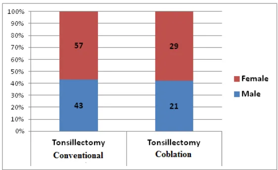
There were 100 patients in Conventional Tonsillectomy group and 50
[image:57.612.109.518.257.388.2]patients in Coblation Tonsillectomy group.
Table 6.1.1 GENDER DISTRIBUTION COLD
Tonsillectomy
HOT
Tonsillectomy Total Male 43 (43.0%) 21 (42.0%) 64 (42.7%) Female 57 (57.0%) 29 (58.0%) 86 (57.3%) Total 100 (100%) 50 (100%) 150 (100%) Chi2 – 0.014 p-Value – 0.907
In the Conventional Tonsillectomy group, 43 (43.0%) were males and
57 (57.0%) were females. In the same manner, in the Coblation
Tonsillectomy group, 21 (42.0%) were males and 29 (58.0%) were females
and so both groups are comparable as evident by p-value > 0.05.
[image:57.612.146.427.503.675.2]
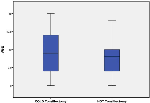
Table 6.1.2 AGE DISTRIBUTION
Category N Mean Standard Deviation COLD
Tonsillectomy 100 9.67 2.69 HOT
Tonsillectomy 50 8.92 2.58
Total 150 9.42 2.67
T – Value 2.66 p-Value – 0.105
The minimum age of the study population is from 5 years to
maximum of 15 years. The mean age was 9.42 years with standard deviation
of 2.67 years. The mean age (± standard deviation) in Conventional
Tonsillectomy group is 9.67 (± 2.69) years while the mean age (± standard
deviation) in Coblation Tonsillectomy group is 8.92 (± 2.58) years. This
difference is not significant as the p-value is > 0.05. So both the groups are
comparable by age too.
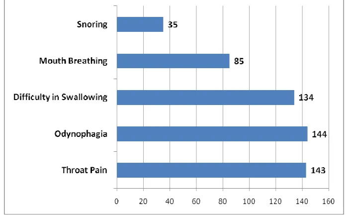
[image:58.612.164.473.462.673.2]6.2 PREOPERATIVE SYMPTOMS Table 6.2 PREOPERATIVE SYMPTOMS
Number Percentage
Throat Pain 143 95.3
Odynophagia 144 96.0
Difficulty in Swallowing 134 89.3 Mouth Breathing 85 56.7
Snoring 35 23.3
Regarding the preoperative symptoms of the study population,
Odynophagia is the most common symptom as seen in 144 (96.0%) patients
followed by Throat pain in 143 (95.3%) and Difficulty in swallowing in 134
(89.3%) patients. Then Mouth breathing is seen in 85 (56.7%) of the
patients and Snoring is seen only 35 (23.3%) patients.
[image:59.612.141.490.458.675.2]IMMEDIATE COMPLICATIONS 6.3 Intraoperative Complications
Table 6.3.1 INTRAOPERATIVE ANAESTHEIC COMPLICATIONS Number Percentage Dislodging of Loose Tooth 9 6.0 Dislodging of Temparo Mandibular
joint 0 0.0
Accidental Extubation 10 6.7 Kinking of ET Tube 19 12.7
Among the Intraoperative Anesthetic complications in the study
populations, Compression of ET tube is the commonest complication as
seen in 19 (12.7%) patients followed by Accidental Extubation seen in 10
(6.7%) patients and Dislodging of Loose Tooth seen in 9 (6.0%) patients.
[image:60.612.155.475.502.689.2]Table 6.3.2 INTRAOPERATIVE SURGICAL COMPLICATIONS
Number Percentage
Primary Hemorrhage 43 28.7
Edema Uvula 39 26.0
Pillar Injury 33 22.0
Regarding Intraoperative Surgical complications, Primary
Hemorrhage is seen in maximum as many as 43 (28.7%) patients followed
by Edema Uvula seen in 39 (26.0%) and Pillar Injury seen in 33 (22.0%)
6.4 Postoperative Complications
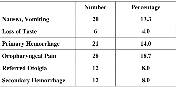
Table 6.4.1 POSTOPERATIVE COMPLICATIONS
Number Percentage Nausea, Vomiting 20 13.3
Loss of Taste 6 4.0
Primary Hemorrhage 21 14.0 Oropharyngeal Pain 28 18.7 Referred Otolgia 12 8.0 Secondary Hemorrhage 12 8.0
Oropharyngeal Pain is the commonest Postoperative Complication in
the study population as seen in 28 (18.7%) followed by Primary
Hemorrhage seen in 21 (14.0%) patients and Nausea, Vomiting seen in 20
(13.3%) patients. Referred Otolgia and Secondary Hemorrhage were
observed in 12 (8.0%) patients respectively each and Loss of Taste was seen
in 6 (4.0%) of the patients.
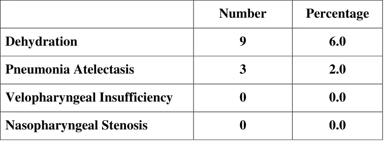
[image:63.612.153.475.477.675.2]Table 6.4.2 POSTOPERATIVE OTHER RARE COMPLICATIONS
Number Percentage
Dehydration 9 6.0
Pneumonia Atelectasis 3 2.0 Velopharyngeal Insufficiency 0 0.0 Nasopharyngeal Stenosis 0 0.0
Regarding Postoperative other rare complications, Dehydration was
observed in 9 (6.0%) and Pneumonia Atelectasis was seen in 3 (2.0%) of the
patients. Velopharyngeal Insufficiency and Nasopharyngeal Stenosis were
not seen in any of the patients.
[image:64.612.145.515.397.616.2]
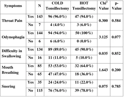
6.5 Comparison of Preoperative Symptoms
Table 6.5 COMPARISION OF PREOPERATIVE SYMPTOMS
Symptoms N COLD Tonsillectomy HOT Tonsillectomy Chi2 Value p- Value Throat Pain
Yes 143 96 (96.0%) 47 (94.0%)
0.300 0.584 No 7 4 (4.0%) 3 (6.0%)
Odynophagia
Yes 144 94 (94.0%) 50 (100%)
3.125 0.077 No 6 6 (6.0%) 0 (0.0%)
Difficulty in Swallowing
Yes 134 89 (89.0%) 45 (90.0%)
0.035 0.852 No 16 11 (11.0%) 5 (10.0%)
Mouth Breathing
Yes 85 53 (53.0%) 32 (64.0%)
1.643 0.200 No 65 47 (47.0%) 18 (36.0%)
Snoring
Yes 35 24 (24.0%) 11 (22.0%)
[image:65.612.108.523.145.470.2]0.075 0.785 No 115 76 (76.0%) 39 (78.0%)
Table 6.5 shows the Comparison of Preoperative symptoms between
both Conventional Tonsillectomy and Coblation Tonsillectomy group of
patients.
Throat Pain was seen in 96 (96.0%) of the Conventional
tonsillectomy patients and 47 (94.0%) of the Coblation Tonsillectomy
patients. This difference is not statistically significance as seen in p-value >
In the same way, Odynophagia was seen in 94 (94.0%) of the
Conventional Tonsillectomy patients and all the 50 (100%) of the Coblation
Tonsillectomy patients. But this difference is not statistically significance as
seen in p-value is 0.077.
Regarding Difficulty in Swallowing, it was seen in 89 (89.0%) of the
Conventional tonsillectomy patients and 45 (90.0%) of the Coblation
Tonsillectomy patients and this difference is not statistically significance
(p-value – 0.852).
Mouth Breathing was seen in 53 (53.0%) of the Conventional
Tonsillectomy patients and in 32 (64.0%) of the Coblation Tonsillectomy
patients. But this difference is not statistically significance as p-value is
0.200.
Snoring was observed in 24 (24.0%) of the Conventional
Tonsillectomy patients and in 11 (22.0%) of the Coblation Tonsillectomy
patients and there is no statistically significant difference between the
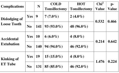
6.6 Comparison of Intraoperative Complications
Table 6.6.1 COMPARISON OF INTRAOPERATIVE ANAESTHEIC COMPLICATIONS
Complications N COLD Tonsillectomy HOT Tonsillectomy Chi2 Value p- Value Dislodging of Loose Tooth
Yes 9 7 (7.0%) 2 (4.0%)
0.532 0.466 No 141 93 (93.0%) 48 (96.0%)
Accidental Extubation
Yes 10 6 (6.0%) 4 (8.0%)
0.214 0.642 No 140 94 (94.0%) 46 (92.0%)
Kinking of ET Tube
Yes 19 15 (15.0%) 4 (8.0%)
1.476 0.224 No 131 85 (85.0%) 46 (92.0%)
Table 6.6.1 shows the comparison of Intra operative Anesthetic
Complications between the two groups.
Dislodging of Loose Tooth was seen in 7 (7.0%) of the Conventional
Tonsillectomy patients but it was observed in only 2 (4.0%) of the Coblation
Tonsillectomy patients but this difference is not statistically significant
(p-value – 0.466).
Accidental Extubation was seen in 6 (6.0%) of the Conventional
Tonsillectomy patients and this difference is not statistically significant
(p-value – 0.642).
Compression of ET Tube was seen in 15 (15.0%) of the Conventional
Tonsillectomy patients but it was seen in only 4 (8.0%) of the Coblational
Tonsillectomy patients but this difference is not statistically significant
(p-value – 0.224).
Table 6.6.2 COMPARISION OF INTRAOPERATIVE SURGICAL COMPLICATIONS
Complications N COLD Tonsillectomy HOT Tonsillectomy Chi2 Value p- Value Primary Hemorrhage
Yes 43 40 (40.0%) 3 (6.0%)
18.844 0.0001 No 107 60 (60.0%) 47 (94.0%)
Edema Uvula
Yes 39 32 (32.0%) 7 (14.0%)
5.613 0.018 No 111 68 (68.0%) 43 (86.0%)
Pillar Injury
Yes 33 19 (19.0%) 14 (28.0%)
[image:68.612.111.522.366.659.2]Table 6.5 compares the Intra Operative Surgical Complications between the groups
Primary Hemorrhage which is the commonest Intra Operative Surgical Complications was observed in 40 (40.0%) of the Conventional
tonsillectomy patients while it was seen in only 3 (6.0%) of the Coblation
Tonsillectomy patients. This is the highly statistically significant
difference as seen in p-value 0.0001.
In the same way, Edema Uvula was observed in 32 (32.0%) of the
Conventional tonsillectomy patients while it was seen in only 7 (14.0%) of
the Coblation Tonsillectomy patients. This difference is also statistically
significant as seen in p-value 0.018.
Pillar Injury was observed in 19 (19.0%) of the Conventional
tonsillectomy patients and 14 (28.0%) of the Coblation Tonsillectomy
Fig. 6.6.2.1 Showing Comparision of Intraoperative Surgical Complication – Primary Hemorrhage
[image:70.612.114.523.135.600.2] [image:70.612.115.518.140.374.2]6.7 Comparison of Postoperative Complications
Table 6.7 COMPARISION OF POSTOPERATIVE COMPLICATIONS
Complications N COLD
Tonsillectomy HOT Tonsillectomy Chi2 Value p- Value Nausea, Vomiting
Yes 20 12 (12.0%) 8 (16.0%)
0.462 0.497
No 130 88 (%) 42 (84.0%)
Loss of Taste
Yes 6 2 (2.0%) 4 (8.0%)
3.125 0.077
No 144 98 (98.0%) 46 (92.0%)
Primary Hemorrhage
Yes 21 12 (12.0%) 9 (18.0%)
0.997 0.318
No 129 88 (88.0%) 41 (82.0%)
Oropharyngeal Pain
Yes 28 19 (19.0%) 9 (18.0%)
0.022 0.882
No 122 81 (81.0%) 41 (82.0%)
Referred Otolgia
Yes 12 4 (4.0%) 8 (16.0%)
6.522 0.011
No 138 96 (96.0%) 42 (84.0%)
Secondary Hemorrhage
Yes 12 0 (0.0%) 12 (24.0%)
26.087 0.0001
No 138 100 (100%) 38 (76.0%)
Table 6.7 compares the Post Operative Complications between the groups.
Nausea, Vomiting was observed in 12 (12.0%) of the Conventional tonsillectomy patients and it was seen in 8 (16.0%) of the Coblation
Tonsillectomy patients. But this is difference is not statistically significant
In the same way, Loss of Taste was observed in 2 (2.0%) of the
Conventional tonsillectomy patients while it was seen in 4 (8.0%) of the
Coblation Tonsillectomy patients. But this difference is also statistically not
significant as seen in p-value 0.077.
Primary Hemorrhage was observed in 12 (12.0%) of the Conventional
tonsillectomy patients and 9 (18.0%) of the Coblation Tonsillectomy
patients but this difference is not statistically significance (p-value – 0.318).
Oropharyngeal pain which was commonest among the post operative
complications was observed in 19 (19.0%) of the Conventional
tonsillectomy patients and it was seen in 9 (18.0%) of the Coblation
Tonsillectomy patients and the difference is not statistically significant as
seen in p-value is only 0.882.
Referred Pain was observed only in 4 (4.0%) of the Conventional
tonsillectomy patients but it was seen in 8 (16.0%) of the Coblation
Tonsillectomy patients. This difference is statistically significant as seen in
Secondary Hemorrhage was not observed in any of the Conventional
tonsillectomy patients but it was observed in 12 (24.0%) of the Coblation
Tonsillectomy patients. This difference is highly statistically significant as
[image:73.612.114.543.218.468.2]seen in p-value – 0.0001.
Fig. 6.7.1 Showing Comparison of Postoperative Complication – Referred Otolgia
Fig. 6.7.3 Showing Comparison of Postoperative Complication – Secondary Hemorrhage
[image:74.612.113.511.67.296.2] [image:74.612.116.508.374.607.2]6. DISCUSSION
Tonsillectomy with or without adenoidectomy is the most commonly
performed pediatric otorhinolaryngological procedure. Variety of
techniques and approaches for adenotonsillectomy have been tested and
tried over the years. Yet post operative complications were mostly noted in
terms of oropharyngeal pain, bleeding, and referred otalgia.
An analytical study to ascertain the incidence of immediate and
delayed complications following adenotonsillectomy during the period from
August 2012 – September 2013 was conducted in Government Stanley
Medical College and Hospital. Since in our hospital we do
adenotonsillectomy by both Conventional and Coblation methods, a
comparison is also made between them on the immediate and delayed
complications.
For the study purpose the complications of adenotonsillectomy are
classified into
1. Intra operative complications
a. Anaesthetic complications
b. Surgical complications.
I. INTRA OPERATIVE COMPLICATIONS
a. ANAESTHETIC COMPLICATIONS
With regard to intra operative anaesthetic complications, it has been
found that the compression and obstruction of the anesthetic tube is the
predominant complication due to selection of faulty size of the blade of
Davis mouth gag33. In our study also compression of the endo tracheal tube
is the most frequently noted complication in the conventional
tonsillectomy group.
Accidental extubation while changing the head position and also
during Doughty’s tongue blade removal is another commonly encountered
anaesthetic complications in both conventional and coblation
tonsillectomy.
b. SURGICAL COMPLICATIONS
The onset of hemorrhage to the procedure defines 2 categories:
I. Primary haemorrhage occuring during surgery.
Tonsillar hemorrhage is defined as continuous bleeding for more than
one hour, or more than 250ml of blood loss regardless of the duration of
II. Delayed (Reactionary) with in 24 hours after surgery
At the minimum, a clinically significant posttonsillectomy
hemorrhage requires return to the operating theatre for control of bleeding,
with the inherent risk of aspiration during anesthesia induction.
Primary haemorrhage is the most commonly recorded intra
operative surgical complication. The reason for primary haemorrhage may
be due to
(a) inadequate pre operative patient preparation,
(b) faulty surgical technique
(c) in advertent injury to superior constrictor muscles and
(d) difficulty in ligating the bleeders in the tonsillar bed especially in
superior and inferior pole.
The intraoperative blood loss is usually measured in terms of weighing
swabs and also by measuring the volume of suction aspirate.
Edema of the uvula is another intra operative complication which is
mainly encountered due to in advertent and frequent suctioning over the
II. POST OPERATIVE COMPLICATIONS
Odynophagia and referred otalgia are the two complications observed
in the immediate post operative period following adenotonsillectomy.
Odynophagia is due to muscle spasm, especially the superior
constrictor fibres and dissection of the tonsil substance. Similar irritation of
the superior constrictor occurs while curreting the adenoids.
Referred otalgia is probably due to close removal of the tonsil from
the tongue base.
DELAYED POST OPERATIVE COMPLICATIONS
Secondary haemorrhage is the most common delayed post operative
complication observed in Coblation tonsillectomy because of dislodgement
COMPARISON STUDY OF IMMEDIATE AND DELAYED
COMPLICATIONS FOLLOWING ADENOTONSILLECTOMY USING
CONVENTIONAL AND COBLATION TECHNIQUE
Immediate
complications Conventional Coblation
Primary Haemorrhage Significant (P < 0.05) Not significant
Odynophagia Significant(p < 0.05) Not significant
Referred Otalgia Not significant Significant
Primary Haemorrhage is less in Coblation technique because
1. Dissection is done extracapsular, hence no surgical trauma to
the para tonsillar vein and the superior constrictor muscle
which lies in the tonsillar bed
2. Hemostasis was possible for vessels less than 1 mm in
diameter24
3. Histopathological thermal injury of only 0.13-mm depth is
reported24.
The mean intraoperative blood loss in the Conventional tonsillectomy
group is more than double that of the Coblation group25. [33.1mL Vs 15.7
Referred otalgia is probably due to close removal of the tonsil from
the tongue base.
Though Oropharyngeal pain is found to be the most
commonest post operative complication which delays the early oral
intake within the first 24 hour post operative period26, it is
statistically insignificant in our study.
According to Wong-Baker Faces Pain Rating Scale,
Derbyshire Children’s Hospital Paediatric Pain Chart and Bieri
Faces Pain Scale27 in Coblation vs Conventional technique, the pain
score in the first week of post operative days is significantly
lower, which is 3.3 in the Coblation group and 3.7 in conventional group.
7. CONCLUSION
Invention of equipments like Coblation, mono / bipolar electrocautery
and Laser have made the outlook better for Adenotonsillectomies with
respect to intraoperative and postoperative complications.
Primary haemorrhage and uvula edema are found to be the statistically
significant complications in Conventional tonsillectomy.
Secondary haemorrhage and referred otalgia, are the statistically
highly significant complications in Coblation tonsillectomies.
Hence training and experience of the surgeon, technical preferences
and its complications, cost-effectiveness should be considered in choosing
the surgical technique.
All trainee surgeons should become competent in Conventional dissection method and in achieving haemostasis using ligatures before
learning other modern techniques.
Irrespective of seniority and experience, every surgeon should undergo
Emphasis must be placed on explaining the risk of post operative
haemorrhage to the patient, teaching the correct technique like checking the
power settings prior to surgery and the potential hazards of Coblation
technique.
Every complication should be recorded and analyzed regularly to
improve the patient’s safety.
Yet it can be concluded that the use of Coblation tonsillectomy is equivalent to the use of Conventional technique in the current scenario with
BIBILOGRAPHY
1. Hellings P,Jorrissen M, Ceuppens JL. The Waldeyers ring. Acta
otorhinolaryngologica Belgica. 2000;54:237-41
2. Kenna MA, Amin A. Anatomy and physiology of the oral cavity. In:
Snow JB, Wackym PA. Ballenger's Otorhinolaryngology Head and Neck
Surgery. 17th ed. Shelton: BC Decker Inc; 2009:769-774.
3. Berkovitz BKB, Holland GR, Moxham BJ.Oral anatomy,histology and
embryology, 3rd edition .London: Mosby;2002
4.Susan S, Harold E, Jermiah CH, David J, Andrew W. Pharynx (chapter
35). In: Gray's Anatomy: The Anatomical Basis of Clinical Practice. 39th
ed. Philadelphia: Elsevier; 2005:619-631.
5. Falagas ME, Vouloumanou EK, Matthaiou DK, Kapaskelis AM,
Karageorgopoulos DE. Effectiveness and safety of short-course vs
long-course antibiotic therapy for group A -hemolytic streptococcal
tonsillopharyngitis: ameta-analysis of randomized trials. Mayo Clin Proc.
2008;83(8):880-889.
6. STJERNQUIST- DESTNIK A. PRENELLER K AND SCHALEN , C
(1991) High recovery of haemophilus influenza and group A streptococci
in recurrent tonsillar infection and hypertrophy as compared with normal
and Nichols M. L . (1990) A comparison of KTP/532 Laser tonsillectomy
vs traditional dissection /snare tonsillectomy. Otolaryngology and Head and
neck surgery 103, 966-971.
7. POLVOGT, L.M and CROWE,S J (1929) Predominating organisms
found in cultures from tonsils and adenoid. Journal of the American Medical
Association 92,962-964.
8. TONER J. G STEWART T.J CAMPBELL., J B HUNTER (1986)
Tonsil flora in the very young tonsillectomy patient .Clinical
otolaryngology. 11. 171-174
9. Woloszko J, Kwende MM, Stalder KR. Coblation in otolaryngology. Proc
SPIE. 2003;4949:341-352.
10. MANGAT..,ORR.C.W and smith R C (1977) Sleep apnea,
hypersomnolence and upper airway obstruction secondary to adenotonsillar
enlargement. Archives of Otolaryngology, 103, 383-386.
11. CROFT, C B BROCKBANK M.J WRIGHT A. SWANSTON A.R
1990 Obstructive sleep apnea in children undergoing routine tonsillectomy
and adenoidectomy clinical otolaryngology 15, 307- 314
12. EVERETT, M T (1979) The cause of tonsillitis. Practitioner, 223,