0022-538X/10/$12.00 doi:10.1128/JVI.01344-09
Copyright © 2010, American Society for Microbiology. All Rights Reserved.
The Range of Human APOBEC3H Sensitivity to
Lentiviral Vif Proteins
䌤
†
Melody M. H. Li,
1,2Lily I. Wu,
2and Michael Emerman
1,2,3*
Department of Microbiology, University of Washington, Seattle,1and Division of Human Biology,2and Division of
Basic Sciences,3Fred Hutchinson Cancer Research Center, Seattle, Washington 98109
Received 30 June 2009/Accepted 7 October 2009
The APOBEC3Hgene is polymorphic in humans, with four major population-dependent haplotypes that encode proteins with different levels of antiviral activity. Haplotype II, present most frequently in African populations, encodes the most stable protein and is most active against human immunodeficiency virus type 1 (HIV-1). In contrast to human APOBEC3G, which can be completely counteracted by HIV-1 Vif, the protein encoded byAPOBEC3Hhaplotype II is only partially sensitive to Vif, while the protein encoded byAPOBEC3H haplotype I is completely resistant to HIV-1 Vif. We mapped a residue on APOBEC3H that determines this partial Vif sensitivity. However, it is unclear how HIV-1 can replicate in vivo without the ability to neutralize APOBEC3H antiviral activity. In order to directly address this question, we clonedvif genes from HIV-1-infected individuals with differentAPOBEC3Hgenotypes and tested them for their ability to inhibit human APOBEC3H. We found that while theAPOBEC3Hgenotype of infected individuals significantly influences the activity of Vif encoded by their virus, none of the Vif variants tested can completely neutralize APOBEC3H as well as they neutralize APOBEC3G. Consistent with this genetic result, APOBEC3H protein expression in human peripheral blood mononuclear cells was below our limit of detection using newly developed antibodies against the endogenous protein. These results demonstrate that human APOBEC3H is not as strong of a selective force for current HIV-1 infections as human APOBEC3G.
APOBEC3 (apolipoprotein B mRNA-editing catalytic poly-peptide) proteins belong to a family of cytidine deaminases that have antiviral and antiretroelement functions (16). APOBEC3 proteins have been shown to restrict various retroviruses by causing cytidine-to-uridine editing in minus-sense viral DNA and by a deaminase-independent mechanism that acts to block the completion of reverse transcription (1, 2, 10). In order to achieve productive infection in cells expressing APOBEC3, all known modern lentiviruses, except equine infectious anemia virus, encode a viral protein called Vif, which counteracts APOBEC3 antiviral activity. Vif binds to APOBEC3 and re-cruits the E3 ubiquitin ligase complex to APOBEC3, which leads to the polyubiquitination and subsequent degradation of APOBEC3 by the proteasome (16).
TheAPOBEC3family of antiviral genes has expanded
dur-ing mammalian evolution. Rodents have a sdur-ingle APOBEC3
gene, whereas other placental mammals encode multiple genes (4, 6, 14). In humans, chromosome 22 carries sevenAPOBEC3
genes:APOBEC3A,APOBEC3B,APOBEC3C,APOBEC3DE,
APOBEC3F, APOBEC3G, andAPOBEC3H. However,
dele-tions ofAPOBEC3Bare common in some human populations (12).APOBEC3Gand otherAPOBEC3genes have been under intense positive selection during primate evolution (19, 23), presumably to adapt to a changing landscape of viral patho-gens that can evade the action of these antiviral proteins.
However, it is not yet known the extent to which the evolution of different APOBEC3 family members has occurred in re-sponse to different viral pathogens.
An important characteristic of the Vif interaction with the APOBEC3 proteins is that it is often species specific. For example, the Vif protein encoded by human immunodefi-ciency virus type 1 (HIV-1) is active against human APOBEC3G but not APOBEC3G from African green mon-key, which is the natural host of the simian immunodefi-ciency virus SIVagm. Similarly, APOBEC3G from African green monkey is sensitive to SIVagm Vif but not HIV-1 Vif due to one amino acid difference in APOBEC3G (3, 17, 24, 33). Compared with human APOBEC3G, human APOBEC3F inhibits HIV-1 with less potency but demon-strates greater resistance to neutralization by Vif (26, 32). Nonetheless, it was previously shown that HIV-1 Vif uti-lizes two distinct regions to counteract APOBEC3G and APOBEC3F, which suggests that HIV-1 has simultaneously evolved to evade at least two different APOBEC3 family members (15, 21, 26, 36).
Previous studies done by our laboratory and other groups have shown thatAPOBEC3H, the most diverged from other members of the APOBEC3 family in primates, is polymorphic in humans (9, 18, 19, 28). Among the four major haplotypes, haplotype II encodes a protein with the longest half-life and is the most active againstvif-deficient HIV-1 and non-long-ter-minal-repeat retrotransposons in tissue culture (9, 18). This is a unique feature ofAPOBEC3Hsince genetic polymorphisms of otherAPOBEC3genes in humans are not known to exhibit such functional dichotomy. The loss of stability of human APOBEC3H proteins can be mapped to two independent polymorphic changes (R105G and Del15N) (18). Interestingly,
* Corresponding author. Mailing address: Division of Human Biol-ogy, Fred Hutchinson Cancer Research Center, 1100 Fairview Ave. N., P.O. Box 10924, Seattle, WA 98109-1024. Phone: (206) 667-5058. Fax: (206) 667-6523. E-mail: [email protected].
† Supplemental material for this article may be found at http://jvi .asm.org/.
䌤Published ahead of print on 14 October 2009.
88
on November 8, 2019 by guest
http://jvi.asm.org/
the protein encoded byAPOBEC3H haplotype II is partially resistant to Vif from HIV-1 LAI and completely resistant to NL4-3 Vif (9, 18). It is not clear, therefore, how HIV-1 over-comes human APOBEC3H in vivo since, at least as measured by PCR, the RNA is expressed in human peripheral blood mononuclear cells (PBMCs) (19).
Here, we examined this question by looking at the sensitivity of human APOBEC3H haplotypes to different vifgenes, in-cluding thevifgenes cloned from HIV-1-infected people with differentAPOBEC3Hgenotypes and from other primate len-tiviruses, and by looking directly at protein expression with newly developed antibodies. We identified a single polymor-phic site in APOBEC3H (amino acid 121) that determines its partial sensitivity to HIV-1 Vif. Moreover,vifvariants isolated from HIV-1-infected individuals with haplotype I or II dem-onstrate differential activity against APOBEC3H. However, no HIV-1 Vif protein is able to completely neutralize APOBEC3H, although HIV-2 Vif can do so. Consistent with these genetic results, we showed that the APOBEC3H protein is below our detection level in human PBMCs with available antibodies. Taken together, these data show that APOBEC3H has less impact on current HIV infections in human T cells than human APOBEC3G.
MATERIALS AND METHODS
APOBEC3HcDNA cloning, expression constructs, and plasmids.The hemag-glutinin tag at the 5⬘ end of the previously described human and macaque APOBEC3HcDNAs (18, 19) was removed by PCR amplifying APOBEC3H cDNA with a primer lacking the hemagglutinin tag sequence and cloning the fragment into the EcoRI/XhoI sites of pcDNA3.1 (Invitrogen). Point mutations were introduced by site-directed mutagenesis using the QuikChange kit (Strat-agene), and the entire insert was resequenced.
Cells, transfections, and Western blot analysis.HEK293T cells were main-tained in Dulbecco’s modified Eagle’s medium–1% penicillin-streptomycin– 10% bovine growth serum at 37°C in a CO2incubator. SupT1 cells were
maintained similarly in RPMI medium–1% penicillin-streptomycin–10% bo-vine growth serum. Human PBMCs were isolated from healthy donors by using Ficoll gradient centrifugation, and whole-cell lysates were prepared from them for immunoblotting. Transfections were performed with TransIT-LT1 transfection reagent (Mirus Bio) at a reagent-to-plasmid DNA ratio of 3:1. Western blot analyses were performed as previously described (18). A 1:2,500 dilution of human APOBEC3H antibodies (P1H6-1 and P1D8-1), a 1:5,000 dilution of human APOBEC3G antibody, and a 1:10,000 dilution of cyclophilin A antibody (Biomol) were used.
HumanAPOBEC3Hgenotyping.Genomic DNA was isolated from human PBMCs with a QIAamp DNA blood minikit (Qiagen). Two different PCR-based approaches were developed for the genotyping of the DelN15 and R105G poly-morphisms. For the Del15N polymorphism, genomic DNA was amplified with fluorescence-labeled primers, and DNA fragment analysis was performed as previously described (18). For the R105G polymorphism, two primer pairs were used in a tetraprimer ARMS-PCR to amplify the two different alleles, respec-tively, of the single nucleotide polymorphism at position 105 (35). The PCR products were then resolved on a 1% agarose gel to visualize the presence of a 191-bp fragment (105R/R), a 158-bp fragment (105G/G), or both fragments (105R/G).
Cloning ofvifgenes.A cloning system was developed for insertingvifgenes from HIV-1 -infected people into a reportervif-deleted viral construct. HIV-1vif sequences were amplified in nested PCRs from genomic DNA with viral loads above 50,000 copies per ml of plasma from the University of Washington CFAR HIV Specimen Repository from HIV-1-infected people who self-identified as being African-Americans. These patients were most likely exposed to a subtype B virus, as subtype B is the predominant virus strain circulating in the United States (29). Thus, the primers for nested PCR were designed from the subtype B viralvifconsensus sequence. The primers used for the first round of PCR amplified the viral genome between the end ofintegraseand the beginning oftat (5⬘-GTC TTA GGC TGA CTT CCT GGA TG and 5⬘-GGA ATA GAT AAG GCC CAA GAA GAA C). The primers used for the second round of PCR
flanked thevifgene with MluI and Xba sites on the 5⬘and 3⬘ends, respectively (5⬘-ATA ACG CGT GGC CAC CAT GGA AAA C and 5⬘-GAT TCT AGA CCT AGT GTC CAT TCA TTG). The nested PCR products were ligated into a TA vector from a Promega pGEM-T Easy kit and sequenced. Thesevifgenes were then ligated into the MluI/Xba sites of a vif-deleted HIV proviral plasmid that contains a deletion in theenvgene and has the firefly luciferase gene inserted into Nef (pLai3⌬envLuc2⌬vifLk). In this construct, all the start codons in the region from the original start ofvifto the end of theintegrase gene were mutated to prevent the expression of wild-typevif. The portion from the end of theintegrasegene to the AvrII site in thevprgene was deleted and replaced by new restriction sites (5⬘-SnaBI MluI XbaI HpaI-3⬘). Wild-typeviffrom LAI (20) was also cloned back into thevif-deleted viral construct to yield pLai3⌬envLuc2⌬vifLk/Lai. vif genes from HIV-2 ROD9 (7) and SIVcpz TAN3.1 (27) were also amplified with specific primers and cloned into pLai3⌬envLuc2⌬vifLk. All constructs were confirmed by sequencing.
APOBEC3H antibodies.APOBEC3Hhaplotype I was cloned into the bacterial expression plasmid pTrC-His (Invitrogen) under an IPTG (isopropyl- -D-thio-galactopyranoside)-inducible promoter with a His tag at the C terminus and transformed into BL21(DE3) RIL cells (Invitrogen). Three hundred milliliters of culture was grown to an optical density at 600 nm of 0.8 and then induced with 1 mM IPTG for 3 h at 37°C. The cell pellet was resuspended in denaturing buffer, freeze-thawed, treated with 1 mg/ml lysozyme on ice for 90 min, and sonicated on ice. The soluble material was then loaded onto a Ni-Sepharose 6 Fast Flow column (GE HealthCare), washed extensively in buffer containing 30 mM imi-dazole, and then eluted in a buffer containing 8 M urea at pH 4.0. The peak fraction showed only one band (20 kDa) on a Coomassie blue-stained gel with a protein concentration of 5.6 mg/ml.
Polyclonal and monoclonal antibodies against human APOBEC3H were gen-erated in RBF/DnJ outbred mice (Jackson Laboratories) as described previously (31). Two mice were injected with 50g of protein and then boosted eight times over a period of 1 year. After a final boost with 100g of protein, polyclonal sera were collected at autopsy, and fusions were generated. Monoclonal antibodies were screened for reactivity to the bacterial APOBEC3H protein by enzyme-linked immunosorbent assay and then rescreened on Western blots containing whole-cell lysates collected from 293T cells transfected with expression plasmids for the human APOBEC3H haplotype I protein, the human APOBEC3H hap-lotype II protein, rhesus macaque APOBEC3H, chimpanzee APOBEC3H, human APOBEC3A, human APOBEC3B, human APOBEC3C, human APOBEC3DE, human APOBEC3F, and human APOBEC3G. Three monoclo-nal antibodies (P1H6-1, P1D8-1, and P5H9-A11) that reacted with only the APOBEC3H proteins were identified.
Viral infectivity assays. Single-round HIV-1 infectivity assays were per-formed as previously described (19, 34). All assays were perper-formed by the transfection of 1.25⫻105
293T cells in 24-well plates with approximately a 1:1 ratio of pcDNA3/APOBEC plasmid (200 to 250 ng) to 250 ng of pLai3⌬envLuc2 (34) or⌬vif proviral plasmid (19). For functional assays of patientvifgenes, 1.25⫻105293T cells in 24-well plates were transfected with
approximately a 1:1 ratio of pcDNA3/APOBEC plasmid (250 ng) to pLai3⌬envLuc2⌬vifLk, pLai3⌬envLuc2⌬vifLk/Lai, or pLai3⌬envLuc2⌬vifLk/ Patient# (cloned patientvifgenes). Virus equivalent to 2 ng of p24CA was used to infect 4⫻104SupT1 cells in a 96-well plate in the presence of 20
g/ml DEAE-dextran. After 48 h, cells from triplicate infections were lysed in 100l of Bright-Glo luciferase assay reagent (Promega) and read on a luminometer.
Packaging assays.Virus stocks (4 ml per sample) were filtered and spun in an ultracentrifuge at 24,000 rpm for 1.5 h at 4°C. The viral pellets were resuspended in loading dye and analyzed by immunoblotting using antibodies specific for APOBEC3H or p24gag (mouse monoclonal antibody 24-2) (25).
RESULTS
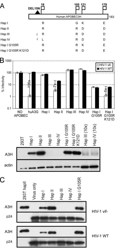
Human APOBEC3H resistance to HIV-1 Vif can be mapped to a single polymorphic amino acid.Previous studies showed that humanAPOBEC3His polymorphic, with four major hap-lotypes in the human population (called haphap-lotypes I, II, III, and IV) (Fig. 1A). Unlike APOBEC3F and APOBEC3G, the protein encoded by humanAPOBEC3H haplotype II is only partially neutralized by Vif of HIV-1 LAI (18) or not at all by Vif of HIV-1 NL4-3 (9). Since those previous studies were done with epitope-tagged APOBEC3H proteins (9, 18), which
on November 8, 2019 by guest
http://jvi.asm.org/
could potentially confound the interactions between the pro-tein and HIV-1 Vif, we developed antibodies to APOBEC3H proteins that allowed us to detect untagged versions of this protein (see Materials and Methods). Consistent with data from previous reports using epitope-tagged proteins, the pro-teins encoded by both haplotypes III and IV are very poorly expressed and are completely inactive against HIV-1 (Fig. 1B). The expression of the protein encoded byAPOBEC3H haplo-type I is more easily detectable than the proteins encoded by haplotypes III and IV and is weakly active against HIV-1 (63% infectivity compared to the no-APOBEC control) (Fig. 1B).
APOBEC3Hhaplotype II yields the highest steady-state levels
of protein after transfection and is highly active against vif -deficient HIV-1 (3% infectivity compared to the no-APOBEC control) (Fig. 1B).
We then compared the Vif sensitivities of untagged APOBEC3H proteins. As a control, HIV-1 restriction by hu-man APOBEC3G was completely rescued by Vif (Fig. 1B). However, the protein encoded byAPOBEC3Hhaplotype II is only partially neutralized by HIV-1 LAI Vif (about sevenfold virus rescue). On the other hand, the small amount of antiviral activity encoded by haplotype I is completely resistant to the effects of HIV-1 LAI Vif (Fig. 1B). Because of the differences in the sensitivities of the proteins encoded by APOBEC3H
haplotypes I and II to HIV-1 Vif, we could determine which of the polymorphic changes in this gene contributes to their re-sistance to Vif. Among the three polymorphic changes that differ between haplotypes I and II, the mutation at position 105 from a glycine (haplotype I) to an arginine (haplotype II) in the protein encoded by APOBEC3H haplotype I was previously shown to greatly increase expression levels (9, 18) and, in our study, the antiviral activity of this protein against HIV-1 LAI (Fig. 1A and B). However, this polymorphism did not affect the sensitivity of APOBEC3H to HIV-1 Vif (Fig. 1B), which dem-onstrates that although position 105 is a determinant for APOBEC3H protein stability, it is not a determinant for APOBEC3H sensitivity to Vif (Fig. 1B).
[image:3.585.43.284.70.590.2]We also analyzed the virion encapsidation of different hu-man APOBEC3H proteins and found that the level of APOBEC3H packaging corresponds to its resistance to HIV-1 Vif. Similar levels of proteins encoded by haplotypes I and II and the haplotype I G105R mutant were packaged into the virions regardless of the presence of Vif, which suggests that HIV-1 Vif is unable to efficiently target these APOBEC3H proteins for degradation by the proteasome (Fig. 1C). On the other hand, we found that mutating position 121 from a lysine (haplotype I) to an aspartic acid (haplotype II) increases APOBEC3H antiviral activity, which is consistent with a pre-vious finding (9). This mutant demonstrates a greater inhibi-tion ofvif-deficient HIV-1 than the protein encoded by the
FIG. 1. Human APOBEC3H resistance to Vif can be mapped to a single polymorphic change at amino acid 121. (A) Schematic of the polymorphic sites in humanAPOBEC3Hhaplotype I (Hap I) to hap-lotype IV and two mutants tested. A flag indicates a polymorphic amino acid (amino acid numbers listed above), while the black triangle represents an amino acid deletion (check marks indicate that the deletion is present). The total protein size is 183 amino acids. Details of humanAPOBEC3Hhaplotypes were previously described (18). (B, top) Sensitivity of human APOBEC3H proteins to HIV-1 Vif as shown by the bars for virus rescue between HIV with a deletion in thevifgene and the wild type (WT) (vif⫹). Averages of triplicate assays (⫾ stan-dard errors of the means) are shown as the percent infectivity relative to the control assay with no APOBEC expression. The open bars are infections with HIV that contains a deletion in thevifgene, and the solid boxes are infections with wild-type HIV (LAI) that contains a
functionalvifgene. Note the log scale on theyaxis. (Bottom) Western blot analysis of human APOBEC3H proteins after transfection in 293T cells. Ten-times-more sample was loaded in the last two lanes since the human APOBEC3H proteins encoded by haplotypes III and IV were undetectable when the same amounts (1⫻) were loaded compared to other samples. huA3G, human APOBEC3G. (C) Western blot analysis of HIV-1vif-deleted and wild-type virions for the presence of human APOBEC3H (A3H) proteins and p24gag.
on November 8, 2019 by guest
http://jvi.asm.org/
haplotype I G105R mutant (Fig. 1B). Most importantly, mu-tating the basic residue at position 121 to an acidic one leads to increased APOBEC3H sensitivity to HIV-1 Vif (about fivefold virus rescue) (Fig. 1B). Thus, the polymorphic change at po-sition 121 is critical for the sensitivity of APOBEC3H to HIV-1 Vif in addition to APOBEC3H antiviral activity.
Differential sensitivity of APOBEC3H tovif genes isolated from HIV-infected humans with different genotypes.We next asked why the protein encoded by humanAPOBEC3H haplo-type II is only partially neutralized by HIV-1 Vif from LAI, while human APOBEC3G is almost completely neutralized by the same Vif (Fig. 1B). If APOBEC3H poses an important block to viral replication, one would expect that HIV-1 would have to overcome it with its viralvifgene in order to replicate efficiently in vivo. We reasoned that thevifgenes in the labo-ratory strains might not reflect the selection for Vif activity in an HIV-infected person who encodes the more active APOBEC3H haplotype II protein (9, 18).APOBEC3H haplo-type II is found mostly in African and African-American pop-ulations but is uncommon among European/Caucasian and Asian populations (18). However, our study and others inves-tigating the APOBEC3H interaction with HIV-1 Vif were done with standard HIV laboratory strains derived from indi-viduals of unknownAPOBEC3Hgenotypes. Therefore, we hy-pothesized that HIV-infected individuals who are homozygous or heterozygous for the stableAPOBEC3Hallele (haplotype II) might harbor viral Vif variants that have stronger antago-nistic activities against the APOBEC3H haplotype II protein than the previously tested Vif proteins. In other words, if APOBEC3H is important for virus replication in patients, these viruses would encode variations of Vif that can neutralize the haplotype II protein better than the viruses infecting indi-viduals that have one of the unstable APOBEC3H proteins.
In order to test the activity of Vif proteins isolated from HIV-infected people who encode the most stable and active form of APOBEC3H, we obtained PBMCs of HIV-1-infected African-Americans who had a viral load of greater than 50,000 copies per ml of plasma and genotyped their polymorphisms of
APOBEC3H(R105G and DelN15) (see Table S1 in the
sup-plemental material). Three individuals were chosen for further study: one individual who has two copies of haplotype II (pa-tient 1203), one individual with one copy of haplotype II and one copy of haplotype I (patient 1440), and one individual with two copies of haplotype I (patient 1393). We then amplifiedvif
variants from genomic DNA isolated from the PBMCs of each of these individuals. The phylogenetic relationships of thevif
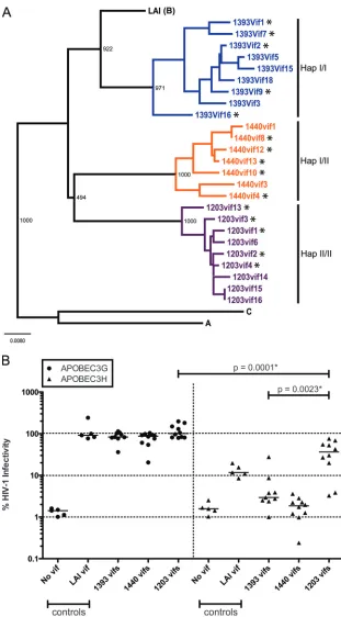
genes from viruses in the PBMCs of these three patients are of subtype B and were not contaminated with our standard lab-oratory strain LAI since thevifsequences from each patient form a separate group (Fig. 2A). Consensus Vif sequences from each patient share about 85 to 91% identity with LAI Vif and 81 to 86% identity with each other (see Fig. S1 in the supplemental material).
Thevifvariants from these three patients were then cloned into avif-deleted HIV-1 luciferase reporter construct in place of the originalvifgene (see Materials and Methods for details). We first tested the ability of the patient Vif variants to neu-tralize human APOBEC3G by determining the viral infectivity measured by the luciferase activity of infected SupT1 cells in the presence of Vif. We tested fivevifclones from each patient
(Fig. 2A), and each one was tested in triplicate in two different experiments. The infectivity data matched with the corre-sponding patientvifgene are shown in Table S2 in the supple-mental material. We found that the proteins encoded by thevif
alleles from all three patients are able to effectively neutralize APOBEC3G and rescue viral infectivity up to a level similar to that of Vif from HIV-1 LAI (Fig. 2B). This indicates that the
vifgenes that we cloned are active and not defective. We next tested the ability ofvifgenes cloned from HIV-1-infected people with differentAPOBEC3Hhaplotypes to neu-tralize the human APOBEC3H haplotype II protein. As shown in Fig. 1B, HIV-1 LAI Vif only partially neutralizes the anti-viral effect of human APOBEC3H (Fig. 2B). Likewise, we found that Vif variants from the patients either showed mini-mal to no virus rescue or showed about the same modest level of rescue as LAI (Fig. 2B). Strikingly, the proteins encoded by thevifgenes isolated from a homozygous haplotype II patient (patient 1203) demonstrate a wider range of activity against APOBEC3H, and some of them are able to rescue virus at up to 50 to 70% infectivity (Fig. 2B).
A one-way analysis of variance (ANOVA) to compare the means of the activities encoded by vifgenes from the three patients against APOBEC3H found that at least one of the patient pairs is significantly different (P⬍0.0001). In particu-lar, a Student’s t test showed that the infectivity of viruses carrying thevifgenes isolated from the homozygous haplotype II patient (patient 1203) is significantly different (P⫽0.0023) from that of viruses carrying thevifgenes isolated from the homozygous haplotype I patient (patient 1393). These data suggest that the haplotype II protein is mostly resistant to inactivation by thevifgenes from viruses infecting patients with one copy of haplotype II or no copies and from the standard HIV-1 laboratory strain. However, the individual carrying two copies of the stable allele ofAPOBEC3Hdoes harbor viral Vif variants that can neutralize the haplotype II protein to a sig-nificantly greater extent. Carrying two copies of haplotype II might confer some advantage against HIV-1 to individuals, although a larger cohort needs to be screened. Interestingly, the activity of the Vif proteins from the homozygous haplotype II patient against APOBEC3G is also significantly different from that against APOBEC3H (P⫽0.0001). In other words, none of the patient Vif variants that we tested are as potent against APOBEC3H as they are against APOBEC3G. There-fore, these data suggest that while theAPOBEC3Hgenotype of HIV-1-infected people significantly influences the activity of the Vif protein from their own virus, the selective pressure on HIV-1 Vif due to APOBEC3H expression is not strong enough to drive the evolution of Vif to neutralize APOBEC3H as well as it neutralizes APOBEC3G.
Expression of endogenous APOBEC3H in PBMCs of hu-mans is undetectable.We hypothesized that perhaps HIV-1vif
genes have not evolved to neutralize APOBEC3H as well as they neutralize APOBEC3G because human APOBEC3H is poorly expressed in target cells of HIV-1. In order to test this hypothesis, we analyzed human PBMC samples for endoge-nous APOBEC3H expression. Two monoclonal antibodies, P1H6-1 and P1D8-1, reacted against human protein expressed in 293T cells with minimal background (Fig. 3, lane 1). We probed whole-cell lysates made from PBMCs of HIV-1-in-fected individuals who were the same ones genotyped for the
on November 8, 2019 by guest
http://jvi.asm.org/
FIG. 2. Function ofvifgenes isolated from HIV-1-infected individuals with differentAPOBEC3Hhaplotypes. (A) Twenty-five independentvif
nucleotide sequences derived from patients 1393, 1440, and 1203 were analyzed with consensus subtype A and Cvifsequences and a reference subtype B sequence (LAI) using CLUSTAL_X (5). The phylogenetic relationships among these sequences were then analyzed by using the neighbor-joining method. Bootstrap values are also shown by the branches. Genotyping of genomic DNA (see Table S1 in the supplemental material) showed that patient 1393 was homozygous forAPOBEC3H haplotype I (Hap I), patient 1440 was heterozygous for APOBEC3H
haplotype I/II, and patient 1203 was homozygous forAPOBEC3Hhaplotype II. The asterisks next to thevifgenes indicate that these sequences were tested for Vif function as described below (B). (B) Activity encoded by thevifgenes cloned from HIV-infected individuals that encode no copy, one copy, or both copies of haplotype II (patients 1393, 1440, and 1203, respectively) against human APOBEC3G and untagged APOBEC3H haplotype II protein. The percent infectivity of HIV in the absence ofAPOBEC3transfection is set at 100% (not shown), and all other values are relative. On the left side of the dotted line, the circles represent infections performed with HIV-1 containing novif, LAIvif, or functionalvifgenes
on November 8, 2019 by guest
http://jvi.asm.org/
data in Fig. 2 and were homozygous for haplotype I (patient 1393) (Fig. 3, lane 2), heterozygous haplotype I and haplotype II (patient 1440) (Fig. 3, lane 3), and homozygous for haplo-type II (patient 1203) (Fig. 3, lane 4). The amount of APOBEC3H protein expression in PBMCs of infected donors was below our level of detection, as no band of 20 kDa was visible, even for the individual that is homozygous for
haplo-type II (patient 1203) (Fig. 3, lane 4). In addition, we probed whole-cell lysates made from PBMCs of healthy donors with different APOBEC3H genotypes (Fig. 3, lanes 5 to 8). Healthy donor 2 carries one copy of haplotype II. The amount of APOBEC3H protein expression was again below our level of detection. We also carried out threefold serial dilutions of whole-cell lysates collected from 293T cells transfected with exogenous APOBEC3H to show that our immunoblotting sys-tem could still detect APOBEC3H in a total protein level that is equivalent to a 1:720 dilution of human PBMC samples loaded. We then stripped the blot and reprobed it with human APOBEC3G antibody. A band of the expected size (about 40 kDa) was detected in the lanes loaded with lysates of patient PMBCs, which suggests that the absence of APOBEC3H in these samples is not due to the degradation of the endogenous protein by HIV-1 Vif. Interestingly, the level of APOBEC3G protein expression is higher in patient PBMCs than in healthy PBMCs, which is consistent with a previous observation of a modest induction of APOBEC3G mRNA levels in CD4⫹ T cells by alpha interferon treatment (13). Thus, the low level or lack of APOBEC3H protein expression in the PBMCs of HIV-1-infected individuals may explain the lack of selective pres-sure for Vif proteins from these individuals to evolve to com-pletely neutralize this antiviral protein.
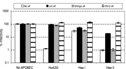
Human APOBEC3H resistance to Vif is specific to viruses from the HIV-1 lineage.A previous study has shown the com-plete inactivation of rhesus macaque APOBEC3H by SIVmac, SIVagm, and HIV-2 Vif proteins (30). Given the differences between the abilities of rhesus and human APOBEC3H pro-teins to be neutralized by SIV and HIV-1 Vif propro-teins, we tested the proteins encoded by APOBEC3H haplotypes against Vif from other lentiviruses that infect primates to see if human APOBEC3H insensitivity to Vif is species specific. We PCR amplified and cloned thevifgenes from the full-length infectious clones of SIVcpz clone TAN3.1 (27) and HIV-2 ROD9 (7) into ourvif-deficient HIV-1 LAI luciferase report-er construct for testing against human APOBEC3G and APOBEC3H. Both the SIVcpz and HIV-2 Vif proteins, along with LAI Vif, are able to completely neutralize human APOBEC3G, which is shown by a virus rescue of 62- to 100-fold (Fig. 4). Similar to our findings with HIV-1 Vif proteins, the human APOBEC3H haplotype I and II proteins are highly resistant to SIVcpz Vif (no virus rescue) (Fig. 4). On the other hand, the proteins encoded by both humanAPOBEC3H hap-lotypes can be neutralized by HIV-2 Vif (5- and 131-fold virus rescues, respectively) (Fig. 4). These results suggest that Vif proteins from the SIVcpz/HIV-1 lineage have specifically lost the ability to neutralize APOBEC3H, while this activity has been retained in another lineage of primate lentiviruses.
[image:6.585.55.266.70.356.2]from infected individuals prepared by the cotransfection of APOBEC3G, while the triangles represent infections performed with HIV-1 prepared by the cotransfection of APOBEC3H (haplotype II). Averages of data for triplicate assays from two independent experiments for patientvifgenes and five independent experiments for novifand LAIvifare shown here, and the horizontal lines represent the median values of virus infectivity for each group of Vif proteins tested. Note the log scale on theyaxis. A one-way analysis of variance was carried out to compare the means for the three patient groups against APOBEC3H, and at least one of the patient pairs is significantly different (P⬍0.0001). Pairwise comparisons (using Bonferroni’s adjustment for multiple comparisons) of virus infectivity with patient 1203 Vif proteins or patient 1393 Vif proteins against APOBEC3H and those with patient 1203 Vif proteins against APOBEC3G or APOBEC3H were performed on individual pairs. Table S2 in the supplemental material shows which data points in B correspond to which sequences in A.
FIG. 3. Endogenous APOBEC3H protein is not detected in the PBMCs of HIV-1-infected individuals with stable haplotype II. Shown is a Western blot analysis of the endogenous APOBEC3H haplotype II (hapII) protein in PBMCs of infected individuals and healthy donors. (Top) Lane 1 represents whole-cell lysates (1g of total protein) collected from 293T cells transfected with humanAPOBEC3H. Lane 2 to 4 represent cell lysates (80 to 90g of total protein) collected from patient PBMCs with or without the most activeAPOBEC3H allele (haplotype II), while the last four lanes were loaded with lysates (20g of total protein) made from PBMCs of healthy donors. Asterisks in-dicate that donor 1 carries one copy of haplotype I and one copy of haplotype I with DEL15N and that donor 2 carries one copy of hap-lotype II and one copy of either haphap-lotype III or IV. The same blot was also stripped and reprobed with human APOBEC3G and cyclophilin A antibodies. (Bottom) Threefold serial dilutions of whole-cell lysates made from 293T cells transfected with humanAPOBEC3H(huA3H) were loaded.
on November 8, 2019 by guest
http://jvi.asm.org/
DISCUSSION
We find thatvifgenes isolated from an HIV-1-infected indi-vidual who is homozygous for the most active form of
APOBEC3H(haplotype II) are significantly better at
antagoniz-ing APOBEC3H than thevifgenes from the heterozygote or the homozygote for the less active form ofAPOBEC3H(haplotype I). Nonetheless, in all cases, human APOBEC3H is less sensitive to neutralization by HIV-1 Vif than is human APOBEC3G. These results suggest that APOBEC3H is not as strong of a selective force on HIV-1vifevolution as APOBEC3G and imply that the APOBEC3H protein is not well expressed in HIV-1 target cells. Indeed, using newly developed monoclonal antibodies against APOBEC3H, we find that protein is undetectable in the PBMCs of HIV-1-infected and uninfected humans. Furthermore, we show that a single polymorphic amino acid at position 121 in human APOBEC3H is the major determinant for the sensitivity of human APOBEC3H to neutralization by HIV-1 Vif. This amino acid is distinct from the polymorphism that was previously shown to be responsible for the differences in protein stabilities among humanAPOBEC3Hhaplotypes, which is determined by R105G and Del15N (18). These results demonstrate that human APOBEC3H does not play a role as important as APOBEC3G in current HIV-1 infections but suggest that this protein may have evolved to counteract other viral pathogens in the past.
APOBEC3H protein expression in primates.We showed in this study that the amount of the APOBEC3H protein was below the detection level in PBMCs of both HIV-1-infected and uninfected humans. This result is consistent with the func-tional data that suggest that none of the Vif variants cloned from individuals with a stableAPOBEC3H allele are able to completely rescue HIV-1 infectivity from restriction by human APOBEC3H. In other words, the endogenous expression of APOBEC3H in PBMCs might be insufficient compared to the
endogenous APOBEC3G expression to completely drive the evolution of HIV-1vifalleles from most infected individuals. On the other hand, we did see a statistically significant differ-ence in the ability of the Vif proteins from an individual ho-mozygous for theAPOBEC3Hhaplotype II (the haplotype that makes the most stable protein) to neutralize APOBEC3H rel-ative to that of the Vif proteins from people with other geno-types. This implies that there is a low level of APOBEC3H in PBMCs that is below our level of detection with our current antibodies or else in an HIV-target cell such as mucosal T cells or macrophages that we have not yet measured. Alternatively,
APOBEC3H haplotype II might be linked to a haplotype of
anotherAPOBEC3gene in the individual homozygous for hap-lotype II (patient 1203) that is exerting evolutionary pressure on thevifgene of his virus.
In contrast to the inability of HIV-1 Vif to encounter human APOBEC3H, SIVmac, SIVagm, and HIV-2 Vif proteins have been shown to efficiently neutralize rhesus macaque APOBEC3H (30). This suggests that these Vif proteins have likely encountered and therefore evolved to inhibit the APOBEC3H proteins from Old World monkeys. One would predict that the APOBEC3H protein is expressed at significant levels in the PBMCs of Old World monkeys even though the expression of APOBEC3H in human PBMCs is undetectable. Moreover, we showed that HIV-2 Vif can counteract human APOBEC3H more effec-tively than can HIV-1 Vif. Since HIV-2 has a more recent origin in Old World monkeys than does HIV-1 (8), these results imply that HIV-2 retains Vif determinants that were useful in antagonizing APOBEC3H that it encountered in sooty mangabeys. Thus, some evolutionary event might have selected for the change in the transcriptional or posttranscrip-tional regulation of this antiviral gene in a species-specific manner that potentially caused the differential expression of the APOBEC3H protein between humans and Old World monkeys. Whatever event this was, its modern consequence is that the antiviral repertoire in human PBMCs has been re-duced compared to that in Old World monkeys. Further stud-ies should be carried out to look at endogenous APOBEC3H expression in macaque PBMCs.
Possible current and ancestral antiviral functions of human APOBEC3H.Apart from the potential species-specific differ-ences in the protein expression level, we also observed func-tional differences between the various versions of human APOBEC3H. We previously showed that the stability of the APOBEC3H protein was lost twice in human evolution (18). Here, we demonstrate that haplotype I carries a basic residue at position 121 (K121) that causes it to be completely resistant to HIV-1 Vif, while haplotype II contains an acidic residue at the same position (D121) that confers partial sensitivity to HIV-1 Vif. Interestingly, the region containing the amino acid responsible for human APOBEC3H sensitivity to HIV-1 Vif can be mapped to homologous regions in the N terminus of APOBEC3G and the C terminus of APOBEC3F that are im-portant for their interaction with Vif (11, 22). This interaction domain appears to be conserved among different APOBEC3 proteins.
[image:7.585.44.285.64.196.2]It is possible that at least one of theAPOBEC3Hhaplotypes evolved to become more resistant to a Vif-like factor of an ancient pathogen that is now extinct. Consistent with this idea, our labo-ratory previously demonstrated that primateAPOBEC3Hgenes
FIG. 4. Human APOBEC3H resistance to Vif is species specific. Shown is the inactivation of human APOBEC3H and human APOBEC3G (HuA3G) by SIVcpz and HIV-2 Vif proteins. Averages of triplicate assays (⫾standard errors of the means) are shown as the percent infectivity relative to the control assay with no APOBEC expression. The open bars represent infections with HIV (LAI) that has a multiple-cloning site in place of thevifgene. The black bars are infections performed with HIV that contains a functional vif gene cloned into the same multiple-cloning site, the gray bars are infections with HIV that contains thevifgene from SIVcpz (TAN3.1), and the stippled bars are infections with HIV that contains thevifgene from HIV-2 Vif (ROD9). Hap I, human APOBEC3H haplotype I protein; Hap II, human APOBEC3H haplotype II protein. Note the log scale on theyaxis.
on November 8, 2019 by guest
http://jvi.asm.org/
have been shown to be under positive selection, which is indica-tive of “genetic conflict” between a host defense gene and viral pathogens (19). Although a role for human APOBEC3H in re-stricting HIV-1 in vivo is yet to be demonstrated since it expres-sion in peripheral blood is low, human APOBEC3H might play a role in inhibiting other viruses that infect and replicate in the tissue types where the APOBEC3H protein is highly expressed. The availability of antibodies against the native protein described in this paper will allow us to describe the tissue expression pat-terns of human APOBEC3H more fully and should provide clues to what other viruses might be targets of this potent antiviral agent. The range of viral targets of APOBEC3H and the possible consequences of the differences in the activities of the protein among human populations have yet to be discovered.
ACKNOWLEDGMENTS
We thank the University of Washington CFAR HIV Specimen Re-pository for human PBMC samples; Sarah Holte of the University of Washington CFAR Biometrics Core for advice on statistical analysis; Elizabeth Wayner of the FHCRC Antibody Development Shared Re-sources, FHCRC Genetic Analysis core; and Jaisri Lingappa for hu-man APOBEC3G antibody. The following reagents were obtained through the NIH AIDS Research and Reference Reagent Program, Division of AIDS, NIAID, NIH: HIV-1 p24 Gag monoclonal antibody (catalog no. 24-2) from Michael H. Malim and SIVcpzTAN3.1 (catalog no. 11498) from Jun Takehisa, Matthias H. Kraus, and Beatrice H. Hahn. We thank Masahiro Yamashita, Semih Tareen, Nisha Duggal, Efrem Lim, Alex Compton, and Molly OhAinle for comments on the manuscript.
This work was supported by NIH grant R37 AI30937.
REFERENCES
1.Bishop, K. N., R. K. Holmes, A. M. Sheehy, N. O. Davidson, S. J. Cho, and M. H. Malim.2004. Cytidine deamination of retroviral DNA by diverse APOBEC proteins. Curr. Biol.14:1392–1396.
2.Bishop, K. N., M. Verma, E. Y. Kim, S. M. Wolinsky, and M. H. Malim.2008. APOBEC3G inhibits elongation of HIV-1 reverse transcripts. PLoS Pathog.
4:e1000231.
3.Bogerd, H. P., B. P. Doehle, H. L. Wiegand, and B. R. Cullen.2004. A single amino acid difference in the host APOBEC3G protein controls the primate species specificity of HIV type 1 virion infectivity factor. Proc. Natl. Acad. Sci. USA101:3770–3774.
4.Bogerd, H. P., R. L. Tallmadge, J. L. Oaks, S. Carpenter, and B. R. Cullen.
2008. Equine infectious anemia virus resists the antiretroviral activity of equine APOBEC3 proteins through a packaging-independent mechanism. J. Virol.82:11889–11901.
5.Chenna, R., H. Sugawara, T. Koike, R. Lopez, T. J. Gibson, D. G. Higgins, and J. D. Thompson.2003. Multiple sequence alignment with the Clustal series of programs. Nucleic Acids Res.31:3497–3500.
6.Conticello, S. G., C. J. Thomas, S. K. Petersen-Mahrt, and M. S. Neuberger.
2005. Evolution of the AID/APOBEC family of polynucleotide (deoxy)cyti-dine deaminases. Mol. Biol. Evol.22:367–377.
7.Guyader, M., M. Emerman, L. Montagnier, and K. Peden. 1989. VPX mutants of HIV-2 are infectious in established cell lines but display a severe defect in peripheral blood lymphocytes. EMBO J.8:1169–1175.
8.Hahn, B. H., G. M. Shaw, K. M. De Cock, and P. M. Sharp.2000. AIDS as a zoonosis: scientific and public health implications. Science287:607–614. 9.Harari, A., M. Ooms, L. C. Mulder, and V. Simon.2009. Polymorphisms and
splice variants influence the antiretroviral activity of human APOBEC3H. J. Virol.83:295–303.
10.Holmes, R. K., F. A. Koning, K. N. Bishop, and M. H. Malim. 2007. APOBEC3F can inhibit the accumulation of HIV-1 reverse transcription products in the absence of hypermutation. Comparisons with APOBEC3G. J. Biol. Chem.282:2587–2595.
11.Huthoff, H., and M. H. Malim.2007. Identification of amino acid residues in APOBEC3G required for regulation by human immunodeficiency virus type 1 Vif and virion encapsidation. J. Virol.81:3807–3815.
12.Kidd, J. M., T. L. Newman, E. Tuzun, R. Kaul, and E. E. Eichler.2007.
Population stratification of a common APOBEC gene deletion polymor-phism. PLoS Genet.3:e63.
13.Koning, F. A., E. N. Newman, E. Y. Kim, K. J. Kunstman, S. M. Wolinsky, and M. H. Malim.2009. Defining APOBEC3 expression patterns in human tissues and hematopoietic cell subsets. J. Virol.83:9474–9485.
14.LaRue, R. S., S. R. Jonsson, K. A. Silverstein, M. Lajoie, D. Bertrand, N. El-Mabrouk, I. Hotzel, V. Andresdottir, T. P. Smith, and R. S. Harris.2008. The artiodactyl APOBEC3 innate immune repertoire shows evidence for a multi-functional domain organization that existed in the ancestor of placen-tal mammals. BMC Mol. Biol.9:104.
15.Liddament, M. T., W. L. Brown, A. J. Schumacher, and R. S. Harris.2004. APOBEC3F properties and hypermutation preferences indicate activity against HIV-1 in vivo. Curr. Biol.14:1385–1391.
16.Malim, M. H., and M. Emerman.2008. HIV-1 accessory proteins—ensuring viral survival in a hostile environment. Cell Host Microbe3:388–398. 17.Mangeat, B., P. Turelli, S. Liao, and D. Trono.2004. A single amino acid
determinant governs the species-specific sensitivity of APOBEC3G to Vif action. J. Biol. Chem.279:14481–14483.
18.OhAinle, M., J. A. Kerns, M. M. Li, H. S. Malik, and M. Emerman.2008. Antiretroelement activity of APOBEC3H was lost twice in recent human evolution. Cell Host Microbe4:249–259.
19.OhAinle, M., J. A. Kerns, H. S. Malik, and M. Emerman.2006. Adaptive evolution and antiviral activity of the conserved mammalian cytidine deami-nase APOBEC3H. J. Virol.80:3853–3862.
20.Peden, K., M. Emerman, and L. Montagnier. 1991. Changes in growth properties on passage in tissue culture of viruses derived from infectious molecular clones of HIV-1LAI, HIV-1MAL, and HIV-1ELI. Virology185:
661–672.
21.Russell, R. A., and V. K. Pathak.2007. Identification of two distinct human immunodeficiency virus type 1 Vif determinants critical for interactions with human APOBEC3G and APOBEC3F. J. Virol.81:8201–8210.
22.Russell, R. A., J. Smith, R. Barr, D. Bhattacharyya, and V. K. Pathak.2009. Distinct domains within APOBEC3G and APOBEC3F interact with sepa-rate regions of human immunodeficiency virus type 1 Vif. J. Virol.83:1992– 2003.
23.Sawyer, S. L., M. Emerman, and H. S. Malik.2004. Ancient adaptive evo-lution of the primate antiviral DNA-editing enzyme APOBEC3G. PLoS Biol.2:E275.
24.Schrofelbauer, B., D. Chen, and N. R. Landau.2004. A single amino acid of APOBEC3G controls its species-specific interaction with virion infectivity factor (Vif). Proc. Natl. Acad. Sci. U. S. A.101:3927–3932.
25.Simon, J. H., R. A. Fouchier, T. E. Southerling, C. B. Guerra, C. K. Grant, and M. H. Malim.1997. The Vif and Gag proteins of human immunodefi-ciency virus type 1 colocalize in infected human T cells. J. Virol.71:5259– 5267.
26.Simon, V., V. Zennou, D. Murray, Y. Huang, D. D. Ho, and P. D. Bieniasz.
2005. Natural variation in Vif: differential impact on APOBEC3G/3F and a potential role in HIV-1 diversification. PLoS Pathog.1:e6.
27.Takehisa, J., M. H. Kraus, J. M. Decker, Y. Li, B. F. Keele, F. Bibollet-Ruche, K. P. Zammit, Z. Weng, M. L. Santiago, S. Kamenya, M. L. Wilson, A. E. Pusey, E. Bailes, P. M. Sharp, G. M. Shaw, and B. H. Hahn.2007. Generation of infectious molecular clones of simian immunodeficiency virus from fecal consensus sequences of wild chimpanzees. J. Virol.81:7463–7475. 28.Tan, L., P. T. Sarkis, T. Wang, C. Tian, and X. F. Yu.2009. Sole copy of Z2-type human cytidine deaminase APOBEC3H has inhibitory activity against retrotransposons and HIV-1. FASEB J.23:279–287.
29.Taylor, B. S., M. E. Sobieszczyk, F. E. McCutchan, and S. M. Hammer.2008. The challenge of HIV-1 subtype diversity. N. Engl. J. Med.358:1590–1602. 30.Virgen, C. A., and T. Hatziioannou.2007. Antiretroviral activity and Vif sensitivity of rhesus macaque APOBEC3 proteins. J. Virol.81:13932–13937. 31.Wayner, E. A., and B. G. Hoffstrom.2007. Development of monoclonal
antibodies to integrin receptors. Methods Enzymol.426:117–153. 32.Wiegand, H. L., B. P. Doehle, H. P. Bogerd, and B. R. Cullen.2004. A second
human antiretroviral factor, APOBEC3F, is suppressed by the HIV-1 and HIV-2 Vif proteins. EMBO J.23:2451–2458.
33.Xu, H., E. S. Svarovskaia, R. Barr, Y. Zhang, M. A. Khan, K. Strebel, and V. K. Pathak.2004. A single amino acid substitution in human APOBEC3G antiretroviral enzyme confers resistance to HIV-1 virion infectivity factor-induced depletion. Proc. Natl. Acad. Sci. U. S. A.101:5652–5657. 34.Yamashita, M., and M. Emerman.2004. Capsid is a dominant determinant
of retrovirus infectivity in nondividing cells. J. Virol.78:5670–5678. 35.Ye, S., S. Dhillon, X. Ke, A. R. Collins, and I. N. Day.2001. An efficient
procedure for genotyping single nucleotide polymorphisms. Nucleic Acids Res.29:e88.
36.Zheng, Y. H., D. Irwin, T. Kurosu, K. Tokunaga, T. Sata, and B. M. Peterlin.
2004. Human APOBEC3F is another host factor that blocks human immu-nodeficiency virus type 1 replication. J. Virol.78:6073–6076.