RESEARCH
A short CD3/CD28 costimulation
combined with IL‑21 enhance the generation
of human memory stem T cells for adoptive
immunotherapy
C. Alvarez‑Fernández
1,2,4, L. Escribà‑Garcia
1,2,4, S. Vidal
2,3, J. Sierra
1,2and J. Briones
1,2*Abstract
Background: Immunotherapy based on the adoptive transfer of gene modified T cells is an emerging approach for the induction of tumor‑specific immune responses. Memory stem T cells, due to their enhanced antitumor and self‑renewal capacity, have become potential candidate for adoptive T cell therapy of cancer. Methods to generate memory stem T cells ex vivo rely on CD3/CD28 costimulation and the use of cytokines such as IL‑7 and IL‑15 during the entire culture period. However, a strong costimulation may induce differentiation of memory stem T cells to effec‑ tor memory T cells. Here we show that manipulation of the length of the costimulation and addition of IL‑21 enhance the ex vivo expansion of memory stem T cells.
Methods: Purified naïve T cells from healthy donors were cultured in the presence of anti‑CD3/CD28 coated beads, IL‑7, IL‑15 and/or IL‑21 (25 ng/ml). T cells phenotype from the different memory and effector subpopulations were analyzed by multiparametric flow cytometry.
Results: A short anti‑CD3/CD28 costimulation of naïve T cells, combined with IL‑7 and IL‑15 significantly increased the frequencies of CD4+ and CD8+ memory stem T cells ex vivo, compared to a prolonged costimulation
(34.6 ± 4.4 % vs 15.6 ± 4.24 % in CD4+; p = 0.008, and 20.5 ± 4.00 % vs 7.7 ± 2.53 % in CD8+; p = 0.02). Moreover, the addition of IL‑21 to this condition further enhanced the enrichment and expansion of CD4+ and CD8+ memory stem T cells with an increase in the absolute numbers (0.7 × 10⁶
± 0.1 vs 0.26 × 10⁶
± 0.1 cells for CD4+; p = 0.002 and 1.1 × 10⁶
± 0.1 vs 0.27 × 10⁶
± 0.1 cells for CD8+; p = 0.0002; short + IL‑21 vs long).
Conclusions: These new in vitro conditions increase the frequencies and expansion of memory stem T cells and may have relevant clinical implications for the generation of this memory T cell subset for adoptive cell therapy of patients with cancer.
Keywords: Immunotherapy, IL‑21, Memory stem T cell
© 2016 The Author(s). This article is distributed under the terms of the Creative Commons Attribution 4.0 International License (http://creativecommons.org/licenses/by/4.0/), which permits unrestricted use, distribution, and reproduction in any medium, provided you give appropriate credit to the original author(s) and the source, provide a link to the Creative Commons license, and indicate if changes were made. The Creative Commons Public Domain Dedication waiver (http://creativecommons.org/ publicdomain/zero/1.0/) applies to the data made available in this article, unless otherwise stated.
Background
Adoptive T cell therapy (ACT) represents a highly prom-ising strategy to treat cancer [1, 2]. While this approach has been mostly based on the use of terminally differ-entiated effector T cells, recent studies indicate that
the use of less differentiated T cells with extensive rep-licative capacity has greater engraftment and antitumor effect [3–6]. In particular, the recently described mem-ory stem T cells (TSCM), defined as CD62L+CCR7+CD
45RA+CD95+CD45RO−CD28+CD27+IL2Rb+IL7Rα+, have enhanced proliferation, self-renewal, persistence and are able to differentiate into central memory, effec-tor memory and effeceffec-tor T cells [6, 7]. Remarkably, stud-ies in murine models have shown that TSCM have greater
Open Access
*Correspondence: [email protected]
1 Hematology Service, Hospital de la Santa Creu i Sant Pau, Mas
Casanovas 90, 08041 Barcelona, Spain
antitumor effect in vivo compared to other more differ-entiated T cell subsets [6, 8].
All these features make TSCM a promising T cell subset
candidate for ACT of cancer. Although TSCM have been
identified in the peripheral blood under basal conditions, the very low amount of these cells pose difficulty to their isolation, limiting their use for ACT of patients with can-cer. Gattinoni et al. showed that CD8+ T
SCM could be
generated through the induction of the Wnt-β-catenin signaling [6]. Further studies demonstrated the gen-eration and expansion of CD8+ T
SCM from naïve T cells
[9–11] through CD3/CD28 costimulation in combina-tion with IL-7 and IL-15 cytokines. However, prolonged in vitro costimulation decrease the expression of memory markers substantially (e.g., CD62L, CCR7 or CD27) lead-ing to a swift to more differentiated T cells [12, 13]. For these reasons, identification of reproducible methods to generate and expand large numbers of TSCM for ACT of
cancer remains a clinical priority.
IL-21, the most recent member of the common γ-chain (γc) receptor cytokine family (that includes IL-2, IL-7, and IL-15), has a role in innate and adaptive immunity [12], and contrary to IL-2, plays a key role in the devel-opment and maintenance of central memory T cells by the induction of an early differentiation phenotype [14]. Importantly, T cells generated under IL-21 showed a superior antitumor effect in vivo in experimental models [15–17]. IL-21 acts synergistically with IL-7 and IL-15 to promote proliferation and survival of both memory and naïve T cells [18, 19]. However, to our knowledge the effects of IL-21 on the TSCM generation have not been
studied so far.
We explored whether manipulating the length of CD3/ CD28 costimulation and/or the addition of IL-21 could enrich for the TSCM population within T cells cultured
under IL-7 and IL-15. We found that both, a very short costimulation and the addition of IL-21 increase the fre-quencies of CD4+ and CD8+ T
SCM and enhance their
proliferative capacity, resulting in a significant increase of TSCM numbers at the end of the culture. Our work
reveals a new method to ex vivo enrich and expand TSCM,
which may have relevant clinical implications for ACT of patients with cancer.
Methods
T‑cell isolation, sorting and culture
Peripheral blood mononuclear cells (PBMCs) were obtained from healthy donors (n = 6) after informed con-sent, isolated by centrifugation through a ficoll-hypaque gradient (lymphoprep, Axis-shield, UK), and frozen until use. Naïve T-cells were enriched with an on demand naïve T-cell enrichment kit (Stem cell technologies, Canada) including CD14, CD16, CD19, CD20, CD36,
CD45RO, CD56, CD66b, CD57, CD94, CD123, CD244, TCRγ/δ, glycophorin A and dextran-coated magnetic particles and according to manufacturer’s instructions.
TSCM, TCM, and TEM were FACS-purified on a FACS
CANTO II cell sorter (BD Bio- sciences, USA) using the fluorescent antibodies CD4, CD8, anti-CD45RA, anti-CD45RO, anti-CD27, anti-CD95 and anti-CCR7.
Primary cells were cultured in RPMI-1640 medium supplemented with 10 % heat inactivated fetal bovine serum (FBS), penicillin (100 U/ml) and streptomycin (100 µg/ml) (ThermoFisher Scientific, USA). All cell cultures were performed at 37 °C in a fully humidified atmosphere with 5 % CO2 in air.
Purified naïve T cells (higher than 95 %) were activated with anti-CD3/CD28 magnetic beads (Life Technologies, USA) in 1:2 bead/T-cell ratio, and then cultured with: IL-7 and IL-15 at 25 ng/ml each [11] (Peprotech, USA) or IL-7, IL-15 and IL-21 at 25 ng/ml each (Peprotech, USA). 2,5 × 10⁵ cells/ml per well were plated in a 24-wells
plate. After day 6, cells were cultured at a concentra-tion of 1 × 10⁶ cells/ml and plated in 12 or 6-well plates
(depending on cell number). Cytokines and medium were replaced every 3–4 days, and cells were counted every 3–4 days by Trypan blue dye exclusion.
All donor’s samples were obtained according to a pro-tocol approved by the Ethics Comittee of Hospital Santa Creu I Sant Pau.
Lentivirus production and transduction
A second generation lentival expression vector pLS-CG was used to generate GFP-encoding lentivirus. Lentivirus was produced in 293T cells cotransfected with the len-tiviral vector and the packaging vectors pCMV-dR8.91 and pCMV-G using jet PRIME (Polyplus Transfection). Medium was changed 4 h after transfection, and lenti-virus collected and concentrated after 48–72 h. Trans-duction was performed 48 h after initial stimulation by 1000 g spinoculation for 2 h at 32 °C in the presence of polybrene (5 μg/ml) (Sigma-Aldrich, USA). GFP expres-sion was detected by flow cytometry in TSCM at day 8 of
culture.
Flow cytometry, and antibodies and intracellular staining
Cells were thawed and labeled with fluorescent anti-bodies against human CD4-APC-Vio-770 (clone VIT4), CD8-Viogreen (clone BW135/80), CD45RA-PerCP-Vio700 (clone T6D11), CD45RO-PE (clone UCHL1), CD27-Vioblue (clone M-T271), CD95-FITC (clone DX2) and CCR7-PE-vio770 (clone REA108) (all from Miltenyi Biotech, Germany).
USA) during 6 h. Brefeldin 1× (eBioscience, USA) was added for the last 4 h. Cells were stained with surface antibodies, washed and fixed with BD Cytofix/Cytoperm (BD Biosciences, USA), and intracellular staining was performed with anti–IL-2, anti-IFN, granzyme-A, and perforin specific-antibodies (miltenyiBiotech, Germany). All antibodies were titrated before use.
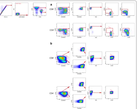
Corresponding isotypes for each antibody were used as controls. Cells were analyzed by flow cytom-etry using a MACS Quant cytometer (Miltenyi Biotech, Germany) and the FlowJo version V10 software (Tree-star, USA). Different T cell subpopulations obtained from fresh blood (Fig. 1a) and cultured cells (Fig. 1b) were determined by a previous published gating strat-egy [11] using fluorescence minus one (FMO) controls for each antibody (i.e., samples stained with all fluoro-chromes except the one of interest). Particularly, TSCM
were identified by a previously described plotting strat-egy [11]. Briefly, CD4+ and CD8+ cells were sorted and a CCR7 against CD45RO plot were used to select CCR7+CD45RO− cells in each subpopulation. In these cells, we identified CCR7+CD45RO−CD45RA+ cells by plotting CCR7 against CD45RA. Then, CD27 was used to detect CCR7+CD45RO−CD45RA+CD27+ cells. Finally, a plot of CD95 against CCR7 was used to identify CCR7+CD45RO−CD45RA+CD27+CD95+ T
SCM (Fig. 1).
Statistical analysis
Statistical analysis was performed with GraphPad Prism 6 (GraphPad Software, USA). Data are shown as the mean ± SEM. Differences were tested for statistical sig-nificance using one-way ANOVA test. A p value <0.05 was considered significant.
Results
Short CD3/CD28 costimulation enriches for memory stem T cells (TSCM) cultured with IL‑7/IL‑15
To assess whether the length of CD3/CD28 costimulation has an impact on the maintenance of the TSCM phenotype
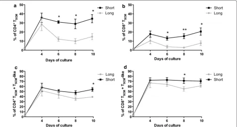
in vitro, naïve T cells were cultured with low doses of IL-7 and IL-15 and activated with magnetic beads coated with anti-CD3/anti-CD28. A short CD3/CD28 costimu-lation (48 h) was compared to a long stimulus (the entire period of cell culture: 10 days) by analyzing individual T-cell subsets at different time points. As shown in Fig. 2a, while the percentage of CD4+ T
SCM at day 4 was
comparable between both conditions (35.64 ± 5.1 % and 28.38 ± 6.9 %; p = 0.42), the short CD3/CD28 costimu-lation led to a significant increase in the frequencies of CD4+ T
SCM after day 4 that was maintained until the end
of the culture (34.6 ± 4.4 % vs 15.6 ± 4.24 % respectively; p = 0.008) (Fig. 2a).
Similar results were obtained in the CD8+ T
SCM
pop-ulation (Fig. 2b). A short costimulation generated a sig-nificant increase of CD8+ T
SCM frequencies compared to
a long costimulation (20.5 ± 4.00 % vs 7.7 ± 2.53 % at day 10, respectively; p = 0.02). Day 10 was selected as an endpoint for culture since a decline in TSCM numbers was
observed after this time (data not shown) and over this time period TSCM expand to numbers considered to be
sufficient for clinical translation.
According to previous data [9], when TSCM are cultured
in vitro they may also acquire the expression of CD45RO, while preserving CD45RA and CCR7+CD27+CD95+ expression (i.e., a TSCM-like phenotype). We found no
differences in the percentage of both CD4+ and CD8+ TSCM-like cells across different time points over the
culture period (19.4 ± 3.06 % vs 24.4 ± 2.6 % in CD4+; p = 0.252 and 49.95 ± 3.6 % vs 53.36 ± 1.04 % in CD8+; p = 0.35).
When total TSCM (i.e., TSCM + TSCM-like
popula-tion) were analyzed (Fig. 2c, d), a higher percentage was observed in the CD4+ population after a short stimula-tion, reaching 54.02 ± 3.837 % at day 10 vs 38.49 ± 1.48 % in the long stimulus condition (p = 0.0054) (Fig. 2c). In the CD8+ population, a trend to higher percentages were found after short costimulation compared to a long costimulation (70.45 ± 4.1 % vs 60.2 ± 3.29 %; p = 0.08) (Fig. 2d).
IL‑21 increases the frequencies of TSCM generated under short CD3/CD28 costimulation
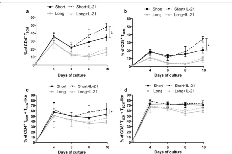
Next, we analyzed the impact of IL-21 on the in vitro generation and maintenance of TSCM. As shown in
Fig. 3a, the addition of IL-21 to the culture from day 0 significantly increased the percentage of CD4+ T
SCM
after a short CD3/CD28 costimulation (47.92 ± 4.10 % vs 34.62 ± 4.44 %; p = 0.006). However, IL-21 had no impact on the percentage of CD4+ T
SCM cultured under
long costimulation (21.1 ± 1.31 % vs 15.6 ± 4.24 %; p = 0.17) (Fig. 3a). The same hold true for CD8+ T
SCM.
IL-21 significantly increased the percentage of CD8+ TSCM cultured under short costimulation (34.15 ± 3.51 %
vs 20.5 ± 4.005 %; p = 0.00039) at day 10 of culture, while adding IL-21 to the long costimulation condition resulted in no significant changes (10.20 ± 2.34 % vs 7.73 ± 3.27 %; p = 0.14) (Fig. 3b).
Finally, we analyzed if IL-21 could increase the percent-age of total TSCM (i.e., TSCM + TSCM-like population). At
the CD8+ subset are enhanced by the addition of IL-21 (74.15 ± 5.26 % vs 60.2 ± 3.29 %; short + IL-21 vs short respectively; p = 0.04), no significant differences were observed in the short stimulation condition by the addi-tion of IL-21 regarding total TSCM (74.15 ± 5.26 % vs
70.45 ± 4.13 %; p = 0.56) (Fig. 3d).
IL-21 is a cytokine with action on multiple T cell sub-sets. To clarify if the short stimulus with IL-21 could affect any of the populations present in the culture, dif-ferent memory T cell subsets were analyzed at day 10
(Fig. 4). The percentages of central memory T cells were 34.27 ± 4.13 % vs 26.4 ± 6.9 %; p = 0.95 in CD4+ and 16.6 ± 8.52 % vs 15.45 ± 9.5 %; p > 0.05 in CD8+ (short vs short + IL-21 respectively). The percentages of effec-tor memory T cells were 7.97 ± 0.12 % vs 5.54 ± 0.07 %; p > 0.05 in CD4+, and 4.8 ± 0.54 % vs 4.15 ± 0.59 %; p > 0.05 in CD8+ (short vs short + IL-21 respectively). In addition, CD4+/CD8+ T
SCM ratio was also analyzed
to explore the possibility of an overexpansion of CD8+ in detriment of CD4+. IL-21 did not preferentially enriched Fig. 1 Gating strategy for the identification of human TSCM. T cells were identified by gating on lymphocytes (SSC versus FSC), singlets (FSC‑H versus
FSC‑A) and live T cells (SSC versus LIVE/DEAD). CD4+ and CD8+ T cells were simultaneously identified with anti‑CD4 and anti‑CD8 antibodies. a
Gating strategy of fresh blood cells. After gating on CD4+ and CD8+ cells, T
CM and TEM subpopulations were identified based on CCR7 and CD45RO
expression. In the gated CCR7+CD45RO− population, cells expressing CD45RA and CD27 were further analyzed. In this later population (CCR7+CD4
5RO−CD45RA+CD27+), T
N and TSCM were identified based on the CD95 expression. TN is defined as CCR7+CD45RO−CD45RA+CD27+CD95− whereas
TSCM subpopulation is defined as CCR7+CD45RO−CD45RA+CD27+CD95+. Red arrows indicate the sequential gating strategy. b Gating strategy of
10 days culture cells. After gating on CD4+ and CD8+ cells, T
CM and TEM subpopulations were identified based on CCR7 and CD45RO expression.
In the gated CCR7+CD45RO− population, cells expressing CD45RA and CD27 were further analyzed. In this later population (CCR7+CD45RO−C
D45RA+CD27+), T
SCM were identified based on the CD95 expression. TSCM subpopulation is defined as CCR7+CD45RO−CD45RA+CD27+CD95+.
Similarly, in the gated CCR7+CD45RO+ population, cells expressing CD45RA, CD27 and CD95+ identify a T
SCM‑like subpopulation, which is defined
[image:4.595.72.541.86.460.2]for the CD8+ population since the CD4+ cells were maintained over the cell culture period in all the con-ditions tested (CD4+/CD8+ T
SCM ratio: 1.03 ± 0.28 vs
1.34 ± 0.19 for long versus long + IL-21 costimulation, and 0.68 ± 0.14 vs 0.72 ± 0.002 for short vs short + IL-21 costimulation; p = 0.23).
Short costimulation combined with IL‑21 enhance TSCM
expansion
Next, we analyzed the effect of IL-21 in TSCM expansion
under the costimulation conditions described. As shown in Fig. 5a, b, the addition of IL-21 to the culture produced a significant increase in the TSCM expansion. The
combi-nation of short costimulation and IL-21 led to the greater TSCM expansion, for both CD4+ and CD8+ subsets,
among all conditions tested (CD4+ T
SCM: 311.34 ± 39
fold vs 192.84 ± 58.61 fold; TSCM: 728.06 ± 53 fold
vs 442.73 ± 122 fold; IL-21 vs no IL-21 respectively; p = 0.040) (Fig. 5a, b). Finally, we analyzed the impact of IL-21 on total TSCM expansion. As shown in Fig. 5c,
d, the combination of short costimulation and IL-21 led to the greatest expansion of total CD4+ and CD8+ T
SCM
(351.79 ± 36 fold vs 251.83 ± 16.6 fold; p = 0.07, and 744.46 ± 101.76 fold vs 592.3 ± 172 fold; short + IL-21 vs long, respectively; p = 0.1) (Fig. 5c, d).
TSCM shows different functional properties
To further define the functional features of T-cells gener-ated under these conditions, we analyzed the expression of IL-2, IFN-γ, granzyme-A and perforin on FACS-puri-fied TSCM, TCM and TEM at day 10 of culture. We found
that both CD4+ and CD8+ T
SCM secrete higher
lev-els of IL-2 compared to TCM (CD4: 84.65 ± 0.75 %
vs 83.3 ± 0.7 %; p = 0.48; CD8: 68.86 ± 1.9 % vs 62.56 ± 0.13 %, p = 0.03) and TEM (CD4: 84.65 ± 0.75 %
vs 80.5 ± 0.75 %; p = 0.057; CD8: 68.86 ± 1.9 % vs 57.8 ± 1.12 %, p = 0.0022) (Additional file 1: Figure S1 A, B). Moreover, we observed lower proportion of CD4+ and CD8+ T
SCM that secrete IFN-γ compared
to TCM (CD4: 8.18 ± 0.16 % vs 13.6 ± 1 %; p = 0.016;
CD8: 18.66 ± 4.2 % vs 48.76 ± 8.2 %, p = 0.0397) and TEM (CD4: 8.18 ± 0.16 % vs 21.55 ± 0.25 %; p = 0.0012;
CD8: 18.66 ± 4.2 % vs 60.86 ± 6.4 %, p = 0.0091) and granzyme-A compared to TCM (CD4: 34.83 ± 8.6 %
vs 55.66 ± 1.53 %; p = 0.0631; CD8: 7.2 ± 3.3 % vs 75.8 ± 0.3 %, p = 0.0004) and TEM (CD4: 34.83 ± 8.6 %
vs 78.13 ± 2.36 %; p = 0.0033; CD8: 7.2 ± 3.3 % vs 78.1 ± 0.5 %, p = 0.0004). We also observed that CD4+ and CD8+ T
SCM stained mainly negative for perforin
(0.024 ± 0.002 % and 0.9 ± 0.3 %, respectively) (Addi-tional file 1: Figure S1 C–H).
Fig. 2 Short CD3/CD28 costimulation increases CD4+ and CD8+ T
SCM frequencies compared with long costimulation. Naïve T cells from healthy
donors (n = 6) were cultured for 10 days with short (48 h) (solid black line) or long (solid grey line) costimulation. a, b Frequency of CD4+ (a) and
CD8+ (b) T
SCM cell subset (mean ± SEM). c, d Frequencies of total TSCM (TSCM+ TSCM‑like) CD4+ (c) and CD8+ (d) (mean ± SEM). *p < 0.05; **p < 0.01;
[image:5.595.60.539.88.345.2]Fig. 3 IL‑21 enhances CD4+ and CD8+ T
SCM frequencies. Naïve T cells from healthy donors (n = 6) were cultured for 10 days with short (solid black line) or long (solid grey line) costimulation and in the presence (black dashed line) or absence (grey dashed line) of IL‑21. a, b Frequencies of CD4+ and
CD8+ T
SCM (mean ± SEM) are shown in (a) and (b), respectively. c, d Frequencies of CD4+ and CD8+ Total TSCM (TSCM+ TSCM‑like) (mean ± SEM) are
shown in (c) and (d), respectively. *p < 0.05; **p < 0.01; ***p < 0.001
Fig. 4 Analysis of CD4+ and CD8+ T cell subsets composition. a, b Column graphs showing the relative frequencies (mean ± SEM) of CD4+ (a) and
CD8+ (b) T cell subsets respectively, for each condition tested (long costimulation, short costimulation, long + IL‑21, and short + IL‑21). T
N (T Naïve),
[image:6.595.58.536.84.401.2] [image:6.595.59.540.472.659.2]Finally, we analyzed the phenotype and self-renewal capacity of FACS-purified TSCM cells after being expanded
for 10 days. We observed that, in contrast to TCM and TEM
subsets, both CD4+ and CD8+ T
SCM maintained their
phe-notype and proliferative capacity in culture for another 10 days after sorting. Remarkably, 93 % of the CD4+ and 88 % of CD8+ T cells displayed a T
SCM phenotype after
20 days in culture with IL-7, IL-15 and IL-21, while TCM
and TEM largely lost their proliferative capacities and
became differentiated (Additional file 2: Figure S2).
CD4+ and CD8+ T
SCM are efficiently transduced
by lentiviral vectors
Unlike activated and dividing cells, quiescent T cells are less permissive to vector transduction. To assess whether TSCM generated under this new conditions could be
genetically modified for adoptive immunotherapy, an encoding-GFP lentivirus was used to transduce TSCM.
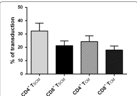
We have analyzed transduction efficiencies (multiplicity of infection (m.o.i.) = 1) following stimulation of naïve T cells for 48 h with magnetic anti-CD3/CD28 beads and IL-7, IL-15 and IL-21. Gene transfer in CD4+and CD8+
cells averaged 28 and 19 % respectively. The transduc-tion efficiency was also analyzed in the different mem-ory subset presents in the culture. We observed similar transduction percentages of TSCM and TCM in both CD4+
and CD8+ population (32.25 ± 5.91 % vs 24.30 ± 4.36 % in CD4+, T
SCM vs TCM respectively; P = 0.056 and
21.29 ± 3.54 % vs 17.96 ± 3.07 % in CD8+, T
SCM vs TCM
respectively; p = 0.5;) (Fig. 6).
Discussion
The generation of TSCM for ACT has become a
clini-cally relevant issue since preclinical studies have shown that the use of the less-differentiated TSCM is associated
with superior T-cell engraftment, persistence, and antitu-mor efficacy [3–6]. Although ACT to treat cancer have experimented significant progress in the past few years, particularly with the chimeric antigen receptor (CAR) approach, improvements on their clinical efficacy are clearly needed. Remarkably, although no ACT clinical trials with TSCM have been done in cancer patients so
far, studies in patients with lymphoma receiving CAR-T cells showed that the infusion of >5 % of cells with a Fig. 5 IL‑21 enhances CD4+ and CD8+ T
SCM expansion. Naïve T cells from healthy donors (n = 3) were cultured for 10 days and fold expansion were
analyzed in short costimulation (solid black line), long costimulation (solid grey line) short + IL‑21 (black dashed line) and long + IL‑21 (grey dashed line) conditions. a, b Absolute fold expansion of CD4+ (a) and CD8+ (b) (mean ± SEM), expressed in increment of cells from the starting culture
time point is shown. c, d Absolute fold expansion of CD4+ (c) and CD8+ (d) total TSCM (TSCM + TSCM‑like) (mean ± SEM). *p < 0.05; **p < 0.01;
[image:7.595.59.539.86.369.2]TSCM phenotype correlated with greater persistence and
expansion in vivo, properties associated with clinical effi-cacy [20].
Previous studies have shown that the combination of IL-7, IL-15 and CD3/CD28 costimulation led to the gen-eration of TSCM from naïve T cells [9]. Interestingly, cells
generated under this condition also express CD45RO, which indicates that the combination of prolonged CD3/CD28 costimulation with IL-7 and IL-15 induce a more differentiated TSCM (TSCM-like) than the originally
[image:8.595.57.290.86.249.2]described [9]. This could be related to the stimulation through CD3 and CD28, which expands T cells efficiently but also causes a drastic change in the cell phenotype, including downregulation of CD45RA, CCR7 and CD62L [6, 13, 21]. Although both subsets have comparable gene expression profiles, it remains to be proven that the TSCM-like subset (i.e., those cells expressing CD45RO in
addition to CD45RA and CCR7) retain similar antitumor efficacy than the less differentiated TSCM. In this regard,
when CAR-T cells were cultured with IL-7/IL-15, only the fraction CD45RA+/CCR7+ retained the ability to expand after repetitive antigen stimulations [20]. Accord-ingly, strategies directed to preserve the phenotype of TSCM during their ex vivo generation should be regarded
a clinical priority.
In this study, we show that a very short (48 h) CD3/ CD28 costimulation increases the percentage of TSCM
within the CD4+ and CD8+ subsets in detriment of a more differentiated phenotypes. Moreover, the contribu-tion of the less differentiated TSCM to the total TSCM
fre-quency is superior in the short costimulation condition compared to a long costimulation, in line with the con-cept that prolonging the CD3/CD28 stimulation in the
culture is associated with T cell differentiation [21, 22]. In our condition, TSCM proliferation was not affected by
the absence of a prolonged CD3/CD28 signal, suggesting that a continued costimulatory signal would not be nec-essary to promote an efficient ex vivo TSCM expansion in
the presence of IL-7 and IL-15 [23–25].
The T-cell receptor signal strength impacts on T cell expansion and differentiation [26]. Methods for the gen-eration of effector T cells based on CD3/CD28 stimula-tion have relied on the use of a high bead:cell ratio (e.g., 3:1). We observed that a high bead-to-cell ratio reduced the frequency of TSCM and did not generate a strong T
cell proliferative response, a fact that may be associated with activation-induced T cell death [21, 27]. In contrast, in our hands, a low bead-to-cell ratio (e.g., 1:2) coupled to a short costimulation was the best condition for pro-moting the generation of higher TSCM frequencies while
maintaining their proliferative capacity. This is in line with recent studies showing that low or equal bead:cell ratios give the higher yields of T cells when cultured in IL-7 and IL-15 [21]. Moreover we observed that the short TCR triggering do not reduced the transduction effi-ciency of TSCM, probably due to the presence of the IL-7,
IL-15 or IL-21 cytokines, which promote long-term sur-vival of resting T cells, while maintaining their matura-tional status [28].
Previous studies have shown that IL-21 synergizes with either IL-7 or IL-15 to regulate CD8+ T cell expansion and to maintain a memory phenotype [18, 19, 29]. How-ever, the potential role of IL-21 in the ex vivo generation of TSCM has not been described. Here, we show for the
first time that the addition of IL-21 significantly increases the percentage of CD4+ and CD8+ T
SCM cultured with a
short CD3/CD28 stimulation and IL-7/IL-15. Remark-ably, addition of IL-21 to that condition further increased the proliferation rate of TSCM, which translated into an
increase of absolute numbers of TSCM at the end of the
culture. Although previous studies have shown that CD3/ CD28 costimulation in combination with IL-7 and IL-15 promote a substantial increment in TSCM expansion [9],
the reduction in the length of costimulation and, particu-larly, the addition of IL-21 produces even higher numbers of TSCM that could be sufficient for clinical translation.
Interestingly, this effect was noted in both CD4+ and CD8+ T
SCM, and although the proliferation rate was
greater in the CD8+ subset compared to that in CD4+, it is remarkable that the CD4+/CD8+ ratios of T
SCM were
preserved over the culture period.
According to previous studies showing a higher expres-sion of IL-21 receptor in activated naïve T cells [30], and that the effect of IL-21 might depend on the T cell differ-entiation status [21, 31], we observed that the impact of Fig. 6 Analysis of transduction efficiency with a GFP‑expressing
lentivirus. Lentivirus transduction efficiency measured in CD4+ and
CD8+ T
SCM and TCM subsets by flow cytometry (% of cells expressing
IL-21 was more pronounced in those T cells with a phe-notype close to naïve T cells, resulting in an enrichment of less differentiated TSCM within the total TSCM subset.
Moreover, we have noticed a higher expression of IL-21R in naïve and TSCM cells compared to more differentiated
T cells subsets (57.5 % of TSCM vs 22 % of TCM in CD4+
and 42.6 % of TSCM vs 5 % of TCM in CD8+) (data not
shown). Interestingly, this difference is more pronounced at day 4 of the culture (in particular in CD8+ T cells), which could increase the effect of IL-21 in the following days.
It has been shown that self-renewal depends on STAT3 (Signal transducer and activator of transcription 3) sign-aling cytokines, including IL-6, IL-10 and IL-21, which inhibit exhaustion and differentiation of T cell [32]. Similarly, T cells expanded in the presence of IL-21 (the unique common γ-chain cytokines to sustain STAT3 activation) prevent the loss of CD45RA, CD62L, CD27, and IL-7Rα, increasing the ability of the cell to release IL-2 [33, 34]. Interestingly, we noticed that TSCM, TCM
and TEM cultured in the presence of IL-21 express high
level of IL-2. In addition, TSCM express low levels of
IFN-γ, granzyme-A and perforin compared to TCM and TEM.
Thus, TSCM display phenotypic and functional
character-istics of early-differentiated cells.
We also observed that highly purified naïve T cells are a more suitable starting population for TSCM generation in
contrast to the use of “bulk” peripheral blood mononu-clear cells [10, 20]. The use of purified naïve T cells may overcome the variability of this subset observed between donors when using whole peripheral blood lymphocytes. Another important issue is the potential deleterious effect that more differentiated memory T cells present in the starting cell population may have on the generation of TSCM. Accordingly, in our experience, TSCM are more
efficiently generated when naïve T cells are used as start-ing population (data not shown), in line with previous data from Bonini´s group [9].
Conclusions
In summary, we demonstrated that when naïve T cells are cultured with IL-7 and IL-15, the use of a very short CD3/CD28 costimulation enhances CD4+ and CD8+ TSCM generation and expansion. Moreover, the addition
of IL-21 to this culture conditions produces a further enrichment and greater expansion of CD4+ and CD8+ TSCM. Importantly, the conditions described here allow
an efficient transduction of both CD4+ and CD8+ T SCM
with lentivirus. These refinements on the in vitro condi-tions may be of clinical relevance for the generation of TSCM for ACT of patients with cancer, and can be easily
adapted to preserve this cell subset in the final product under GMP conditions.
Abbreviations
ACT: adoptive T cell therapy; TCM: central memory T cells; CAR: chimeric
antigen receptor; TEM: effector memory T cells; TEMRA: effector memory‑RA + T
cells; FMO: fluorescence minus one; TSCM: memory stem T cells; m.o.i: multiplic‑
ity of infection; TN: naïve T cells; PBMCs: peripheral blood mononuclear cells;
γc: γ‑chain; IFN‑γ: interferon gamma; STAT3: signal transducer and activator of transcription 3.
Authors’ contributions
CAF: Design, experimental and analytical performance. LEG: Experimental per‑ formance. SV: Design performance. JS: Analytical performance. JB: Study’s con‑ ception, design, and analytical performance. All authors read and approved the final manuscript.
Author details
1 Hematology Service, Hospital de la Santa Creu i Sant Pau, Mas Casanovas
90, 08041 Barcelona, Spain. 2 Autonomous University, Barcelona, Spain. 3 IIB‑Institut Recerca Hospital de la Santa Creu I Sant Pau, Barcelona, Spain. 4 Laboratory of Experimental Hematology–IIB, Institut Recerca Hospital de la
Santa Creu I Sant Pau, Barcelona, Spain.
Acknowledgements
The authors thank Dr. Sonia Guedan for helpful discussions and critical review of the manuscript.
Competing interests
The authors declare that they have no competing interests.
Ethics approval and consent to participate
All donor’s samples were obtained according to a protocol approved by the Ethics Comittee of Hospital Santa Creu I Sant Pau.
Funding
This work was supported by Fundació La Marató TV3, Cellex Research Founda‑ tion (Barcelona), AGAUR 2014SGR‑1281, and ISCIII RD12/0036/0071.
Received: 5 April 2016 Accepted: 11 July 2016
References
1. June CH. Principles of adoptive T cell cancer therapy. J Clin Invest. 2007;117:1204–12.
2. Restifo NP, Dudley ME, Rosenberg SA. Adoptive immunotherapy for cancer: harnessing the T cell response. Nat Rev Immunol. 2012;12:269–81. 3. Hinrichs CS, Borman ZA, Cassard L, Gattinoni L, Spolski R, Yu Z, Sanchez‑
Perez L, Muranski P, Kern SJ, Logun C, et al. Adoptively transferred effector cells derived from naive rather than central memory CD8+ T cells mediate superior antitumor immunity. Proc Natl Acad Sci USA. 2009;106:17469–74.
Additional files
Additional file 1: Figure S1. TSCM functional properties. A‑F Frequen‑
cies of CD4+ and CD8+ T
SCM, TCM, and TEM cytokine‑producing cells
(mean ± SEM). Quantitation of IL‑2 (A), IFN‑γ (C), granzyme‑A (E) and perforin (G) production by CD4+ cells and IL‑2 (B), IFN‑γ (D), granzyme‑ A (F) and perforin (H) production by CD8+ cells. *p < 0.05; **p < 0.01; ***p < 0.001.
Additional file 2: Figure S2. Long term culture and proliferative capacity of TSCM cells. Isolated naïve T cells were cultured under IL‑7, IL‑15, IL‑21 and
short costimulation until day 10. Selected TSCM were sorted and cultured
for another 10 days. Column graphs showing the relative frequencies of CD4+ and CD8+ T cell subsets from day 0 to day 10 after T
SCM sorting (total
• We accept pre-submission inquiries
• Our selector tool helps you to find the most relevant journal • We provide round the clock customer support
• Convenient online submission • Thorough peer review
• Inclusion in PubMed and all major indexing services • Maximum visibility for your research
Submit your manuscript at www.biomedcentral.com/submit
Submit your next manuscript to BioMed Central
and we will help you at every step:
4. Zhou X, Cui Y, Huang X, Yu Z, Thomas AM, Ye Z, Pardoll DM, Jaffee EM, Cheng L. Lentivirus‑mediated gene transfer and expression in established human tumor antigen‑specific cytotoxic T cells and primary unstimu‑ lated T cells. Hum Gene Ther. 2003;14:1089–105.
5. Berger C, Jensen MC, Lansdorp PM, Gough M, Elliott C, Riddell SR. Adoptive transfer of effector CD8+ T cells derived from central memory cells establishes persistent T cell memory in primates. J Clin Invest. 2008;118:294–305.
6. Gattinoni L, Lugli E, Ji Y, Pos Z, Paulos CM, Quigley MF, Almeida JR, Gostick E, Yu Z, Carpenito C, et al. A human memory T cell subset with stem cell‑ like properties. Nat Med. 2011;17:1290–7.
7. Lugli E, Dominguez MH, Gattinoni L, Chattopadhyay PK, Bolton DL, Song K, Klatt NR, Brenchley JM, Vaccari M, Gostick E, et al. Superior T memory stem cell persistence supports long‑lived T cell memory. J Clin Invest. 2013;123:594–9.
8. Klebanoff CA, Gattinoni L, Palmer DC, Muranski P, Ji Y, Hinrichs CS, Borman ZA, Kerkar SP, Scott CD, Finkelstein SE, et al. Determinants of successful CD8+ T‑cell adoptive immunotherapy for large established tumors in mice. Clin Cancer Res. 2011;17:5343–52.
9. Cieri N, Camisa B, Cocchiarella F, Forcato M, Oliveira G, Provasi E, Bon‑ danza A, Bordignon C, Peccatori J, Ciceri F, et al. IL‑7 and IL‑15 instruct the generation of human memory stem T cells from naive precursors. Blood. 2013;121:573–84.
10. Gomez‑Eerland R, Nuijen B, Heemskerk B, van Rooij N, van den Berg JH, Beijnen JH, Uckert W, Kvistborg P, Schumacher TN, Haanen JB, Jorritsma A. Manufacture of gene‑modified human T‑cells with a memory stem/ central memory phenotype. Hum Gene Ther Methods. 2014;25:277–87. 11. Lugli E, Gattinoni L, Roberto A, Mavilio D, Price DA, Restifo NP, Roederer M.
Identification, isolation and in vitro expansion of human and nonhuman primate T stem cell memory cells. Nat Protoc. 2013;8:33–42.
12. Teschner D, Wenzel G, Distler E, Schnurer E, Theobald M, Neurauter AA, Schjetne K, Herr W. In vitro stimulation and expansion of human tumour‑ reactive CD8+ cytotoxic T lymphocytes by anti‑CD3/CD28/CD137 magnetic beads. Scand J Immunol. 2011;74:155–64.
13. Li Y, Kurlander RJ. Comparison of anti‑CD3 and anti‑CD28‑coated beads with soluble anti‑CD3 for expanding human T cells: differing impact on CD8 T cell phenotype and responsiveness to restimulation. J Transl Med. 2010;8:104.
14. Hinrichs CS, Spolski R, Paulos CM, Gattinoni L, Kerstann KW, Palmer DC, Klebanoff CA, Rosenberg SA, Leonard WJ, Restifo NP. IL‑2 and IL‑21 confer opposing differentiation programs to CD8+ T cells for adoptive immuno‑ therapy. Blood. 2008;111:5326–33.
15. Wang G, Tschoi M, Spolski R, Lou Y, Ozaki K, Feng C, Kim G, Leonard WJ, Hwu P. In vivo antitumor activity of interleukin 21 mediated by natural killer cells. Cancer Res. 2003;63:9016–22.
16. Moroz A, Eppolito C, Li Q, Tao J, Clegg CH, Shrikant PA. IL‑21 enhances and sustains CD8+ T cell responses to achieve durable tumor immunity: com‑ parative evaluation of IL‑2, IL‑15, and IL‑21. J Immunol. 2004;173:900–9. 17. Di Carlo E, Comes A, Orengo AM, Rosso O, Meazza R, Musiani P, Colombo
MP, Ferrini S. IL‑21 induces tumor rejection by specific CTL and IFN‑ gamma‑dependent CXC chemokines in syngeneic mice. J Immunol. 2004;172:1540–7.
18. Zeng R, Spolski R, Finkelstein SE, Oh S, Kovanen PE, Hinrichs CS, Pise‑ Masison CA, Radonovich MF, Brady JN, Restifo NP, et al. Synergy of IL‑21 and IL‑15 in regulating CD8+ T cell expansion and function. J Exp Med. 2005;201:139–48.
19. Liu S, Lizee G, Lou Y, Liu C, Overwijk WW, Wang G, Hwu P. IL‑21 synergizes with IL‑7 to augment expansion and anti‑tumor function of cytotoxic T cells. Int Immunol. 2007;19:1213–21.
20. Xu Y, Zhang M, Ramos CA, Durett A, Liu E, Dakhova O, Liu H, Creighton CJ, Gee AP, Heslop HE, et al. Closely related T‑memory stem cells correlate with in vivo expansion of CAR.CD19‑T cells and are preserved by IL‑7 and IL‑15. Blood. 2014;123:3750–9.
21. Shi Y, Wu W, Wan T, Liu Y, Peng G, Chen Z, Zhu H. Impact of polyclonal anti‑CD3/CD28‑coated magnetic bead expansion methods on T cell proliferation, differentiation and function. Int Immunopharmacol. 2013;15:129–37.
22. Trickett A, Kwan YL. T cell stimulation and expansion using anti‑CD3/ CD28 beads. J Immunol Methods. 2003;275:251–5.
23. Ramanathan S, Gagnon J, Dubois S, Forand‑Boulerice M, Richter MV, Ilangumaran S. Cytokine synergy in antigen‑independent activation and priming of naive CD8+ T lymphocytes. Crit Rev Immunol. 2009;29:219–39. 24. Ramanathan S, Gagnon J, Ilangumaran S. Antigen‑nonspecific activation
of CD8+ T lymphocytes by cytokines: relevance to immunity, autoim‑ munity, and cancer. Arch Immunol Ther Exp (Warsz). 2008;56:311–23. 25. Tan JT, Dudl E, LeRoy E, Murray R, Sprent J, Weinberg KI, Surh CD. IL‑7 is
critical for homeostatic proliferation and survival of naive T cells. Proc Natl Acad Sci USA. 2001;98:8732–7.
26. Sallusto F, Geginat J, Lanzavecchia A. Central memory and effector memory T cell subsets: function, generation, and maintenance. Annu Rev Immunol. 2004;22:745–63.
27. Weiss EM, Schmidt A, Vobis D, Garbi N, Lahl K, Mayer CT, Sparwasser T, Ludwig A, Suri‑Payer E, Oberle N, Krammer PH. Foxp3‑mediated suppres‑ sion of CD95L expression confers resistance to activation‑induced cell death in regulatory T cells. J Immunol. 2011;187:1684–91.
28. Cavalieri S, Cazzaniga S, Geuna M, Magnani Z, Bordignon C, Naldini L, Bonini C. Human T lymphocytes transduced by lentiviral vectors in the absence of TCR activation maintain an intact immune competence. Blood. 2003;102:497–505.
29. Alves NL, Arosa FA, van Lier RA. IL‑21 sustains CD28 expression on IL‑ 15‑activated human naive CD8+ T cells. J Immunol. 2005;175:755–62. 30. Battaglia A, Buzzonetti A, Baranello C, Fanelli M, Fossati M, Catzola V,
Scambia G, Fattorossi A. Interleukin‑21 (IL‑21) synergizes with IL‑2 to enhance T‑cell receptor‑induced human T‑cell proliferation and coun‑ teracts IL‑2/transforming growth factor‑beta‑induced regulatory T‑cell development. Immunology. 2013;139:109–20.
31. Mehta DS, Wurster AL, Grusby MJ. Biology of IL‑21 and the IL‑21 receptor. Immunol Rev. 2004;202:84–95.
32. Gattinoni L, Klebanoff CA, Restifo NP. Paths to stemness: building the ultimate antitumour T cell. Nat Rev Cancer. 2012;12:671–84. 33. Li Y, Bleakley M, Yee C. IL‑21 influences the frequency, phenotype,
and affinity of the antigen‑specific CD8 T cell response. J Immunol. 2005;175:2261–9.