0022-538X/11/$12.00 doi:10.1128/JVI.00549-11
Copyright © 2011, American Society for Microbiology. All Rights Reserved.
Nipah Virus Uses Leukocytes for Efficient Dissemination
within a Host
䌤
Cyrille Mathieu,
1† Christine Pohl,
1† Judit Szecsi,
1Selena Trajkovic-Bodennec,
1Se
´verine Devergnas,
1Herve
´ Raoul,
2Franc
¸ois-Loïc Cosset,
1Denis Gerlier,
1T. Fabian Wild,
1and Branka Horvat
1*
INSERM, U758, Ecole Normale Supe´rieure de Lyon, Lyon, F-69007 France, and IFR128 BioSciences Lyon-Gerland Lyon-Sud, University of Lyon 1, 69365 Lyon, France,1and Laboratory P4-Jean Me´rieux, INSERM, 69365 Lyon, France2
Received 17 March 2011/Accepted 6 May 2011
Nipah virus (NiV) is a recently emerged zoonotic paramyxovirus whose natural reservoirs are several species ofPteropusfruit bats. NiV provokes a widespread vasculitis often associated with severe encephalitis, with up to 75% mortality in humans. We have analyzed the pathogenesis of NiV infection, using human leukocyte cultures and the hamster animal model, which closely reproduces human NiV infection. We report that human lymphocytes and monocytes are not permissive for NiV and a low level of virus replication is detected only in dendritic cells. Interestingly, despite the absence of infection, lymphocytes could efficiently bind NiV and transfer infection to endothelial and Vero cells. This lymphocyte-mediated transinfection was inhibited after proteolytic digestion and neutralization by NiV-specific antibodies, suggesting that cells could transfer infec-tious virus to other permissive cells without the requirement for NiV internalization. In NiV-infected hamsters, leukocytes captured and carried NiV after intraperitoneal infection without themselves being productively infected. Such NiV-loaded mononuclear leukocytes transfer lethal NiV infection into naïve animals,
demon-strating efficient virus transinfectionin vivo. Altogether, these results reveal a remarkable capacity of NiV to
hijack leukocytes as vehicles to transinfect host cells and spread the virus throughout the organism. This mode of virus transmission represents a rapid and potent method of NiV dissemination, which may contribute to its high pathogenicity.
Nipah virus (NiV) is a highly pathogenic zoonotic paramyxo-virus that emerged in 1998 as the causal agent of a respiratory disease and an acute febrile encephalitis in humans (9, 10). Together with Hendra virus (HeV), identified in Australia in 1994 (31), and based on unique genetic characteristics distinct from those of other paramyxoviruses, NiV is classified in the genusHenipavirus, (15). Further NiV outbreaks have regularly been documented in Bangladesh and India since 2001 (7, 25). The human death rates varied from 40% in Malaysia to 75% in Bangladesh and India, and a number of outbreaks were asso-ciated with interhuman transmission (21, 27). Several species of fruit bats, primarily of the genusPteropus, widely distributed in Australia, Southeast Asia, India, and Africa, appear to be theHenipavirusreservoir, maintaining a permanent risk of new outbreaks (11, 22, 38).
The site of primary replication of NiV and the mode of virus propagation throughout the organism remain unknown. The incubation period varies from 4 to 60 days and is shorter in Bangladeshi than in Malaysian patients, potentially reflecting differences between two viral strains (24, 43). In humans, the blood vessels appear to be one of the early targets of infection, with the central nervous system (CNS) being the most severely affected, although lung, kidney, and other organs are also in-fected (43). The majority of human infections led to acute
encephalitis with vasculitis-induced thrombosis in the brain and, in some patients, atypical pneumonia and respiratory distress. A late-onset encephalitis could arise up to several months or even years after the initial infection, and a relapsed encephalitis has occurred in patients who had previously re-covered from acute encephalitis (39).
The pathogenesis of Nipah virus infection is poorly under-stood. We have analyzed the permissiveness of human leuko-cytes to NiV infection. Only dendritic cells (DC) support a productive NiV infection, although all leukocyte types are ca-pable of binding NiV and transmitting it to susceptible cellsin vitro. Accordingly, in the hamster infection model, virus is found associated with leukocytes without signs of infection, and the transfer of these cells resulted in the infection of naïve animals, demonstrating efficient virus transinfection in vivo. We propose that leukocytes act as vehicles for NiV spread within the organism to permissive tissues, contributing to the pathogenesis of NiV infection.
MATERIALS AND METHODS
Cell culture.Vero E6, 293T, and human U373 astroglioma cells were main-tained in Dulbecco’s modified Eagle’s medium (DMEM) (Invitrogen) supple-mented with 10% fetal calf serum (FCS), 100 U/ml penicillin, 0.1 mg
strepto-mycin, 10 mM HEPES, and 2 mML-glutamine at 37°C in 5% CO2. HPMEC-ST1
(human pulmonary microvascular endothelial cells) (26) were grown in Endo-thelial Cell Growth Medium (Promocell) on culture dishes precoated with 0.2% gelatin.
Human peripheral blood was obtained from 20 different healthy donors from the Blood Transfusion Centre (Lyon, France). Peripheral blood mononuclear cells (PBMC) were isolated by Ficoll-Hypaque density gradient centrifugation and then centrifuged through a 50% Percoll gradient (Pharmacia Fine
Chemi-* Corresponding author. Mailing address: INSERM U758, 21 Ave-nue Tony Garnier, 69007 Lyon, France. Phone: 33 4 3728 2392. Fax: 33 4 3728 2391. E-mail: [email protected].
† C.M. and C.P. contributed equally to this study.
䌤Published ahead of print on 18 May 2011.
7863
on November 7, 2019 by guest
http://jvi.asm.org/
cals, Uppsala, Sweden) for 20 min at 400⫻g. Peripheral blood lymphocytes (PBLs) were recovered from the high-density fraction and monocytes from the
low-density fraction at the interface. CD3⫹and CD19⫹lymphocytes and CD14⫹
monocytes were isolated from the high- and low-density fractions, respectively, using microbeads (Miltenyi Biotech) and magnetic cell separation with a MACS
Separator. DC were generatedin vitrofrom the adherent fraction of purified
monocytes, treated for 6 days at 5⫻105monocytes/ml with interleukin 4 (IL-4)
(250 U/ml; Peprotech) and graulocyte-macrophage colony-stimulating factor (GM-CSF) (500 U/ml; Peprotech). Macrophages were derived by growth in
M-CSF (50 ng/ml; Peprotech) at 37°C-5% CO2in 6-well plates. PBLs were
stimulated overnight with IL-2 (100 U/ml; Abcys) and phytohemagglutinin
(PHA) (2g/ml; Sigma). The maturation of DC was induced by
lipopolysaccha-ride (LPS) (100 ng/ml; Sigma) for 24 h. Cell purity was verified by flow cytometry after cell labeling with dye-conjugated monoclonal antibodies specific for CD14, CD1, CD11c, CD3, and CD19 (Becton Dickinson) using a FACSCalibur 3C and CellQuestPro software (Becton Dickinson).
Splenocytes were harvested from hamsters and stimulated in culture with
concanavalin A (2g/ml; Sigma) or left unstimulated. Both human and hamster
cells were cultured in complete RPMI medium supplemented with 10% FCS, 100
U/ml penicillin, 0.1 mg streptomycin, 10 mM HEPES, and 2 mML-glutamine at
37°C in 5% CO2.
Virus infection and titration. Nipah virus (isolate UMMC1; GenBank AY029767) (8), recombinant NiV (rNiV), and rNiV-enhanced green fluorescent protein (EGFP) (44) were prepared on Vero-E9 cells as described previously (19). Leukocytes were infected at a multiplicity of infection (MOI) of 1, washed twice, and observed by inverted and/or fluorescence microscopy every day postin-fection (p.i.) or harvested for RNA isolation or for use in transinpostin-fection assays.
At the indicated times p.i., 150l of cell culture supernatant was collected and
frozen prior to viral titration. Viral titration was performed as detailed elsewhere (19). The viral infection in cocultures of leukocytes with Vero cells was deter-mined using a previously described infectious-center assay (23).
RNA isolation and reverse transcription-quantitative PCR (RT-qPCR).RNA was isolated from cells and plasma using an RNeasy Mini Kit (Qiagen) in either RLT or AVL buffer, according to the manufacturer’s instructions. Reverse
tran-scription was performed on 0.5g of total RNA using oligo(dT) and
random-hexamer oligonucleotide primers (iScript cDNA synthesis kit; Bio-Rad) and run in a Biometra T-Gradient PCR device, and cDNAs were diluted 1/10.
Quantitative PCR was performed with all cDNA samples using Platinum SYBR green qPCR SuperMix-UDG with a ROX kit (Invitrogen). qPCR was run on the ABI 7000 PCR system (Applied Biosystems) as follows: 95°C for 5 min and 40 cycles of 95°C for 15 s and 60°C for 1 min, followed by a melting curve up to 95°C at 0.8°C intervals. All samples were run in duplicate, and the results were analyzed using ABI Prism 7000 SDS software. The glyceraldehyde 3-phosphate dehydrogenase (GAPDH) gene was used as a housekeeping gene to normalize the samples. GAPDH and standard references for the corresponding genes were included in each run to check for RNA integrity, RNA load, and inter-PCR variations. After normalization, the results were expressed as the ratio of mRNA
copy numbers to the number of copies at time zero (t⫽0) (the fold change); in
some experiments, the number of copies of the gene of interest was expressed
perg of analyzed RNA. All calculations were done using the 2⌬⌬CT
model (36), and experiments were performed according to the MIQE guideline (4). The primers used were designed using Beacon 7.0 software and validated for their efficacy close to 100%: EFNB2 forward, TCGGGCTAGTTAAGGTGTGC, and reverse, ATGAGTGTTCCATGAGTGATGC; EFNB3 forward, TCACCCTCT TGGCTTCTTATCC, and reverse, GGGGAGTGGTTGGTATGAGAG; NiV N forward, GGCAGGATTCTTCGCAACCATC, and reverse, GGCTCTTGG GCCAATTTCTCTG; NiV M forward, AACGGCTGTTTGCTCAAATGGG, and reverse, GCTGCTACTCGGCTGATCTCAC; NiV F forward, GCAGGGC AATCTCACAATCAGG, and reverse, GGACCGATAGCAATGCCTTCAG; NiV G forward, AGGTTCAAAGATCAGCCAGTCG, and reverse, AAAGGG AGTGGGTTAGGACAAG; NiV L forward, ATGGTGCTGTGCTGTCTC AGG, and reverse, AGCCGACATTTCTTGACAACCC; and hGAPDH forwa rd, CACCCACTCCTCCACCTTTGAC, and reverse, GTCCACCACCCTGTT GCTGTAG.
Production of envelope glycoprotein-pseudotyped virus.NiV G- and F- and control vesicular stomatitis virus (VSV) G-pseudotyped Friend murine leukemia virus (MLV) particles were generated as described elsewhere (32). Briefly, 293T cells were transfected with the expression vectors pTG534 and pTG13077 (MLV-based transfer vectors coding for GFP), together with either phCMV-VSV-G
plasmid coding for VSV G or phCMV-NiV G⌬20 and phCMV-NiV F⌬24
plas-mids coding for NiV G with its first N-terminal 20 amino acids (aa) truncated and NiV F with its last C-terminal 24 aa truncated, respectively. Pseudoparticles were
concentrated on a 20% sucrose cushion by ultracentrifugation (110,000⫻gfor
2 h; Beckman SW28). The pseudoparticles were used at an MOI of 1 to trans-duce PHA and IL-2-activated PBLs and cultured in 24-well plates for 48 to 120 h with daily observation under light and fluorescence microscopes.
Transinfection assay.DC, PBLs, and monocytes were prepared from fresh blood and infected for 1 h with NiV or rNiV-EGFP at either 4°C or 37°C in the
presence or absence of 50 M 5-(N-ethyl-N-isopropyl) amiloride (EIPA)
(Sigma). In some experiments, 106PBLs were preincubated with 100g/ml of
EphB4 soluble Fc fusion protein (ligand for ephrinB2; R&D Systems) for 45 min, with 1 mg/ml of pronase (Roche) or 2 mg/ml of trypsin (Sigma) for 30 min at 37°C, or with 5 mM EDTA or 5 mM EGTA (Sigma) for 10 min at room
temperature; washed; and infected with NiV. In other experiments, 106PBLs
were stripped after infection for 30 min at 4°C with 200g/ml pronase.
Alter-natively, 106PBLs were saturated 24 h p.i. with the anti-NiV G monoclonal
antibody (MAb) Nip GIP 1.7 and the anti-NiV F MAb Nip GIP 21 (20) for 30 min at room temperature. After two washes with cold phosphate-buffered saline (PBS), the cells were cultured at 37°C for up to 96 h and then collected and washed, and 10-fold serial dilutions were added to reporter cell monolayers of either endothelial HPMEC cells or Vero cells for determination of cell-associ-ated infectious NiV, using the infectious-center assay (23). All experiments were performed with cells obtained from 3 to 7 different donors and analyzed sepa-rately.
Infection of hamsters.Eight-week-old golden hamsters (Mesocricetus auratus; Janvier, France) were anesthetized and infected intraperitoneally (i.p.) with 0.4 ml of NiV or rNiV-EGFP in a biosafety level 4 (BSL-4) laboratory. Blood cells and plasma collected retro-orbitally at 1, 2, 6, and 7 days p.i. were separated by centrifugation and analyzed by RT-PCR for the NiV N gene content and/or determination of infectious NiV. For adoptive transfer, mononuclear spleen leukocytes, obtained after purification on a Ficoll gradient (Eurobio) of spleno-cytes from infected hamsters (4 days p.i.), were washed and observed under the fluorescence microscope, and the content of cell-associated infectious NiV was followed by coculturing them with Vero cells. In addition, purified cells were
injected i.p. into 6 naive hamsters (25⫻105
per hamster). A control group of animals received purified spleen leukocytes from naive hamsters. The animals were followed daily for 2 weeks. All animals were handled in strict accordance with good animal practice as defined by the French national charter on the ethics of animals, and all efforts were made to minimize suffering.
RESULTS
NiV replication in human leukocytes. NiV is thought to
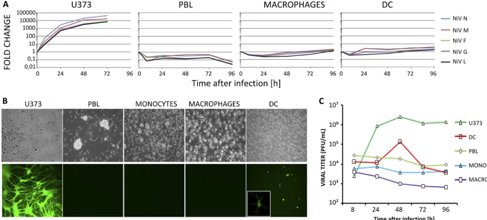
infect lymphoid cells as members of the closest genus, Morbil-livirus, do, although convincing evidence is rather sparse (28). The permissiveness of human lymphocytes, monocytes, mac-rophages, and DC to NiV infection was investigated by mea-suring NiV RNA synthesis, expression of a viral EGFP re-porter gene, and production of infectious virus particles. Human astroglioma U373 cells, used as controls for productive infection, produced increasingly high levels of NiV RNA for every gene with the expected decreasing transcription gradient from N to L genes (Fig. 1A). Early after infection, DC showed an initial increase of viral-RNA production up to 24 h p.i. Then, viral RNA remained stable. Unexpectedly, NiV infec-tion of lymphocytes resulted in an apparent lack of productive infection, since the level of viral RNA slowly decreased with time (Fig. 1A). Accordingly, after infection with rNiV-EGFP, EGFP expression was observed in most U373 cells and a few DC, but not in monocytes, macrophages, and lymphocytes (ei-ther resting or activated by PHA and/or IL-2) at any time p.i. (Fig. 1B and data not shown). Likewise, cytopathic effects were observed only in U373 cells. They produced high levels of infectious NiV detectable at 24 h p.i. DC produced much lower viral titers detectable only at 48 h p.i., and their maturation with LPS did not increase the virus yield (Fig. 1C and data not shown). Monocytes, macrophages, and lymphocytes did not produce virus particles during the course of infection (Fig. 1B and C).
7864 MATHIEU ET AL. J. VIROL.
on November 7, 2019 by guest
http://jvi.asm.org/
Expression of NiV receptors by human leukocytes.The ex-pression of the two known NiV receptors, ephrinB2 and ephrinB3 (1, 33, 34), on human leukocytes and astroglioma U373 was then evaluated from their transcript levels, since currently available anti-ephrinB2/B3 antibodies are rather un-reliable. Low basal expression of ephrinB2 mRNA was de-tected in all cell types analyzed, whereas the level of expression was 1 to 2 log units higher in U373 cells (Fig. 2A). EphrinB3 mRNA expression was detected principally in U373 cells (Fig. 2B). Maturation of DC or activation of lymphocytes with PHA, IL-2, or tumor necrosis factor alpha (TNF-␣) did not signifi-cantly change ephrinB2 and -B3 mRNA expression. To test the ability of NiV to enter different leukocyte types, MLV-based particles pseudotyped with NiV F and G glycoproteins and expressing the GFP reporter gene (NiV-PP) were tested for their capacity to transduce lymphocytes, macrophages, and DC (Fig. 2C). DC, but neither lymphocytes nor macrophages, were transduced with NiV-PP (Fig. 2C). As both lymphocytes and macrophages were readily transduced with the control VSV G-pseudotyped particles (Fig. 2C), this suggests that NiV could enter DC but not the other leukocyte types, which is in agree-ment with the permissiveness of each cell subtype to NiV infection described above.
We next determined the abilities of cells to bind to NiV. Viral genomic RNAs were quantified from cells incubated for 1 h with NiV using a quantitative N gene-specific RT-PCR. Despite their low expression of ephrinB2 mRNA, lymphocytes displayed the strongest ability to bind NiV particles. Surpris-ingly, the highest ephrinB2 mRNA expresser, U373, had the lowest binding capacity detected by this assay (Fig. 2D). Inter-estingly, these results mirrored the various viral titers mea-sured at 8 h p.i. for each cell subset (Fig. 1C). This suggested that the infectious virus detected at 8 h p.i. could represent
cell-bound virus that was not yet engaged in infection but that had survived the cell washes. These results suggest that all human leukocytes appear able to bind NiV efficiently, but the binding level does not correlate with the expression level of ephrinB2 or ephrinB3 mRNA or with permissiveness to NiV infection.
Transinfection mediated by human leukocytes.NiV binding
[image:3.585.41.539.70.295.2]to leukocytes without any detectable productive infection sug-gested the possibility of virus transmission from leukocytes to permissive cells. Indeed, when either DC or lymphocytes were incubated with NiV, washed, cultured for 24 h, and then trans-ferred to endothelial cell monolayers, these cells became in-fected with cell foci observed 48 to 96 h later (Fig. 3B). The transfer of NiV infection by macrophages and monocytes was also observed but was rather low (Fig. 3D and data not shown). To distinguish between an infection occurring incis by NiV issuing from virus replication by infected cells and that occur-ring intransby virus particles transferred from a cell-attached virus, additional controls were made (Fig. 3). The amiloride derivative EIPA (35) was used to block NiV entry during the primary infection. As expected, treatment with EIPA blocked NiV replication in DC (Fig. 3C). In contrast, NiV transmission by lymphocytes remained intact upon their incubation with NiV in the presence of EIPA over more than 4 days (Fig. 3D). Likewise, incubation of lymphocytes with NiV at 4°C did not affect virus transmission, at least for the first 24 h. When DC were loaded with NiV either in the presence of EIPA or at 4°C, their ability to transfer NiV infection to Vero cells was strongly inhibited, as expected for virus replication blockade by EIPA or in the cold for DC (Fig. 3D). Thus, DC appeared to transmit infection mostly by ade novopathway (in cis), i.e., being in-fected themselves and producing infectious viral particles (Fig. 1C). The loss of infectivity of NiV bound to DC in the presence
FIG. 1. NiV replication in human leukocytes. (A) Kinetics of the expression of NiV N, M, F, G, and L genes in PBLs, DC, macrophages, and U373 cells infected with NiV after analysis by RT-qPCR. (B) NiV-EGFP replication in human cells 48 h p.i. observed under light and fluorescence microscopes. (C) Kinetics of NiV production by human leukocytes. The results are from 1 representative experiment out of 2 to 5 performed with different blood donors.
on November 7, 2019 by guest
http://jvi.asm.org/
of EIPA or in the cold suggests that the virus quickly becomes inactivated in the absence of early entry and/or replication. In contrast, lymphocytes could bind NiV without being produc-tively infected (Fig. 1C) and were capable of infecting permis-sive Vero cells intranseven 4 days after virus capture (Fig. 3).
Characterization of lymphocyte-mediated transinfection.
When peripheral blood lymphocytes were separated into CD3⫹T cells and CD19⫹B cells, the two cell types showed similar capacities for transinfection (Fig. 4C). Although only 1 to 10% of the initially loaded virus bound stably to lympho-cytes, the transmission of virus occurred rapidly upon cell-to-cell contact, since similar efficiencies were observed after in-cubation of NiV-loaded lymphocytes with Vero cells for 1 h and for 3 days (data not shown). Activation of lymphocytes with PHA and IL-2 did not affect their capacity for transinfec-tion (data not shown). Surprisingly, up to 5% of NiV bound to lymphocytes remained infectious in the transinfection assay for up to 5 days of culture at 37°C in the absence of the permissive Vero cells, whereas the same amount of virus kept under cell-free culture conditions lost its infectivity much more rap-idly: less than 1% remained after 4 days, and it was undetect-able on the 5th day (Fig. 4B). The stundetect-able carryover of NiV by lymphocytes over several days and the increase of NiV endur-ance in comparison to the cell-free virus indicated virus pro-tection mediation by attachment to cells.
[image:4.585.43.280.66.732.2]To investigate the nature of the NiV attachment receptor, human lymphocytes were pretreated with proteolytic enzymes or with chelators of divalent ions that are required for the structural integrity of the carbohydrate receptor C lectins (17). While EGTA did not affect the transinfection ability of lym-phocytes (Fig. 4E), pretreatment of cells with either trypsin or pronase caused 30% and 95% inhibition, respectively (Fig. 4E), showing that the NiV attachment receptor has a protein component. We then checked if the ephrinB2 receptor could mediate the NiV transinfection of lymphocytes. Cells were incubated with the ephrinB2 cognate ligand EphB4, known to block NiV-induced cell-cell fusion (33). Lymphocyte-mediated NiV transmission to Vero cells was not inhibited when NiV loading was performed in the presence of EphB4 (Fig. 4E). Furthermore, CHO cells, which are resistant to NiV infection, as they express neither ephrinB2 nor -B3 (44), were capable of mediating transinfection at a level similar to that of lympho-cytes (Fig. 4D), arguing once again for a cell surface protein distinct from ephrinB2 and -B3 that serves as a NiV attach-ment molecule on cells.
FIG. 2. Expression of cellular receptors for NiV by human leuko-cytes. (A and B) Expression of ephrinB2 (A) and ephrinB3 (B) mRNAs in human leukocytes. The results are presented as copy numbers of ephrinB2 or ephrinB3/g of RNA, normalized with GAPDH; the average was calculated for 3 to 5 donors for each cell type. MACRO, macrophage; MONO, monocyte. (C) Transduction of human leukocytes with GFP-encoding MLV-based pseudotyped virus bearing either NiV glycoproteins G and F or VSV attachment protein G (MOI⫽1) and observed under light (left) and fluorescence (right) microscopes at 72 h p.i. (D) Cells were purified from human blood, DC, and macrophages generated from monocytes and put in contact with NiV for 1 h, washed twice, harvested for RNA isolation and determination of the cell-retained NiV genome material by RT-qPCR, and analyzed as described in Materials and Methods.
7866 MATHIEU ET AL. J. VIROL.
on November 7, 2019 by guest
http://jvi.asm.org/
We next analyzed whether viral particles are internalized by lymphocytes before being transmitted to Vero cells. NiV transinfection was strongly inhibited when virus-loaded lym-phocytes were treated with pronase (Fig. 4F). Furthermore, NiV-loaded lymphocytes treated with anti-NiV monoclonal antibodies for 30 min, followed by washing, were no longer able to transfer infection to Vero cells (Fig. 4F). Altogether, these data support the idea that infectious NiV virions origi-nate from the cell surface.
Hamster leukocytes capture and transmit NiVin vivo.
Ham-sters are highly susceptible to NiV infection, with clinical signs mimicking the human pathology (42), and thus, this model was used to further evaluate the transinfection capacity of leuko-cytesin vivo. Like human lymphocytes, hamster splenocytes, infected with rNiV-EGFP at an MOI of 1, did not show any signs of infection or cytopathic effectin vitroup to 4 days p.i., and concavalin A-activated and nonactivated cells were simi-larly nonpermissive (Fig. 5A and B).
The kinetics of viremia in hamsters infected i.p. with NiV was determined in blood cells and plasma for 1 week, at which time most of the infected hamsters had died. From day 2 to day 7, all but one of the animals tested scored positive for viral RNA in blood cells, while plasma samples remained negative throughout the experiment (Table 1). When hamsters were infected with rNiV-EGFP, green cells were not observed in the blood by fluorescence microscopy throughout the infection, suggesting the absence of productive NiV infection, in agree-ment with ourin vitroobservations (Fig. 5A and B). Although
we did not observe any infectious NiV in sera obtained from infected animals, peripheral blood cells from infected hamsters efficiently transmitted NiV to Vero cells, with maximal titers at 5 days p.i. (Fig. 5C).
Finally, the ability of leukocytes from infected animals to trans-fer NiV to another animalin vivowas tested. Hamsters used for adoptive transfer were infected with NiV-EGFP and sacrificed at 4 days p.i., and mononuclear cells were purified from spleens using Ficoll gradient, washed, and analyzed for the presence of EGFP-expressing green cells by fluorescence microscopy. In agreement with the absence of a productive NiV infection in blood, green cells were not observed among spleen leukocytes obtained from infected hamsters (Fig. 5D and E). However, the same cells could rapidly transinfect Vero cells (Fig. 5F and G), documenting the presence of NiV carried over with the purified mononuclear splenocytes. Upon i.p. transfer to naïve animals, these NiV-loaded spleen leukocytes induced mortality in 50% of the animals, with the development of a neurological syndrome, typical of NiV infection in hamsters (Fig. 5H). Altogether, these results show that leukocytes can capture and carry NiVin vivo
without themselves being productively infected and that they can act as cell carriers to disseminate the virus to permissive host tissues during the course of infection.
DISCUSSION
[image:5.585.62.524.71.337.2]We report here that DC are the only human leukocyte type permissive to NiV infection. NiV infection was inhibited by
FIG. 3. Transinfection capacity of human leukocytes. (A) Experimental protocol. PBLs, DC, or monocytes were infected with NiV-EGFP at either 37°C or 4°C in the presence or absence of EIPA, an inhibitor of NiV entry; washed; and returned to culture. (B) Light and fluorescence observation after 4 days of coculture of NiV-infected DC or PBLs (105cells/well) for 24 h with endothelial HPMEC as cell indicators. (C) Kinetics of cell-free NiV production by PBLs and DC infected at 4°C or 37°C in the presence or absence of EIPA. Supernatants were taken at different time points and titrated by plaque assay. (D) Amounts of infectious NiV associated with PBLs, DC, or monocytes collected at 24 or 96 h p.i. and measured by infectious-center assay after 72 h of coculture with Vero cells.
on November 7, 2019 by guest
http://jvi.asm.org/
FIG. 4. Characterization of lymphocyte-mediated transinfection. (A) Experimental protocol; lymphocytes were treated or not with trypsin, pronase, EGTA, or EphB4-Fc and then infected with NiV, washed, and further cultured for 24 to 120 h. Lymphocyte-bound NiV was additionally analyzed after treatment with pronase or anti-NiV antibodies. (B) Stability of NiV in culture. Lymphocytes (106) were incubated with NiV (106PFU) for 1 h and then washed twice before further incubation in 2 ml of medium (grey bars). As a control, the same amount of virus was directly incubated in 2 ml of medium in the absence of any cells (black bars). The virus titer was determined after the indicated periods of culture by plaque essay using a Vero cell monolayer. The data are expressed as percentages of the initial titer determined after 1 h of incubation of either free virus or cell-bound virus. (C) Levels of infectious NiV bound to PBLs, purified CD3⫹T cells, and CD19⫹B cells after 24 h and 96 h of incubation as measured by infectious-center assay. (D) Lymphocytes were infected as in panel B, and CHO cells were infected as an adherent cell monolayer and detached with trypsin 24 h after infection. Cell-bound infectious virus was determined after coculture with a Vero cell monolayer. The data are expressed as PFU/ml and presented as averages of 3 different experiments using lymphocytes from different donors. (E) Sensitivity of NiV binding ability of PBLs to pretreatment with either pronase, trypsin, EGTA, or soluble EphB4-Fc. (F) Sensitivity of NiV bound to PBLs to stripping with pronase or neutralization by anti-NiV MAbs. The results in panels D to F are expressed as percentages of inhibition compared to untreated lymphocytes. The vertical bars indicate standard deviations.
7868
on November 7, 2019 by guest
http://jvi.asm.org/
EIPA, suggesting that virus entry into DC occurs via macro-pinocytosis, an entry pathway recently shown for this virus (35). The unique permissiveness of DC among the leukocytes is not associated with higher expression of NiV entry receptors, ephrinB2 and ephrinB3 (1, 33, 34), at least at the transcrip-tional level, since lymphocytes display higher levels of both mRNAs, but it may reflect constitutive macropinocytosis ac-tivity of DC. Although only low levels of infectious virus were observed in DC, they may play a key role in NiV pathogenesis, owing to their high capacity for migration from different viral entry sites to draining lymph nodes, where DC-produced NiV will find new permissive target cells.
The nonpermissiveness of human lymphocytes, monocytes, and macrophages to NiV infection is supported by (i) the lack of accumulation of any NiV gene during infection, (ii) the absence of viral progeny, (iii) the lack of expression of the GFP reporter gene from infectious rNiV-GFP, and (iv) the inability of MLV pseudotypes with NiV G and F to enter these cells. This is in agreement with the inability of the closely related HeV G and F proteins to induce fusion of human lymphocytes or monocytes (2). Our data suggest that human lymphocytes, monocytes, and macrophages resist NiV infection due to a block at the level of virus entry. NiV infection of macrophages has previously been suggested (40, 42, 43), but based only on cell morphology, which can hardly exclude DC. Whether mac-rophages are permissive to NiV infectionin vivoremains to be documented.
Although NiV binds to lymphocytes, it is not internalized, since it can be stripped off by proteolysis and remains sensitive to neutralization by antibodies. The virus stays bound at the cell surface for several days without much loss of infectivity compared to cell-free virus, as if stabilization occurs upon contact with the plasma membrane. Upon contact, cell-bound virus is quickly transferred to a permissive cell. Although monocytes and macrophages attach NiV, their lower level of transinfection may be associated with the high phagocytosis capacity of these cell types, leading to the decreased cell sur-face exposure of viral particles and their likely inactivation upon endocytosis.
[image:7.585.61.267.72.625.2]Facilitation of viral spread between different cell types has been reported for other viruses via targeting of C-type lectins, including DC-SIGN, expressed by dendritic cells (41), as well as L-SIGN, Langerin, and Dectin 1 (5). It is unlikely that this class of receptors mediates NiV transinfection by lymphocytes because of the resistance of this NiV transmission pathway to calcium chelation and because of the lack of expression of DC-SIGN on T lymphocytes. Our results do not favor a role for ephrinB2 in transinfection and suggest the existence of an alternative attachment membrane protein for NiV that does not seem to induce any internalization signaling upon virus
FIG. 5. Transinfection of NiVex vivoandin vivo. (A and B) Ham-ster splenocytes are not permissive to infectionin vitro with rNiV-EGFP, as observed under light (A) and fluorescence (B) microscopes at 4 days p.i. (C to I) Hamsters were infected with 103PFU of rNiV-EGFP, and either blood or spleen leukocytes were taken and analyzed. (C) The titer of NiV bound to blood cells taken from infected hamsters was measured by incubating the cells with a Vero cell monolayer for 4 days. The results are presented as numbers of PFU obtained from 1 to 4 animals for each time point, and the error bars correspond to stan-dard deviations. (D to G) Spleens were taken from rNiV-EGFP-in-fected hamsters 4 days p.i., mononuclear leukocytes were isolated, and
106cells were cultured either alone (D and E) or with Vero cells (F and G) and observed by fluorescence microscope by 4 days p.i. (H) In parallel, 25⫻106purified spleen leukocytes isolated either from rNiV-EGFP-infected hamsters (circles) or from noninfected hamsters (tri-angles) were injected i.p. into naïve animals. Control animals received 103PFU of rNiV-EGFP i.p. (squares), and all animals were followed for signs of infection for 2 weeks. The results are expressed as per-centages of surviving animals in each group.
on November 7, 2019 by guest
http://jvi.asm.org/
binding. NiV binding to a receptor different from the entry receptor may facilitate its cell membrane localization by avoid-ing cell fusion and macropinocytosis. Similar cell surface local-ization of virus during transinfection was demonstrated forin vitro-derived DC, which transmitted HIV-1 virions to T cells (6). External HIV virions may remain deeply enmeshed in membrane protrusions and microvilli of the plasma membrane but remained infectious and could be transmitted to target cells without prior internalization (16). In addition, human B lymphocytes were shown to efficiently transfer cell membrane-bound Epstein-Barr virus to epithelial cells (37). While DC, macrophages, and B lymphocytes have been demonstrated to mediate virus transinfection, this is, to our knowledge, the first report that T lymphocytes could perform it, as well.
Although transinfection by several viruses of different cell types has been illustratedin vitro, itsin vivorelevance has yet to be demonstrated. The rapid course of NiV infection, affect-ing multiple organs, indicates dissemination of the virus via the bloodstream. The present observations argue in favor of leu-kocytes acting as virus carriers, mediating transinfection of permissive host target cells. Detailed follow-up of virus dissem-ination within the host after transfer of virus-loaded leukocytes should allow further analysis of leukocytes in NiV spread. Accordingly, in experimentally infected squirrel monkeys, NiV is also detected only associated with PBMC and is not found in
plasma (29). However, occasionally, virus was found in both blood cell and plasma compartments from African green mon-keys and cats infected by NiV, although virus detection re-quired RT-PCR with 50 cycles of amplification, suggesting that there was only a very small amount of free virus (18, 30). In humans, NiV has been isolated from urine and cerebrospinal fluid from infected patients (12, 13, 43), but there is no report of isolation from serum. Thus, PBMC appear much more ap-propriate than serum for NiV diagnosis and virus isolation from infected patients. The lymphocyte-bound NiV particles are remarkably stable and retain infectivity for a prolonged period. The ability of leukocytes to transfer virus from infected to noninfected hamsters argues for thein vivo relevance of cell-mediated transinfection for virus spread throughout the organism. The high trafficking capability of lymphocytes and their constant interaction by rolling onto the endothelial cells could explain disseminated microvascular NiV infection in multiple organs in the absence of detectable viremia. This method of virus propagation may not be unique to NiV but could be used for dissemination of other viruses, such as the closely related Hendra virus, contributing to their high patho-genicity.
These results shed new light on NiV pathogenesis, and we propose a model of its transmission within the host (Fig. 6). NiV may initially enter the respiratory or digestive tract,
po-TABLE 1. Cell-associated viremia in hamstersa
NiV dose (PFU)
Viremia on day p.i.b
:
1 2 6 7
Plasma Cells Plasma Cells Plasma Cells Plasma Cells
10,000 0/2 0/2 0/2 2/2 0/1 1/1 0/2 2/2
1,000 0/2 0/2 0/2 2/2 0/2 2/2 0/1 0/1
a
Detection of NiV by RT-qPCR in the cellular blood compartment, but not in the plasma of infected animals. Hamsters were infected i.p. with the indicated doses of NiV, and blood samples were taken at different times p.i.; cells and plasma were separated and analyzed by RT-PCR for the expression of the NiV N gene.
b
[image:8.585.136.450.507.659.2]Results are presented as the number of positive RT-PCR results/total number of animals tested.
FIG. 6. Model of NiV dissemination within a host. (1) NiV initially enters the host using an ill-defined crossing pathway and subsequently infects local DC within the epithelium of the digestive or respiratory tract. (2) Infected DC migrate via lymphatic vessels to draining lymphoid organs. (3) There, they produce NiV particles, which can bind to surrounding lymphocytes. (4) Lymphocytes enter blood vessels, where they continue interacting and rolling over the endothelium. This intimate and dynamic contact favors the transfer of the lymphocyte virus load to the endothelial cells (E.C.), which initiate virus amplification and infection of the underlying smooth muscle cells (S.M.), a characteristic histopatho-logical feature of NiV infection (20). (5) From the infected vessels, NiV invades underneath tissues and organs that are permissive to the infection. Such a mechanism may explain virus passage through the blood brain barrier and the development of fatal encephalitis.
7870 MATHIEU ET AL. J. VIROL.
on November 7, 2019 by guest
http://jvi.asm.org/
tentially via abrasions and breaks in the mucosal surface or skin, where it could infect local DC. Infected DC migrate to regional lymph nodes via the lymphatic vessels, as described for measles, another paramyxovirus, (14). There, locally pro-duced virus binds to lymphocytes, which subsequently exit the lymph nodes and act as passive vehicles for NiV spread to susceptible cells of the blood vessels of many target organs and possibly enable NiV to cross the blood brain barrier to cause fatal encephalitis. NiV binding to lymphocytes may provide protection to virions and eventually allow their more efficient transport to different tissues, thus playing a critical role in NiV pathogenesis. The leukocyte-bound virus represents a poten-tial target for therapeutic intervention, particularly in the early stages of infection. Indeed, the efficiency of anti-NiV antibod-ies has been demonstrated in hamsters (19) and ferrets (3). Identification of the NiV attachment receptor may provide additional information to better understand NiV-host interac-tion and potentially open novel therapeutic approaches for this emergent, highly lethal infection.
ACKNOWLEDGMENTS
The work was supported by INSERM, ANR MIME, and ANR-09-MIEN-018-01, and C.P. was supported by ANR MIME and a research fellowship from the Deutsche Forschungsgemeinschaft. D.G. is Re-search Director at the CNRS.
We thank F. Jacquot and other biosafety team members from BSL-4 Jean Me´rieux for their assistance. We are also grateful to V. Krump-Kovalinkova and C. J. Kirkpatrick from the Pathology Institute, Mainz, Germany, for providing us the HPMEC-ST1 line; M. Yoneda from Tokyo University for generating recombinant NiV-EGFP in our lab-oratory; J. L. Darlix for valuable comments on the manuscript; and G. Gourru and other members of the group for their help during the realization of the study.
REFERENCES
1.Bonaparte, M. I., et al.2005. Ephrin-B2 ligand is a functional receptor for
Hendra virus and Nipah virus. Proc. Natl. Acad. Sci. U. S. A.102:10652–
10657.
2.Bossart, K. N., L. F. Wang, B. T. Eaton, and C. C. Broder.2001. Functional expression and membrane fusion tropism of the envelope glycoproteins of
Hendra virus. Virology290:121–135.
3.Bossart, K. N., et al.2009. A neutralizing human monoclonal antibody protects against lethal disease in a new ferret model of acute Nipah virus
infection. PLoS Pathog.5:e1000642.
4.Bustin, S. A., et al.2009. The MIQE guidelines: minimum information for
publication of quantitative real-time PCR experiments. Clin. Chem.55:611–
622.
5.Cambi, A., M. Koopman, and C. G. Figdor.2005. How C-type lectins detect
pathogens. Cell Microbiol.7:481–488.
6.Cavrois, M., J. Neidleman, J. F. Kreisberg, and W. C. Greene.2007. In vitro derived dendritic cells trans-infect CD4 T cells primarily with surface-bound
HIV-1 virions. PLoS Pathog.3:e4.
7.Chadha, M. S., et al.2006. Nipah virus-associated encephalitis outbreak,
Siliguri, India. Emerg. Infect. Dis.12:235–240.
8.Chan, Y. P., K. B. Chua, C. L. Koh, M. E. Lim, and S. K. Lam.2001. Complete nucleotide sequences of Nipah virus isolates from Malaysia.
J. Gen. Virol.82:2151–2155.
9.Chua, K. B., et al.2000. Nipah virus: a recently emergent deadly
paramyxo-virus. Science288:1432–1435.
10.Chua, K. B., et al.1999. Fatal encephalitis due to Nipah virus among
pig-farmers in Malaysia. Lancet354:1257–1259.
11.Chua, K. B., et al.2002. Isolation of Nipah virus from Malaysian Island
flying-foxes. Microbes Infect.4:145–151.
12.Chua, K. B., et al.2001. The presence of Nipah virus in respiratory secretions and urine of patients during an outbreak of Nipah virus encephalitis in
Malaysia. J. Infect.42:40–43.
13.Chua, K. B., et al.2000. High mortality in Nipah encephalitis is associated
with presence of virus in cerebrospinal fluid. Ann. Neurol.48:802–805.
14.de Swart, R. L., et al.2007. Predominant infection of CD150⫹lymphocytes and dendritic cells during measles virus infection of macaques. PLoS Pathog.
3:e178.
15.Eaton, B. T., C. C. Broder, and L. F. Wang.2005. Hendra and Nipah viruses:
pathogenesis and therapeutics. Curr. Mol. Med.5:805–816.
16.Garcia, E., D. S. Nikolic, and V. Piguet.2008. HIV-1 replication in dendritic cells occurs through a tetraspanin-containing compartment enriched in
AP-3. Traffic9:200–214.
17.Gardner, J. P., et al.2003. L-SIGN (CD 209L) is a liver-specific capture
receptor for hepatitis C virus. Proc. Natl. Acad. Sci. U. S. A.100:4498–4503.
18.Geisbert, T. W., et al. Development of an acute and highly pathogenic
nonhuman primate model of Nipah virus infection. PLoS One5:e10690.
19.Guillaume, V., et al.2004. Nipah virus: vaccination and passive protection
studies in a hamster model. J. Virol.78:834–840.
20.Guillaume, V., et al.2009. Acute Hendra virus infection: analysis of the pathogenesis and passive antibody protection in the hamster model. Virology
387:459–465.
21.Gurley, E., et al.2007. Person-to-person transmission of Nipah virus in a
Bangladeshi community. Emerg. Infect. Dis.13:1031–1037.
22.Halpin, K., P. L. Young, H. E. Field, and J. S. Mackenzie.2000. Isolation of Hendra virus from pteropid bats: a natural reservoir of Hendra virus. J. Gen.
Virol.81:1927–1932.
23.Horvat, B., et al.1996. Transgenic mice expressing human measles virus (MV) receptor CD46 provide cells exhibiting different permissivities to MV
infections. J. Virol.70:6673–6681.
24.Hossain, M. J., et al.2008. Clinical presentation of Nipah virus infection in
Bangladesh. Clin. Infect. Dis.46:977–984.
25.Hsu, V. P., et al.2004. Nipah virus encephalitis reemergence, Bangladesh.
Emerg. Infect. Dis.10:2082–2087.
26.Krump-Konvalinkova, V., et al.2001. Generation of human pulmonary
mi-crovascular endothelial cell lines. Lab. Invest.81:1717–1727.
27.Luby, S. P., E. S. Gurley, and M. J. Hossain.2009. Transmission of human
infection with Nipah virus. Clin. Infect. Dis.49:1743–1748.
28.Maisner, A., J. Neufeld, and H. Weingartl.2009. Organ- and endotheliotro-pism of Nipah virus infections in vivo and in vitro. Thromb. Haemost.
102:1014–1023.
29.Marianneau, P., et al.2010. Experimental infection of squirrel monkeys with
Nipah virus. Emerg. Infect. Dis.16:507–510.
30.Mungall, B. A., et al.2006. Feline model of acute Nipah virus infection and
protection with a soluble glycoprotein-based subunit vaccine. J. Virol.80:
12293–12302.
31.Murray, K., et al.1995. A morbillivirus that caused fatal disease in horses
and humans. Science268:94–97.
32.Negre, D., et al.2000. Characterization of novel safe lentiviral vectors de-rived from simian immunodeficiency virus (SIVmac251) that efficiently
transduce mature human dendritic cells. Gene Ther.7:1613–1623.
33.Negrete, O. A., et al.2005. EphrinB2 is the entry receptor for Nipah virus, an
emergent deadly paramyxovirus. Nature436:401–405.
34.Negrete, O. A., et al.2006. Two key residues in ephrinB3 are critical for its
use as an alternative receptor for Nipah virus. PLoS Pathog.2:e7.
35.Pernet, O., C. Pohl, M. Ainouze, H. Kweder, and R. Buckland.2009. Nipah
virus entry can occur by macropinocytosis. Virology395:298–311.
36.Pfaffl, M. W.2001. A new mathematical model for relative quantification in
real-time RT-PCR. Nucleic Acids Res.29:e45.
37.Shannon-Lowe, C. D., B. Neuhierl, G. Baldwin, A. B. Rickinson, and H. J. Delecluse.2006. Resting B cells as a transfer vehicle for Epstein-Barr virus
infection of epithelial cells. Proc. Natl. Acad. Sci. U. S. A.103:7065–7070.
38.Stone, R.2011. Epidemiology breaking the chain in Bangladesh. Science
2011:1128–1131.
39.Tan, C. T., et al.2002. Relapsed and late-onset Nipah encephalitis. Ann.
Neurol.51:703–708.
40.Torres-Velez, F. J., et al.2008. Histopathologic and immunohistochemical characterization of Nipah virus infection in the guinea pig. Vet. Pathol.
45:576–585.
41.van Kooyk, Y., and T. B. Geijtenbeek.2003. DC-SIGN: escape mechanism
for pathogens. Nat. Rev. Immunol.3:697–709.
42.Wong, K. T., et al.2003. A golden hamster model for human acute Nipah
virus infection. Am. J. Pathol.163:2127–2137.
43.Wong, K. T., et al.2002. Nipah virus infection: pathology and pathogenesis
of an emerging paramyxoviral zoonosis. Am. J. Pathol.161:2153–2167.
44.Yoneda, M., et al.2006. Establishment of a Nipah virus rescue system. Proc.
Natl. Acad. Sci. U. S. A.103:16508–16513.