Copyright © 1998, American Society for Microbiology. All Rights Reserved.
The Replication Protein A Binding Site in Simian Virus 40 (SV40)
T Antigen and Its Role in the Initial Steps
of SV40 DNA Replication
KLAUS WEISSHART,
1POONAM TANEJA,
2ANDELLEN FANNING
2*
Institute for Molecular Biotechnology, 07745 Jena, Germany,
1and Department of Molecular
Biology, Vanderbilt University, Nashville, Tennessee 37235, and Vanderbilt
Cancer Center, Nashville, Tennessee 37232-6838
2Received 6 July 1998/Accepted 4 September 1998
Physical interactions of simian virus 40 (SV40) large tumor (T) antigen with cellular DNA polymerase
a
-primase (Pol/Prim) and replication protein A (RPA) appear to be responsible for multiple functional
inter-actions among these proteins that are required for initiation of viral DNA replication at the origin, as well as
during lagging-strand synthesis. In this study, we mapped an RPA binding site in T antigen (residues 164 to
249) that is embedded within the DNA binding domain of T antigen. Two monoclonal antibodies whose epitopes
map within this region specifically interfered with RPA binding to T antigen but did not affect T-antigen
bind-ing to origin DNA or Pol/Prim, ATPase, or DNA helicase activity and had only a modest effect on origin DNA
unwinding, suggesting that they could be used to test the functional importance of this RPA binding site in the
initiation of viral DNA replication. To rule out a possible effect of these antibodies on origin DNA unwinding,
we used a two-step initiation reaction in which an underwound template was first generated in the absence of
primer synthesis. In the second step, primer synthesis was monitored with or without the antibodies.
Alterna-tively, an underwound primed template was formed in the first step, and primer elongation was tested with or
without antibodies in the second step. The results show that the antibodies specifically inhibited both primer
synthesis and primer elongation, demonstrating that this RPA binding site in T antigen plays an essential role
in both events.
Simian virus 40 (SV40) DNA replication is carried out
en-tirely by host cell replication proteins, with the exception of
one essential viral protein, large tumor (T) antigen (4, 6, 29,
40). The use of a cell-free SV40 DNA replication system and
fractionated cell extracts has led to the identification and
char-acterization of 10 cellular factors necessary and sufficient to
reconstitute the process (5, 6, 79, 87, 88). Two of these
essen-tial cellular proteins, replication protein A (RPA) (27, 95, 97)
and DNA polymerase
a
-primase complex (Pol/Prim) (42, 50,
63, 95), act together with T antigen and topoisomerase I or II
(100) during the initiation step (56, 84, 89). RPA and Pol/Prim,
probably guided by physical protein-protein interactions with T
antigen (2, 15, 23–25, 30, 31, 60, 67, 69, 72), are thought to
form a preinitiation complex (66, 69, 71) after or perhaps
concomitantly with assembly of T antigen as a double hexamer
on its recognition site (18, 22, 55, 93). T antigen distorts the
origin region locally and catalyzes bidirectional unwinding of
the template DNA, forming an underwound intermediate that
represents the template for the first primer synthesis (4, 6–8,
40). In the absence of other replication proteins, RPA can be
replaced in the unwinding reaction by Escherichia coli
single-stranded DNA (ssDNA) binding protein (SSB) or other
ssDNA binding proteins that do not support SV40 DNA
rep-lication, except for T4 gene 32 protein, implying that its DNA
binding activity is probably required simply to stabilize the
single-stranded regions (3, 27, 95–97). However, in the
pres-ence of crude cellular protein extracts, unwinding is limited to
the origin-proximal region, and subsequent primer synthesis
initiates on the lagging-strand template in sequences outside
but very close to the core origin (7–10, 20). These studies and
others (65, 83) suggested that unwinding and initiation of DNA
synthesis are coupled, but the mechanisms and factors that
limit the extent of unwinding in crude extracts have not been
determined. As unwinding becomes more extensive, primer
synthesis on the lagging-strand template occurs at sites
pro-gressively farther away from the core origin (20).
The fact that RPA of metazoan origin is required to support
SV40 DNA replication (96) suggests that specific
protein-pro-tein interactions between RPA and other replication proprotein-pro-teins
are responsible for functional interactions among these
pro-teins during replication. RPA specifically stimulates Pol/Prim
during elongation (26, 45, 46, 96). RPA inhibits primer
syn-thesis by Pol/Prim on M13 template, and T antigen partially
relieves the inhibition (14, 60, 64). The presence of Pol/Prim
was also reported to stimulate assembly of T antigen on the
origin, and together, Pol/Prim and RPA slowed T-antigen
translocation during unwinding, an interaction that is likely to
play a role in coupling unwinding with primer synthesis (65,
66).
The sites of interaction of T antigen with Pol/Prim have been
localized to two regions in T antigen, a weak site at the amino
terminus that is not essential for viral DNA replication and a
strong site in the carboxy-terminal region (4, 6, 25, 29, 30, 72,
90). SV40 T antigen was shown to bind directly to specific
sequences in both the p180 and p68 subunits of Pol/Prim (15,
23, 25). Monoclonal antibodies against T antigen (Pab414 and
Pab204) abrogate its physical interaction with Pol/Prim, its
ability to stimulate primer synthesis and elongation by purified
Pol/Prim, and also SV40 DNA replication in crude extracts
(14, 25, 31, 72). Hence, T-antigen association with Pol/Prim
* Corresponding author. Mailing address: Department of Molecular
Biology, Vanderbilt University, Box 1820 B, Nashville, TN 37235.
Phone: (615) 343-5677. Fax: (615) 343-6707. E-mail: FANNINE
@ctrvax.vanderbilt.edu.
9771
on November 9, 2019 by guest
http://jvi.asm.org/
was concluded to be essential for viral replication. Human
RPA subunits p70 and p34 have been reported to interact
physically with T antigen (2, 49, 91), while yeast RPA did not
(60), implying that specific T antigen-RPA interactions play a
role in viral replication. However, the T-antigen sequences that
bind to RPA have not been mapped, nor has the functional
relevance of the sites of interaction in the human RPA
polypeptides been tested.
In this study, we have sought to define the T-antigen region
responsible for physical interaction with RPA and to verify its
relevance in the functional interactions between these proteins
in the replication of SV40 DNA. We report here a sequence of
85 residues in the DNA binding domain of T antigen (44) that
is sufficient for RPA binding. In addition, by screening a panel
of T-antigen-specific monoclonal antibodies, we demonstrate
that two antibodies whose epitopes map within the DNA
bind-ing domain, Pab220 and Pab221, specifically disrupt T
anti-gen’s ability to form complexes with RPA. These monoclonal
antibodies have been used to test the physiological importance
of this RPA binding site in the early steps of viral DNA
rep-lication. Except for weak inhibition of the unwinding of
super-coiled SV40 DNA, Pab220 and Pab221 had little effect on
other biochemical activities of T antigen that are required for
replication. To circumvent possible effects of Pab220 and Pab221
on origin DNA unwinding, we have used a two-step initiation
reaction. In the first step, formation of an underwound
tem-plate was permitted in the absence of primer synthesis. In the
second step, primer synthesis was monitored after addition of
ribonucleoside triphosphates with or without Pab220 or Pab221,
or other monoclonal antibodies. Alternatively, a primed
tem-plate was formed in the absence of deoxyribonucleotides in the
first step, and primer elongation was measured with or without
antibodies in the second step. The results show that Pab220
and Pab221 specifically inhibited both primer synthesis and
primer elongation, demonstrating that RPA binding to T
an-tigen plays an essential role in both events.
MATERIALS AND METHODS
Protein purification.SV40 T antigen (25), human RPA (78, 91), and the human Pol/Prim (76, 86) were expressed in Spodoptera frugiperda Sf9 cells in-fected with recombinant baculoviruses and purified as described elsewhere. T antigen was stored in 20 mM HEPES-KOH (pH 8.5)–50 mM NaCl–0.1 mM EDTA–10% glycerol. Glutathione S-transferase (GST) fusion proteins were expressed and purified on glutathione-agarose as described previously (74). Ex-pression plasmids for GST-T antigen fusion proteins were kindly provided by A. Wildeman, A. Arthur, and I. Moarefi. If the fusion protein, T antigen, or Pol/Prim was to be used for protein-protein interaction studies, the protein was nuclease treated during purification by incubation of the immunoaffinity matrix or the glutathione-agarose beads in a buffer containing benzonase nuclease mixture (0.05 U/ml; Merck) in 30 mM HEPES-KOH (pH 7.8)–5 mM MgCl2–1 mM dithiothreitol (DTT) at room temperature for 30 min prior to elution. After elution from the ssDNA column, RPA was dialyzed against the same buffer and treated with benzonase before MonoQ chromatography (78). Topoisomerase I, purified by the method of Strausfeld and Richter (80) from calf thymus, was kindly provided by I. Moarefi. E. coli SSB was purified from bacterial extracts as described previously (51) and was the kind gift of V. Podust. Monoclonal anti-bodies from hybridoma culture medium and polyclonal antianti-bodies from serum were purified by ammonium sulfate precipitation and protein A-agarose chro-matography as described previously (25) and dialyzed against 20 mM HEPES-KOH (pH 7.8)–50 mM NaCl–0.1 mM EDTA. Isolation and epitope mapping of monoclonal antibodies Pab101 and -108 (35, 36), Pab419, -416, and -414 (37), Pab220 and -221 (62), Pab204 (13), and KT3 (53) against T antigen have been described elsewhere. Monoclonal antibody 70C against the largest RPA subunit was previously characterized (2, 45).
Protein affinity pull-down assay.A column containing 0.2 ml of glutathione-agarose, to which a GST-T antigen fusion protein had been adsorbed (approx-imately 1 mg/ml of bed volume), was equilibrated by gravity flow in binding buffer (30 mM HEPES-KOH [pH 7.9]–50 mM KCl–7 mM MgCl2–0.25% inositol–0.25 mM EDTA–0.05% Nonidet P-40 [NP-40]); 20mg of soluble RPA, diluted to 0.1 mg/ml in binding buffer, was passed three times over the column by gravity flow. The column was washed with 10 column volumes of wash buffer (30 mM HEPES-KOH [pH 7.9], 100 mM KCl, 7 mM MgCl2), and bound RPA was eluted with 5
column volumes of elution buffer (30 mM HEPES-KOH [pH 7.9]–1% sodium dodecyl sulfate [SDS], 300 mMb-mercaptoethanol). The eluted protein was concentrated by adsorption to 20ml of StrataClean resin (Stratagene), which was then suspended in SDS sample buffer (47). The resin was loaded on a 10% denaturing gel (47), and the proteins were separated by electrophoresis. RPA was detected by immunoblotting (81) using the Amersham enhanced chemilu-minescence detection system.
Immunoprecipitation.Twenty microliters of 50% (vol/vol) protein G-agarose beads and 5mg of specific monoclonal antibodies were incubated with 2mg of T antigen for 1 h. After three washes, the beads were resuspended in 100ml of binding buffer (50 mM HEPES-KOH [pH 7.9], 100 mM KCl, 7 mM MgCl2, 0.25% inositol, 0.25 mM EDTA, 0.05% NP-40, 2% bovine serum albumin [BSA]) and incubated with 1mg of RPA for 1 h at 4°C. The beads were washed four times with 1 ml of wash buffer (30 mM HEPES-KOH [pH 7.9], 100 mM KCl, 7 mM MgCl2) and boiled in 20ml of sample buffer. Proteins were electropho-resed on a 10% denaturing gel, transferred to a nitrocellulose filter, and detected by immunoblotting with specific antibody and the enhanced chemiluminescence system. Before being reprobed with a different antibody, the filter was stripped of the first antibody as suggested by the manufacturer.
ELISA. Enzyme-linked immunosorbent assays (ELISAs) were carried out essentially as described previously (25). Wells of a microtiter ELISA plate were coated with 1mg of purified protein in 50ml of phosphate-buffered saline (PBS) for 1 h, washed three times with PBS, blocked with 300ml of 3% BSA in PBS, and washed again. To screen for the influence of antibodies specific for the solid-phase protein, the wells were incubated with 10mg of murine monoclonal antibodies in 50ml of PBS for 1 h. After being washed three times, wells were incubated with 2mg of a soluble second protein for 2 h at room temperature and then washed again. Binding of the soluble protein was detected by incubation with 20mg of polyclonal rabbit antibody that had been conjugated with horse-radish peroxidase (Zymed, San Francisco, Calif.) according to the supplier’s instructions and a chromogenic substrate and then quantitated spectrophoto-metrically at 405 nm.
DNA substrates.pUC-HS DNA (69), containing the complete SV40 origin of DNA replication, was purified by isopycnic centrifugation in CsCl-ethidium bro-mide gradients and used for unwinding assays with supercoiled template and for in vitro replication assays. pUCmori, containing the minimal SV40 origin, was obtained by insertion of the EcoRI/HindIII fragment of pOR1 (19) into pUC19. For DNA unwinding assays with linear templates, pUCmori was digested with XmnI, NdeI, and HindIII and 59end labeled, and the 330-bp origin-containing and 575-bp nonspecific DNA fragments were isolated. For helicase assays, a 59-end-labeled 30-mer oligodeoxyribonucleotide was hybridized to M13mp18 ssDNA (Pharmacia), and the partial duplex DNA was isolated by agarose gel electrophoresis.
Band shift assays.The 59-end-labeled origin-containing 81-bp EcoRI/HindIII fragment of pOR1 was used in band shift experiments. Eight femtomoles of labeled, origin-containing DNA fragment (specific activity, 2,000 cpm/fmol) in 10
ml of 30 mM HEPES-KOH (pH 7.8)–7 mM MgCl2–1 mM DTT–40 mM creatine phosphate–2mg of creatine kinase–4 mM AMP-PNP–100 pg of pBluescript KSII competitor DNA–1mg of BSA was incubated with 50 ng of T antigen for 30 min at 37°C (85). Where indicated, 10mg of monoclonal antibody was present in the reaction. Proteins were cross-linked to DNA by addition of glutaraldehyde to an end concentration of 0.2% and a further 5-min incubation. The reaction was supplemented with 1/5 volume of loading buffer (10 mM HEPES-KOH [pH 7.8], 25% Ficoll 400, 0.2% bromophenol blue, 0.2% xylene cyanol), and protein-DNA complexes were separated by electrophoresis in a 3.5% native polyacrylamide gel in TBE (89 mM Tris-borate, 89 mM boric acid, 0.2 mM EDTA) at 200 V. The gel was dried and autoradiographed. Bound DNA was quantitated by densitom-etry of the autoradiogram.
ATPase assay.To measure ATPase activity, 600 ng of T antigen was added to a 20-ml assay mixture containing 50 pmol of ATP and 0.4mCi of [g-32P]ATP (3,000 Ci/mmol; ICN) in ATPase buffer (50 mM Tris-HCl [pH 8], 10 mM NaCl, 7 mM MgCl2, 0.05% NP-40, 1 mM DTT). Where stated, 10mg of the indicated monoclonal antibody was present in the reaction. The ATPase reaction was terminated after 10 min at 37°C by addition of 1ml of 0.5 M EDTA, 1ml of the reaction mixture was spotted onto polyethyleneimine-cellulose F thin-layer chro-matography plates (Merck), and the plates were developed in 0.75 M NaH2PO4. After drying of the plates, released phosphate (Pi) was quantitated with a Phos-phorImager.
Helicase assay.Helicase assays were performed with 300 ng of T antigen and 10 fmol (corresponding to about 2.5 ng) of oligonucleotide-hybridized M13mp18 DNA (specific activity of 1,000 cpm/ng) in 10ml of ATPase buffer. Where stated, 10mg of monoclonal antibody was included in the reaction. After 30 min at 37°C, 2ml of loading buffer (20 mM HEPES-KOH [pH 7.8], 25% Ficoll 400, 0.01% bromophenol blue, 1% SDS) was added, and the sample was immediately elec-trophoresed in an 8% polyacrylamide gel in TBE at 80 V until the bromophenol blue marker had migrated 2 cm into the gel. The gel was dried and exposed to X-ray film. Displaced oligonucleotide was quantitated by densitometry of the autoradiogram.
DNA unwinding assays.Unwinding assays with linear DNA template con-tained 600 ng of T antigen and 5 fmol each of a 330-bp origin-containing DNA fragment and a 575-bp nonspecific fragment (specific activity, 2,000 cpm/fmol) in 30ml of ATPase buffer. Where stated, 10 mg of monoclonal antibody was
on November 9, 2019 by guest
http://jvi.asm.org/
included. After 60 min at 37°C, 10ml of loading buffer (20 mM HEPES-KOH [pH 7.8], 25% Ficoll 400, 0.01% bromophenol blue, 1% SDS) was added, and the sample was immediately electrophoresed in an 8% polyacrylamide gel in TBE at 80 V until the bromophenol blue marker had reached the bottom of the gel. The gel was dried and exposed to X-ray film, and the unwound DNA was quantitated by densitometry.
Unwinding reactions with supercoiled closed circular DNA (total volume of 20
ml) were performed with 200 ng of pUC-HS DNA, 40 mM HEPES-KOH (pH 7.9), 0.5 mM DTT, 8 mM MgCl2, 4 mM ATP, 40 mM creatine phosphate, 0.5mg of creatine kinase, 2mg of BSA, 120 ng of topoisomerase I, and 250 ng of E. coli SSB and were started by adding 800 ng of T antigen. Where indicated, 10mg of monoclonal antibody or antibody buffer was present. After 1 h at 37°C, the mixture was incubated in 0.2% SDS–400 ng of proteinase K at 37°C for 30 min and then ethanol precipitated. The samples were redissolved in 10 mM EDTA– 2% Ficoll–2% sucrose–0.01% bromophenol blue–0.1% SDS and electropho-resed in 1.5% agarose gels. The gel was stained with ethidium bromide and photographed. Unwound DNA fragments were quantitated by densitometry.
SV40 DNA replication.In vitro replication reactions were carried out essen-tially as described previously (61), with slight modifications. The reaction mixture (60ml) contained 30 mM HEPES-KOH (pH 7.8), 7 mM magnesium acetate, 1 mM EGTA, 0.5 mM DTT, 4 mM ATP, 0.2 mM each CTP, GTP, and UTP, 0.1 mM each dGTP and dATP, 0.05 mM each dCTP and dTTP, 5mCi each of [a-32P]dCTP and [a-32P]dTTP, 40 mM creatine phosphate, 4.8mg of creatine kinase, 100 ng of pUC-HS DNA, 600 ng of T antigen, and 190mg of S100 extract prepared from human 293S cells. Where stated, 10mg of monoclonal antibody was included. After 90 min at 37°C, 5ml of the reaction mixture was spotted on DE81 paper to quantitate incorporated nucleotides (54). EDTA, SDS, and proteinase K were added to final concentrations of 20 mM, 0.65%, and 1.7 mg/ml, respectively, and incubation was continued for another 30 min. The sample was extracted once with phenol-chloroform, and the DNA was passed over a Sephadex G-50 spin column (Boehringer Mannheim) equilibrated in TE buffer (10 mM Tris-HCl [pH 8], 1 mM EDTA). DNA was ethanol precipitated and dissolved in 20ml of TE buffer. Then 5-ml aliquots were digested with EcoRI or EcoRI/DpnI, and reaction products were separated by 0.8% agarose gel electrophoresis in TBE. The gel was dried and exposed to X-ray film.
Initiation assays.Initiation reaction mixtures (69) (40ml) contained 30 mM HEPES-KOH (pH 7.8), 7 mM magnesium acetate, 1 mM EGTA, 0.5 mM DTT, 4 mM ATP, 0.2 mM each GTP and UTP, 2mM CTP, 10mCi of [a-32P]CTP, 40 mM creatine phosphate, 0.4mg of creatine kinase, 10mg of BSA, 400 ng of RPA, 600 ng of T antigen, 300 ng of topoisomerase I, 400 ng of Pol/Prim (8 primase units and 15.2 polymerase units [69]), and 100 ng of pUC-HS DNA. Where stated, 10mg of monoclonal antibody was included. After 60 min at 37°C, 5ml of the reaction were spotted on DE81 paper to quantitate incorporated nucleotides (54). Reaction products were precipitated in the presence of 0.8 M LiCl–10 mM MgCl2–10mg of yeast tRNA. The precipitate was dissolved in 35% formamide–8 mM EDTA–0.1% bromophenol blue–0.1% xylene cyanol FF for 30 min at 65°C, heated for 3 min at 95°C, and electrophoresed in 20% denaturing polyacrylamide gels at 600 V until the bromophenol blue had migrated to the bottom of the gel. The gel was exposed wet to an X-ray film.
To uncouple initial unwinding from primer synthesis, a two-step procedure was used. In the first step (unwinding reaction), a 20-ml initiation assay mixture was assembled as described above except that CTP, GTP, UTP and [a-32P]CTP were omitted. After 30 min at 37°C, the reaction mixture was supplemented in the second step (primer synthesis) with the missing nucleotides, adjusting the reaction volume to 40ml; 10mg of antibody was added at the beginning of step 1 or step 2, as indicated in the figure legends. After 60 min at 37°C, reaction products were analyzed as described above.
The monopolymerase system.The monopolymerase system was set up essen-tially as described elsewhere (66). The standard reaction mixture (40ml) con-tained 30 mM HEPES-KOH (pH 7.8), 7 mM magnesium acetate, 1 mM EGTA, 0.5 mM DTT, 4 mM ATP, 0.2 mM each CTP, GTP, and UTP, 0.1 mM each dATP, dGTP, and dTTP, 2mM dCTP, 10mCi of [a-32P]dCTP, 40 mM creatine phosphate, 0.4mg of creatine kinase, 10mg of BSA, 400 ng of RPA, 600 ng of T antigen, 300 ng of topoisomerase I, 400 ng of Pol/Prim, and 100 ng of pUC-HS DNA. After 60 min at 37°C, 5ml of the mixture was spotted on DE81 paper to quantitate incorporated nucleotides (54). EDTA, SDS, and proteinase K were added to final concentrations of 20 mM, 0.65%, and 1.7 mg/ml, respectively, and incubation was continued for another 30 min. The sample was extracted once with phenol-chloroform and DNA was passed over a G-50 spin column (Boehr-inger Mannheim) equilibrated in TE buffer to remove unincorporated nucleo-tides. DNA was ethanol precipitated in the presence of 10mg of yeast tRNA, dissolved in 20ml of alkaline loading buffer (50 mM NaOH, 1 mM EDTA, 5% Ficoll 400, 0.025% bromocresol green), and electrophoresed at 4°C in alkaline 1.5% agarose gels in 50 mM NaOH–1 mM EDTA for 10 h at 150 mA with circulating buffer. The gel was fixed in 10% trichloroacetic acid, dried, and exposed to X-ray film.
To uncouple unwinding/initiation from the elongation reaction, a two-step procedure was used. A 40-ml initiation assay mixture containing 0.2 mM CTP and four times the normal amounts of proteins and DNA, but no labeled CTP, was first assembled. After 30 min at 37°C, unincorporated nucleotides were removed by gel filtration on G-50 spin columns (Boehringer Mannheim). (addition of labeled CTP to the DNA complex recovered after gel filtration and further
incubation did not result in any significant incorporation of radioactivity, dem-onstrating efficient removal of nucleoside triphosphates). In the second step, a 40-ml elongation reaction mixture was assembled as described above except that no nucleoside triphosphates were added and the naked DNA was replaced with one-fourth of the DNA complex recovered after gel filtration. Fresh proteins at the standard concentrations were included, since they increased incorporation rates five- to sevenfold (data not shown).
RESULTS
Physical interaction of RPA with T-antigen sequences
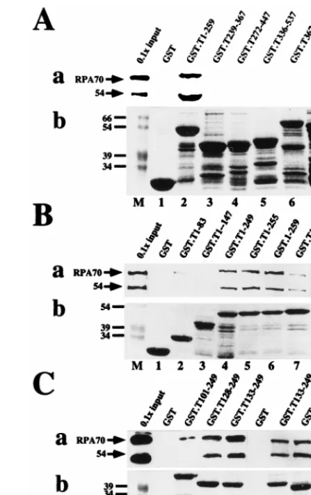
with-in the DNA bwith-indwith-ing domawith-in.
To map the site(s) of interaction
[image:3.612.314.538.70.425.2]of human RPA with T antigen, GST-T antigen fusion proteins
bound to glutathione-agarose were tested for the ability to bind
to RPA in a protein affinity pull-down assay (Fig. 1).
Immu-noblotting of the bound material with a monoclonal antibody
against RPA70 was used to detect bound RPA. Coarse
map-ping using large fusion peptides indicated that RPA bound to
T-antigen sequences within residues 1 to 259 but not to
C-terminal regions of T antigen or to GST used as a negative
control (Fig. 1A, panel a). Since the proteins had been treated
FIG. 1. Mapping the RPA binding sequences of SV40 T antigen. The indi-cated residues of T antigen were expressed as GST fusion proteins and adsorbed to glutathione-agarose. Fusion protein-bound beads were incubated with puri-fied RPA in a pull-down assay. (a) After washing, bound RPA was detected by denaturing gel electrophoresis and immunoblotting with RPA antibody 70C and chemiluminescence (lanes 1 to 7 [A and B] or 1 to 8 [C]). As a marker, 1/10 of the input RPA (lanes M) was analyzed in parallel. Positions of the 70-kDa subunit and a 54-kDa degradation product are indicated by arrows. (b) A 10-ml sample of beads bearing each fusion protein was analyzed by denaturing gel electrophoresis and detected by Coomassie staining (lanes 1 to 7 and 1 to 8). Lanes M show prestained marker proteins. Only the relevant portions of the gels are shown.on November 9, 2019 by guest
http://jvi.asm.org/
with nucleases during their purification, this interaction was
unlikely to be due to bridging by nucleic acids present in the
protein preparations. Furthermore, inclusion of 50
m
g of
ethi-dium bromide per ml in the binding reaction (48) did not
prevent RPA binding to the fusion proteins (data not shown).
Coomassie blue staining of the fusion proteins bound to the
beads demonstrated that all of them were present in similar
amounts (Fig. 1A, panel b). Fine mapping of the N-terminal
259 residues of T antigen was then performed to define the site
of interaction more closely. RPA bound relatively well to
T-antigen residues 1 to 249 but poorly to 1 to 83 and 1 to 147
(Fig. 1B, panel a), suggesting that its binding site could be
lo-cated between residues 147 and 249. Indeed fusion proteins
bearing T-antigen residues 128 to 249, 133 to 249, 145 to 249,
and 164 to 249 bound well to RPA (Fig. 1C, panel a),
demon-strating that a site sufficient for RPA binding resides within the
C-terminal portion of the T-antigen DNA binding domain (44).
To confirm the location of the T-antigen binding site for
RPA, we used a panel of monoclonal antibodies against T
an-tigen whose epitopes had been mapped (Fig. 2A) to
immuno-precipitate T antigen and then test for its ability to bind RPA.
We reasoned that monoclonal antibodies whose epitopes map
outside the region of RPA binding should not interfere with
the interaction, while those with epitopes close to or
overlap-ping the RPA binding site might inhibit the interaction.
Im-munoprecipitation of T antigen was observed with each of the
antibodies used (Fig. 2B, panel b). Two antibodies whose
epi-topes mapped within the T-antigen DNA binding domain,
Pab220 and Pab221, precipitated slightly less T antigen than
the other antibodies but noticeably diminished the amount of
RPA that bound to the T antigen (Fig. 2B, panel a, lanes 8 and
9). This result is consistent with the RPA binding site defined
by using T-antigen fusion proteins. However, RPA binding was
also inhibited by Pab204, whose epitope was mapped in the C
terminus of T antigen well outside the RPA binding site
de-fined by using the fusion proteins (panel a, lane 10). Although
this observation was initially surprising, Pab204 was also found
to inhibit every other biochemical activity of T antigen that was
tested (see Fig. 3 and 4), suggesting that it drastically disrupted
the overall structure of the protein.
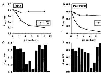
ELISAs were carried out to verify that Pab220 and Pab221
specifically inhibited RPA binding of T antigen. T antigen was
immobilized on ELISA plates and incubated with increasing
amounts of monoclonal antibody Pab220, Pab414, or Pab101,
or buffer as a control (Fig. 3A and B). After washing and
in-cubation with RPA or Pol/Prim, bound protein was detected
using peroxidase-conjugated polyclonal rabbit antibodies against
either RPA (Fig. 3A) or Pol/Prim (Fig. 3B) and a chromogenic
substrate. Maximal inhibition of both RPA binding and Pol/
Prim binding to T antigen was observed with 10
m
g of
mono-clonal antibodies Pab220 and Pab414, respectively, while Pab101
displayed little inhibition of either interaction. We then tested
the ability of 10
m
g of each monoclonal antibody in the panel
to inhibit T-antigen interactions with RPA and Pol/Prim (Fig.
3C and D). Pab220 and Pab221 again impaired T-antigen
in-teractions with RPA (Fig. 3C, columns 5 and 6) but had no
effect on its interactions with Pol/Prim (Fig. 3D, columns 5 and
6). In agreement with previous reports (14, 25, 31, 69, 72),
T-antigen interactions with Pol/Prim were impaired by Pab414
(Fig. 3C and D, columns 8). Pab204 inhibited T-antigen
bind-ing to both proteins (columns 7), while the other monoclonal
antibodies had little effect on these protein-protein
interac-tions. These results confirm that T-antigen binding to RPA was
specifically impaired by Pab220 and Pab221.
Effect of Pab220 and Pab221 on other biochemical activities
of T antigen.
The ability of Pab220 and Pab221 to specifically
[image:4.612.53.289.65.323.2]block T-antigen interaction with RPA might provide a way to
test the functional relevance of the RPA binding site defined
above in viral DNA replication. A clear link between any
interference of Pab220 and Pab221 in viral DNA replication
with a block in T antigen-RPA binding, however, would
re-quire that these antibodies not interfere with other
biochemi-cal activities of T antigen. Since Pab220 and Pab221 epitopes
map within the DNA binding domain of T antigen, which is
involved in multiple functions of the protein (6, 99), specific
binding of T antigen to the viral origin of DNA replication and
assembly as a double hexamer on the origin seemed the most
likely activity with which the antibodies might interfere. An
electrophoretic mobility shift assay was used to test binding of
T antigen to a labeled origin DNA fragment (Fig. 4A). T
antigen-origin DNA complexes migrated more slowly than free
DNA (compare lanes 1 and 2). Addition of monoclonal
anti-bodies supershifted the complexes to even lower mobility
(lanes 3 to 7 and 9 to 11), except for Pab204, which prevented
or disrupted T antigen-origin DNA complex formation (lane
8). The results indicate that Pab220 and Pab221 did not impair
origin DNA binding activity of T antigen and that the epitopes
were still available for binding in the T antigen-DNA complex.
The ATPase activity of T antigen has been mapped to the
C-terminal region of the protein (6, 29) and hence was not
expected to be affected by Pab220 or Pab221. In fact, none of
FIG. 2. Coimmunoprecipitation of RPA with T antigen. (A) A schematicdiagram depicting the amino acid (aa) regions in T antigen (open box) to which the epitopes for the monoclonal antibodies indicated below were mapped. The minimal origin DNA binding domain (44) is indicated by a thick line above the T-antigen diagram. The binding site for RPA determined in Fig. 1 is shown as a hatched box. (B) T antigen was bound to the indicated monoclonal antibody adsorbed to protein G-agarose, and the beads were incubated with RPA. (a) Bound RPA was eluted (lanes 5 to 13), separated by denaturing gel electro-phoresis, and detected by immunoblotting with the 70-kDa protein-specific monoclonal antibody 70C. The input T antigen (Tag) and 1/10 of the input RPA were run on the same gel (lane 4). On a separate gel, controls with Pab419 beads loaded with T antigen (lane 1) and without T antigen (lane 2) were analyzed together with a duplicate input control (lane 3). Positions of the 70-kDa subunit (RPA) and the antibody heavy chain (IgH) are indicated. (B) The same blots reprobed with the T-antigen-specific antibody Pab419. Positions of T antigen (Tag) and the heavy chain (IgH) are indicated.
on November 9, 2019 by guest
http://jvi.asm.org/
the monoclonal antibodies in this panel except Pab204
inhib-ited the ATPase activity of T antigen (Fig. 4B). Hydrolysis of
ATP was reduced to about one-third of the control by Pab204
(compare lanes 2 and 8), in agreement with earlier reports (30,
94). A modest stimulation of ATPase activity was observed
with antibodies Pab419, Pab416, and Pab414 (lanes 4, 5, and
9).
The DNA helicase activity of T antigen (6, 29) requires
sequences within the origin DNA binding domain (98),
sug-gesting that it might be affected by Pab220 or Pab221.
How-ever, in reactions with a partial duplex DNA template, the
helicase activity of T antigen was only marginally inhibited by
Pab220 or Pab221 (Fig. 4C; compare lanes 6 and 7 with lane 2).
Strong inhibition was observed in the presence of Pab204 (lane
8) and Pab414 (lane 9). None of the other antibodies affected
helicase activity.
Bidirectional unwinding of SV40 origin DNA requires the
coordinated functioning of multiple domains of T antigen:
specific binding of T antigen to the origin, assembly as a double
hexamer, DNA helicase activity, and probably interactions
be-tween the two hexamers (6, 28, 57, 58, 61, 73, 85, 92, 93). The
effect of monoclonal antibodies on bidirectional origin DNA
unwinding was tested in two different assays, one using linear
DNA fragments (Fig. 4D) and one using closed circular
super-coiled DNA carrying the origin of replication (Fig. 4E). Since
both Pab204 and Pab414 impaired the helicase activity of T
antigen, it was not unexpected that they also suppressed origin
DNA unwinding in both assays (Fig. 4D, lanes 8 and 9; Fig. 4E,
lanes 9 and 10). Four antibodies that had little effect on
heli-case activity impaired origin DNA unwinding. Pab419 did not
inhibit unwinding of linear DNA but did inhibit unwinding of
supercoiled DNA (Fig. 4D, lane 4; Fig. 4E, lane 5). Pab416
slightly inhibited unwinding of the linear origin DNA fragment
(Fig. 4D, lane 5) and strongly inhibited unwinding of
super-coiled DNA (Fig. 4E, lane 6). Pab220 and Pab221 reduced
unwinding of the linear origin DNA fragment slightly and also
partially inhibited unwinding of supercoiled DNA (Fig. 4D
[compare lanes 6 and 7 with lane 2] and 4E [compare lanes 7
and 8 with lane 2]). The other antibodies had no effect on
unwinding in either assay.
The effects of this panel of monoclonal antibodies on the
biochemical activities of T antigen are summarized in Table 1.
Effects of monoclonal antibodies on the early steps in SV40
DNA replication.
The results presented above suggested that
with the possible exception of Pab108, Pab101, and KT3, each
of the antibodies would be expected to interfere with SV40
DNA replication at one or several of the early steps. Indeed,
each of the antibodies in the panel except these three did
significantly block SV40 DNA replication in vitro in crude cell
extracts (data not shown). However, the functional relevance
of the RPA binding site that is blocked by Pab220 and Pab221
cannot be deduced from these experiments, since these
anti-bodies not only inhibited T-antigen interaction with RPA but
also partially inhibited origin DNA unwinding, which is known
to be independent of a direct physical interaction between T
antigen and RPA (3, 14, 45, 60, 97). To distinguish between the
effects of Pab220 and Pab221 on origin unwinding and on
subsequent steps in replication, we sought to uncouple these
events, allowing unwinding to proceed in the absence of
anti-body and then testing the effect of antianti-body in subsequent
events.
[image:5.612.122.476.68.325.2]As a foundation for this strategy, conventional coupled
ini-tiation reactions containing purified T antigen, RPA, Pol/Prim,
and topoisomerase I (56, 84, 89) were first carried out in the
presence and absence of each antibody (Fig. 5A). Labeled
RNA primers were synthesized in the presence of Pab108,
FIG. 3. Effects of T-antigen-specific monoclonal antibodies on complex formation with cellular initiation proteins. (A and B) T antigen coupled to wells of an ELISA plate was treated with the indicated amounts of monoclonal antibody Pab101 or Pab220 (A) or Pab101 or Pab414 (B). After washing, the wells were incubated with either RPA (A) or Pol/Prim (B). The bound RPA or Pol/Prim was detected by incubation with the corresponding peroxidase-coupled polyclonal antibodies and a chromogenic substrate and then quantitated spectrophotometrically at 405 nm. (C and D) T antigen bound to the wells of the ELISA plate was incubated with T-antigen buffer (column 1), with 10mg of the indicated monoclonal antibody (columns 2 to 10), or with antibody buffer (column 11). After addition of either RPA (C, columns 1 to 11) or Pol/Prim (D, columns 1 to 11) or neither (column 12), the bound RPA or Pol/Prim was detected as in panels A and B.
on November 9, 2019 by guest
http://jvi.asm.org/
Pab101, and KT3 in amounts similar to those in control
reac-tions (Fig. 5A; compare lanes 3, 10, and 11 with lanes 2 and
12). In contrast, primer synthesis was markedly reduced in the
presence of Pab419 and Pab416 (lanes 4 and 5) and nearly
absent in the presence of Pab220, Pab221, Pab204, and Pab414
(lanes 6 to 9).
[image:6.612.61.312.67.487.2]To determine whether these antibodies interfered with
ini-tiation by blocking origin binding and unwinding, or at a later
step, the initiation reaction was carried out in two sequential
steps (7, 22, 27, 83). In the first step, T antigen, RPA, Pol/Prim,
topoisomerase I, and DNA were preincubated with ATP to
allow formation of an underwound DNA template, but without
the other ribonucleoside triphosphates to prevent primer
syn-thesis. In the second step, ribonucleotides were added in the
TABLE 1. Inhibition of biochemical activities of SV40 T antigen
by T-antigen-specific monoclonal antibodies
Antibody
Inhibition ofa:
SV40 DNA
binding ATPase Helicase
Unwinding formationComplex
Linear CCS RPA Pol/Prim
108
2
2
2
2
2
2
2
419
2
2
2
2
1
2
2
416
2
2
2
p
1
2
2
220
2
2
2
p
p
1
2
221
2
2
2
p
p
1
2
204
1
1
1
1
1
1
1
414
2
2
1
1
1
2
1
101
2
2
2
2
2
2
2
KT3
2
2
2
2
2
2
2
[image:6.612.309.547.537.677.2]aPurified monoclonal antibodies were tested for the ability to inhibit SV40 origin DNA binding (Fig. 4A); ATPase activity (Fig. 4B); helicase activity using an oligonucleotide-primed M13mp18 ssDNA (Fig. 4C); SV40 origin DNA un-winding activity on linear and closed circular supercoiled (CCS) templates (Fig. 4D and E and data not shown); and complex formation with RPA and Pol/Prim (Fig. 3).1, inhibition; p, partial inhibition;2, lack of inhibition.
FIG. 4. Effects of T-antigen-specific monoclonal antibodies on biochemical activities of T antigen. (A) T-antigen binding to a labeled SV40 origin DNA frag-ment was tested in a band shift in the presence of the indicated monoclonal anti-bodies (lanes 3 to 11), without antibody (lane 2), or without T antigen (lane 1). (B) ATPase reactions were carried out without T antigen (lane 1), with T antigen (lane 2), or with T antigen in the presence of the indicated monoclonal antibody (lanes 3 to 11) or buffer (lane 12). The reaction products were separated by ascend-ing thin-layer chromatography. Helicase reactions were performed with M13 DNA annealed to a labeled primer (C), and unwinding reactions were performed with a labeled duplex origin DNA fragment (ori) and a labeled nonspecific DNA fragment (ns) (D), in the presence of the indicated monoclonal antibodies (lanes 3 to 11) or buffer (lanes 12). Negative control reactions were performed without T antigen (lanes 1); positive controls were performed with T antigen and without antibod-ies (lanes 2) as indicated. (C and D) The substrate DNA in the native confor-mation (lane N) or after heat denaturation (lane D) was electrophoresed in parallel. Quantitative evaluation of the autoradiograms for each reaction is given below each lane ss and ds, single-stranded and double-stranded DNA, respectively. (E) SV40 DNA unwinding assays contained closed circular supercoiled pUC-HS DNA, T antigen (TAg; lanes 2 to 10), topoisomerase I, and E. coli SSB. Reactions were carried out in the presence of monoclonal antibodies as indicated (lanes 4 to 10) or antibody buffer (lane 3). Reaction products were analyzed by electrophoresis and ethidium bromide staining. Form U, underwound covalently closed circular DNA.
on November 9, 2019 by guest
http://jvi.asm.org/
presence or absence of each monoclonal antibody to assess
primer synthesis (Fig. 5B). Primer synthesis was detected at
levels resembling the controls in reactions containing Pab108,
Pab101, and KT3 (lanes 3 and 10 to 12), and little or no primer
synthesis was observed in reactions containing Pab220, Pab221,
and Pab414 (lanes 6, 7, and 9). These results were thus largely
independent of the time of addition of the antibody to the
reaction. Interestingly, however, primer synthesis in the
pres-ence of Pab419, Pab416, and Pab204 was clearly less sensitive
to inhibition when the antibodies were added after formation
of an underwound template DNA (lanes 4, 5, and 8).
Quanti-tative estimates of primer synthesis in three independent
ex-periments with each antibody added to the reaction either
prior to origin binding and unwinding, or afterwards, were
averaged to give the results depicted in Fig. 5C and D. These
results thus separate the antibodies that impaired origin DNA
unwinding (Fig. 4D and E) into two classes: those that
inhib-ited DNA replication primarily at the unwinding step and
significantly less in primer synthesis (Pab419, Pab416, and
Pab204) and those that inhibited both steps (Pab220, Pab221,
and Pab414).
To test whether elongation of RNA primers was also
sensi-tive to inhibition by these monoclonal antibodies, an
elonga-tion reacelonga-tion was carried out in two steps. In the first, origin
DNA binding, unwinding, and primer synthesis were permitted
in the absence of deoxyribonucleoside triphosphates, and the
primed unwound template was isolated by gel filtration to
remove unincorporated ribonucleoside triphosphates.
Addi-tion of labeled CTP to this isolated template did not support
[image:7.612.79.515.71.373.2]synthesis of labeled products (data not shown), indicating that
ribonucleotides had been removed. In the second step,
de-oxyribonucleoside triphosphates were added to permit primer
elongation, either in the presence or in the absence of each
monoclonal antibody. Supplementation of the reaction with
fresh replication proteins in the second step stimulated
incor-poration five- to sevenfold, while fresh Pol/Prim alone
stimu-lated incorporation three- to fivefold (data not shown),
sug-gesting that some Pol/Prim, and possibly other proteins, had
dissociated from the prereplication complex during gel
filtra-tion. The reactions shown here were therefore supplemented
with fresh proteins prior to elongation. Primer elongation in
the presence of Pab108, Pab101, and KT3 was nearly as
effi-cient in as the control reactions (Fig. 6A; compare lanes 3, 10,
and 11 with lanes 2 and 12). Pab419, Pab416, and Pab204
reduced primer elongation to about half of the level observed
in the control reactions (lanes 4, 5, and 8). In contrast, primer
elongation in the presence of Pab220, Pab221, and Pab414 was
almost completely blocked (lanes 6, 7, and 9). Quantitative
estimates of primer elongation products formed in three
inde-pendent experiments in the presence and absence of each
antibody were averaged (Fig. 6B) and confirmed this
conclu-sion. These results demonstrate that Pab419, Pab416, and
Pab204 inhibited DNA replication primarily at the
origin-un-winding step and significantly less in primer synthesis and
elon-gation, while Pab220, Pab221, and Pab414 essentially abolished
primer synthesis and elongation even when added after origin
unwinding.
FIG. 5. Delineation of the step in SV40 initiation at which antibodies interfere. Uncoupled initiation reactions were performed in which antibodies (lanes 3 to 11) or buffer (lanes 12) were added before (A) or after (B) the origin DNA-unwinding step. Negative control reactions contained no T antigen (lanes 1), and positive controls contained no antibodies (lanes 2). NMP, nucleoside monophosphate. (C and D) Mean results from three independent initiation experiments performed as for panels A and B. Brackets indicate the average error of the mean. Incorporation in reactions with antibodies (column 2 to 10) or buffer (column 11) was expressed as a percentage of the value in a reaction without antibody (column 1), defined as 100%.
on November 9, 2019 by guest
http://jvi.asm.org/
DISCUSSION
SV40 T antigen has been previously shown to interact
phys-ically and functionally with RPA during initiation of viral DNA
replication and lagging-strand DNA synthesis (2, 14, 24, 26, 56,
60, 65, 67). Here we have used fusion peptides of T-antigen
and anti-T-antigen monoclonal antibodies whose epitopes
have been mapped to localize the sequences in T antigen that
interact with RPA and to confirm the functional relevance of
this binding site in viral DNA replication.
The region of T antigen (residues 164 to 249) that binds to
RPA is localized within the DNA binding domain of T antigen
(Fig. 1 and 2). Genetic evidence has implicated the DNA
binding domain in multiple functions of T antigen (99).
Bio-chemical studies demonstrate that it not only is essential for
sequence-specific binding to the SV40 control region DNA but
also participates in multiple interactions with host cell
pro-teins. Among the proteins known to bind within this region of
T antigen are the transcription factors TATA binding protein
(TBP), TFIIB, several TBP-associated factors, TEF-1, Sp1,
RNA polymerase II, and topoisomerase I (1, 16, 34, 39, 43, 71).
Finally, functional interactions between T-antigen hexamers
during bidirectional origin DNA unwinding appear to require
sequences within the DNA binding domain (58, 92).
Compe-tition studies indicate that not all of the transcription factors
can bind to T antigen at once, suggesting that some of the
binding sites for these proteins may overlap (43). However, at
least several separate protein interaction sites appear to reside
within the DNA binding domain. Preliminary evidence from
competition experiments suggests that RPA binds to a region
of T antigen that does not overlap with the binding site for
TBP or TEF-1 (39). The observation that monoclonal
antibod-ies Pab220 and Pab221 block RPA binding but not DNA
bind-ing to T antigen (Fig. 2, 3, and 4A) indicates that the RPA and
DNA binding surfaces are unlikely to overlap. However, it
remains possible that the topoisomerase I binding site of T
antigen may overlap the RPA binding site.
The solution structure of the DNA binding domain of T
antigen was recently determined by nuclear magnetic
reso-nance spectroscopy, and on the basis of spectroscopic and
genetic data, the origin DNA binding surface has been
mod-eled to include two neighboring loops containing residues 152
to 155 and 203 to 207 (44, 52, 70, 99). A mutation at residue
189 (S189N) impairs T-antigen binding to TEF-1, activation of
transcription by TEF-1, stimulation of quiescent cells, and cell
transformation by T antigen (1, 21). Residue 189 is located in
a loop between
b
strands B and C that resides on the opposite
side of the DNA binding domain from the proposed DNA
binding surface (52) and that may comprise part of the TEF-1
binding site. Mutations at residues 173 and 174 (K173A and
K174A) were reported to prevent T-antigen interaction with
several transcription factors and to block transactivation by T
antigen (43). Also, a small in-frame insertion mutation at
res-idue 168 was shown to significantly reduce transactivation
ac-tivity (16). These three residues are all located in
a
helix B on
one surface of the DNA binding domain (52), which may
constitute part of a binding surface for transcription factors
that is distinct from that for origin DNA.
Based on the genetic and biochemical evidence above, we
suggest that the RPA binding surface is unlikely to overlap
with those for either the transcription factors or the viral
ori-gin. Functional interactions of RPA with the T-antigen-related
proteins, polyomavirus T antigen, and bovine papillomavirus
E1 protein, as well as with EBNA-1, have been observed (4, 59,
101) and may reflect similar binding sites in these proteins for
RPA. Although there is little homology between SV40 T
an-tigen and EBNA-1, comparison of the RPA binding region of
SV40 T antigen with the entire sequences of polyomavirus T
antigen and E1 reveals short regions of homology that
corre-spond to SV40 T antigen residues 194 to 199 and 196 to 200,
which are located at the junction between
b
strand C and the
second loop postulated for the DNA binding surface (52).
Most of the residues between 194 to 200 are not exposed on
the surface (52), but it will be interesting to determine whether
mutations in this region or neighboring sequences in the
three-dimensional structure affect RPA binding activity.
T antigen has recently been reported to associate with the
large RPA subunit RPA70 within residues 1 to 326, but not 1
to 168 or 237 to 616 (2), suggesting that the T-antigen binding
site probably resides between residues 168 and 237. We
pre-viously demonstrated that T antigen associated with native
trimeric RPA but not with recombinant RPA70 expressed as
an insoluble protein in bacteria (24). However, our more
re-cent studies performed with soluble RPA70 expressed as a
fusion protein confirm that RPA70 is sufficient by itself to bind
to T antigen (91). Since RPA70 is poorly soluble (32, 38), it
seems likely that the concentration of the resolubilized RPA70
used in our early experiments was too low to detect the
inter-action with T antigen. Consistent with this interpretation,
re-cent evidence from surface plasmon resonance experiments
indicates that T-antigen affinity for RPA is about an order of
magnitude weaker than its affinity for Pol/Prim (33).
FIG. 6. Influence of antibodies on primer elongation in the absence of primer synthesis. (A) Standard initiation reactions were performed except that T antigen was omitted from the negative control reaction (lane 1). After the reaction, unincorporated ribonucleoside triphosphates were removed by gel fil-tration, and the primed DNA-protein complex was recovered. Elongation reac-tions containing the primed DNA-protein complex supplemented with additional proteins and deoxyribonucleoside triphosphates were carried out in the presence of the indicated antibodies (lanes 3 to 11) or buffer (lane 12). A positive control reaction contained no antibody (lane 2). dNMP, deoxynucleoside monophos-phate. (B) Quantitation of results of three independent experiments as in panel A. The mean is shown as a percentage of the positive control, which was set at 100%. The brackets indicate the average error of the mean.
on November 9, 2019 by guest
http://jvi.asm.org/
[image:8.612.50.287.73.355.2]RPA binding to T antigen was specifically inhibited by
mono-clonal antibodies Pab220 and Pab221 (Fig. 2 and 3). These
an-tibodies recognize native but not denatured sequences within
the DNA binding domain of T antigen (62). These antibodies
had little effect on other biochemical functions of T antigen
that are known to play a role in viral DNA replication (Fig. 4).
The partial inhibition of T-antigen-mediated unwinding of
closed circular origin DNA detected in the presence of Pab220
and Pab221 may be due to partial interference with the
hex-amer-hexamer interactions that are implicated in origin DNA
unwinding (12, 57, 58, 61, 73, 85, 92, 93). Several other
mono-clonal antibodies inhibited unwinding of closed circular origin
DNA essentially completely (Fig. 4E). Pab419, whose epitope
mapped within the J domain at the N terminus of T antigen
(Fig. 2) (11, 75), strongly inhibited unwinding, as did Pab416,
whose epitope mapped between the J domain and the DNA
binding domain. Pab414, whose epitope mapped to the C
ter-minus, inhibited not only unwinding but also DNA helicase
activity (Fig. 4C to E) and binding to Pol/Prim (14, 25, 72).
Pab204 inhibited virtually every replication-related activity of
T antigen (Fig. 2 to 6 and reference 94), suggesting that it
prob-ably disrupts the global structure of the protein. All of these
antibodies were also potent inhibitors of initiation of SV40
DNA replication (Fig. 5A). Interestingly, when Pab419, -416,
or -204 was added to the initiation reaction after origin DNA
unwinding, the inhibition was significantly relieved (Fig. 5B),
suggesting either that these epitopes were masked in the origin
DNA-protein complex or that further unwinding of the
tem-plate DNA was not required to observe primer synthesis (Fig.
5B) or primer elongation (Fig. 6).
In contrast with these antibodies, Pab414 strongly interfered
with primer synthesis and primer elongation even when added
to the assays after origin DNA unwinding or after unwinding
and primer synthesis (Fig. 5B and 6). This interference may
reflect the essential role of T-antigen binding to Pol/Prim in
primer synthesis and elongation (14, 15, 23–25, 30, 31, 69, 72,
77), but interference with unwinding during primer synthesis
and elongation is difficult to rule out (Fig. 4C to E). Recent
evidence from fluorescence spectroscopy indicates that the
stoichiometry of Pol/Prim binding to T-antigen monomers in
solution is 1:6 (41), consistent with a model in which one
Pol/Prim would be located on each lagging-strand template,
tethered to a T-antigen double hexamer associated with both
replication forks (22, 28, 40, 55, 61, 73, 93). Pab220 and Pab221
interfered with primer synthesis and elongation even when
added to the reactions after template unwinding or unwinding
and primer synthesis (Fig. 5B and 6), implying that T
antigen-RPA interactions are required for both primer synthesis and
elongation. These results are consistent with previous data that
yeast RPA failed to bind to T antigen and to support primer
synthesis on ssDNA (60). The critical role of RPA-T antigen
interactions in primer elongation was more unexpected,
par-ticularly since much of the Pol/Prim that synthesized primers in
the first step of the assay apparently dissociated from the
primed unwound template during gel filtration (Fig. 6). T
an-tigen, once assembled as a double hexamer active in
unwind-ing, has been shown to be processive (65), probably due to the
toroidal structure of each hexamer encircling the DNA (68).
RPA was probably retained on the unwound template DNA
during gel filtration through its strong DNA binding affinity
(96). Nevertheless, prevention or disruption of RPA binding to
T antigen on the primed unwound template appeared to be
sufficient to prevent primer elongation by freshly added Pol/
Prim (Fig. 6). These observations suggest the existence of a
multiprotein complex involving multiple protein-protein
inter-actions in lagging-strand synthesis. This report defining the
RPA binding site in T antigen represents one further step
to-ward a clearer understanding of how this complex works.
ACKNOWLEDGMENTS
We thank A. Brunahl for help with antibody purification and
ELISAs, I. Moarefi, A. Arthur, and A. Wildeman for sharing plasmids
and topoisomerase I, V. Podust for purified SSB, D. von Winkler for
antiserum against RPA, H.-P. Nasheuer for antiserum against Pol/
Prim, and M. Kenny, J. Hurwitz, D. P. Lane, E. Harlow, E. Gurney,
and G. Walter for monoclonal antibodies. We thank T. Melendy and
F. Grosse for communication of unpublished data, and we thank V.
Podust and U. Herbig for criticism of the manuscript.
The financial support of the NIH (GM52948), Vanderbilt
Univer-sity, and the NSF (Shared Instrumentation grant BIR-9419667) is
gratefully acknowledged. These studies were begun with a grant from
the German Science Foundation to E.F.
REFERENCES
1. Berger, L. C., D. B. Smith, I. Davidson, J.-J. Hwang, E. Fanning, and A. G.
Wildeman.1996. Interaction between T antigen and the TEA domain of the factor derepresses simian virus 40 late promoter in vivo: identification of the T-antigen domains important for transcriptional control. J. Virol. 70: 1203–1212.
2. Braun, K. A., Y. Lao, Z. He, J. Ingles, and M. S. Wold. 1997. Role of protein-protein interactions in the function of replication protein A (RPA): RPA modulates the activity of DNA polymerase alpha by multiple mech-anisms. Biochemistry 36:8443–8454.
3. Brill, S. J., and B. Stillman. 1989. Yeast replication factor-A functions in the unwinding of the SV40 origin of DNA replication. Nature 342:92–95. 4. Brinton, B., and J. A. Hassell. 1996. SV40 and polyoma DNA replication, p. 639–677. In M. L. DePamphilis (ed.), DNA replication in eukaryotic cells. Cold Spring Harbor Laboratory Press, Plainview, N.Y.
5. Brush, G. S., and T. J. Kelly. 1996. Mechanisms for replicating DNA, p. 1– 43. In M. L. DePamphilis (ed.), DNA replication in eukaryotic cells. Cold Spring Harbor Laboratory Press, Plainview, N.Y.
6. Bullock, P. A. 1997. The initiation of simian virus 40 DNA replication in vitro. Crit. Rev. Biochem. Mol. Biol. 32:503–568.
7. Bullock, P. A., Y. S. Seo, and J. Hurwitz. 1989. Initiation of simian virus 40 DNA synthesis in vitro: pulse-chase experiments identify the first labeled species as topologically unwound. Proc. Natl. Acad. Sci. USA 86:3944–3948. 8. Bullock, P. A., Y. S. Seo, and J. Hurwitz. 1991. Initiation of simian virus 40
DNA synthesis in vitro. Mol. Cell. Biol. 11:2350–2361.
9. Bullock, P. A., S. Tevosian, C. Jones, and D. Denis. 1994. Mapping initia-tion sites for simian virus 40 lagging-strand DNA synthesis events in vitro. Mol. Cell. Biol. 14:5043–5055.
10. Bullock, P. A., and D. Denis. 1995. DNA synthesis generally initiates out-side of the simian virus 40 core origin in vitro. Mol. Cell. Biol. 15:173–178. 11. Campbell, K., K. Mullane, I. Aksoy, H. Stubdal, J. Pipas, P. Silver, T.
Roberts, B. Schaffhausen, and J. DeCaprio.1997. DnaJ/hsp40 chaperone domain of SV40 large T antigen promotes efficient viral DNA replication. Genes Dev. 11:1098–1110.
12. Cegielska, A., I. Moarefi, E. Fanning, and D. M. Virshup. 1994. T-antigen kinase inhibits simian virus 40 DNA replication by phosphorylation of intact T antigen on serines 120 and 123. J. Virol. 68:269–275.
13. Clark, R., D. P. Lane, and R. Tjian. 1981. Use of monoclonal antibodies as probes of simian virus 40 T antigen ATPase activity. J. Biol. Chem. 256: 11854–11858.
14. Collins, K. L., and T. J. Kelly. 1991. The effects of T antigen and replication protein A on the initiation of DNA synthesis by DNA polymerasea -pri-mase. Mol. Cell. Biol. 11:2108–2115.
15. Collins, K. L., A. A. R. Russo, B. Y. Tseng, and T. J. Kelly. 1993. The role of the 70 kDa subunit of human DNA polymeraseain DNA replication. EMBO J. 12:4555–4566.
16. Damania, B., and J. C. Alwine. 1996. TAF-like function of SV40 large T antigen. Genes Dev. 10:1369–1381.
17. Dean, F. B., P. A. Bullock, Y. Murakami, C. R. Wobbe, L. Weissbach, and
J. Hurwitz.1987. Simian virus 40 (SV40) DNA replication: SV40 large T antigen unwinds DNA containing the SV40 origin of replication. Proc. Natl. Acad. Sci. USA 84:16–20.
18. Dean, F. B., M. Dodson, H. Echols, and J. Hurwitz. 1987. ATP-dependent formation of a specialized nucleoprotein structure by simian virus 40 (SV40) large tumor antigen at the SV40 replication origin. Proc. Natl. Acad. Sci. USA 84:8981–8985.
19. DeLucia, A. L., S. Deb, K. Partin, and P. Tegtmeyer. 1986. Functional interactions of the simian virus 40 core origin of replication with flanking regulatory sequences. J. Virol. 57:138–144.
20. Denis, D., and P. A. Bullock. 1993. Primer-DNA formation during simian virus 40 DNA replication in vitro. Mol. Cell. Biol. 13:2882–2890. 21. Dickmanns, A., A. Zeitvogel, F. Simmersbach, R. Weber, A. K. Arthur, S.
on November 9, 2019 by guest
http://jvi.asm.org/
Dehde, A. G. Wildeman, and E. Fanning.1994. The kinetics of simian virus 40-induced progression of quiescent cells into S phase depend on four independent functions of large T antigen. J. Virol. 68:5496–5508. 22. Dodson, M., F. B. Dean, P. Bullock, H. Echols, and J. Hurwitz. 1987.
Unwinding of duplex DNA from the SV40 origin of replication by T anti-gen. Science 238:964–968.
23. Dornreiter, I., W. C. Copeland, and T. S.-F. Wang. 1993. Initiation of simian virus 40 DNA replication requires the interaction of a specific domain of human DNA polymeraseawith large T antigen. Mol. Cell. Biol. 13:809– 820.
24. Dornreiter, I., L. F. Erdile, I. U. Gilbert, D. von Winkler, T. J. Kelly, and E.
Fanning.1992. Interaction of DNA polymerasea-primase with cellular replication protein A and SV40 T antigen. EMBO J. 11:769–776. 25. Dornreiter, I., A. Ho¨ss, A. K. Arthur, and E. Fanning. 1990. SV40 T antigen
binds directly to the large subunit of DNA polymerase alpha. EMBO J. 9: 3329–3336.
26. Erdile, L. F., W. D. Heyer, R. Kolodner, and T. J. Kelly. 1991. Character-ization of a cDNA encoding the 70-kDa single-stranded DNA-binding subunit of human replication protein A and the role of the protein in DNA replication. J. Biol. Chem. 266:12090–12096.
27. Fairman, M. P., and B. Stillman. 1988. Cellular factors required for mul-tiple stages of SV40 replication in vitro. EMBO J. 7:1211–1218. 28. Fanning, E. 1994. Control of SV40 DNA replication by protein
phosphor-ylation: a model for cellular DNA replication? Trends Cell Biol. 4:250–255. 29. Fanning, E., and R. Knippers. 1992. Structure and function of simian virus
40 large tumor antigen. Annu. Rev. Biochem. 61:55–85.
30. Gannon, J. V., and D. P. Lane. 1987. p53 and DNA polymeraseacompete for binding to SV40 T antigen. Nature (London) 329:456–458.
31. Gannon, J. V., and D. P. Lane. 1990. Interactions between SV40 T antigen and DNA polymerasea. New Biol. 2:84–92.
32. Gomes, X. V., and M. S. Wold. 1995. Structural analysis of human replica-tion protein A: mapping funcreplica-tional domains of the 70-kDa subunit. J. Biol. Chem. 270:4534–4543.
33. Grosse, F. Personal communication.
34. Gruda, M., J. Zabolotny, J. Xiao, I. Davidson, and J. Alwine. 1993. Tran-scriptional activation by simian virus 40 large T antigen: interactions with multiple components of the transcription complex. Mol. Cell. Biol. 13:961– 969.
35. Gurney, E. G., R. O. Harrison, and J. Fenno. 1980. Monoclonal antibodies against simian virus 40 T antigens: evidence for distinct subclasses of large T antigen and for similarities among nonviral T antigens. J. Virol. 34:752– 763.
36. Gurney, E. G., D. Tamowsky, and W. Deppert. 1986. Antigenic binding sites of monoclonal antibodies specific for simian virus 40 large T. J. Virol. 57: 1168–1172.
37. Harlow, E., L. V. Crawford, D. C. Pim, and N. M. Williamson. 1981. Monoclonal antibodies specific for simian virus 40 tumor antigen. J. Virol.
39:861–869.
38. Henricksen, L. A., C. B. Umbricht, and M. S. Wold. 1994. Recombinant replication protein A: expression, complex formation, and functional char-acterization. J. Biol. Chem. 269:11121–11132.
39. Herbig, U., K. Weisshart, P. Taneja, and E. Fanning. Interaction of the transcription factor TFIID with SV40 large T antigen interferes with rep-lication of SV40 DNA in vitro. Submitted for pubrep-lication.
40. Herendeen, D., and T. J. Kelly. 1996. SV40 DNA replication, p. 29–65. In J. J. Blow (ed.), Eukaryotic DNA replication. Oxford University Press, New York, N.Y.
41. Huang, S.-G., K. Weisshart, I. Gilbert, and E. Fanning. Stoichiometry and mechanism of assembly of SV40 T antigen complexes with the viral origin of DNA replication and DNA polymerasea-primase. Biochemistry, in press.
42. Ishimi, Y., A. Claude, P. Bullock, and J. Hurwitz. 1988. Complete enzy-matic synthesis of DNA containing the SV40 origin of replication. J. Biol. Chem. 263:19723–19733.
43. Johnston, S. D., X.-M. Yu, and J. E. Mertz. 1996. The major transcriptional transactivation domain of simian virus 40 large T antigen associates non-concurrently with multiple components of the transcriptional preinitiation complex. J. Virol. 70:1191–1202.
44. Joo, W. X., X. Luo, D. Denis, H. Y. Kim, G. J. Rainey, C. Jones, K. R.
Sreekumar, and P. A. Bullock.1997. Purification of the simian virus 40 (SV40) T antigen DNA-binding domain and characterization of its inter-actions with the SV40 origin. J. Virol. 71:3972–3985.
45. Kenny, M. K., S.-H. Lee, and J. Hurwitz. 1989. Multiple functions of human single-stranded DNA binding protein in simian virus 40 DNA replication: single-strand stabilization and stimulation of DNA polymeraseaandd. Proc. Natl. Acad. Sci. USA 86:9757–9761.
46. Kenny, M. K., U. Schlegel, H. Furneaux, and J. Hurwitz. 1990. The role of human single-stranded DNA binding protein and its individual subunits in simian virus 40 DNA replication. J. Biol. Chem. 265:7693–7700. 47. Laemmli, U. K. 1970. Cleavage of structural proteins during the assembly of
the head of bacteriophage T4. Nature (London) 227:680–685.
48. Lai, J., and W. Herr. 1992. Ethidium bromide provides a simple tool for
establishing genuine DNA-independent protein associations. Proc. Natl. Acad. Sci. USA 89:6958–6962.
49. Lee, S.-H., and D. K. Kim. 1995. The role of the 34-kDa subunit of human replication protein in simian virus 40 DNA replication in vitro. J. Biol. Chem. 270:12801–12807.
50. Lee, S.-H., T. Eki, and J. Hurwitz. 1989. Synthesis of DNA containing the simian virus 40 origin of replication by the combined action of DNA polymeraseaandd. Proc. Natl. Acad. Sci. USA 86:7361–7365. 51. Lohman, T. M., J. M. Green, and R. S. Beyer. 1986. Large-scale
overpro-duction and rapid purification of the Escherichia coli ssb gene product. Expression of the ssb gene underlPLcontrol. Biochemistry 25:21–25. 52. Luo, X., D. G. Sanford, P. A. Bullock, and W. W. Bachovchin. 1996.
Solu-tion structure of the origin specific DNA binding domain from simian virus 40 T-antigen. Nat. Struct. Biol. 3:1034–1039.
53. MacArthur, H., and G. Walter. 1984. Monoclonal antibodies specific for the carboxy terminus of simian virus 40 large T antigen. J. Virol. 52:483–491. 54. Maniatis, T., E. F. Fritsch, and J. Sambrook. 1989. Molecular cloning: a laboratory manual, 2nd ed. Cold Spring Harbor Laboratory, Cold Spring Harbor, N.Y.
55. Mastrangelo, I. A., P. V. C. Hough, V. G. Wilson, J. S. Wall, J. F. Hainfeld,
and P. Tegtmeyer.1989. ATP-dependent assembly of double hexamers of SV40 T antigen at the viral origin of DNA replication. Nature (London)
338:658–662.
56. Matsumoto, T., T. Eki, and J. Hurwitz. 1990. Studies on the initiation and elongation reactions in the simian virus 40 DNA replication system. Proc. Natl. Acad. Sci. USA 87:9712–9716.
57. McVey, D., S. Ray, Y. Gluzman, L. Berger, A. G. Wildeman, D. R. Marshak,
and P. Tegtmeyer.1993. cdc2 phosphorylation of threonine 124 activates the origin-unwinding functions of simian virus 40 T antigen. J. Virol. 67: 5206–5215.
58. McVey, D., B. Woelker, and P. Tegtmeyer. 1996. Mechanisms of simian virus 40 T-antigen activation by phosphorylation of threonine 124. J. Virol.
70:3887–3893.
59. Melendy, T. Personal communication.
60. Melendy, T., and B. Stillman. 1993. An interaction between replication protein A and SV40 T antigen appears essential for primosome assembly during SV40 DNA replication. J. Biol. Chem. 268:3389–3395.
61. Moarefi, I. F., D. Small, I. Gilbert, M. Hoepfner, S. K. Randall, C.
Schnei-der, A. R. R. Russo, U. Ramsperger, A. K. Arthur, H. Stahl, T. J. Kelly, and E. Fanning.1993. Mutation of the cyclin-dependent kinase phosphorylation site in simian virus 40 (SV40) large T antigen specifically blocks SV40 origin DNA binding. J. Virol. 67:4992–5002.
62. Mole, S. E., J. V. Gannon, M. J. Ford, and D. P. Lane. 1987. Structure and function of large T antigen. Philos. Trans. R. Soc. Lond. Ser. B 317:455– 469.
63. Murakami, Y., C. R. Wobbe, L. Weissbach, F. B. Dean, and J. Hurwitz. 1986. Role of DNA polymeraseaand DNA primase in simian virus 40 DNA replication in vitro. Proc. Natl. Acad. Sci. USA 83:2869–2873. 64. Murakami, Y., T. Eki, and J. Hurwitz. 1992. Studies on the initiation of
simian virus 40 replication in vitro: RNA primer synthesis and its elonga-tion. Proc. Natl. Acad. Sci. USA 89:952–956.
65. Murakami, Y., and J. Hurwitz. 1993. Functional interactions between SV40 T antigen and other replication proteins at the replication fork. J. Biol. Chem. 268:11008–11017.
66. Murakami, Y., and J. Hurwitz. 1993. DNA polymeraseastimulates the ATP-dependent binding of simian virus 40 tumor (T) antigen to the SV40 origin of replication. J. Biol. Chem. 268:11018–11027.
67. Nasheuer, H.-P., D. von Winkler, C. Schneider, I. Dornreiter, I. Gilbert,
and E. Fanning.1992. Purification and functional characterization of bo-vine RP-A in an in vitro SV40 DNA replication system. Chromosoma 102: S52–S59.
68. San Martin, M. C., C. Gruss, and J. M. Carazo. 1997. Six molecules of SV40 large T antigen assemble in a propeller-shaped particle around a channel. J. Mol. Biol. 268:15–20.
69. Schneider, C., K. Weisshart, L. A. Guarino, I. Dornreiter, and E. Fanning. 1994. Species-specific functional interactions of DNA polymerasea -pri-mase with simian virus 40 (SV40) T antigen require SV40 origin DNA. Mol. Cell. Biol. 14:3176–3185.
70. Simmons, D. T., G. Loeber, and P. Tegtmeyer. 1990. Four major sequence elements of simian virus 40 large T antigen coordinate its specific and nonspecific DNA binding. J. Virol. 64:1973–1983.
71. Simmons, D. T., T. Melendy, D. Usher, and B. Stillman. 1996. Simian virus 40 large T antigen binds to topoisomerase I. Virology 222:365–374. 72. Smale, S. T., and R. Tjian. 1986. T-antigen-DNA polymerase alpha
com-plex implicated in simian virus 40 DNA replication. Mol. Cell. Biol. 6:4077– 4087.
73. Smelkova, N. V., and J. A. Borowiec. 1997. Dimerization of simian virus 40 T-antigen hexamers activates T-antigen DNA helicase activity. J. Virol. 71: 8766–8773.
74. Smith, D. B., and K. S. Johnson. 1988. Single-step purification of polypep-tides expressed in Escherichia coli as fusions with glutathione S-transferase. Gene 67:31–40.
on November 9, 2019 by guest
http://jvi.asm.org/
75. Srinivasan, A., A. J. McClellan, J. Vartikar, I. Marks, P. Cantalupo, Y. Li,
P. Whyte, K. Rundell, J. L. Brodsky, and J. M. Pipas.1997. The amino-terminal transforming region of simian virus 40 large T and small t antigens functions as a J domain. Mol. Cell. Biol. 17:4761–4773.
76. Stadlbauer, F., A. Bru¨ckner, C. Rehfuess, C. Eckershorn, F. Lottspeich, V.
Fo¨rster, B.-Y. Tseng, and H.-P. Nasheuer.1994. DNA replication in vitro by recombinant DNA-polymerasea–primase. Eur. J. Biochem. 222:781–793. 77. Stadlbauer, F., C. Voitenleitner, A. Bru¨ckner, E. Fanning, and H.-P.
Nasheuer.1996. Species-specific replication of simian virus 40 DNA in vitro requires the p180 subunit of human DNA polymerasea-primase. Mol. Cell. Biol. 16:94–1104.
78. Stigger, E., F. Dean, J. Hurwitz, and S.-H. Lee. 1994. Reconstitution of functional human single-stranded DNA-binding protein from individual subunits expressed by recombinant baculoviruses. Proc. Natl. Acad. Sci. USA 91:579–583.
79. Stillman, B. 1996. Comparison of DNA replication in cells from prokarya and eukarya, p. 435–460. In M. L. DePamphilis (ed.), DNA replication in eukaryotic cells. Cold Spring Harbor Laboratory Press, Plainview, N.Y. 80. Strausfeld, U., and A. Richter. 1989. Simultaneous purification of DNA