Simultaneously Regulate HBV Replication and Hepatocyte Survival
Siddhartha Rawat,aMichael J. Bouchardb
Graduate Program in Molecular and Cellular Biology and Genetics, Drexel University College of Medicine, Philadelphia, Pennsylvania, USAa; Department of Biochemistry and Molecular Biology, Drexel University College of Medicine, Philadelphia, Pennsylvania, USAb
ABSTRACT
Chronic infection with hepatitis B virus (HBV) is a risk factor for developing liver diseases such as hepatocellular carcinoma (HCC). HBx is a multifunctional protein encoded by the HBV genome; HBx stimulates HBV replication and is thought to play an important role in the development of HBV-associated HCC. HBx can activate the phosphatidylinositol 3-kinase (PI3K)/AKT signaling pathway in some cell lines; however, whether HBx regulates PI3K/AKT signaling in normal hepatocytes has not been
evaluated. In studies described here, we assessed HBx activation of PI3K/AKT signaling in anex vivomodel of cultured primary
hepatocytes and determined how this HBx activity affects HBV replication. We report that HBx activates AKT in primary hepa-tocytes and that the activation of AKT decreases HBV replication and HBV mRNA and core protein levels. We show that the
transcription factor hepatocyte nuclear factor 4␣(HNF4␣) is a target of HBx-regulated AKT, and we link HNF4␣to
HBx-regu-lated AKT modulation of HBV transcription and replication. Although we and others have shown that HBx stimulates and is likely required for HBV replication, we now report that HBx also activates signals that can diminish the overall level of HBV rep-lication. While this may seem counterintuitive, we show that an important effect of HBx activation of AKT is inhibition of apop-tosis. Consequently, our studies suggest that HBx balances HBV replication and cell survival by stimulating signaling pathways that enhance hepatocyte survival at the expense of higher levels of HBV replication.
IMPORTANCE
Chronic hepatitis B virus (HBV) infection is a common cause of the development of liver cancer. Regulation of cell signaling pathways by the HBV HBx protein is thought to influence the development of HBV-associated liver cancer. HBx stimulates, and may be essential for, HBV replication. We show that HBx activates AKT in hepatocytes to reduce HBV replication. While this seems contradictory to an essential role of HBx during HBV replication, HBx activation of AKT inhibits hepatocyte apoptosis, and this may facilitate persistent, noncytopathic HBV replication. AKT regulates HBV replication by reducing the activity of the
transcription factor hepatocyte nuclear factor 4␣(HNF4␣). HBx activation of AKT may contribute to the development of liver
cancer by facilitating persistent HBV replication, augmenting the dedifferentiation of hepatocytes by inhibiting HNF4␣
func-tions, and activating AKT-regulated oncogenic pathways. AKT-regulated factors may provide therapeutic targets for inhibiting HBV replication and the development of HBV-associated liver cancer.
C
hronic infection with hepatitis B virus (HBV) remains a majorglobal health problem. Despite the availability of effective
HBV vaccines, there are⬃350 million people worldwide who are
chronically infected with HBV. Chronic HBV infection is the ma-jor cause of the development of hepatocellular carcinoma (HCC)
(1). The level of HBV replication in a chronically HBV-infected
individual can vary throughout the course of the infection, and the rise and fall of HBV replication during a chronic infection may
affect disease progression (2, 3). There are limited therapeutic
options for treating a chronic HBV infection, and the emergence of HBV mutants that are resistant to available therapies is mon. Moreover, available anti-HBV drugs typically do not com-pletely eliminate HBV in chronically infected individuals due to the presence and stability of covalently closed circular DNA (cccDNA), a replicative intermediate of HBV that is localized to
the nucleus of HBV-infected hepatocytes (4–7). Consequently,
there is continued interest in understanding the mechanisms that regulate HBV replication and could serve as potential therapeutic targets.
HBV is an enveloped, partially double-stranded DNA virus
that belongs to theHepadnaviridaefamily of viruses (8). Although
HBV is a DNA virus, it replicates through reverse transcription of
an RNA intermediate. HBV replication occurs within the viral capsid in the cytosol of hepatocytes; the capsid is composed of the viral core protein. After the entry of HBV into hepatocytes, the partially double-stranded genome of the virus is delivered to the nucleus, where the genome is repaired by host cell DNA repair machinery to generate cccDNA. cccDNA forms a minichromo-some and is the template for all HBV RNA transcripts. Pre-genomic RNA (pgRNA), one of the longest HBV transcripts, is enclosed in capsids; pgRNA is covalently linked to the viral DNA
Received22 August 2014 Accepted23 October 2014
Accepted manuscript posted online29 October 2014
CitationRawat S, Bouchard MJ. 2015. The hepatitis B virus (HBV) HBx protein activates AKT to simultaneously regulate HBV replication and hepatocyte survival. J Virol 89:999 –1012.doi:10.1128/JVI.02440-14.
Editor:G. McFadden
Address correspondence to Michael J. Bouchard, [email protected].
Copyright © 2015, American Society for Microbiology. All Rights Reserved.
doi:10.1128/JVI.02440-14
on November 7, 2019 by guest
http://jvi.asm.org/
polymerase/reverse transcriptase (RT), which reverse tran-scribes the first DNA strand of the HBV genome by using pgRNA as the template. For reasons that remain incompletely understood, the second, cDNA strand is synthesized to various lengths, giving rise to the partially double-stranded genome of
HBV (9–11). The interaction between numerous host cell
fac-tors and HBV proteins governs HBV replication in hepatocytes and likely plays an important role in the development of
HBV-associated HCC (3,8,11,12).
The shortest open reading frame (ORF) and mRNA transcript
of the HBV genome encodes the HBx protein (11,13). HBx
stim-ulates HBV replication in multiple experimental systems, includ-ing cultured primary rat and human hepatocytes, some liver cell lines, livers of normal mice, and chimeric mice with humanized
livers (14–19). HBx is also thought to play an important role in the
development of HBV-associated HCC due to the regulatory ef-fects of HBx on cellular signaling pathways, DNA damage repair
mechanisms, the cell cycle, and apoptosis (14,20–24). Some HBx
activities, such as its role in regulating apoptosis, appear to be context dependent and have varied depending upon the model
system that was used to assess this HBx activity (8,11,25). While
studies conducted with various cell lines have provided important insights concerning the effect of HBx expression on cellular signal transduction pathways, the influence of HBx expression on cellu-lar signal transduction pathways in normal hepatocytes, and in the context of an HBV infection, remains poorly understood. Recent
studies withex vivo-cultured primary hepatocyte model systems
andin vivomodels have begun to identify HBx effects on normal hepatocytes and how these HBx activities influence HBV
replica-tion and affect hepatocyte physiology (8,11,14,23,26,27).
The phosphatidylinositol 3-kinase (PI3K)/AKT pathway is a cell signal transduction pathway that can regulate numerous
cel-lular processes, including celcel-lular apoptotic pathways (28,29).
The PI3K/AKT pathway is regulated by growth factors that acti-vate PI3K, which in turn phosphorylates phosphatidylinositol-4,5-bisphosphate (PIP2) to form phosphatidylinositol-3,4,5-tris-phosphate (PIP3). PIP3 acts as a docking site for AKT and facilitates the subsequent phosphorylation of AKT at threonine 308 (T308) by phosphoinositide-dependent kinase 1 (PDK1). The complete activation of AKT also requires phosphorylation at
ser-ine 473 (S473) of AKT (28,29). The PI3K/AKT pathway is
upregu-lated in many cancers, and many viruses encode proteins that can
activate this pathway (30–33).
Hepatocyte nuclear factor 4␣(HNF4␣) belongs to the nuclear
receptor superfamily and binds to DNA as a homodimer (34).
HNF4␣is expressed in hepatocytes and is important for the
tran-scription of many hepatocyte-specific genes that affect liver
devel-opment and hepatocyte differentiation (35,36). In hepatocytes,
HNF4␣is mostly nuclear and can undergo covalent modifications
such as acetylation and phosphorylation (37–39). HNF4␣is
phos-phorylated at many serine, threonine, and tyrosine residues; how-ever, the identity and location of all phosphorylated amino acids
of HNF4␣and the kinases responsible for phosphorylation
re-main incompletely characterized (40,41). AKT can phosphorylate
HNF4␣, which causes HNF4␣to exit the nucleus and reduces the
transcription of HNF4␣-dependent genes (42).
In the present study, we assessed the effect of HBx on the reg-ulation of the PI3K/AKT pathway in cultured primary rat and human hepatocytes and analyzed how the PI3K/AKT pathway affects HBV replication. Although previous studies that were
con-ducted with transformed or immortalized cells demonstrated that HBx can activate the AKT/PI3K pathway, those studies were con-ducted when HBx was expressed on its own and not in the context of the HBV genome. Moreover, those studies did not assess the
effect of AKT activation on HBV replication (31–33). Separate
studies that did demonstrate that the AKT/PI3K signaling path-way can influence HBV replication did not, in turn, determine whether HBV replication or expression of HBV proteins affected
the PI3K/AKT pathway (43,44). In addition to the HBx protein,
the HBV large surface antigen has also been shown to activate AKT
in hepatoma cells (45). In our studies, we now demonstrate that
HBx activates AKT in cultured primary human and rat hepato-cytes, both when HBx is expressed on its own and in the context of the HBV genome. Because HBV naturally infects hepatocytes, our
ex vivohepatocyte systems likely represent more biologically rele-vant models for studying HBV and HBx effects on hepatocyte signal transduction pathways than similar types of studies with established cell lines. We also show that the activation of either AKT1 or AKT2 diminishes HBV replication in cultured primary hepatocytes; previous studies tested AKT1 regulation of HBV
rep-lication in transformed cells only (43,44). Moreover, we
demon-strate that the transcription factor HNF4␣ is a target of
HBx-activated, AKT-mediated regulation of HBV replication. We also show that HBx activation of AKT, both when HBx is expressed on its own and in the context of the HBV genome, prevents apoptosis in cultured primary rat hepatocytes. Finally, we demonstrate that the activation of AKT signaling by HBx is important for inhibiting apoptosis in HBV-expressing hepatocytes. We and others have previously shown that HBx stimulates, and is likely required for,
HBV replication (14–19). Therefore, our studies described here in
conjunction with previous work suggest that HBx activates mul-tiple cellular signaling pathways to balance HBV replication. Since HBV replicates well in primary hepatocytes, the stimulatory ef-fects of HBx on HBV replication are dominant over the inhibitory effects, but HBx-induced signals that inhibit HBV replication, such as the activation of AKT, may create a cellular environment that prolongs HBV replication by enhancing the survival of HBV-infected cells. We propose that HBx activation of AKT is a critical controller of the noncytopathic characteristics of an HBV infec-tion and that by activating AKT to inhibit apoptosis, HBV sacri-fices higher levels of replication to allow hepatocyte survival and, possibly, viral persistence. Our study provides novel insights into the interplay of the PI3K/AKT pathway, HBx expression, and
HBV replication in a biologically relevant,ex vivohepatocyte
sys-tem.
MATERIALS AND METHODS
Animal studies.Surgery and isolation of hepatocytes from rats were ap-proved by the Institutional Animal Care and Use Committee of the Drexel University College of Medicine and complied with the Animal Welfare Act, thePublic Health Service Policy on Humane Care and Use of Laboratory Animals, and the NIHGuide for the Care and Use of Laboratory Animals (46,47).
Isolation and maintenance of primary rat hepatocytes.Primary rat hepatocytes were isolated by a two-step perfusion method as previously described (48). The hepatocytes were plated onto 6-well (34.8-mm) col-lagen-coated tissue culture plates at⬃1.5⫻106cells/well. The cells were
maintained in Williams E medium supplemented with 1 mM sodium pyruvate, 2 mML-glutamine, 1g/ml insulin-transferrin-selenium (ITS), 5 ng/ml epidermal growth factor (EGF), and 1g/ml hydrocortisone at 37°C in 5% CO2. Hepatocyte morphology was monitored, and reverse
Rawat and Bouchard
on November 7, 2019 by guest
http://jvi.asm.org/
transcriptase PCR (RT-PCR) was performed, as previously described, to assess the expression of known hepatocyte differentiation markers and to demonstrate that hepatocytes remain differentiated throughout the time course of our experiments (14,23,26).
Maintenance of primary human hepatocytes.Normal primary hu-man hepatocytes in suspension were obtained through the Liver Tissue Cell Distribution System, Pittsburgh, PA, which is funded by NIH con-tract number HHSN276201200017C. Cultured primary human hepato-cytes were maintained under conditions similar to those used for cultured primary rat hepatocytes. The morphology of cultured primary human hepatocytes was monitored, and RT-PCR was performed, as previously described, to assess the expression of known hepatocyte differentiation markers and to demonstrate that cultured primary human hepatocytes remained differentiated throughout the time course of our experiments (26).
Transfections and reagents.Cultured primary rat hepatocytes were transfected by using Lipofectamine 2000 (Invitrogen, Carlsbad, CA), ac-cording to the manufacturer’s instructions. Primary rat hepatocytes were transfected 24 h after plating; 30% to 40% transfection efficiency was regularly obtained, as monitored by cotransfection with a green fluores-cent protein (GFP) expression plasmid. Small interfering RNAs (siRNAs) were transfected by using Oligofectamine reagent (Invitrogen, Carlsbad, CA), according to the manufacturer’s instructions. Tumor necrosis factor alpha (TNF-␣) was purchased from Calbiochem (La Jolla, CA), and LY294002 was purchased from Promega (Madison, WI).
Antibodies.The anti-AKT, anti-phospho-AKT (pAKT), anti-his-tone H3, anti-glyceraldehyde-3-phosphate dehydrogenase (GAPDH), and anti-cleaved caspase-3 antibodies were purchased from Cell Sig-naling (Danvers, MA). The anti-HBx antibody was purchased from Affinity BioReagents (Golden, CO); the anti--actin antibody was pur-chased from Sigma-Aldrich (St. Louis, MO); the anti-HBV core antibody was purchased from Dako (Carpentaria, CA); the anti-HBsAg antibody was purchased from Meridian Life Science, Inc. (Memphis, TN); and the anti-HNF4␣and anti-cytochrome c(A-8) antibodies were purchased from Santa Cruz Biotechnology (Santa Cruz, CA).
Plasmids.The FL1-154 HBx expression plasmid has full-length HBx cloned into the pcDNA3.1(⫺) vector and a Flag tag at the N terminus of HBx and was previously described (49). The constitutively active myris-toylated AKT1 (mAKT1) and mAKT2 plasmids express AKT1 or -2 that has an N-terminal hemagglutinin (HA) tag and is myristoylated at the N terminus; these plasmids were generated by William Sellers (Addgene plasmid numbers 9008 and 9016, respectively). pGEMHBV (payw1.2) and pGEMHBV (HBx⫺)/payw*7, which expresses all HBV proteins ex-cept HBx, were previously described (50–52).
Recombinant adenovirus.HBV has a narrow host range and natu-rally infects only human hepatocytes. Some studies, such as HBV replica-tion analyses with cultured primary rat hepatocytes, require that 100% of hepatocytes express the HBV genome, and therefore, for these studies, we used a recombinant adenovirus that expresses a greater-than-unit-length copy of the HBV genome (AdHBV) to infect hepatocytes. The construc-tion of AdHBV was previously described (14). AdHBV also expresses GFP to monitor infection efficiency and to ensure that 100% of hepatocytes were infected.
Cell collection and Western blot analysis.After a specified time fol-lowing transfection or AdHBV infection, cells were washed with cold phosphate-buffered saline (PBS), scraped into PBS, pelleted by centrifu-gation at 2,000 rpm at 4°C for 5 min, and lysed in 0.8% sodium dodecyl sulfate (SDS) buffer (0.8% SDS, 240 mM Tris [pH 6.8], 10% glycerol). The protein concentration of each sample was determined by using the Bio-Rad protein assay (Bio-Bio-Rad, Hercules, CA) according to the manufactur-er’s directions, and equal amounts of protein were subjected to SDS-polyacrylamide gel electrophoresis (PAGE). Proteins were then transferred onto a nitrocellulose membrane and blocked for 1 h in 5% nonfat milk. The nitrocellulose membrane was incubated with primary antibody overnight at 4°C, after which the membrane was washed 3 times
with Tris-buffered saline (TBS) containing 0.1% Tween 20 (TBST) and incubated with secondary antibody for 1 h. For analyses of AKT, pAKT, HBV core, histone H3, GAPDH, cleaved caspase-3,-actin, and HBx expression and levels, Alexa Fluor-conjugated secondary antibodies were used, and the proteins were visualized and quantified by using the Odyssey infrared imaging system (Licor Biosciences, Lincoln, NE). For analyses of cytosolic cytochromecand HNF4␣expression and levels, horseradish peroxidase-conjugated secondary antibodies were used, and the proteins were visualized by enhanced chemiluminescence and quantified with ImageJ software. For detection of cleaved caspase-3 as an apoptotic marker, cultured primary hepatocytes were scraped into the cell growth medium so that both live and dead cells were collected. The cells were then processed for SDS-PAGE and Western blot analysis as described above.
Northern blot analysis.Cultured primary hepatocytes were washed with cold PBS at the indicated times following AdHBV infection. The cells were then collected, and total RNA was isolated by using TRIzol reagent according to the manufacturer’s instructions (Invitrogen, Carlsbad, CA). mRNA was then isolated from total RNA by using oligo(dT)-cellulose columns (Molecular Research Center, Inc., Cincinnati, OH) according to the manufacturer’s instructions, and Northern blot analysis was per-formed as previously described (53).
HBV replication assay.Cultured primary hepatocytes were washed with cold PBS at the indicated times post-AdHBV infection; HBV repli-cation was analyzed by Southern blotting as previously described (54).
Analysis of HNF4␣nuclear localization.Cultured primary hepato-cytes were washed with cold PBS, scraped into PBS, and pelleted at 2,000 rpm for 5 min at 4°C. The cells were then suspended in cold buffer A (10 mM HEPES [pH 7.9], 1.5 mM MgCl2, 10 mM KCl, 0.1 mM EDTA, 1 mM
dithiothreitol [DTT]) and incubated on ice for 15 min with occasional mixing, after which NP-40 (0.1%, vol/vol) was added to the cells. The cells were vortexed for 10 s and pelleted at 14,000 rpm for 10 s, and the super-natant was discarded. The pellet was then resuspended in buffer B (20 mM HEPES [pH 7.9], 1.5 mM MgCl2, 0.4 M NaCl, 0.2 mM EDTA, 25% [vol/
vol] glycerol, 1 mM DTT) and incubated on ice for 20 min with occasional mixing. The cells were then pelleted at 14,000 rpm for 2 min, and the nuclear extract supernatant was collected for subsequent SDS-PAGE and Western blot analyses and quantification as described above.
Cytochrome c release assay. Cultured primary hepatocytes were scraped into the culture medium, and both living and dead cells were collected. The cells were then pelleted by centrifugation at 2,000 rpm for 5 min, and the supernatant was discarded. The cytosolic and mitochondrial fractions were separated by using the Mitochondria Isolation kit for mam-malian cells (Thermo Scientific, Rockford, IL) according to the manufac-turer’s instructions. Cytosolic cytochromeclevels were then analyzed by Western blotting and quantified as described above.
HBV secretion assay.Enveloped HBV particles were captured, and quantitative real-time PCR (qRT-PCR) was performed as previously de-scribed (55). Briefly, 96-well plates were coated overnight with 250 ng of HBsAg antibody at 4°C, and the plate was then washed three times with TBST. The plates were blocked with 2% bovine serum albumin (BSA) for 1 h at 37°C and again washed with TBST. The antibody-coated plates were then incubated overnight with HBV-containing sample medium at 4°C. The sample medium was removed, and the plate was washed three times with TBST. qRT-PCR was performed on enveloped HBV that remained in the plates; the PCR mixture contained a forward primer (5=-ACTCGTG GTGGACTTCTCTC-3=), a reverse primer (5=-AAGATGAGGCATAGCA GCAGG-3=), and the Power SYBR green PCR master mix (Applied Bio-systems, Warrington, United Kingdom), and PCR was conducted according to the master mix manufacturer’s instructions. Known copy numbers of HBV plasmid DNA were used in qRT-PCR to generate a standard curve for the quantification of secreted HBV.
Real-time PCR of HNF4␣targets.Cultured primary rat hepatocytes were transfected with the control vector, the pGEMHBV expression plas-mid, or the mAKT2 expression plasmid. The cells were harvested 48 h after transfection, and total RNA was isolated by using TRIzol reagent
on November 7, 2019 by guest
http://jvi.asm.org/
(Invitrogen, Carlsbad, CA) according to the manufacturer’s instructions. mRNA was converted to cDNA by using Moloney murine leukemia virus (M-MuLV) reverse transcriptase (New England BioLabs, Inc., Ipswich MA) according to the manufacturer’s instructions. qRT-PCR was carried out to amplify cDNA by using Power Sybr green PCR master mix (Applied Biosystems, Warrington, United Kingdom) according to the manufacturer’s directions. Three specific HNF4␣targets, aldolase B (ALDOB), cytochrome p450 family 1 subfamily A polypeptide 2 (CYP1A2), and glucose-6-phospha-tase (G6P), were amplified by using the following primers: ALDOB forward
primer 5=-AGGTGCCCCGCTTGCAGGAAC-3=and reverse primer 5=-GC
TGGCGTAGCGAGCCAGAGC-3=, CYP1A2 forward primer 5=-GCGCCCT
GTTCAAGCACAGTGAGAA-3=and reverse primer 5=-GCCAATCACCGT
GTCCAGCTCCT-3=, and G6P forward primer 5=-AGTCTTGTCAGGCAT
TGCTGTGGC-3=and reverse primer
5=-CCCACTCGGGGCGCTCACAC-3=. The fold difference in the expression levels of HNF4␣target mRNAs was calculated by the delta-delta threshold cycle (CT) method (56).
RESULTS
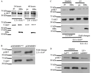
HBx activates AKT in cultured primary hepatocytes.We first
tested whether HBx activates AKT in cultured primary rat hepa-tocytes. Twenty-four hours after isolation and plating, cultured primary rat hepatocytes were transfected with an HBx expression plasmid or the pcDNA control vector. Rat hepatocytes were har-vested at 24 and 48 h posttransfection, and Western blot analysis
with a phosphospecific AKT antibody was performed to check AKT activation. Phosphorylation of AKT at S473 is a marker for
AKT activation (28, 29). HBx-transfected cultured primary rat
hepatocytes had higher levels of phosphorylated AKT (S473) than did control-transfected cells, indicating that HBx activates AKT in primary rat hepatocytes. In addition, HBx activation of AKT in primary rat hepatocytes was sustained for at least 48 h after
trans-fection (Fig. 1A). Similar results were obtained with cultured
pri-mary rat hepatocytes infected with a recombinant adenovirus that expresses HBx, demonstrating that, when necessary to achieve expression of HBx or HBV in 100% of the cultured hepatocytes, recombinant adenoviruses that express HBV or HBx can be used to study HBV- or HBx-specific effects in cultured primary hepa-tocytes (J. C. Garner and M. J. Bouchard, unpublished results). These observations are also consistent with our previously re-ported studies that compared HBx activities in cells transfected with an HBx expression plasmid to those in cells infected with an
HBx-expressing recombinant adenovirus (26,27).
Cultured primary rat hepatocytes were next transfected with HBV expression plasmid pGEMHBV or mutant HBV expression plasmid pGEMHBV*7, which cannot express HBx due to a stop codon in-serted in the HBx gene. Expression of HBV from pGEMHBV resulted
FIG 1HBx activates AKT in primary rat hepatocytes. (A) Primary rat hepatocytes were transfected with the HBx expression plasmid or the pcDNA control vector. The cells were collected at different time points, and the levels of pAKT (Ser473) were measured by Western blot analyses. T AKT, total AKT. (B) Primary rat hepatocytes were transfected with the pGEMHBV or pGEMHBV*7 (HBx-deficient HBV) expression plasmid. Western blot analysis was performed to check the levels of pAKT in pGEMHBV- and pGEMHBV*7-transfected cells. (C) Primary rat hepatocytes were transfected with the HBx expression plasmid or the pcDNA control vector. The cells were treated with either the DMSO control or LY294002 (LY) (PI3K inhibitor), and Western blot analysis was performed to check the levels of pAKT in HBx- and pcDNA-transfected cells. (D) Primary human hepatocytes were infected with AdHBV or Ad*7 (without HBx) and then treated with either the vehicle control or LY294002. Western blot analysis was conducted to assess the levels of AKT activation. Each Western blot shows one representative result from at least two independent experiments. The band intensities in each Western blot were measured by using ImageJ software; the band intensities of pAKT were divided by the band intensities of total AKT, and the ratios are represented in the terms of fold differences. The differences indicated are average fold changes from three independent experiments⫾standard errors. An asterisk represents aPvalue of⬍0.05, determined by Student’sttest. Rawat and Bouchard
on November 7, 2019 by guest
http://jvi.asm.org/
[image:4.585.108.476.66.362.2]in the activation of AKT compared to pGEMHBV*7-transfected
pri-mary rat hepatocytes (Fig. 1B). Expression of all HBV mRNAs,
in-cluding HBx, is under the control of their own promoters in the pGEMHBV vector. HBx expression from pGEMHBV in cultured primary rat hepatocytes is below detectable levels by standard West-ern analysis techniques, similar to observations of HBx expression during an authentic HBV infection; however, we have previously shown that HBx expression can be detected from pGEMHBV when the lysates of a large number of cells are concentrated and analyzed by
Western blotting (49,57). Therefore, the observed activation of AKT
in both pGEMHBV- and HBx-transfected cultured primary rat hepa-tocytes confirmed that that activation of AKT in HBx-transfected primary rat hepatocytes is not an artifact of HBx overexpression and that AKT is activated when HBx is expressed both alone and in the context of the HBV genome. Activation of AKT by HBx in cultured primary rat hepatocytes was blocked by LY294002, a general PI3K
inhibitor (Fig. 1C). Although there were variations in the levels of
AKT activation in different experiments, they likely reflect slight dif-ferences in the transfection efficiencies of different hepatocyte sam-ples and the fact that the hepatocytes were isolated from outbred rats. Importantly, AKT activation was always higher in HBx-expressing hepatocytes than in controls.
We also analyzed the effect of HBx expression on AKT activa-tion in cultured primary human hepatocytes. Cultured primary human hepatocytes were infected with a recombinant adenovirus expressing HBV (AdHBV) or a recombinant adenovirus express-ing an HBx-defective mutant HBV (Ad*7). Although cultured primary human hepatocytes can be directly infected with HBV, for these studies and subsequent studies described below, we used AdHBV for infecting cultured primary human hepatocytes be-cause our goal was to directly compare HBx effects in the human and rat primary hepatocyte systems using the recombinant ade-novirus system. We also noted in our primary rat hepatocyte stud-ies that in order to detect the consistent but moderate effect of HBx on AKT signaling, a high percentage of cells must express HBx, either alone or in the context of the HBV genome, which can be difficult to achieve by the less-efficient, direct HBV infection of cultured primary human hepatocytes. Finally, due to the ex-tremely low level of replication of HBV*7 (HBx-deficient HBV), isolation and purification of high-titer samples of this HBx mu-tant HBV are technically difficult, which hampers a comparison of primary human hepatocytes that are directly infected with HBV or HBV*7. AdHBV-infected primary human hepatocytes had higher levels of AKT phosphorylation (S473) than did Ad*7-infected pri-mary human hepatocytes. LY294002 blocked the increase in AKT phosphorylation in AdHBV-infected cultured primary human
hepatocytes (Fig. 1D); these observations confirmed that AKT can
be activated by HBV, and specifically by HBx, in primary human hepatocytes. In addition to demonstrating HBV activation of AKT in human hepatocytes, the natural site of an HBV infection, these studies also confirmed, as we previously reported, that cultured primary rat hepatocytes can serve as a surrogate system for study-ing HBV and HBx effects on cellular signal transduction pathways
in human hepatocytes (26).
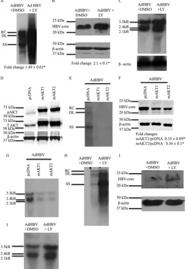
PI3K/AKT signaling reduces HBV replication and HBV
mRNA levels.To examine the effect of PI3K/AKT signaling on
HBV replication in primary hepatocytes, 24 h after isolation and plating, cultured primary rat hepatocytes were infected with AdHBV; at 12 h post-AdHBV infection, the cells were treated with either LY294002 or the dimethyl sulfoxide (DMSO) vehicle
con-trol. Because recombinant adenoviruses can infect 100% of cul-tured primary rat hepatocytes, the use of AdHBV in our studies facilitates the detection of HBV replication, which can be difficult to detect with the lower level of cultured primary rat hepatocytes that are transfected with the pGEMHBV expression plasmid. AdHBV-infected primary rat hepatocytes were harvested 48 h af-ter AdHBV infection, and HBV replication was analyzed by Southern blotting of cytosolic, encapsidated HBV replicative
in-termediates, as previously described (54). LY294002 inhibition of
PI3K/AKT signaling significantly enhanced HBV replication in
cultured primary rat hepatocytes (Fig. 2A). The level of HBV core
protein was also elevated when PI3K/AKT signaling was inhibited
by LY294002 (Fig. 2B). Overall, these results indicate that
inhibi-tion of the PI3K/AKT pathway in cultured primary rat hepato-cytes elevates HBV core protein levels and stimulates HBV repli-cation.
We also compared our observations of primary rat hepatocytes to the effect of AKT signaling on HBV replication in cultured primary human hepatocytes. We observed similar effects of PI3K/ AKT signaling on HBV replication in cultured primary human hepatocytes; inhibition of PI3K/AKT signaling enhanced HBV
replication and elevated HBV core protein levels (Fig. 2HandI).
These results confirm that PI3K/AKT signaling also modulates HBV replication in primary human hepatocytes and again con-firm that cultured primary rat hepatocytes can serve as a surrogate model for studying factors that regulate HBV replication in hu-man hepatocytes.
Because PI3K/AKT inhibition increased HBV core protein lev-els, we speculated that inhibition of the PI3K/AKT pathway might also result in increased transcription of HBV RNAs. Therefore, we examined the effect of the inhibition of PI3K/AKT signaling on HBV mRNA levels. Primary rat hepatocytes were treated as
de-scribed in the legend ofFig. 2A, and Northern blot analysis was
performed to analyze HBV mRNA levels. Inhibition of PI3K/AKT signaling by LY294002 elevated the levels of HBV mRNAs in
cul-tured primary rat hepatocytes (Fig. 2C). We observed similar
PI3K/AKT-dependent effects on HBV mRNA levels when PI3K/ AKT signaling was inhibited in AdHBV-infected cultured primary
human hepatocytes (Fig. 2J).
LY294002 is a general inhibitor of the PI3K/AKT pathway; therefore, to specifically analyze the role of AKT in the regulation of HBV mRNA levels and HBV replication, we utilized
constitu-tively active AKT1 and AKT2 (58). The addition of a
myristoyl-ation signal to AKT targets AKT to the plasma membrane, which increases basal AKT phosphorylation and constitutively activates AKT so that AKT initiates downstream signaling in the absence of
upstream activators (59). Myristoylated AKT1 (mAKT1) and
mAKT2 were used as the constitutively active forms of AKT. We did not assess the effect of the third AKT isoform, AKT3, in our studies because the expression level of AKT3 in liver is very low
compared to the expression levels of AKT1 and AKT2 (60). We
first confirmed that the mAKT1 and mAKT2 expression plasmids
express constitutively active AKT1 and AKT2 (Fig. 2D).
We next examined the effect of mAKT1 or mAKT2 on HBV replication. Cultured primary rat hepatocytes were transfected with mAKT1, mAKT2, or pcDNA, and at 6 h posttransfection, primary rat hepatocytes were infected with AdHBV. Primary rat hepatocytes were collected 48 h after AdHBV infection, and HBV replication was analyzed by Southern blotting. mAKT1 and mAKT2 reduced the levels of HBV DNA compared to the levels in
on November 7, 2019 by guest
http://jvi.asm.org/
FIG 2AKT/PI3K signaling reduces HBV replication and HBV mRNA levels. (A) Primary rat hepatocytes were infected with AdHBV, and the cells were treated with either LY294002 or the vehicle control. Southern blot analysis of core particles that were isolated from the infected hepatocytes was performed to assess HBV replication. RC, relaxed circular; DL, double-stranded linear; SS, single stranded. (B) Western blot analysis was performed on the samples from panel A to check the levels of HBV core protein. Southern and Western data shown here are one representative result of three independent experiments, each with duplicate samples. The difference indicated below the Western blot is the average fold change from three independent experiments⫾standard error. An asterisk represents aPvalue of⬍0.05, determined by using Student’sttest. (C) Primary rat hepatocytes were infected with AdHBV, and the cells were treated with either LY294002 or the vehicle control. Northern blot analysis was performed to ascertain the levels of HBV mRNAs. (D) Western blot analysis to check the expression of pAKT in primary rat hepatocytes transfected with mAKT1 or mAKT2 expression plasmids (constitutively active forms of AKTs). T AKT, total AKT. (E) Primary rat hepatocytes were transfected with the pcDNA control vector or the mAKT1 or mAKT2 expression vector and were also infected with AdHBV. Southern blot analysis was performed to check the levels of HBV replication. (F) Western blot analysis was performed on the samples from panel E to check the levels of HBV core protein. (G) Primary rat hepatocytes were treated as described above for panel E, and Northern blot analysis was performed to determine the levels of HBV mRNA. (H) Primary human hepatocytes were treated as described above for panel A, and Southern blot analyses were performed to assess HBV replication. (I) Western blot analyses were performed on the samples from panel H to check the levels of HBV core protein. (J) Primary human hepatocytes were treated as described above for panel A, and Northern blot analyses were performed to check the levels of HBV RNA. The blots shown in panels C to G are representative of at least 2 independent experiments, each with duplicate samples. The differences indicated in panel F are average fold changes from 2 independent experiments⫾ standard errors. An asterisk represents aPvalue of⬍0.05, determined by using Student’sttest.
on November 7, 2019 by guest
http://jvi.asm.org/
[image:6.585.111.476.37.562.2]control-transfected cells (Fig. 2E). HBV core protein levels were
also reduced by mAKT1 and mAKT2 (Fig. 2F). We also analyzed
HBV mRNA levels by Northern blot analyses; mAKT1 and mAKT2 decreased HBV mRNA levels in cultured primary rat
hepatocytes (Fig. 2G). We were unable to repeat similar
transfec-tion experiments in cultured primary human hepatocytes because transfection efficiencies of primary human hepatocytes are ex-tremely low; however, our previously reported studies and those with cultured primary human hepatocytes described here strongly suggest that HBV and HBx effects are identical in cultured
pri-mary human and rat hepatocytes (23,26,27). Overall, the results
of these AKT studies demonstrate that the activation of either AKT1 or AKT2 decreases HBV mRNA and core protein levels and HBV replication in cultured primary rat hepatocytes.
HNF4␣acts downstream of AKT to decrease HBV mRNA
levels.The observed downregulation of HBV mRNA levels
sug-gested that the activation of AKT by HBx might modulate the activity of one or more transcription factors that regulate HBV
RNA levels. HNF4␣was previously shown to be one transcription
factor that can influence the level of HBV replication (61). HNF4␣
is also a target of AKT; activated AKT can phosphorylate HNF4␣,
causing translocation of HNF4␣out of the nucleus (42). We
an-alyzed the levels of HNF4␣in the nucleus of cultured primary rat
hepatocytes that were transfected with either pGEMHBV or pGEMHBV*7. Cultured primary rat hepatocytes were harvested
at 24 h posttransfection, and the level of HNF4␣in nuclear
ex-tracts was determined by Western blot analysis.
pGEMHBV-transfected primary rat hepatocytes had lower levels of HNF4␣
in the nucleus than did pGEMHBV*7-transfected primary rat
hepatocytes (Fig. 3A). Histone H3 was used as the loading control
and nuclear marker, and GAPDH was used as the cytosolic marker and indicator of fraction purity.
To further confirm the regulation of HNF4␣by HBV in
cul-tured primary hepatocytes, we analyzed mRNA expression levels
of three specific HNF4␣target genes, ALDOB, CYP1A2, and G6P,
which were previously described to be HNF4␣-specific target
genes (62). Cultured primary rat hepatocytes were transfected
with the control vector pGEM, pGEMHBV, or the mAKT2 ex-pression plasmid, and the cells were harvested 48 h after transfec-tion. Because we observed similar effects on HBV replication in cells transfected with either the mAKT1 or mAKT2 expression plasmids, we used only the mAKT2 expression plasmids in the studies described here. qRT-PCR was performed to determine the
mRNA expression levels of the HNF4␣target genes; HBV
de-creased the expression levels of ALDOB, CYP1A2, and G6P, sup-porting the notion that HBV negatively regulates the activity of HNF4␣(Fig. 3B). As expected, mAKT2 also decreased the
expres-sion levels of HNF4␣targets. This indicates that the activation of
AKT in hepatocytes inhibits the transcriptional activity of HNF4␣
and suggests that HBV could regulate HNF␣by activating AKT.
To determine whether HNF4␣acts downstream of AKT to
regulate HBV replication, we next used siRNAs targeting HNF4␣
(siHNF4␣) to decrease the expression of HNF4␣in cultured
pri-mary rat hepatocytes. At 100 nM siHNF4␣, we saw efficient
knockdown of HNF4␣compared to that in nontargeting siRNA
(siNT)-transfected, cultured primary rat hepatocytes (data not shown). Cultured primary rat hepatocytes were transfected with
100 nM siHNF4␣or siNT, and at 24 h posttransfection, primary
rat hepatocytes were infected with AdHBV. At 12 h post-AdHBV infection, primary rat hepatocytes were treated with either
LY294002 or the vehicle control. The cells were harvested 36 h after LY294002 treatment, and Western blot analyses was
per-formed to assess the efficiency of HNF4␣knockdown (Fig. 3C).
We also determined whether HNF4␣knockdown prevented an
increase in the HBV mRNA level when AdHBV-infected primary rat hepatocytes were treated with LY294002. Cultured primary rat
hepatocytes were treated as described in the legend ofFig. 3C, and
HBV RNA levels were analyzed by Northern blotting. In the pres-ence of siNT and LY294002 treatment, HBV mRNA levels in Ad-HBV-infected primary rat hepatocytes were higher than those in
vehicle control-treated cells (Fig. 3D, lanes 1 and 3). However,
when HNF4␣ was knocked down and cells were treated with
LY294002, the level of HBV mRNA was not as high as that in
siNT-transfected cells that were treated with LY294002 (Fig. 3D,
lanes 3 and 4). In a similar experiment, we also analyzed HBV core
protein levels after HNF4␣knockdown. LY294002 treatment
in-creased the HBV core protein levels in AdHBV-infected primary
rat hepatocytes (Fig. 3E, lanes 1 and 3); however, when HNF4␣
was knocked down, we did not see an increase in HBV core
pro-tein levels after LY294002 treatment (Fig. 3E, lanes 3 and 4). We
also analyzed HBV replication by Southern blot analysis of cyto-solic, encapsidated HBV replicative intermediates in
AdHBV-in-fected cultured primary rat hepatocytes. When HNF4␣ was
knocked down and primary rat hepatocytes were treated with
LY294002, the knockdown of HNF4␣ expression blocked the
LY294002-mediated increase in HBV replication (Fig. 3F).
Al-though the knockdown of HNF4␣in AdHBV-infected cells that
were treated with LY294002 did not reduce HBV replication to control levels (AdHBV, siNT, and DMSO), this is most likely due
to an incomplete knockdown of HNF4␣. It is also possible that
there is more than one factor downstream of AKT that inhibits HBV replication. Overall, the results of these studies suggest that
HBx activation of AKT decreases HNF4␣ nuclear localization,
causing decreases in HBV mRNA levels, core protein levels, and HBV replication.
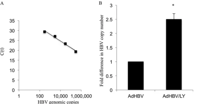
Inhibition of the PI3K/AKT pathway does not inhibit HBV
secretion.Our results described above indicated that the
inhibi-tion of the AKT pathway increased the levels of HBV replicative intermediates contained within cytosolic core particles, which is the focus of our standard HBV replication assay. We next deter-mined if the increase in the level of cytosolic HBV replicative in-termediates reflected a true measure of the overall production of enveloped HBV or an accumulation of cytosolic HBV DNA-con-taining particles resulting from a defect in HBV secretion caused by the inhibition of AKT. Cultured primary rat hepatocytes were infected with AdHBV and then treated with LY294002 or the ve-hicle control. Primary rat hepatocyte cell culture medium super-natants were collected 48 h after treatment, and enveloped HBV particles were captured by using an anti-HBs antibody as
previ-ously described (55). The captured HBV particles were then
sub-jected to real-time PCR with HBV-specific primers to quantify the levels of enveloped HBV particles in the supernatant. A standard
curve was made by plotting the change in theCTvalues with
in-creasing numbers of HBV genomic copies (Fig. 4A).
AdHBV-in-fected primary rat hepatocytes that were treated with LY294002 had higher levels of enveloped HBV particles in the supernatant than did control primary rat hepatocyte medium supernatants (Fig. 4B). The HBV copy number in the supernatant was
calcu-lated on the basis of the standard curve shown inFig. 4A. This
result demonstrates that the increased cytosolic core particle HBV
on November 7, 2019 by guest
http://jvi.asm.org/
Rawat and Bouchard
on November 7, 2019 by guest
http://jvi.asm.org/
DNA level in primary rat hepatocytes after inhibition of the PI3K/ AKT pathway is not due to a defect in the secretion of HBV and that the standard cytosolic core particle HBV replication assay, in this case, accurately reflects overall levels of HBV replication.
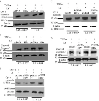
HBx activates AKT to inhibit apoptosis in cultured primary
rat hepatocytes.HBx activates AKT in cultured primary rat
hepa-tocytes, even though activation of PI3K/AKT signaling inhibits HBV replication. This led us to question why HBV would activate a pathway that decreases its own replication. The PI3K/AKT
path-way is a cell survival pathpath-way (63). We previously demonstrated
that HBx inhibits spontaneous and TNF-␣-mediated apoptosis in
cultured primary rat hepatocytes (14). Therefore, we next
deter-mined whether HBx activation of AKT in cultured primary rat hepatocytes is linked to HBx inhibition of apoptosis. Cultured primary rat hepatocytes were transfected with the HBx expression plasmid or the pcDNA control, and at 6 h posttransfection,
pri-mary rat hepatocytes were treated with TNF-␣and LY294002 or
the vehicle control. Primary rat hepatocytes were harvested after 36 h of treatment, and mitochondrial and cytosolic fractions were separated. The cytosolic fractions were then analyzed by Western
blotting to determine the levels of cytosolic cytochromec.
Cyto-chromecis a mitochondrial protein that is released from
mito-chondria into the cytosol upon activation of the apoptotic
path-ways; the level of cytosolic cytochrome c can be used as an
indicator of the level of activation of apoptosis (14,64). In
agree-ment with our previously reported observations, HBx-expressing cultured primary rat hepatocytes had lower levels of cytosolic
cy-tochromecthan did control cells, indicating that HBx inhibited
TNF-␣-induced apoptosis (Fig. 5A) (14). However, when the
ac-tivation of AKT was blocked by LY294002, HBx was no longer able
to inhibit TNF-␣-mediated apoptosis in cultured primary rat
hepatocytes, and both HBx- and vector control-transfected cells
had similar levels of cytosolic cytochromec(Fig. 5A). To further
confirm our apoptosis results, we analyzed levels of cleaved caspase-3, another apoptosis marker. The levels of cleaved caspase-3 were analyzed by Western blot analysis of cultured pri-mary rat hepatocytes that were treated the same way as described
in the legend ofFig. 5A. As shown inFig. 5B, HBx-expressing
primary rat hepatocytes had lower levels of cleaved caspase-3 in
response to TNF-␣treatment, indicating inhibition of apoptosis,
but this inhibition of apoptosis by HBx was lost upon LY294002 treatment. To determine whether similar effects on apoptosis were observed in cultured primary rat hepatocytes transfected with the entire HBV genome, we also analyzed the consequence of apoptosis after blocking the PI3K/AKT pathway in pGEMHBV-transfected cultured primary rat hepatocytes; we previously dem-onstrated that inhibition of apoptosis in pGEMHBV-transfected
hepatocytes was directly linked to HBx expression (14). Cultured
primary rat hepatocytes were transfected with either pGEMHBV or the pGEM control, and 6 h after transfection, the cells were
FIG 3HNF4␣acts downstream of AKT to decrease HBV mRNA levels. (A) Primary rat hepatocytes were transfected with the pGEMHBV or pGEMHBV*7 expression plasmid, and nuclear extracts (NE) were prepared after harvesting of the cells. Western blot analysis of the nuclear extracts was performed to determine the levels of HNF4␣. Histone H3 was used as the nuclear marker and the loading control, and GAPDH was used as the cytosolic marker. The data are representative of 3 independent experiments, and the difference indicated is the average fold change⫾standard error. WCL, whole-cell extracts. (B) Primary rat hepatocytes were transfected with either the pGEM control vector or the pGEMHBV or mAKT2 expression vector. Real-time PCR was conducted by using primers for three HNF4␣direct-target genes. ALDO, aldolase; CYP1A2, cytochrome p450 family 1 subfamily A polypeptide 2; G6P, glucose 6 phosphatase. (C) Primary rat hepatocytes were transfected with either siNT (100 nM) or siHNF4␣(100 nM), and the cells were infected with AdHBV. The cells were treated with either the vehicle control (DMSO) or LY294002 (LY), and Western blot analysis was performed to check the efficiency of HNF4␣knockdown. (D) Primary rat hepatocytes were treated as described above for panel C, and Northern analysis was performed to determine the levels of HBV mRNA. (E) Primary rat hepatocytes were treated as described above for panel C, and Western blot analyses was performed to check the levels of HBV core protein. (F) Primary rat hepatocytes were treated as described above for panel C, and Southern blot analysis was performed to ascertain the levels of HBV replication. The data shown in panels B to F are representative of at least two independent experiments. The differences indicated in panel E are average fold changes from the three independent experiments⫾ standard errors. Statistical analysis was conducted by using Student’sttest, where * and ** represent aPvalue of⬍0.05.
FIG 4Inhibition of the AKT pathway does not inhibit HBV secretion. (A) Standard curve showing the change inCTvalues with increasing numbers of HBV
genomic copies. (B) The supernatant from AdHBV-infected primary rat hepatocytes that were treated with either the vehicle control or LY294002 was used to detect secreted, enveloped HBV particles by qRT-PCR. The copy number of the HBV genome in the supernatant was calculated based upon the standard curve shown in panel A and is represented as the fold difference. The graph represents averages of data from 4 independent experiments, each with triplicate samples. Statistical analysis was conducted by using Student’sttest, where the asterisk represents aPvalue of⬍0.05.
on November 7, 2019 by guest
http://jvi.asm.org/
[image:9.585.136.459.66.234.2]treated with TNF-␣and LY294002 or the vehicle control. Cul-tured primary rat hepatocytes were harvested after 16 h of treat-ment, and either the cells were processed to separate the cytosolic fraction from mitochondria to determine the levels of cytosolic
cytochromecor the samples were directly analyzed to determine
the levels of cleaved caspase-3. HBV-expressing cells had lower
levels of cleaved caspase-3 and cytosolic cytochromec, indicating
that HBV inhibits TNF-␣-mediated apoptosis; however, HBV was
not able to inhibit TNF-␣-mediated apoptosis when AKT
activa-tion was blocked by LY294002 (Fig. 5CandD). We also compared
the regulation of apoptosis in pGEM*7- and pGEMHBV-trans-fected primary rat hepatocytes to more specifically assess HBx regulation of apoptosis in pGEMHBV-expressing cells; the results
were similar to those shown inFig. 5C(Fig. 5E). Overall, these
studies demonstrate that HBV, and specifically HBx, activation of the PI3K/AKT pathway in hepatocytes inhibits apoptosis.
DISCUSSION
The HBV HBx protein can modulate numerous cell signal
transduction pathways and stimulate HBV replication (27,54,
65, 66). While some HBx-regulated cell signaling pathways
such as the Src–mitogen-activated protein kinase (MAPK) pathway and calcium signaling stimulate HBV replication,
oth-ers, such as the NF-B signaling pathway, can inhibit HBV
replication in certain cellular contexts (27,65–67). HBx
eleva-tion of cytosolic calcium levels activates Src kinases, which in turn activate the HBV polymerase/reverse transcriptase to
stimulate HBV replication (65,67,68). HBx also activates the
transcription factor NF-B; however, activation of NF-B
in-hibits HBV replication in HepG2 cells by decreasing the
stabil-ity or formation of HBV core particles (66). The regulation of
HBV replication by various cell signal transduction pathways has been studied mostly with immortalized or transformed cell lines, and the results of many of these studies suggest that some HBx activities may be cell context dependent. Therefore, the results of studies conducted with immortalized or transformed cell lines may or may not be relevant to HBx activities in nor-mal hepatocytes and the associated effects on HBV replication
(11, 44). Cultured primary rat and human hepatocytes are
FIG 5HBx activates the AKT pathway to inhibit apoptosis in primary hepatocytes. (A) Primary rat hepatocytes were transfected with either the pcDNA control vector or the HBx expression plasmid. The cells were then treated with TNF-␣and LY294002 or the DMSO vehicle control. The cells were harvested, and Western blot analysis was performed to check the levels of cytosolic cytochromec. (B) Primary rat hepatocytes were transfected and treated as described above for panel A, and Western blot analysis was performed to check the levels of cleaved caspase-3. (C and D) Primary rat hepatocytes were transfected with either pGEMHBV or the pGEM control vector and treated as described above for panel A. Western analysis was performed to check the levels of cytosolic cytochromec(C) and cleaved caspase-3 (D). (E) Primary rat hepatocytes were transfected with either the pGEM*7 control vector or the pGEMHBV expression plasmid, and the cells were treated as described above for panel A. Western blot analysis was performed to check the levels of cytosolic cytochromec. The blots shown are representative of data from 2 independent experiments. The differences indicated are the average fold changes from the 2 independent experiments⫾standard errors. An asterisk represents aPvalue of⬍0.05, determined by using Student’sttest.
Rawat and Bouchard
on November 7, 2019 by guest
http://jvi.asm.org/
[image:10.585.125.459.66.407.2]model systems for studying HBx and HBV effects in normal,
differentiated hepatocytes (11,14,26,27,49).
In the present study, we demonstrated that HBx activates AKT inex vivo, cultured primary human and rat hepatocyte model systems and that the activation of AKT decreased the transcription of HBV RNAs and HBV replication in these cells. We observed similar HBx activation of AKT when HBx was expressed alone or in the context of the HBV genome. It was previously reported that HBx expression levels might differentially affect transcription
pathways and apoptosis in various experimental systems (69–71).
In a chronically HBV-infected individual, the HBx expression
level is low (72); therefore, expressing HBx in the context of the
HBV genome was important to rule out the possibility of an arti-fact due to HBx overexpression. The mechanism(s) that underlies HBx activation of AKT in primary hepatocytes is under investiga-tion. Previous studies conducted with transformed cell lines have shown that HBx activation of the AKT pathway could be linked to decreases in phosphatase and tensin homolog (PTEN) expression
levels (73). PTEN dephosphorylates PI3K, and a reduction in the
level of PTEN would facilitate AKT phosphorylation and activa-tion. However, in our studies of cultured primary rat hepatocytes, we did not see any HBx-induced change in PTEN levels, suggest-ing that the activation of AKT in primary hepatocytes is not due to a reduction in PTEN expression levels (data not shown). Whether HBx affects PTEN activity in normal hepatocytes will be the sub-ject of future studies.
HBV and HBx expression also decreased nuclear levels of
HNF4␣, which we linked to the activation of AKT. Blocking of the
PI3K/AKT signaling pathway increased HBV replication in pri-mary hepatocytes, and this effect was countered by knocking
down HNF4␣ expression, indicating that AKT targeting of
HNF4␣can decrease HBV replication. HNF4␣is a transcription
factor that controls the expression of a number of genes in hepa-tocytes and is regulated by various posttranslational modifications (37–41). Previous studies have also shown that HNF4␣binds to transcription enhancers in the HBV genome that stimulate the
transcription of HBV mRNAs (61,74). It was demonstrated
pre-viously that AKT can phosphorylate HNF4␣to reduce HNF4␣
nuclear localization (42). Our studies also demonstrate that HBx
decreases the nuclear localization of HNF4␣and that transcript
levels of HNF4␣target genes are decreased by HBV, suggesting
that the AKT-mediated decrease in HBV replication is linked to
the reduction in HNF4␣nuclear levels and the associated
de-creased levels of HBV mRNAs. In support of this notion, inhibi-tion of the PI3K/AKT pathway increased HBV replicainhibi-tion, but
knockdown of HNF4␣in hepatocytes prevented the increase of
HBV replication that was observed when PI3K/AKT signaling was inhibited. Cumulatively, our studies suggest that the activation of
AKT by HBx results in decreased HNF4␣nuclear levels and
de-creased HBV replication, whereas the inhibition of the PI3K/AKT
pathway allows increased nuclear HNF4␣ levels and increased
HBV replication (Fig. 6). Although HNF4␣ knockdown in
LY294002-treated cells did not bring the levels of HBV mRNA and HBV replication to the levels in control hepatocytes, we believe
that this was caused by the incomplete knockdown of HNF4␣in
cultured primary rat hepatocytes due to the lower transfection efficiency of primary hepatocytes combined with the very high AdHBV infection efficiency of cultured primary hepatocytes. It is also possible that additional factors downstream of AKT regulate HBV replication, which will be the focus of future studies.
Although our observation that the activation of AKT inhibits HBV replication suggests that the replication of an HBx-deficient HBV (HBV*7) might be enhanced by the inhibition of AKT, anal-ysis of the effect of inhibiting AKT on the replication of an HBx-deficient HBV is challenging and perhaps not informative for a number of reasons. The level of HBV replication from HBV*7 is
extremely low in cultured primary rat hepatocytes (14), even
though, in the absence of HBx, HBV*7 failed to activate AKT (Fig.
1B). In HBV*7-expressing cells, the effect of inhibiting AKT with
LY294002 would show the consequence of inhibiting endogenous levels of active AKT on HBV replication rather than the effect of inhibition of HBx-specific activation of AKT. Moreover, this anal-ysis would be conducted with cells where requisite HBx activities that stimulate HBV replication would not be present; HBV*7 does not replicate well in primary hepatocytes because signaling path-ways that are activated by HBx to stimulate HBV replication
re-main inactive (14). We therefore did not explore the effect of the
PI3K/AKT pathway on the replication of HBx-deficient HBV. The observation that HBx activation of AKT decreases HBV replication seems counterintuitive considering that many previ-ous studies, including our own, have shown that HBx stimulates
HBV replication (14). The PI3K/AKT pathway is an important cell
survival pathway and has been shown to inhibit apoptosis in
im-mortalized hepatocytes in the context of HBx expression (31,33).
We observed that HBx activation of AKT inhibits apoptosis in cultured primary rat hepatocytes. It was previously reported that inhibition of apoptosis in hepatocytes is important for HBV to
release infectious progeny (14,75). HBV is a noncytopathic virus,
and inhibition of apoptosis in HBV-infected hepatocytes might be important for persistent replication of HBV in hepatocytes. Our
FIG 6Model. HBx activates AKT in primary hepatocytes, causing a reduction in HNF4␣transcription activity. HNF4␣is an important transcription factor for the transcription of the HBV genome, and a reduction in HNF4␣ transcrip-tion activity decreases HBV transcriptranscrip-tion and, consequently, HBV replicatranscrip-tion. However, activation of AKT by HBx is important for the inhibition of apop-tosis in HBV-infected hepatocytes. Overall, activation of the PI3K/AKT path-way decreases HBV replication but inhibits apoptosis, likely providing persis-tent, noncytopathic replication of HBV in hepatocytes.
on November 7, 2019 by guest
http://jvi.asm.org/
[image:11.585.301.542.65.287.2]ability to detect elevated levels of HBV replication in hepatocytes where the PI3K/AKT pathway was inhibited likely reflects the short time period of our assays; cells were collected at a time in which apoptotic signals had been activated but the cells had not yet progressed to complete apoptosis. During chronic HBV infec-tion, the short-term HBV replication benefit that might result from not activating AKT would likely be balanced by the long-term benefit of a noncytopathic infection that could facilitate the persistence of HBV infection and the prolonged production of infectious progeny.
Development of HCC in an individual with chronic HBV in-fection can take decades. Although the underlying causes of HBV-associated HCC remain incompletely understood, three mecha-nisms that are not mutually exclusive have been proposed as contributors to the development of HBV-associated HCC: recur-rent immune-mediated destruction of HBV-infected hepatocytes followed by compensatory liver regeneration; integration of HBV DNA into the host genome, which could alter the expression of cellular factors and, in turn, the control of normal cell functions; and modulation of hepatocyte physiology by HBV proteins such
as HBx (8,76–79). Although HBx is directly oncogenic in some
HBx-transgenic mice (80,81), many studies suggest that HBx has
a cofactor role in hepatocyte transformation (11, 82–84). It is
likely that subtle and moderate HBx activities, such as modulation of cellular signaling pathways, contribute to processes that link an HBV infection to the development of HCC. The subtlety of HBx effects and the cell-specific consequences of some HBx activities have sometimes made it difficult to precisely define HBx activities in an authentic HBV infection and to directly link these activities to hepatocyte transformation; however, these moderate effects of
HBx, such as the⬃2-fold activation of AKT by HBx observed in
our studies with cultured primary hepatocytes, are, in fact, con-sistent with the biology of an HBV infection, including the non-cytopathic nature of the infection and the long time frame re-quired for progression from a chronic HBV infection to the
development of HCC (11,85).
Activation of AKT and downregulation of HNF4␣
transcrip-tional activity could be independent or interconnected
contribu-tors to the development of HBV-associated HCC. HNF4␣is an
important transcription factor for liver development, and
knock-out of HNF4␣in mice is embryonically lethal (86,87). HNF4␣
expression maintains hepatocyte differentiation, and the loss of
HNF4␣transcriptional activity can lead to the dedifferentiation
of hepatocytes and hepatocyte proliferation (36). Inhibition of
HNF4␣can initiate transformation in immortalized hepatocytes
through a microRNA (miRNA) inflammatory feedback loop; per-turbation of this feedback loop was also found for human
hepa-tocellular carcinoma (62). The loss of HNF4␣has been directly
associated with the development of HCC, and HNF4␣expression
suppressed the development of HCC in a mouse model system
(88,89). AKT is an oncogene and is upregulated in many cancers,
providing additional signaling pathways that could link HBx
ex-pression to the development of HCC (90–93). Finally, by lowering
the level of HBV replication, HBx activation of AKT might influ-ence HBV evasion of immune-mediated destruction of HBV-in-fected hepatocytes as an additional means to facilitate persistent HBV replication, a chronic HBV infection, and the eventual de-velopment of HCC.
Overall, the results of our studies with cultured primary rat and human hepatocytes demonstrate that HBx activation of the PI3K/
AKT signaling pathway decreases HBV replication and cellular apoptosis. Although it is now well established that HBx is required for efficient HBV replication, our findings here suggest that HBx activates pathways that both stimulate and diminish HBV replica-tion. While some of these cellular signaling pathways, such as the Src/MAPK and calcium signaling pathways, are important for stimulating HBV replication, others, such as the PI3K/AKT path-way, diminish HBV replication while enhancing cell survival. Therefore, we propose that HBx can function as a rheostat that balances cell survival and HBV replication, possibly to allow for persistent HBV replication. HBx activation of AKT and inhibition
of HNF4␣might also influence the development of
HBV-associ-ated HCC by facilitating the establishment of a chronic HBV in-fection, enhancing the dedifferentiation of hepatocytes, and acti-vating AKT-regulated oncogenic pathways. Modulation of PI3K/ AKT-regulated factors may provide therapeutic targets for inhibiting HBV replication and the development of HBV-associ-ated HCC.
ACKNOWLEDGMENTS
We thank members of the Bouchard lab as well as Jan Hoek, Patrick Loll, Bradford Jameson, and Mauricio Reginato for advice and many helpful discussions.
This project was supported by NIH grant AI064844 to M.J.B.
REFERENCES
1.Nguyen VT, Law MG, Dore GJ.2009. Hepatitis B-related hepatocellular carcinoma: epidemiological characteristics and disease burden. J Viral Hepat16:453– 463.http://dx.doi.org/10.1111/j.1365-2893.2009.01117.x. 2.Ganem D, Prince AM.2004. Hepatitis B virus infection—natural history
and clinical consequences. N Engl J Med350:1118 –1129.http://dx.doi .org/10.1056/NEJMra031087.
3.Fattovich G, Bortolotti F, Donato F.2008. Natural history of chronic hepatitis B: special emphasis on disease progression and prognostic fac-tors. J Hepatol48:335–352.http://dx.doi.org/10.1016/j.jhep.2007.11.011. 4.Cheng PN, Liu WC, Tsai HW, Wu IC, Chang TT, Young KC.2011. Association of intrahepatic cccDNA reduction with the improvement of liver histology in chronic hepatitis B patients receiving oral antiviral agents. J Med Virol83:602– 607.http://dx.doi.org/10.1002/jmv.22014. 5.Ghany MG, Doo EC.2009. Antiviral resistance and hepatitis B therapy.
Hepatology49:S174 –S184.http://dx.doi.org/10.1002/hep.22900. 6.Perrillo RP.2005. Current treatment of chronic hepatitis B: benefits and
limitations. Semin Liver Dis25(Suppl 1):S20 –S28.http://dx.doi.org/10 .1055/s-2005-915647.
7.Zoulim F.2004. Mechanism of viral persistence and resistance to nucle-oside and nucleotide analogs in chronic hepatitis B virus infection. Anti-viral Res64:1–15.http://dx.doi.org/10.1016/j.antiviral.2004.07.003. 8.Bouchard MJ, Navas-Martin S.2011. Hepatitis B and C virus
hepatocar-cinogenesis: lessons learned and future challenges. Cancer Lett305:123– 143.http://dx.doi.org/10.1016/j.canlet.2010.11.014.
9.Seeger C, Mason WS.2000. Hepatitis B virus biology. Microbiol Mol Biol Rev64:51– 68.http://dx.doi.org/10.1128/MMBR.64.1.51-68.2000. 10. Locarnini S, Zoulim F.2010. Molecular genetics of HBV infection.
An-tivir Ther15(Suppl 3):S3–S14.http://dx.doi.org/10.3851/IMP1619. 11. Rawat S, Clippinger AJ, Bouchard MJ.2012. Modulation of apoptotic
signaling by the hepatitis B virus X protein. Viruses4:2945–2972.http://dx .doi.org/10.3390/v4112945.
12. Kremsdorf D, Soussan P, Paterlini-Brechot P, Brechot C.2006. Hepa-titis B virus-related hepatocellular carcinoma: paradigms for viral-related human carcinogenesis. Oncogene 25:3823–3833. http://dx.doi.org/10 .1038/sj.onc.1209559.
13. Bouchard MJ, Schneider RJ.2004. The enigmatic X gene of hepatitis B virus. J Virol78:12725–12734.http://dx.doi.org/10.1128/JVI.78.23.12725 -12734.2004.
14. Clippinger AJ, Gearhart TL, Bouchard MJ.2009. Hepatitis B virus X protein modulates apoptosis in primary rat hepatocytes by regulating both NF-kappaB and the mitochondrial permeability transition pore. J Virol
83:4718 – 4731.http://dx.doi.org/10.1128/JVI.02590-08. Rawat and Bouchard