synthesis, and pharmacologic
evaluation tf peptide RGS4
inhibitors
H. Zhong
J.R. Omnaas
R.R. Neubig
H.I. Mosberg
Authors' affiliations:
Y. Jin, J.R. Omnaas and H.I. Mosberg, Department of Medicinal Chemistry, The University of Michigan, Ann Arbor, MI
48109, USA.
H. Zhong and R.R. Neubig, Department of Pharmacology, The University of Michigan, Ann Arbor, MI48109, USA.
R.R. Neubig, Department of Internal Medicine/ Hypertension, The University of Michigan, Ann Arbor, MI48109-1065, USA.
Correspondence to: Henry I. Mosberg
Department of Medicinal Chemistry College of Pharmacy
The University of Michigan Ann Arbor
MI48109-1065 USA
Tel.:734-764-8117 Fax:734-763-5595 E-mail: [email protected]
Key words:cyclic peptides; G-proteins; GTPase-activating proteins; protein–protein interactions; regulator of G-protein signaling
Abstract:Regulators of G-protein signaling (RGS) proteins form a
multifunctional signaling family. A key role of RGS proteins is
binding to the G-protein Ga-subunit and acting as GTPase-activating proteins (GAPs), thereby rapidly terminating G
protein-coupled receptor (GPCR) signaling. Using the published RGS4–Gia1
X-ray structure we have designed and synthesized a series of cyclic
peptides, modeled on the GiaSwitch I region, that inhibit RGS4
GAP activity. These compounds should prove useful for elucidating
RGS-mediated activity and serve as a starting point for the
development of a novel class of therapeutic agent.
Abbreviations:CHO, Chinese hamster ovary; GAP,
GTPase-activating proteins; GPCR, G protein-coupled receptor; GST,
glutathione S-transferase; RGS, regulator of G-protein signaling;
RP-HPLC, reverse-phase high-performance liquid chromatography.
Dates:
Received31October2003 Revised1December2003 Accepted6December2003
To cite this article:
Jin, Y.,Zhong, H.,Omnaas, J.R.,Neubig, R.R. & Mosberg, H.I.Structure-based design, synthesis, and pharmacologic evaluation tf peptide RGS4 inhibitors.
J. Peptide Res.,2004,63,141–146. Copyright Blackwell Munksgaard,2004
Regulators of G-protein signaling (RGS) proteins are a family of highly diverse, multifunctional signaling proteins. This distinct mammalian family was recognized when a
120-residue homologous domain (defined as the RGS
domain) was described in several proteins at approximately
the same time (1,2) and was demonstrated to be responsible
for binding of the RGS protein to the G-proteina-subunit.
Upon this binding, RGS proteins act as GTPase-activating proteins (GAPs) to limit the lifetime of bound guanosine triphosphate (GTP), thus inhibiting G-protein signaling by the rapid turnoff of the G protein-coupled receptor (GPCR)
signaling pathway (3–5). Thus, RGS proteins represent a
novel drug target, as inhibitors of RGS acceleration of GTP
hydrolysis by Ga could, in principle, potentiate the effects
In1997, Tesmer and colleagues (8) reported a2.8A˚
reso-lution crystal structure of the RGS protein, RGS4,
com-plexed with Gia1-Mg2þ-GDP-AlF4. In this complex the
GDP-AlF
4 mimics the transition state of GTP during its
hydrolysis to guanosine diphosphate (GDP), inducing the
conformation of Gathought to be stabilized by RGS-binding.
The core domain of RGS4forms a ninea-helix bundle that
binds to the three distinct ‘Switch’ regions of Gia1, so named
because they undergo large conformational changes during
the transition from the inactive GDP-bound form of the Ga
subunit to the active GTP-bound form (9). These
conform-ational changes allow the Switch regions to contact the
c-phosphate of GTP and play an essential role in GTP
hydrolysis by Ga. The crystal structure of the complex reveals
that RGS4does not contribute catalytic residues that directly
interact with either GDP or AlF
4. Instead, RGS4appears to
promote rapid hydrolysis of GTP primarily by stabilizing the
Switch regions of Gia1in the transition state conformation.
Analysis of the RGS4–Gia1 crystal structure provides
opportunities for the structure-based design of inhibitors of
the RGS interaction with Ga-subunits. Such inhibitors
would be expected to block RGS GAP activity and thus
increase the transduction efficiency of the associated Ga
-subunit. These types of RGS inhibitors could then be used as potentiators of endogenous agonist function similar to the action of benzodiazepines at the ionotropic GABA-A
receptor (10). We describe here initial results of
structure-based inhibitors of RGS4GAP activity that demonstrate the
feasibility of this approach.
Materials and Methods
Solid phase peptide synthesis
All peptides were synthesized by solid-phase methods on an
ABI Model 431A solid phase peptide synthesizer (Applied
Biosystems, Foster City, CA, USA), using Fmoc-protected amino acids obtained from Advanced ChemTech (ville, KY, USA). Rink resin (Advanced ChemTech, Louis-ville, KY, USA) was used as the solid support for C-terminal carboxamide peptides and pre-loaded PEG-PS resin (Applied Biosystems) was used for the C-terminal carboxylic acid peptide. Peptide elongation on the peptide–resin involved treating resin with piperidine (Aldrich, Milwaukee, WI, USA) to cleave the Fmoc-protecting group, followed by
coupling of the next amino acid with o-benzotriazol-1
-yl-N,N,N0,N0-tetramethyl uronium hexafluorophosphate
(HBTU) and 1-hydroxybenzotriazole (HOBt) (Applied
Biosystems). Trifluoroacetic acid/H2O/dithioethane (9:
0.5:0.5, v/v/v) was used to cleave the linear peptide from
the resin and simultaneously remove the side chain pro-tecting groups. The peptide solution was filtered from the resin and then subjected to preparative reverse-phase high-performance liquid chromatography (RP-HPLC) to afford the linear disulfhydryl-containing peptide.
General method for disulfide cyclization of peptides
To obtain disulfide cyclized peptide, linear
disulfhydryl-containing peptide was dissolved in a1% (v/v) HOAc in H2O
solution (saturated with N2) at5C (1mg linear peptide/mL
of aqueous HOAc solution). The pH of the peptide solution
was raised to8.5using NH4OH, followed by the addition of
4mol equivalents of K3Fe(CN)6. The reaction mixture was
stirred for1min, then quenched by adjusting the pH to3.5
with HOAc. The mixture was then subjected to preparative RP-HPLC to afford the disulfide-cyclized peptide.
General method for dithioether cyclization of peptides
To form dithioether-containing cyclic peptides, linear di-sulfhydryl peptide was added to dimethylformamide and
maintained at 5C under a N2 atmosphere (0.1mg linear
peptide/mL dimethylformamide). Five mole equivalents of
potassiumtert-butoxide were added to the peptide solution,
followed by the addition of 2.5mol equivalents of
Br-(CH2)n-Br (n¼1, 2, or 3). The reaction was quenched
with2mL HOAc after2h and the solvent was removed in
vacuo. The residue was dissolved in water, filtered, and then subjected to preparative RP-HPLC to afford the alkyl dithioether-cyclized peptide.
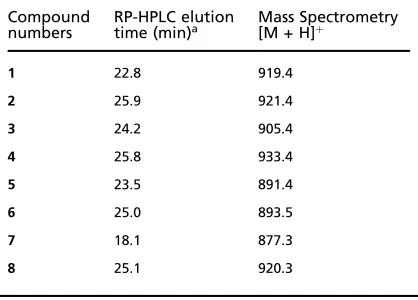
All final product peptides were >95% pure as assessed by
RP-HPLC on a Vydac218TP C-18column (The Nest Group,
Southboro, MA, USA) using the solvent system 0.1%
tri-fluoroacetic acid (TFA) in water/0.1% TFA in acetonitrile
by a gradient of0–40% organic component in40min, and
all peptides displayed the appropriate molecular weights as determined by mass spectrometry. Physicochemical data
for product peptides are summarized in Table1.
Biological materials
[c-32P]GTP (30Ci/mmol) was from New England Nuclear
hamster ovary (CHO) cell line with stable expression of an
Hemaglutinin (HA)-epitope tagged porcine a2aAR
adreno-ceptor (a2aAR-CHO,10–20pmol/mg) was cultured and cell
membranes prepared as described (11). Glutathione
S-transferase (GST) fusion protein containing rat RGS4was
prepared as described (12).
[32P]GTPase assay
The ability of RGS proteins to stimulate steady-state
[32P]GTPase activity of receptor-stimulated G-proteins in
CHO cell membranes expressing high (>5pmol/mg)
con-centrations ofa2aAR was recently described by Zhonget al.
(13). To assess peptide inhibition of RGS-stimulated
[32P]GTPase, measurements were performed in a reaction
mixture (100lL) containing 0.2mm ATP, 1lm GDP,
50units/mL creatine phosphokinase,50mm
phosphocrea-tine, 20mm NaCl, 2mm MgCl2, 0.2mm
ethylenediam-inetetraacetic acid (EDTA), 10mm Tris/HCl, 1mm
dithiothreitol (DTT), and 0.1lm [32P]GTP (pH 7.6). All
components were pre-incubated forc.10min on ice along
with 4lg of a2aAR-CHO membrane protein, the a2aAR
adrenoceptor agonist, UK 14 304, (10lm), 1–300lm
syn-thesized peptide, with or without300nmGST-RGS4
pro-tein. The reaction was started by addition of [c-32P]GTP to
the pre-incubation mixture and GTP hydrolysis was
allowed to proceed for10min at30C. The reaction was
then terminated by adding 1mL of 50% (w/v) ice-cold
activated charcoal slurry in 20mm phosphoric acid,
fol-lowed by incubation on ice for30min. Reaction tubes were
then centrifuged at4000gfor20min at4C and200lL
of supernatant fluid containing the free [32P
i] was
with-drawn and counted by liquid scintillation counting. Each peptide was tested in three separate experiments. Data were fit by nonlinear least-squares analysis in Graph Pad Prism
3.0(San Diego, CA, USA) to the equation
Y¼YmaxþðYminYmaxÞ=ð1þ10logIC50XÞ
where,Xis the logarithm of peptide concentration,Yis the
GTPase activity, Ymax is the maximum and Ymin is
minimum GTPase activity.Ymin was constrained to equal
the GTPase activity in the absence of RGS4.
Results and Discussion
From the crystal structure of the RGS4–Gia1-Mg2þ
-GDP-AlF
4 complex (Protein Data Bank (PDB) file1agr) it can be
seen that the functional binding site for RGS4on the surface
of Gia1 is formed by residues in the three Switch regions of
Gia1: residues 179–185 in Switch I, residues 204–213 in
Switch II, and residues 235–237 in Switch III. Of these
regions, Switch I of Gia1interacts with three-fourths of the
RGS4-binding pocket (8). Furthermore, two surface residues
of Switch I (Thr182 and Gly183) appear to be essential for
high-affinity Ga–RGS interaction (8,14). Therefore, the
Switch I region was chosen as the starting point for the development of small peptide ligands designed to bind to
the RGS4protein and inhibit its GAP activity by preventing
the RGS4–Gia1interaction.
As depicted in Fig.1, the RGS4-binding region of Switch
I, residues179–185, has the amino acid sequence
Val-Lys-Thr-Thr-Gly-Ile-Val. Additionally, Glu186, which is
con-served in most Ga-subunits, is positioned such that it may
interact with nearby positively charged residues of RGS4
(Arg172and Arg167). Therefore, we chose the octa-peptide,
Val1-Lys-Thr-Thr-Gly-Ile-Val-Glu8, as the starting point for
design of analogs of the Gia1 Switch I region. As linear
peptides based upon the RGS-binding region of Switch 1
would be expected to be very flexible, they would be unlikely to highly populate the desired native conformation
observed in the RGS4–Gia1 X-ray structure. Indeed, the
linear Switch I peptide has been prepared and was found to be devoid of inhibitory activity (K.-L. Lan and R. R. Neubig, unpublished data). Hence, our approach was to focus on cyclic peptide analogs, as cyclization is expected to reduce conformational freedom and enhance binding affinity by mimicking or inducing bound structure motifs. From the
crystal structure (Fig.1), it can be seen that the side chains
[image:3.595.50.259.67.217.2]of Thr181and Val185of Gia1are pointing toward each other
Table 1.Physicochemical data for peptide inhibitors of RGS4 GAP activity
Compound
numbers RP-HPLC elutiontime (min)a Mass Spectrometry[M + H]þ
1 22.8 919.4
2 25.9 921.4
3 24.2 905.4
4 25.8 933.4
5 23.5 891.4
6 25.0 893.5
7 18.1 877.3
8 25.1 920.3
a. See Methods for RP-HPLC details.
and have no direct interaction with RGS4. These residues
are thus ideal candidates for substitution by amino acids that allow side chain–side chain cyclization. Accordingly, we designed an initial peptide in which cysteine is
substi-tuted for the corresponding Thr3 and Val7 residues in the
model octa-peptide. Cyclization can be affected by linking these two Cys-residues through a disulfide or
dithioether-bridge. Figure2depicts a model of the ethylene
dithioether-containing cyclic analog, 1, in the RGS4-binding site.
Although cyclization via an ethylene dithioether provides
the best spatial fit to the observed distance between thea
carbons Thr181and Val185in the X-ray structure (8.665A˚
in X-ray vs.8.486A˚ in the modeled ethylene dithioether)
the optimal geometry for a small peptide ligand may differ from that of the corresponding region of the much larger
Ga-subunit. Consequently a more structurally diverse set
of peptide scaffolds was sampled for possible mimics of the Gia1
Switch1region:
where, n¼0–3. Amino-terminal acetylation and
C-ter-minal amidation were chosen to best mimic the corres-ponding region of Gia1.
Figure3 depicts the concentration dependence of the
inhibition of RGS4GAP activity exhibited by compound1–5
and Table2 summarizes the inhibitory potencies of these
peptides. As seen in Table2, compound1, the lead ethylene
dithioether-containing analog, does indeed inhibit RGS Figure1. Interface between regulator of pro-tein signaling (RGS4) (thin lines) and Gia1 Switch I (thick lines) from X-ray structure of Tesmeret al. (8).
Figure2. Interface between regulator of protein signaling (RGS4) (thin lines) and designed ethylene-bridged dithioether peptide,1(thick lines). Cysteine residues are indicated by their sequence positions,
acceleration of Gia1GTPase activity, with an IC50of88lm.
Compound 1 did not inhibit GTPase activity of
receptor-membranes alone indicating that the effect is on RGS4and
not on receptor or G-protein. Consistent with the earlier observation that the linear octa-peptide corresponding to the
Switch I region is inactive, compound2, the linear analog of1
(with each Cys-sulfur converted to a methyl thioether) dis-plays no inhibition of GTPase activity at the highest
con-centration tested,300lm. Compounds3–5further examine
the effect of ring size in the cyclic RGS inhibitor series. As
seen from Table2,3, the methylene dithioether
(dithioac-etal), in which the ring is one carbon smaller, displays
com-parable inhibitory potency as1. In contrast4, the propylene
dithioether, with ring size one carbon larger than1, displays
considerably lower inhibitory potency (61% inhibition at
300lm). Interestingly, 5, the disulfide-containing analog,
whose ring size is two carbon atoms smaller than the lead
compound 1, displays c. threefold higher potency
(IC50¼26lm). To examine the possibility that the improved
potency of5is due to the reduced, linear
sulhydryl-contain-ing species,6, the free sulfhydryl-containing precursor of1,
3–5, was evaluated. As shown in Table2,6displayed no RGS
inhibitory activity at 300lm, the highest concentration
tested, indicating that contribution of the free sulfhydryl
form of the peptide to the observed potency of5is unlikely.
Finally, the design assumption that N-acetylation and C-terminal amidation represent the optimal starting point for analogs in this series was evaluated by preparing and testing
the free amino containing analog,7, and the free carboxylate
containing analog,8. As expected, neither of these analogs
inhibited RGS GAP activity at300lm.
These results support the hypothesis that structure-based design of inhibitors of RGS protein GAP activity modeled
upon the RGS-binding conformation of Gia1 is a viable
approach and, further, represent the first examples of any rationally designed RGS inhibitors. Efforts to improve potency within this series are in progress.
Acknowledgements:Authors are grateful to Dr Irina Pogozheva
for many helpful discussions and Leighton Janes for assistance with GTPase assays. This study was supported by National Institutes of
Health grants DA03910 (HIM) and GM39561 (RRN). Figure3. Inhibition by peptides1–5of regulator of protein signaling
[image:5.595.50.289.48.212.2](RGS) stimulation of GTPase activity. Pi release as a function of peptide concentration in the presence (closed symbols) or absence (open symbols) of300nmglutathione S-transferase (GST)-RGS4protein is shown.
Table 2.IC50values of peptide inhibitors of RGS4 GAP activity
Numbers Structure IC(l50M) SEM
1 Ac-Val-Lys-Cys-Thr-Gly-Ile-Cys-Glu-NH2
(Et)a
88 12
2
Ac-Val-Lys-Cys(Me)-Thr-Gly-Ile-Cys(Me)-Glu-NH2
300
3 Ac-Val-Lys-Cys-Thr-Gly-Ile-Cys-Glu-NH2
(Me)
79 6
4 Ac-Val-Lys-Cys-Thr-Gly-Ile-Cys-Glu-NH2
(Pr)
61% @ 300lM
5 Ac-Val-Lys-Cys-Thr-Gly-Ile-Cys-Glu-NH2
(SS)
26 2
6
Ac-Val-Lys-Cys(SH)-Thr-Gly-Ile-Cys(SH)-Glu-NH2
300
7 H-Val-Lys-Cys-Thr-Gly-Ile-Cys-Glu-NH2
(Et)
300
8 Ac-Val-Lys-Cys-Thr-Gly-Ile-Cys-Glu-OH
(Et)
300
a. Cyclization type between the two Cys-residues denoted in paren-theses:
(SS), -SS-; (Me), -SCH2S-; (Et), -SCH2CH2S-; (Pr), -SCH2CH2CH2S-; RGS4,
regulator of protein signaling; GAP, GTPase-activating proteins.
References
1. Druey, K.M., Blumer, K.J., Kang, V.H. & Kehrl, J.H. (1996) Inhibition of G-protein mediated MAP kinase activation by a new mammalian gene family.Nature379, 742–746.
2. Koelle, M.R. & Horvitz, H.R. (1996) EGL-10 regulates G protein signaling in the C. elegansnervous system and shares a conserved domain with many mammalian proteins.Cell84,115–125.
[image:5.595.51.289.326.585.2]4. Hepler, J.R. (1999) Emerging roles for RGS proteins in cell signaling.Trends Pharmacol. Sci.20,376–382.
5. De Vries, L., Zheng, B., Fischer, T., Elenko, E. & Farquhar, M.G. (2000) The regulator of G protein signaling family.Annu. Rev. Pharmacol. Toxicol.40,235–271. 6. Zhong, H.L. & Neubig, R.R. (2001) RGS
proteins: novel multifunctional drug targets. J. Pharmacol. Expl. Ther.297,837–845. 7. Neubig, R.R. & Siderovski, D.P. (2002) Regulators of G-protein signaling as new central nervous system drug targets.Nat. Rev. Drug Discov.1,189–199.
8. Tesmer, J.J.G., Berman, D.M., Gilman, A.F. & Sprang, S.R. (1997) Structure of RGS4 bound to AlF4-activated G(i alpha1): stabilization of the transition state for GTP hydrolysis.Cell89,251–261.
9. Lambright, D.G., Noel, J.P., Hamm, H.E. & Sigler, P.B. (1994) Structural determinants for activation of the alpha-subunit of a heterotrimeric G-protein.Nature369, 621–628.
10. Macdonald, R.L. & Olsen, R.W. (1994) GABA (A) receptor channels.Annu. Rev. Neurosci.17,569–602.
11. Wade, S.M., Lim, W., Lan, K.L., Chung, D.A., Nanamori, M. & Neubig, R.R. (1999) Gi activator region of2A-adrenergic receptors: distinct basic residues mediate Gi versus Gs activation.Mol. Pharmacol.56, 1005–1013.
12. Lan, K.L., Zhong, H.L., Nanamori, M. & Neubig, R.R. (2000) Rapid kinetics of regulator of G-protein signaling (RGS)-mediated Gi and Go deactivation: G specificity of RGS4and RGS7.J. Biol. Chem. 275,33497–33503.
13. Zhong, H., Wade, S.M., Woolf, P.J., Linderman, J.J., Traynor, J.R. & Neubig, R.R. (2003) A spatial focusing model for G protein signals: regulator of G protein signaling (RGS) protein-mediated kinetic scaffolding. J. Biol. Chem.278,7278–7284.

![(E) 4 {[(Morpholin 4 yl)imino]methyl}benzonitrile](data:image/gif;base64,R0lGODlhAQABAIAAAP///wAAACH5BAEAAAAALAAAAAABAAEAAAICRAEAOw==)