R E S E A R C H A R T I C L E
Open Access
Diagnostic accuracy of semi-quantitative and
quantitative culture techniques for the diagnosis
of catheter-related infections in newborns and
molecular typing of isolated microorganisms
Danilo Flávio Moraes Riboli
1*, João César Lyra
2, Eliane Pessoa Silva
1, Luisa Leite Valadão
1, Maria Regina Bentlin
2,
José Eduardo Corrente
3, Ligia Maria Suppo de Souza Rugolo
2and Maria de Lourdes Ribeiro de Souza da Cunha
1Abstract
Background:Catheter-related bloodstream infections (CR-BSIs) have become the most common cause of healthcare-associated bloodstream infections in neonatal intensive care units (ICUs). Microbiological evidence implicating catheters as the source of bloodstream infection is necessary to establish the diagnosis of CR-BSIs. Semi-quantitative culture is used to determine the presence of microorganisms on the external catheter surface, whereas quantitative culture also isolates microorganisms present inside the catheter. The main objective of this study was to determine the sensitivity and specificity of these two techniques for the diagnosis of CR-BSIs in newborns from a neonatal ICU. In addition, PFGE was used for similarity analysis of the microorganisms isolated from catheters and blood cultures.
Methods:Semi-quantitative and quantitative methods were used for the culture of catheter tips obtained from newborns. Strains isolated from catheter tips and blood cultures which exhibited the same antimicrobial susceptibility profile were included in the study as positive cases of CR-BSI. PFGE of the microorganisms isolated from catheters and blood cultures was performed for similarity analysis and detection of clones in the ICU.
Results:A total of 584 catheter tips from 399 patients seen between November 2005 and June 2012 were analyzed. Twenty-nine cases of CR-BSI were confirmed. Coagulase-negative staphylococci (CoNS) were the most
frequently isolated microorganisms, includingS. epidermidisas the most prevalent species (65.5%), followed by
S. haemolyticus(10.3%), yeasts (10.3%),K. pneumoniae(6.9%),S. aureus(3.4%), andE. coli(3.4%). The sensitivity of the semi-quantitative and quantitative techniques was 72.7% and 59.3%, respectively, and specificity was 95.7% and 94.4%. The diagnosis of CR-BSIs based on PFGE analysis of similarity between strains isolated from catheter tips and blood cultures showed 82.6% sensitivity and 100% specificity.
Conclusion:The semi-quantitative culture method showed higher sensitivity and specificity for the diagnosis of CR-BSIs in newborns when compared to the quantitative technique. In addition, this method is easier to perform and shows better agreement with the gold standard, and should therefore be recommended for routine clinical laboratory use. PFGE may contribute to the control of CR-BSIs by identifying clusters of microorganisms in neonatal ICUs, providing a means of determining potential cross-infection between patients.
Keywords:Semiquantitative culture, Quantitative culture, Catheter-related bloodstream infections, Sensitivity and specificity
* Correspondence:[email protected]
1
Departamento de Microbiologia e Imunologia, Instituto de Biociências, UNESP - Univ Estadual Paulista, Botucatu, SP, Brasil
Full list of author information is available at the end of the article
Background
Catheter-related bloodstream infections (CR-BSIs) have become the most common cause of healthcare-associated bloodstream infections and are associated with substantial morbidity and mortality and excessive hospital costs [1,2]. In Brazil, CR-BSIs are the leading infection in neonatal in-tensive care units (ICUs) [3].
The diagnosis of CR-BSIs continues to be a challenge. Fever and chills are common signs of infection, but are not specific, and the sensitivity of inflammation at the site of catheter insertion is 8% or less [4]. Therefore, micro-biological evidence implicating the catheter as a source of bloodstream infection is necessary to establish the diagno-sis of CR-BSIs.
In view of the lack of a gold standard, the microbio-logical criterion is a subject of intense research and its clinical relevance is frequently discussed by experts. The semi-quantitative method proposed by Maki et al. in 1977 [5] continues to be the international reference diagnos-tic method and is used as the gold standard in studies comparing different diagnostic methods. In this culture method, a segment of the catheter tip is rolled across a blood agar plate to detect the presence of bacteria on the external catheter surface. A count of 15 colony-forming units (CFU) or more on the plate indicates colonization of the catheter [5,6]. Subsequently, an endoluminal culture system was developed by Cleri et al. in 1980 [7] to avoid the loss of intraluminal microorganisms. A modification of the procedure was proposed by Brun-Buisson et al. in 1987 [8], which consisted of diluting the bacteria in sterile water followed by vortexing for determination of the pres-ence of microorganisms on the external and internal sur-face of the catheter. The threshold for a significant count is 103CFU/mL [7,8].
According to the CDC [6], an infection can only be defined as catheter related if, in addition to a result of≥ 15 CFU in semi-quantitative culture of the catheter tip or of 103CFU/mL in quantitative culture, the same micro-organism is isolated from a peripheral blood culture and there are clinical signs of sepsis. For the elucidation of CR-BSI, it is fundamental to determine similarities be-tween microbial strains isolated from catheter tips and blood cultures. Biochemical tests are helpful to determine the genus and species of the microorganism and antibiotic susceptibility testing is used to evaluate similarities be-tween strains isolated from catheters and blood cultures based on their resistance profile. These approaches con-tribute not only to the treatment of CR-BSIs, but also to the diagnosis of these infections.
In an attempt to develop more specific and sensitive methods for the diagnosis of CR-BSIs that are able to establish genetic relationships between isolates obtained from catheters and blood cultures, molecular typing techniques may provide an additional discriminatory
potential, especially in the case of infections in which the pathogen is part of the normal microbiota. The un-derstanding of the relationship between microorgan-isms is fundamental for the elucidation of CR-BSIs. Pulsed-field gel electrophoresis (PFGE) is a molecular typing method that is based on the digestion of DNA with restriction enzymes which generates different geno-type profiles after pulsed-field electrophoresis. This method is the gold standard for the typing of micro-organisms involved in hospital outbreaks and in sur-veillance studies to detect the presence of circulating clones [9].
Since the semi-quantitative culture method only detects the presence of microorganisms on the external catheter surface, whereas quantitative culture also isolates microor-ganisms present inside the catheter, the main objective of this study was to determine the accuracy of the semi-quantitative technique proposed by Maki et al. [5] and of the quantitative technique described by Brun-Buisson et al. [8] for the diagnosis of CR-BSIs in newborns from a neonatal ICU. In addition, PFGE was used for similarity analysis of the microorganisms isolated from catheters and blood cultures.
Methods
The microorganisms studied were isolated from catheter tips and blood cultures of newborns hospitalized in the neonatal ICU of the University Hospital of the Botucatu Medical School (Hospital das Clínicas, Faculdade de Med-icina de Botucatu-HC-FMB) between November 2005 and June 2012.
The University Hospital of FMB is a tertiary care hos-pital. The neonatal ICU currently has 17 beds and offers high-complexity service, attending mainly preterm in-fants weighing less than 1500 g. The maternity unit is a referral center for high-risk pregnancies and receives pa-tients born at the hospital and also from other secondary services in the region.
The present study was approved by Committee for Ethics in Research (“Comitê de Ética em Pesquisa”from“Faculdade de Medicina de Botucatu”, Botucatu, São Paulo State, Brazil-Protocol 3522–2010).
All catheter tips showing growth by at least one of the culture techniques and with at least one peripheral blood culture collected from newborns 7 days before or after removal of the catheter were included in the study. Excluded were catheter tips of newborns for whom no clinical and laboratory records comprising the period be-tween one week before and one week after the date of device removal were available.
exhibiting growth≥15 CFU by the semi-quantitative technique of Maki et al. [5] and/or≥1,000 CFU/mL by the quantitative technique of Brun-Buisson et al. [8] had the same antimicrobial susceptibility profile. In addition, the newborn should present at least one of the following signs and symptoms: thermal instability, bradycardia, apnea, food intolerance, worsening of respiratory distress, glucose intolerance, hemodynamic instability, and hypoactivity/ lethargy in the absence of any other recognized non-infectious cause and of any relationship with infection at another site [10].
Collection and culture of the catheter tips, as well as the collection of clinical data, were done prospectively; however, some clinical data were obtained retrospect-ively from the medical records.
Catheter tips were cultured using the semi-quantitative method proposed by Maki et al. [5] and the quantitative method proposed by Brun-Buisson et al. [8]. The catheters were removed aseptically by the medical team of the hos-pital and two tips measuring approximately 5 cm were transferred to a sterile dry tube and transported immedi-ately to the laboratory for processing. For the semi-quantitative technique, distal segments were rolled across the surface of a blood agar plate and incubated for 72 h at 37°C. The plates were examined daily and colonies were counted as soon as growth was observed. The results are expressed as CFU. Proximal segments of the catheter tip were cultured using a quantitative method. For this pur-pose, the catheter was flushed with 1 mL sterile distilled water, the solution was vortexed for 1 min, and an aliquot of 0.1 mL was spread on a blood agar plate with a Drigalski spatula. The plates were incubated for 72 h at 37°C, examined daily, and colonies were counted as soon as growth was observed. The results are expressed as CFU. The catheter tip cultures were always prepared by the same previously trained researcher. The findings were discussed with the coordinator of the study and the re-sults of the two techniques were analyzed concomitantly. Blood was collected and cultured using the automated Bactec system according to the recommendations of Koneman et al. [11].
After the observation of growth on the blood agar plate, the microorganisms were submitted to Gram stain-ing for the analysis of purity of the strain, morphology, and specific staining. The microorganisms were iden-tified as described by Koneman et al. [11]. Gram-positive cocci were submitted to the catalase test for
differen-tiation of the genera Staphylococcus and Streptococcus.
The coagulase test was used for the identification of
Staphylococcus aureus. CoNS were identified using a simplified scheme of biochemical tests as proposed by Cunha et al. [12], including the utilization of xylose, sucrose, trehalose, mannitol and maltose, anaerobic growth on thioglycolate, nitrate reduction, and presence
of urease and ornithine decarboxylase. Gram-negative bacilli were submitted to manual biochemical tests, in-cluding glucose fermentation, gas release, production of H2S, urease, L-tryptophane deaminase, motility test, indol, production of lysine decarboxylase, and growth on citrate.
Antimicrobial susceptibility of the microbial isolates was tested by the agar disk diffusion method according to the criteria of the Clinical Laboratory Standards Insti-tute (2013). Disks impregnated with the following drugs were used: penicillin (10 μg), oxacillin (1 μg), cefoxitin (30 μg), erythromycin (15μg), cephalothin (30μg), gen-tamicin (30μg), and rifampicin (30μg). The susceptibil-ity profile of yeasts was tested using amphotericin B (100μg) and fluconazole (25μg) disks [13].
PFGE ofStaphylococcusspp. andKlebsiella pneumoniae
strains isolated from catheters and blood cultures was per-formed according to protocols modified from McDougal et al. [14] and Durmaz et al. [15], respectively.
For restriction of genomic DNA, 2μL ofSmaI(Fast Digest
SmaI, Fermentas Life Science, Canada) for Staphylococcus
spp. and of XbaI for Klebsiella pneumoniae were used.
Electrophoresis was performed in a CHEF-DR III System (BioRad Laboratories, USA) using 1% agarose gel (Pulsed Field Certified Agarose, BioRad Laboratories, USA) pre-pared in 0.5 × TBE under the following conditions: pulse
time interval of 5 to 40 s for 21 h for Staphylococcus
spp. and 5 to 90 s for 23 h forKlebsiella pneumoniae;
linear ramp; 6 V/cm; angle of 120°; 14°C; 2.2 L of 0.5 × TBE as running buffer. The Lambda Ladder PFG Marker (New England BioLabs, Canada) was used as molecu-lar weight marker. The gels were stained with GelRed
(400 mL distilled water and 30 μL GelRed 10,000X in
water; Biotium, USA) for 1 h and photographed under UV transillumination.
The BioNumerics software (version 6.1; Applied Maths, Belgium) was used for similarity analysis, calculation of the Dice correlation coefficient, and generation of a den-drogram by the UPGMA method. Band position tolerance and optimization were adjusted to 1.25 and 1%, respect-ively. A similarity coefficient of 80% was chosen for cluster definition [14].
Results
Sample collection
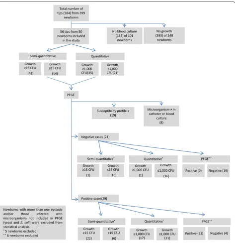
A total of 584 catheter tips from 399 newborns, hospital-ized in the neonatal ICU of HC-FMB between November 2005 and June 2012, were analyzed. Of these, 56 cath-eter tips from 50 newborns were included in the study according to the inclusion criteria. Twenty-nine of the 50 newborns were classified as having CR-BSI ac-cording to the gold standard, i.e., isolation of the same microorganism (the same species and antimicrobial susceptibility profile) from catheter and blood cultures
[image:4.595.64.538.229.718.2]and diagnosis of sepsis according to clinical criteria (Figure 1).
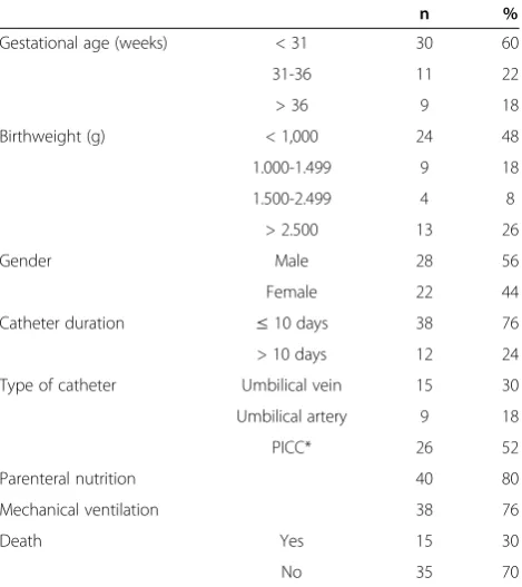
Table 1 shows the personal and clinical data of the new-borns included in the study. Most newnew-borns were pre-term, had a low birth weight, and had received parenteral nutrition, mechanical ventilation and a short-term cath-eter (up to 10 days).
Culture of catheter tips
Forty-two of the 56 catheter tips analyzed exhibited
growth≥15 CFU, indicating colonization of the catheter
according to the criteria proposed by Maki et al. [5],
and 35 exhibited growth≥1,000 CFU/mL according
to the quantitative technique of Brun-Buisson et al. [8] (Table 2).
Twenty-three (79.3%) of the 29 cases of CR-BSI tested positive by the semi-quantitative technique, with growth≥
15 CFU, and 18 (62.1%) exhibited growth≥1,000 CFU by
the quantitative technique. In four cases, growth was≥
15 CFU in semi-quantitative culture, but < 1,000 CFU in quantitative culture. There was one case in which no growth was observed in the quantitative culture. Compari-son of the semi-quantitative technique with the gold standard revealed five cases of growth < 15 CFU and one case in which no growth was observed in the semi-quantitative culture. Of these six cases, three did not grow in quantitative culture and the three other cases exhibited
growth < 1,000 CFU. Therefore, all cases of CR-BSI diag-nosed by quantitative culture were also positive by semi-quantitative culture.
CoNS were the most common microorganisms (n = 17, 73.9%) detected by the semi-quantitative technique.
S. epidermidis was isolated from 14 (61%) of the 23
cases of CR-BSI and S. haemolyticus from 3 (13%). S.
aureus was isolated from only one case of CR-BSI (4.3%). Gram-negative bacteria were isolated from 3 (13%) cases
of CR-BSI, including K. pneumoniae from 2 (8.7%) and
E. coli from one (4.3%). Yeasts were isolated from 2 (8.7%) cases (Table 3).
Semi-quantitative culture did not detect growth in one
case of CR-BSI caused by S. epidermidis and growth
below the cut-off (< 15 CFU) was observed in five cases,
four caused byS. epidermidisand one by yeast (Table 2).
No growth of microorganisms was detected by the quan-titative technique in four episodes, including three
caused by S. epidermidis and one by yeast. Growth in
quantitative culture below the cut-off (< 1,000 CFU) was
observed in six episodes caused byS. epidermidisand in
one caused by yeast (Table 3).
Only 25 of the 29 cases of CR-BSI were submitted to
PFGE since E. coli and yeasts were not analyzed by this
technique. PFGE confirmed the similarity of microorganisms isolated from catheter and blood cultures in 21 (84%) of the 25 cases analyzed. Considering the 23 cases of CR-BSI diag-nosed by the semi-quantitative technique and excluding the
two episodes caused by yeast and one episode caused byE.
colithat were not submitted to molecular typing by PFGE,
confirmation was obtained in 18 (90%) of 20 cases.
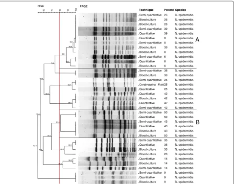
PFGE confirmed 12 of the 14 cases caused by S.
[image:5.595.57.292.112.376.2]epi-dermidis (patients 6, 8, 9, 14, 25, 26, 35, 38, 39, 42, 43, and 50) (Figure 2). Among these cases, there was a pre-dominant cluster (group A) which exhibited similarity > 80% in four cases of CR-BSI, including patients 6, 8, 26
Table 1 Personal and clinical data of the newborns included in the study (n = 50)
n %
Gestational age (weeks) < 31 30 60
31-36 11 22
> 36 9 18
Birthweight (g) < 1,000 24 48
1.000-1.499 9 18
1.500-2.499 4 8
> 2.500 13 26
Gender Male 28 56
Female 22 44
Catheter duration ≤10 days 38 76
> 10 days 12 24
Type of catheter Umbilical vein 15 30
Umbilical artery 9 18
PICC* 26 52
Parenteral nutrition 40 80
Mechanical ventilation 38 76
Death Yes 15 30
No 35 70
*Peripherally inserted central catheter.
Table 2 Diagnosis of catheter-related bloodstream infec-tion by the semi-quantitative and quantitative
techniques
Semi-quantitative technique
Quantitative technique
≥15 CFU ≤15 CFU ≥1,000 CFU ≤1,000 CFU
n % n % n % n %
Positive catheters (n = 56)
42 75.0 14 25.0 35 62.5 21 37.5
CR-BSI (n = 29) 23 79.3 6 26.0 18 54.5 11 37.9
[image:5.595.304.540.570.724.2]CR-BSI: catheter-related bloodstream infection.
Table 3 Incidence of microorganisms associated with catheter-related bloodstream infections detected by semi-quantitative and quantitative culture
Semi-quantitative culture
Quantitative culture
Microorganism ≥15 CFU < 15 CFU ≥1,000 CFU < 1,000 CFU
S. epidermidis
(n = 19)
14 5* 10 9**
S. haemolyticus
(n = 3)
3 0 3 0
S. aureus(n = 1) 1 0 1 0
Klebsiella pneumoniae(n = 2)
2 0 2 0
Yeast (n = 3) 2 1 1 2*
E. coli(n = 1) 1 0 1 0
Total (n = 29) 23 6 18 11
*
[image:5.595.56.292.640.725.2]and 39 from which the same strain was isolated in 2006, 2007, 2009 and 2010, respectively. Analysis of the den-drogram also revealed similarity > 80% between microor-ganisms isolated from patients 43 and 50 (group B) in 2011 and 2012, respectively.
The three cases of CR-BSI caused byS. haemolyticuswere also confirmed by PFGE (patients 2, 22, and 49) (Figure 3). Two catheter tips from patient 22 obtained on different days (interval of 2 days) were cultured and both showed confluent growth by the two methods. However, the same microorganism was isolated from blood culture in only one case. The microorganisms isolated from patients 2 and 22 in 2006 and 2008, respectively, showed 100% similarity.
The only confirmed case of CR-BSI caused byS.
aur-eus was also confirmed by PFGE, showing 100%
simi-larity (Figure 4).
The two cases of infection caused by K. pneumoniae
were confirmed by PFGE (100% and 90.9% similarity).
However, analysis of the dendrogram revealed two dis-tinct clones (Figure 5).
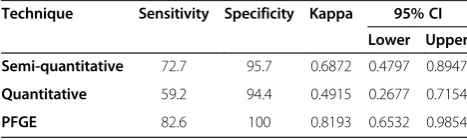
The sensitivity of the semi-quantitative and quantita-tive methods was 72.7% and 59.28%, respecquantita-tively, and specificity was 95.7% and 94.4% (see Additional file 1). The diagnosis of CR-BSIs based on the similarity of iso-lates from catheter tips and blood cultures determined by PFGE genotyping showed a sensitivity of 82.6% and specificity of 100%. The kappa coefficient of agreement between the techniques and the gold standard was 0.6872 (substantial agreement) for the semi-quantitative technique, 0.4915 (moderate agreement) for the quanti-tative technique, and 0.8193 (excellent agreement) for PFGE (Table 4).
Discussion
There are several comparative studies of semi-quantitative and quantitative culture of catheter tips for the diagnosis
[image:6.595.60.539.88.464.2]B
A
98.1 96.8 93.3 88.4 74.4 95.7 96.0 68.0 60.8 81.3 96.3 86.4 70.3 96.6 84.1 59.8 56.6 52.9 PFGE 100 90 80 70 60 PFGE . . . . . . . . . . . . . . . . . . . . . . . . . . . . . . . . . . . . . . . . . . . . . . . . . . . . . . . . . . . . . . . . . . . . . . . . . . . . . . . . . . . . . . . . . . . . . . . . . . . . . . . . . . . . . . . . . . . . . . . . . . . . . . . . . . . . . . . . . . . . . . . . . . . . . . . . . . . . . . . . . . . . . . . . . . . . . . . . . . . . . . . . . . . . . . . . . . . . . . . . . . . . . . . . . . . . . . . . . . . . . Technique Semi-quantitative Blood culture Blood culture Semi-quantitative Quantitative Quantitative Semi-quantitative Blood culture Blood culture Semi-quantitative Quantitative Blood culture Semi-quantitative Blood culture Semi-quantitative Cerebrospinal Fluid Quantitative Quantitative Blood culture Quantitative Semi-quantitative Semi-quantitative Quantitative Semi-quantitative Quantitative Blood culture Blood culture Semi-quantitative Quantitative Blood culture Blood culture Quantitative Blood culture Semi-quantitative Semi-quantitative Quantitative Blood culture Patient 26 26 26 39 39 8 8 39 8 6 6 6 38 38 25 25 25 42 42 42 42 50 50 43 43 43 50 35 35 35 26 14 14 14 9 9 9 Species S. epidermidis S. epidermidis S. epidermidis S. epidermidis S. epidermidis S. epidermidis S. epidermidis S. epidermidis S. epidermidis S. epidermidis S. epidermidis S. epidermidis S. epidermidis S. epidermidis S. epidermidis S. epidermidis S. epidermidis S. epidermidis S. epidermidis S. epidermidis S. epidermidis S. epidermidis S. epidermidis S. epidermidis S. epidermidis S. epidermidis S. epidermidis S. epidermidis S. epidermidis S. epidermidis S. epidermidis S. epidermidis S. epidermidis S. epidermidis S. epidermidis S. epidermidis S. epidermidisof CR-BSIs [16,17]. However, these studies always involved adult patients. We conducted this study in view of the lack of specific studies in the literature comparing these culture methods to improve the diagnosis of CR-BSIs in newborns.
In the present study, the semi-quantitative technique identified more cases of CR-BSI (n = 23) than the quanti-tative culture method (n = 18). Using quantiquanti-tative culture, growth below the cut-off (< 103CFU/mL) was observed in one case caused by yeast and in three cases caused by
S. epidermidis. No growth was observed in one case asso-ciated with S. epidermidis. In contrast, growth > 15 CFU of these five isolates was detected by the semi-quantitative culture method. These results may be explained by the contamination with microorganisms adhered only to the external surface of the device and by the fact that short-term catheters (up to 10 days) were used in most cases. Contamination of the external catheter surface has been reported as the main route of infection of short-term catheters, whereas intraluminal contamination is an important route of infection in long-term catheterization [18]. In addition, the density of colonizing bacteria is higher on the external than on the internal surface of the catheter [6,19].
The sensitivity of the semi-quantitative and quantita-tive techniques was 72.7% and 59.28%, respecquantita-tively, and specificity was 95.7% and 94.4%. There were six episodes
of CR-BSI that were not detected by semi-quantitative
or quantitative culture, five of them associated with S.
epidermidis and one with yeast. One limitation of the semi-quantitative culture method is that it only detects colonization of the external catheter surface and not intraluminal colonization. Furthermore, the lower sensi-tivity of semi-quantitative and, particularly of quantita-tive culture, observed in the present study may be explained by the fact that antibiotic therapy is frequently administered to preterm newborns in ICUs. This treat-ment reduces the yield of bacterial culture of both the external and internal surface of catheters. Since antibi-otics are administered through a central venous catheter, microorganisms present inside the catheter are exposed to higher concentrations of the antibiotic than bacteria found on the external surface. Therefore, previous ad-ministration of antibiotics can influence the sensitivity of detection methods, particularly quantitative culture.
In a meta-analysis comparing six culture techniques, Siegman-Igra et al. [20] reported the superiority of quan-titative culture methods of catheter segments. The authors concluded that quantitative culture of catheter segments shows greater precision in the diagnosis of CR-BSIs and is the only method with sensitivity and specificity higher than 90% and the best cost-benefit relationship [20]. However, according to Bouza et al. [17], quantitative techniques are not superior to the method of Maki et al. [5] when used
32.0
PFGE
100
80
60
40
PFGE
. . . . . . . . . . . . . . . . . . . . . . . . . . . . . . . . . . . . . . . . . . . . . . . . . . . . . . . . . . . . . . .
Technique
Semiquantitative Blood culture Quantitative Semiquantitative Quantitative Blood culture Blood culture Quantitative Semiquantitative
Patient
49 49 49 22 22 22 2 2 2
Species
S. haemolyticus S. haemolyticus
S. haemolyticus S. haemolyticus
S. haemolyticus
S. haemolyticus S. haemolyticus
[image:7.595.58.539.89.208.2]S. haemolyticus S. haemolyticus
Figure 3Dendrogram generated by Dice/UPGMA analysis (Bionumerics, Applied Maths) ofSmaIPFGE profiles ofS. haemolyticus isolated from catheters by semi-quantitative and quantitative culture and from blood cultures of confirmed cases of catheter-related bloodstream infection (similarity≥80%).
100
PFGE
.
.
.
. .
.
.
. ...
.
.
. .
.
.
. .
.
.
.
Technique
Blood culture
Semiquantitative
Quantitative
Blood culutre
Patient
16
16
16
16
Species
S. aureus
S. aureus
S. aureus
S. aureus
[image:7.595.58.539.622.704.2]under the same conditions. Therefore, the sensitivity, spe-cificity and simplicity of the semi-quantitative technique proposed by Maki et al. [5] makes it the method of choice for routine use in clinical microbiology laboratories.
According to the National Nosocomial Infections Sur-veillance (NNIS) [21], CoNS were responsible for 37.7% of bloodstream infections in pediatric ICUs between 1992 and 1999. More recent data reported by the National Healthcare Safety Network (NHSN) of the CDC also show that CoNS rank first place as the etiological agent of
CR-BSIs (34.1%), followed by Enterococcus spp. (16%),
Candidaspp. (11.8%) andS. aureus(9.9%) in fourth place. Gram-negative microorganisms including enterobacteria such as E. coli, Enterobacter, Klebsiella pneumoniae and
Klebsiella oxytocawere responsible for 12.4% of these in-fections [22]. In the present study, the only case of CR-BSI caused by E. coli resulted in the death of the patient in 2009. Although rare, CR-BSIs caused by these etiological agents are severe and are associated with a high rate of le-thality (40-90%) since non-maternal strains ofE. coli(from another patient or nosocomial) can cause severe invasive disease [23]. Considering the immature defense system of preterm newborns, the impact of infections caused by these microorganisms is even greater.
In addition to their virulence and ability to acquire
antibiotic resistance, Klebsiella and E. coli are able to
survive on the skin and moist surfaces, withstanding drying, and are easily transferred through equipment and the hands of the medical team [24]. Neonatal ICUs are the ideal place for the dissemination of these
microorganisms as a result of extended hospital stay and prolonged use of antimicrobial agents, as well as the use of invasive devices [25].
The higher frequency of S. epidermidis in CR-BSIs is
expected since it is the predominant species in the microbiota of newborns. This predominant colonization and the higher pathogenicity of some strains may
ex-plain the fact that S. epidermidis is the most common
species associated with infectious processes in newborns as demonstrated by Cunha et al. [26,27]. The presence of clusters of S. epidermidis, with the isolation of strains from different newborns over a period of 4 years (2006
to 2010), and ofS. haemolyticus(2006 to 2008) indicates
the existence of prevalent clones of these CoNS species in neonatal ICUs. These microorganisms can colonize catheters and invade the bloodstream, causing systemic infection in this group of patients submitted to invasive procedures since most of them are preterm and low-birth weight newborns whose immature and deficient immune system contributes to infection. The presence of S. haemolyticus clones highlights the importance for control measures of CR-BSIs since this species has been described to be the most resistant to different antimicro-bial agents and is an important carrier of resistance genes that can be transferred to other CoNS species causing CR-BSIs [28,29].
Conclusion
The semi-quantitative culture technique showed higher sensitivity and specificity in the diagnosis of CR-BSIs in newborns when compared to quantitative culture. In addition, this method is simpler and shows better agree-ment with the gold standard and should therefore be rec-ommended for routine use in clinical laboratories. PFGE permits to confirm whether the strain isolated from the catheter and blood of the patient is the same and may contribute to the control of CR-BSIs by identifying clus-ters of microorganisms in neonatal ICUs, providing a means of determining potential cross-infection between patients.
90.9
55.6
PFGE XbaI
100
80
60
PFGE XbaI
. . . . . . . . . . . . . . . . . . . . . . . . . . . . . .
Technique
Semiquantitative Quantitative Blood culture Quantitative Semiquantitative Blood culture
Patient
46 46 46 36 36 36
Species
Klebsiella pneumoniae
Klebsiella pneumoniae
Klebsiella pneumoniae
Klebsiella pneumoniae
Klebsiella pneumoniae
[image:8.595.58.537.90.180.2]Klebsiella pneumoniae
Figure 5Dendrogram generated by Dice/UPGMA analysis (Bionumerics, Applied Maths) ofXbaIPFGE profiles ofK. pneumoniae isolated from catheters by semi-quantitative and quantitative culture and from blood cultures of newborns included in the study (similarity≥80%).
Table 4 Sensitivity and specificity of the techniques analyzed and kappa coefficient
Technique Sensitivity Specificity Kappa 95% CI
Lower Upper
Semi-quantitative 72.7 95.7 0.6872 0.4797 0.8947
Quantitative 59.2 94.4 0.4915 0.2677 0.7154
[image:8.595.56.290.664.734.2]Additional file
Additional file 1: Table S1.Determination of the sensitivity and specificity of the semi-quantitative technique.Table S2.Determination of the sensitivity and specificity of the quantitative technique.Table S3.
Determination of the sensitivity and specificity of PFGE.
Competing interests
The authors declare that they have no competing interests.
Authors’contributions
DFMR: participated in the conception and design of the study, carried out the microbiological tests, and wrote the paper. EPS: participated in the conception and design of the study, carried out the microbiological tests, analyzed the clinical data, and participated in the writing of the paper. LLV: helped with the microbiological tests. JCL (physician of the neonatal ICU): provided the clinical material and helped with the collection of clinical data. MRB (physician of the neonatal ICU): provided the clinical material and helped with the collection of clinical data. JEC (statistics consultant of the Botucatu Medical School): performed the statistical analysis of the results. LMSSR (head of the neonatal ICU): coordinated the collection of clinical material and analysis of clinical data. MLRSC: responsible for the conception and design of the study, coordination of laboratory work, data analysis, and manuscript writing. All authors read and approved the final manuscript.
Acknowledgements
Fundação de Amparo a Pesquisa do Estado de São Paulo (FAPESP) (grants 2010/14581-7 and 2014/07631-9).
Author details 1
Departamento de Microbiologia e Imunologia, Instituto de Biociências, UNESP - Univ Estadual Paulista, Botucatu, SP, Brasil.2Departamento de
Pediatria, Faculdade de Medicina, UNESP - Univ Estadual Paulista, Botucatu, SP, Brasil.3Departamento de Bioestatística, Instituto de Biociências,
UNESP - Univ Estadual Paulista, Botucatu, SP, Brasil.
Received: 31 July 2013 Accepted: 14 April 2014 Published: 22 May 2014
References
1. McGee DC, Gould MK:Preventing complications of central venous catheterization.N Engl J Med2003,348:1123–1133.
2. Rosenthal VD, Gusman S, Migone O, Crnich CJ:The attributable cost, length of hospital stay, and mortality of central line-associated bloodstream infection in intensive care departments in Argentina: a prospective, matched analysis.Am J Infect Control2003,31:475–8080. 3. Pessoa-Silva CL, Richtmann R, Calil R, Santos RM, Costa MLM, Frota ACC,
Wey SB:Healthcare-associated infections among neonates in neonatal units in Brazil.Infect Control Hosp Epidemiol2004,25:772–777.
4. Safdar N, Maki DG:Inflammation at the insertion site is not predictive of catheter-related bloodstream infection with short-term, non cuffed central venous catheters.Crit Care Med2002,30:2632–2635.
5. Maki DG, Weise CE, Sarafin HW:A semiquantitative culture method for identifying intravenous-catheter-related infection.New Engl J Med1977,
296:1305–1309.
6. O’Grady NP, Alexander M, Dellinger EP, Gerberding JL, Heard SO, Maki DG, Masur H, McCormick RD, Mermel LA, Pearson ML, Raad II, Randolph A, Weinstein RA:CDC Guidelines for the prevention of intravascular catheter-related infections.MMWR Recomm Rep2011,51:1–29. 7. Cleri D, Corrado M, Seligman S:Quantitative culture of intravenous
catheters and other intravascular inserts.J Infect Dis1980,
141(Suppl 6):781–786.
8. Brun-Buisson C, Abrouk F, Legrand P, Huet Y, Larabi S, Rapin M:Diagnosis of central venous catheter-related sepsis: critical level of quantitative tip cultures.Arch Intern Med1987,147(Suppl 5):873–877.
9. Tenover FC, Arbeit RV, Goering PA, Mickelsen BE, Murray BE, Persing DH, Swaminathan B:Interpreting chromosomal DNA restriction patterns produced by pulsed-field gel electrophoresis: criteria for bacterial strain typing.J Clin Microbial1995,33:2233–2239.
10. Associação Paulista de Estudos e Controle de Infecção Hospitalar:
Diagnóstico e Prevenção de IRAS em Neonatologia.2011.
11. Koneman EW, Allen SD, Janda WM, Schreckenberger PC, Winn WC Jr:Color
atlas and textbook of diagnostic microbiology.Philadelphia: JB Lippincott; 1997.
12. Cunha MLRS, Sinzato YK, Silveira LVA:Comparison of methods for the identification of coagulase-negativeStaphylococcus.Mem Inst Oswaldo Cruz2004,99:855–860.
13. CLSI:Performance standards for antimicrobial susceptibility testing. CLSI approved standard M100-S21, Volume 31. Wayne, PA: Clinical and Laboratory Standards Institute; 2011.
14. McDougal LK, Steward CD, Killgore GE, Chaitram JM, McAllister SK, Tenover FC:Pulsed-field gel electrophoresis typing of oxacillin-resistant Staphylococcus aureusisolates from the United States: establishing a national database.J Clin Microbiol2003,41:5113–5120.
15. Durmaz R, Otlu B, Koksal F:The optimization of a rapid pulsed-field gel electrophoresis protocol for the typing ofAcinetobacter baumannii,Escherichia coliandKlebsiellaspp.Jpn J Infect Dis2009,
62:372–377.
16. Storti A, Manzato AJ, Pizzolitto AC, Pizzolitto EL:Assessment of central venous catheter-associated infections using semi-quantitative or quantitative culture methods.Revista de Ciências Farmacêuticas Básica e
Aplicada2009,27(Suppl 3):213–220.
17. Bouza E, Alvarado N, Alcala L, Sánchez-Conde M, Pérez MJ, Muñoz P, Martín-Rabadán P, Rodríguez-Créixems M:A prospective, randomized, and comparative study of 3 different methods for the diagnosis of intravascular catheter colonization.Clin Infect Dis2005,40:1096–1100. 18. Mermel LA:What is the predominant source of intravascular catheter
infections?Clin Infect Dis2011,52:211–212.
19. Koh DBC, Robertson IK, Watts M, Davies AN:Density of microbial colonization on external and internal surfaces of concurrently placed intravascular devices.Am J Crit Care May2012,21:162–171.
20. Siegman-Igra Y, Anglim AM, Shapiro DE, Adal KA, Strain BA, Farr BM:
Diagnosis of vascular catheter-related bloodstream infection: a meta analysis.J Clin Microbiol1997,35(Suppl 4):928–936.
21. National Nosocomial Infections Surveillance (NNIS):System report, data summary from January 1992 through June 2004, issued October 2004.
Am J Infect Control2004,32:470–485.
22. Hidron AI, Edwards JR, Patel J, Horan TC, Sievert DM, Pollock DA, Fridkin SK, NHSN:Annual update: antimicrobial-resistant pathogens associated with healthcare-associated infections: annual summary of data reported to the national healthcare safety network at the centers for disease control and prevention, 2006–2007.Infect Control Hosp Epidemiol2008,29(Suppl 11):996–1011. 23. Dekema DJ, Barr J, Boyken LD, Buschelman BJ, Jones RN, Pfaller MA,
Herwaldt LA:A cluster of seriousEscherichia Coliinfections in a neonatal intensive-care unit.Infect Control Hosp Epidemiol1997,18:774–776. 24. Jain A, Roy I, Gupta MK, Kumar M, Agarwal SK:Prevalence of extended
spectrumβlactamase producing Gram-negative bacteria in septicemic neo-nates in a tertiary care hospital.J Med Microbiol2003,52:421–425. 25. Trecarichi EM, Tumbarello M, Spanu T, Caira M, Fianchi L, Chiusolo P,
Fadda G, Leone G, Cauda R, Pagano L:Incidence and clinical impact of extended-spectrum-β-lactamase (ESBL) production and fluoroquinolone resistance in bloodstream infections caused byEscherichia coliin patients with hematological malignancies.J Infect2009,58:299–307. 26. Cunha MLRS, Lopes CAM, Rugolo LMSS, Chalita LVAS:Significância clínica
de estafilococos coagulase-negativa isolados de recém-nascidos.J Pediatr 2002,78:279–288.
27. Martins A, Cunha MLRS:Methicillin resistance inStaphylococcus aureus and coagulase-negative staphylococci: epidemiological and molecular aspects.Microbiol Immunol2007,51:787–795.
28. Bouchami O, Hassen AB, Lencastre H, Miragaia M:High prevalence ofmec complex C andccrCis independent of SCCmectype V inStaphylococcus haemolyticus.Eur J Clin Microbiol Infect Dis2012,31:605–614.
29. Barrosa EM, Ceottob H, Bastosb MCF, Santos KRN, Giambiagi-deMarval M:
Staphylococcus haemolyticusas an important hospital pathogen and carrier of methicillin resistance genes.J Clin Microbiol2012,50:166–168.
doi:10.1186/1471-2334-14-283
Cite this article as:Riboliet al.:Diagnostic accuracy of