University of South Carolina
Scholar Commons
Theses and Dissertations
2016
The Identification Of The Direct And Indirect
Pathways Through Which Leptin Facilitates
Synaptic Plasticity In The Hippocampus
Catherine Van DoornUniversity of South Carolina
Follow this and additional works at:https://scholarcommons.sc.edu/etd
Part of theBiomedical and Dental Materials Commons
This Open Access Thesis is brought to you by Scholar Commons. It has been accepted for inclusion in Theses and Dissertations by an authorized administrator of Scholar Commons. For more information, please [email protected].
Recommended Citation
THE IDENTIFICATION OF THE DIRECT AND INDIRECT PATHWAYS THROUGH
WHICH LEPTIN FACILITATES SYNAPTIC PLASTICITY IN THE HIPPOCAMPUS
by
Catherine Van Doorn
Bachelor of Science
University of South Carolina, 2009
Submitted in Partial Fulfillment of the Requirements
For the Degree of Master of Science in
Biomedical Science
School of Medicine
University of South Carolina
2016
Accepted by:
Lawrence Reagan, Director of Thesis
Claudia Grillo, Reader
Jim Fadel, Reader
Angela Murphy, Reader
ACKNOWLEDGEMENTS
I would like to thank my mentor Dr. Larry Reagan for all of your advice,
guidance and patience. Without you, this undertaking would not only have been highly
improbable, but impossible. I would also like to thank my committee members for
generously donating your time for meetings and for reading this thesis. And to my
family- Mh, Ddkns, Pilly & Paprika- you have been with me since the beginning- thank
ABSTRACT
Leptin, a peptide synthesized by adipocytes in the periphery, has been shown to
play significant roles in feeding and energy expenditure mediated by the hypothalamus.
Growing evidence supports the role of leptin in influencing synaptic plasticity in the
hippocampus in that leptin dose-dependently enhances LTP, alters morphology and
neurogenesis, facilitates spatial learning and memory, as well as memory retention.
Models of leptin deficiency and resistance have further supported the importance of
leptin in synaptic plasticity by exhibiting deficits in electrophysiological, morphological
and behavioral tests that are improved after leptin restoration. The effects of leptin when
applied directly into the hippocampus have clear effects on synaptic plasticity; however,
indirect pathways may also be activated by leptin that result in synaptic plasticity changes
in the hippocampus. Due to the nature of the release of leptin from the peripheral
adipocytes, the interconnectivity of the brain, and the localization of leptin receptors
throughout many brain regions, support the hypothesis that leptin could mediate synaptic
plasticity changes in the hippocampus through multi-synaptic circuitry in addition to its
direct effects. A variety of brain regions that express leptin receptors project both directly
and indirectly to the hippocampus, including many nuclei of the hypothalamus, raphe
nucleus, locus coeruleus and ventral tegmental area and therefore pose as potential
starting points for brain-wide networks that leptin could influence for the regulation of
TABLE OF CONTENTS
ACKNOWLEDGEMENTS ... iii
ABSTRACT ... iv
CHAPTER 1 DIRECT EFFECTS OF LEPTIN IN THE HIPPOCAMPUS ...1
1.1SYNAPTIC PLASTICITY: AN INTRODUCTION ...1
1.2 LEPTIN, LEPTIN RECEPTORS & SIGNALING ...2
1.3THE HIPPOCAMPUS ...6
CHAPTER 2 LEPTIN & SYNAPTIC PLASTICITY IN THE HIPPOCAMPUS ...8
2.1 LEPTIN & LTP ...8
2.2 LEPTIN & MORPHOLOGY ...18
2.3 LEPTIN & BEHAVIOR ...27
2.4 ANIMAL MODELS OF LEPTIN DYSFUNCTION ...31
2.5 ANIMAL MODELS AND SYNAPTIC PLASTICITY DEFICITS ...32
2.6 LEPTIN IN DEPRESSION, ANXIETY & STRESS ...36
2.7 LEPTIN & ALZHEIMER’S DISEASE ...43
CHAPTER 3CIRCUITRY: AN INTRODUCTION TO NEURONAL ACTIVATION ...50
3.1NEURONAL ACTIVATION ...51
3.2 EXCITATION & INHIBITION BY LEPTIN ...54
3.3 LEPTIN & OREXIN: APPETITIVE PEPTIDES WITH INDIRECT CIRCUITRY ...58
4.2LEPTIN RECEPTORS IN THE LOCUS COERULEUS ...67
4.3 LEPTIN RECEPTORS IN THE RAPHE NUCLEUS ...68
4.4 LEPTIN RECEPTORS IN THE LATERAL HYPOTHALAMUS ...71
4.5 LEPTIN RECEPTORS IN THE PARAVENTRICULAR HYPOTHALAMUS ...74
4.6 LEPTIN RECEPTORS IN THE DORSOMEDIAL HYPOTHALAMUS ...78
4.7 LEPTIN RECEPTORS IN THE VENTROMEDIAL HYPOTHALAMUS ...80
CHAPTER 5 CONCLUSIONS & FUTURE DIRECTIONS ...85
CHAPTER 1
DIRECT EFFECTS OF LEPTIN IN THE HIPPOCAMPUS
1.1 SYNAPTIC PLASTICITY: AN INTRODUCTION
Synaptic plasticity may be defined as a modification in synaptic strength after a
repeated stimulus. Changes in synaptic plasticity form the basis for how the brain is able
to adapt itself in response to stimuli and is part of the proposed mechanism for learning
and memory. The phenomenon of synaptic plasticity is important because it results in a
change in efficacy in how neurons respond to a stimulus and therefore the response to the
stimulus changes. Synaptic plasticity is thought to underlie certain types of learning and
memory and this is demonstrated in the hippocampus, a well-studied structure shown to
be integral in particular spatial learning and memory.
Synaptic plasticity has many aspects that can be observed and regulated, both
positively and negatively. Factors such as stress (Gould et al, 1997; Schoenfeld and
Gould, 2012; Yap et al, 2006) and aging (Kuhn et al, 1996; Seki and Arai, 1995) can
detrimentally affect learning and memory. Conversely, estrogen (Gould, 1999), the
neurotransmitter serotonin (Brezun and Daszuta, 1999), an enriched environment
(Nilsson et al, 1999) and exercise (Praag et al, 1999) can positively modulate synaptic
plasticity. The 167 amino acid peptide leptin has also been shown to influence synaptic
plasticity (Maffei et al, 1995).
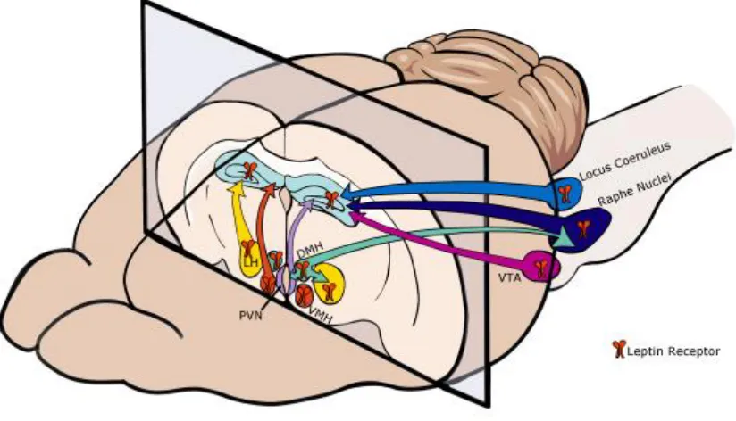
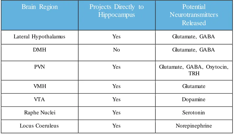
However, synaptic plasticity also be indirectly affected, through receptors in different
brain areas projecting to others in such a way that initiation at one area eventually leads
to the hippocampus to alter synaptic activity. This multi-synaptic circuitry must operate
through a series of neurons connected by synapses that are inhibited or excited by
neurotransmitters. Leptin receptors are expressed in regions that have projections to the
hippocampus and in this manner leptin may be uniquely positioned to directly and
indirectly affect synaptic plasticity in the hippocampus.
1.2 LEPTIN, LEPTIN RECEPTORS & SIGNALING
Leptin, the protein product of the obesity (ob) gene, is predominantly synthesized by adipocytes, proportional to the amount of adipocytes in the body (Frederich et al,
1995). Although leptin is primarily synthesized in the periphery, there is some evidence
that it can be synthesized in the brain as there is evidence of the presence of leptin mRNA
and protein in some brain regions, including the cerebral cortex and hypothalamus
(Elmquist et al, 1998). Leptin mRNA transcription in the hypothalamus has been shown
to be blunted when fasting occurs and leptin levels have also been shown to fall during
starvation (Morash et al, 1999; Frederich et al, 1995).
Once leptin is released into circulation, for example after eating, leptin can act in
the periphery to play roles in immunity, (Lord et al, 1998; Oomura et al, 2006), inhibit
insulin secretion, (Bjorbaek and Kahn, 2004), control sex hormone release (Yu et al,
1997; Ahima et al, 1996) and lipolysis in adipocytes (Siegrist-Kaiser et al, 1997; Wang et
al, 1999). Leptin receptors in the body have been found in skeletal muscle, heart, lungs,
al, 1996; Fei et al, 1997; Ghilardi and Skoda, 1997; Hoggard et al 1997; Bjorbaek and
Kahn, 2004). Peripheral leptin receptors are expressed as the short form of the receptor,
ObRa, and while the exact functions of peripheral ObRa are still not known, there is
evidence of signaling in pancreatic β cells, pituitary cells, hepatocytes, muscle cells and
adipocytes (Hileman et al, 2002; Bjorbaek and Kahn, 2004). Leptin has been shown to
activate PI3K pathways for signaling in insulinoma cells (Harvey et al, 2000),
hepatocytes (Zhao et al, 2000), and muscle cells (Berti et al, 1997).
Leptin can also cross the blood-brain barrier (BBB) through a saturable transport
system to then act in the central nervous system (CNS) (Banks et al, 1996; Hileman et al,
2002; Banks et al, 2006). The role of leptin in the CNS has been described as being
regulatory, where leptin acts as a retrograde modulator (Harvey et al, 2007; Ludwig and
Pittman, 2003). In the CNS, leptin activates the long isoform of the leptin receptor, the
ObRb, a class 1 cytokine receptor encoded by the diabetes (db) gene (Tartaglia et al, 1995). There are six leptin receptor isoforms; ObRa, ObRb, ObRc, ObRd, ObRe and
ObRf but only the long isoform, ObRb, is capable of complete JAK/STAT signal
transduction (Baumann et al, 1996; Ghilardi et al, 1996; Bahrenberg et al, 2002; Tartaglia
et al, 1995).
The ObRb is often paired with the JAK/STAT specific subtypes, JAK2/STAT3,
JAK2/STAT5, and JAK2/STAT6 (Ghilardi et al, 1996) although other pathways can be
used including the MAPK, and PI3K pathways (Bjorbaek et al, 2004; Niswender et al,
2001; Zhao et al, 2002; Rahmouni et al, 2003; Cota et al, 2006, 2008).
In JAK/STAT signaling, leptin binds to its receptor and initiates a conformational
stimulates phosphorylation of tyrosine residues on the ObRb receptor, which then recruits
Src-homology2 and Src-homology 3 that contain within them signal transducer and
activator of transcription 5 (STAT5) and STAT3. STAT5 and STAT3 are then in turn
phosphorylated by JAK2 and STAT5 and STAT3 can now separate off of the ObRb and
dimerize, moving into the nuclear envelope for gene transcription regulation (Marwarha
and Ghribi, 2012). Phosphorylated tyrosine residues can recruit STAT family
transcription factors, phosphoinositide 3-kinase and proteins in the MAPK/Ras Raf
signaling pathways (Tartaglia et al, 1995; Harvey, 2003).
The MAPK pathway also involves phosphorylation of tyrosine residues and
downstream processes similar to JAK/STAT signaling and can similarly regulate gene
expression in the CNS (Marwarha and Ghribi, 2012). The PI3K pathway is also activated
by leptin signaling with the ObRb and utilizes the phosphorylation of Akt at serine
residues in a similar process to that of JAK/STAT signaling (Vecchoine et al, 2002).
Studies have also indicated that leptin can activate the AMPK-SIRT1 pathway
(Yamagishi et al, 2001; Minokoshi et al, 2002; Suzuki et al, 2007).
JAK/STAT pathway has been shown to be critical in the actions of leptin in body
weight regulation (Bates et al, 2003). PI3K signaling is proposed to be involved in
leptin-stimulated reductions in food intake as well as activation of sympathetic nervous system
activity (Zhao et al, 2002b; Rahmouni et al, 2003).
Leptin receptor mRNA has been found in many different brain regions including
the pituitary, arcuate nucleus, dorsomedial hypothalamus (DMH), ventromedial nucleus
(VMH), ventral premammillary nucleus, lateral hypothalamus, thalamic nuclei,
1999). Using in situ hybridization, it was found that specific areas of the hypothalamus show high levels of leptin receptor expression, including the arcuate nucleus, DMH and
VMH (Schwartz et al, 1996). Additionally, specific areas of the hippocampus also have
high levels of immunoreactivity of the leptin receptor, including areas CA1, CA2, CA3
and the dentate gyrus in particular on neurons and glial cells (Ur et al, 2002; Mercer et al,
1996; Hakansson et al, 1998; Huang et al, 1996; Shanley et al, 2002). Hippocampal cell
culture studies have allowed for the localization of leptin receptor expression and have
revealed that leptin receptors are expressed on the dendrites and axons of neurons, in
close proximity to NMDA receptors (Shanley et al, 2002b; O’Malley et al, 2007).
In contrast to the ObRb’s role in signaling, the ObRa short-form leptin receptor
may be involved in receptor-mediated transport of leptin across the BBB (Hileman et al,
2002). The ObRa mRNA is found in high levels in the choroid plexus and brain
microvessels of the BBB (Tartaglia et al, 1995; Bjorbaek et al, 1998). In support of this
hypothesis, knock out mice for short form leptin receptors resulted in decreased leptin
transport across the BBB (Bjorbaek et al, 1998; Hileman et al, 2002). These results
suggest that the ObRb is the main signaling receptor isoform in the brain while the ObRa
is used for transport across the BBB.
1.2.1 LEPTIN IN THE HYPOTHALAMUS: FUNCTIONS IN FEEDING & ENERGY
EXPENDITURE
There are many well-studied functions that leptin regulates through activation of
leptin receptors expressed in the hypothalamus, including regulation of metabolism,
in amounts associated with how much food is being consumed and the amount of stored
triglycerides determine how much leptin mRNA and plasma leptin protein is produced
(Kulcsar et al, 2005). As such, leptin serves as an adiposity indicator, secreted in
proportion to the amount of fat stored and higher levels of leptin decrease food intake and
increase energy expenditure (Schwartz et al, 2000). Direct injection of leptin in animals
lacking leptin or proper leptin receptor functioning into the hypothalamus resulted in a
significant decrease in feeding as well as fat and weight loss (Zhang et al, 1994; Morton
et al, 2006).
Leptin suppresses food-seeking behaviors by inhibiting neuropeptide Y
(NPY)/AgRP and orexin neurons and activating POMC/CART neurons in the arcuate
nucleus (Huang et al, 1996; Cowley et al, 2001). These populations of neurons project to
the lateral hypothalamus for well-studied control of food intake (Elias et al, 1999; Mercer
et al, 1996a; Cheung et al, 1997). These roles of leptin in the regulation of feeding and
energy metabolism mediated through the lateral and arcuate nuclei of hypothalamus lend
understanding to the functional activity of leptin outside of the hypothalamus in brain
regions such as the hippocampus.
1.3 THE HIPPOCAMPUS
As noted above, the ObRb mRNA and protein are expressed in many brain
regions including the hippocampus (Hakansson et al, 1998; Elmquist et al, 1998; Haung
et al, 1996). The hippocampus, part of the limbic system involved in learning and
memory, is comprised of layers of pyramidal cells which are then divided into different
that information flows in one direction and follows a series of pathways. Information
enters the hippocampus from the entorhinal cortex through the perforant path to the
dentate gyrus. Axons project from the dentate gyrus, projections called the Mossy Fiber
Pathway, to area CA3 of the hippocampus, also pyramidal neurons. Projections from area
CA3, the Schaffer Collaterals/Commissural Pathway, project to area CA1 which then
projects to the subiculum and entorhinal cortex and exit the hippocampus. Leptin
receptors are found in areas CA1, CA3 and the dentate gyrus, areas where synaptic
CHAPTER 2
LEPTIN & SYNAPTIC PLASTICITY IN THE HIPPOCAMPUS
2.1 LEPTIN & LTP
Long-term potentiation (LTP) and long-term depression (LTD) are
activity-dependent forms of hippocampal synaptic plasticity and are considered as being cellular
correlates for learning and memory, or the most fundamental units of measuring learning
and memory. LTP is a greater response and LTD is a lesser response of a neuron that
occurs after high-frequency stimulation, as assessed using electrophysiological
techniques (Bliss and Lomo, 1973; Bliss and Gardner-Medwin, 1973). Electrophysiology
is a technique used to measure electrical activity in neurons or cells. A glass micropipette
filled is inserted into a neuron or cell and another electrode is inserted into the
surrounding fluid around the cell for reference. Voltage can be clamped to hold the
membrane potential so that current can be measured. Current can be clamped in a similar
fashion so that current is applied and membrane potential can be measured. Using
different electrophysiology clamping and recording techniques, it is possible to determine
when channels are opened by a stimulus or when neurons are activated.
LTP is important in learning and memory, as evidenced by when LTP induction is
blocked, impairments in memory are seen (Grant and Silva, 1994). Mutations in a gene
encoding a particular tyrosine kinase involved in LTP resulted in impairments in LTP in
area CA1 as well as spatial learning deficits, demonstrating the importance of LTP in
LTP occurs at all excitatory synapses in the hippocampus and other areas of the
brain (Doyere et al, 1992). LTP has been shown to last for hours in hippocampal slices
and days in animals that have had LTP induced (Bliss and Collingridge, 1993). The
enhanced response seen by a neuron after high-frequency stimulation occurs is proposed
as being a combination of several events, which may include increased neurotransmitter
release by the presynaptic neuron, increased number of receptors localized to the
activation of the synapse and less uptake of neurotransmitter by nearby glial cells (Bliss
and Collingridge, 1993).
LTP can be N-methyl-D-aspartate (NMDA) receptor-dependent in the Perforant pathway and Schaffer collaterals in the hippocampus and also NMDA
receptor-independent in the Mossy Fiber pathways (Bliss and Collingridge, 1993). The NMDA
receptor is a type of glutamate receptor and is involved in synaptic plasticity in the
hippocampus (Lisman et al, 2012). NMDA antagonists block LTP induction,
demonstrating the necessity of NMDA receptors in initializing LTP (Bliss and
Collingridge, 1993).
NMDA receptor activation is critical for LTP to occur, the more the NMDA
receptors involved in propagating signal, the more likely that LTP will initiate in the
perforant pathway and Schaffer collaterals, those pathways whose LTP is NMDA
receptor-dependent (Malenka, 1994; Collingridge et al, 1983). NMDA-dependent LTP is
made possible due to several unique properties of the receptor itself. NMDA receptors
contain a magnesium plug that blocks the channel until the membrane potential is
sufficiently depolarized and thus the NMDA receptor is referred to as being
NMDA receptor must occur and so the NMDA receptor is a ‘coincidence detector’ due to
the several conditions that must be met at once for the channel to open (Ascher and
Nowak, 1988). Once the channel is open, calcium flows through and increases
intracellular calcium, beginning a cascade of signaling events that lead to LTP. As
intracellular calcium levels increase, calcium binds to calmodulin and this complex
activates calcium/calmodulin-dependent protein kinase II (CaMKII), which has been
identified as an important component of NMDA-mediated LTP (Lisman et al, 2012).
CaMKII is important in the induction of LTP and knockdown of an isoform of CaMKII
has been shown to impair hippocampal slice LTP (Silva et al, 1992a; Malinow et al,
1989). Leptin can influence many aspects of LTP and LTD mediated through ObRb
receptors in the CA1, CA3 and the dentate gyrus regions of the hippocampus (Mercer et
al, 1996; Oomura et al, 2006; Shanley et al, 2001; Shanley et al, 2002; Xu et al, 2008).
Leptin application shifts short-term potentiation (STP) to LTP (Shanley et al,
2001). STP is similar to LTP in that it is an increased response to tetanus stimulation,
however it persists for only seconds to minutes. Hippocampal slices were used to show
STP conversion after stimulation of the Schaffer collateral commissural pathway
(Shanley et al, 2001; Harvey et al, 2007). STP was evoked in hippocampal slices that
lasted about 30 minutes (Shanley et al, 2001). If the slice was incubated with leptin
immediately preceding the primed burst stimulation to evoke STP, then STP was
converted into LTP that lasted for an hour (Shanley et al, 2001).
Leptin enhances and suppresses NMDA receptor-dependent LTP in a
dose-dependent manner (Oomura et al, 2006; Wayner et al, 2004). Electrophysiology studies at
enhancement of LTP by 163% compared to controls (Oomura et al, 2006). Leptin
application dose-dependently enhanced LTP with the lowest and highest doses resulting
in reducing LTP, resulting in an inverted U dose response (Oomura et al, 2006). Leptin
also enhances LTP in other areas of the hippocampus, including the dentate gyrus
(Wayner et al, 2004). Hippocampal slice studies show similar results, LTP was induced
by bath administration of leptin (Li et al, 2002; Shanley et al, 2001; Wayner et al, 2004).
Interestingly, age can affect the ability of leptin to increase excitatory transmission. In
this regard, it was shown that in hippocampus slices from juvenile rodents that leptin
decreases excitatory transmission, which is contrary to what occurs in adults (Moult et al,
2010; Moult and Harvey, 2011). This has been proposed to being related to the process of
brain development (Irving and Harvey, 2014).
In addition to changes in LTP, changes in excitatory postsynaptic potentials
(EPSCs) were found with leptin application. Leptin receptor signaling resulted in
increases in NMDA receptor excitatory postsynaptic currents (EPSCs) in area CA1 but
decreases AMPA receptor EPSCs (Shanley et al, 2001). In further support,
electrophysiological studies for field potentials in CA1 pyramidal neurons revealed
significant increases in mEPSC amplitude through NMDA receptors dependent on the
dose of leptin (Oomura et al, 2006). It has been shown that leptin receptors are required
for this effect by using Xenopus oocytes with variable NMDA receptor composition and the ObRb.
Leptin enhanced NMDA EPSCs with the NR1/NR2A subunits of the NMDA
receptor with the ObRb present (Shanley et al, 2001). When the ObRb was eliminated,
applied (Shanley et al, 2001). It has been proposed that leptin may traffic NMDA
receptors to the membrane to affect the increased current through NMDA receptors, just
as insulin has been shown to do (Shanley et al, 2001; Harvey et al, 2007; Skeberdis et al,
2001). These studies collectively show that leptin and the leptin receptor directly effect
LTP and EPSCs through several mechanisms in the hippocampus.
Leptin enhances calcium influx through NMDA receptors and also increases the
activity of CaMKII (Shanley et al, 2001; Oomura et al, 2006). Calcium influx into
postsynaptic neurons is vital in the induction step of LTP and leptin is able to increase
calcium flux into hippocampal CA1 neurons via NMDA receptors (Bliss and
Collingridge, 1983; Oomura et al, 2006). Calcium concentration was measured in single
neurons from area CA1 of the hippocampus in response to leptin. Leptin application
dose-dependently resulted in increases in calcium in the same inverted U dose response
as seen with LTP (Oomura et al, 2006).
It was also determined that leptin increases calcium flow through NMDA
receptors as part of the induction phase of LTP (Shanley et al, 2001). Hippocampal cell
cultures showed that leptin quickly enhanced inward flow of calcium through NMDA
receptors in postsynaptic neurons after NMDA was applied (Shanley et al, 2001). To
further support the ability of leptin to increase calcium mediated by NMDA receptors, it
was shown that without the NMDA receptor agonist NMDA, leptin did not enhance
calcium levels (Shanley et al, 2001). Additionally, leptin had no effect on calcium
increase facilitated by AMPA receptors (Shanley et al, 2001). To determine which
signaling pathways were involved, PI3K signaling was blocked with LY 294002 and
calcium influx into neurons. Similar experiments blocking MAPK signaling with PD
98059 and U0126 resulted in a reduction of the ability of leptin to enhance NMDA
currents as well as calcium influx (Shanley et al, 2001). This supports the idea that leptin
increases calcium through MAPK and PI3K-dependent mechanisms.
CaMKII is an enzyme involved in signal transduction in LTP and has been shown
to be involved in LTP maintenance as well as play critical roles in learning and memory
(Fukunaga et al, 1993; Silva et al, 1992; Oomura et al, 2006). CaMKII activity was
assessed using an assay and results showed that leptin at the same dose that increased
calcium concentration increases CaMKII activity, whereas this effect on CaMKII was not
seen at other doses of leptin (Oomura et al, 2006). These data collectively show that
leptin increases calcium influx through NMDA receptors in the postsynaptic neuron and
increases CaMKII activity dose-dependently (Oomura et al, 2006).
Leptin can also reverse LTP, a process called depotentiation, at area CA1.
Depotentiation is important in preventing potentiated synapses from becoming saturated
and to aid neurons in storing information (Harvey, 2013; Moult et al, 2009). The ability
of leptin to depotentiate synapses is dependent on the dose administered and time course
(Moult et al, 2009). Rapid depotentiation was seen with the highest doses of leptin
compared to lowest doses of leptin. The time after LTP induction is also important, 30
minutes after LTP was induced, leptin was given and LTP was completely reversed.
However, leptin administrated after 50 minutes did not change LTP (Moult et al, 2009).
This reversal is NMDA-receptor dependent and involves rectification of currents through
AMPA receptors which has been proposed as being a result of an alteration of subunits of
Leptin can also induce NMDA receptor-dependent LTD when excitability is
enhanced (Durakoglugil et al, 2005). LTD is the weakening of synapses after repeated
stimulus and has been identified as an important aspect of some forms of learning and
memory processing (Bear and Abraham, 1996). Part of the mechanism of the lesser
response with LTD has been proposed to involve withdrawing AMPA receptors from the
membrane and the reduction of dendritic spines (Nagerl et al, 2004). GABA A receptors
normally inhibit the activation of NMDA receptors due to its ability to increase the
intensity of the magnesium blocking of the NMDA receptor. In hippocampal slices with
magnesium removed or with picrotoxin, a GABA A receptor antagonist, leptin
application resulted in LTD. In the same study, it was shown that NMDA receptors are
required for the LTD to occur (Durakoglugil et al, 2005). Additionally, wortmannin and
LY294002, blockers of PI3K signaling, further increased the depressive response induced
by leptin. It can then be inferred that PI3K signaling is involved in inhibiting
leptin-induced LTD (Durakoglugil et al, 2005).
Leptin binding to its receptors can activate three different signaling pathways
linked to the ObRb: JAK/STAT, PI3K and MAPK (Ghilardi et al, 1996; Banks et al,
2000; Bjorbaek et al, 2000; Niswender et al, 2001; Zhao et al, 2002; Rahmouni et al,
2003; Cota et al, 2006, 2008). However, in regard to synaptic plasticity changes, the
JAK/STAT pathway is the unlikely candidate for the signaling pathway because
JAK/STAT initiates gene transcription and synaptic changes occur rapidly. Instead, there
is evidence that leptin utilizes the PI3K and MAPK pathways for signaling that alters
responses (Shanley et al, 2001). In the hippocampus, neurons express PI3K (Folli et al,
1994) and MAPK signaling (Fiore et al, 1993).
PI 3-kinase inhibitors LY 294002 and wortmannin with leptin decreased the
ability of leptin to facilitate NMDA-receptor dependent calcium increases (Shanley et al,
2001). This demonstrates that the ability of leptin to enhance the increase of calcium
through NMDA receptors is dependent on PI3K signaling. MAPK signaling pathways
have been shown to play a role in hippocampal synaptic plasticity (Impey et al, 1999;
Rosenblum et al, 2000). One study inhibited MEK activity using PD 98059 and U0126 to
study if MAPK pathways were being utilized by leptin for some of the rapid NMDA
EPSC changes. Blocking MEK with either PD 98059 or U0126 decreased the
enhancement of NMDA receptor currents by leptin (Shanley et al, 2001). From these
studies that inhibited either PI3K or MEK in the MAPK pathways, it can be concluded
that leptin uses these pathways for at least some of the rapid synaptic plasticity changes
seen in the hippocampus.
To further support this idea, experiments were completed in the arcuate nucleus to
demonstrate the rapid effects of PI3K signaling by leptin to depolarize POMC neurons.
PI3K is activated by phosphorylated IRS proteins so that the PI3K enzyme can in turn
phosphorylate phosphatidylinositol to have downstream effects (Shyng and Nichols,
1998). Loose-patch recordings of POMC neurons showed that leptin application
increases firing rate, as seen in other experiments (Hill et al, 2008; Williams and Smith,
2006; Williams et al, 2007; van den Top et al, 2004; Munzberg et al, 2007). Transgenic
mice with impaired PI3K signaling in POMC neurons provided additional evidence that
increase firing rates of POMC neurons (Hill et al, 2008). Additionally, wortmannin
blocked the effects of leptin on firing rate of POMC neurons whereas a MAPK inhibitor
did not (Hill et al, 2008). Therefore, leptin signals with the PI3K pathway in the
hypothalamus to rapidly depolarize POMC neurons. Although this study was conducted
in the hypothalamus, the data show that leptin can signal quickly with PI3K which is
similar to what has been found in the hippocampus (Shanley et al, 2001).
Enhancing NMDA responses, as leptin has been shown to do, involves PI3K,
MAPK signaling and Src tyrosine kinases (Shanley et al, 2001). This is evidenced by
experiments showing that the inhibition of tyrosine kinases blocks the induction of LTP
(Salter, 1998; O’Dell et al, 1991). Phosphorylation and Src tyrosine kinases are involved
in the improvement of NMDA function (Yu et al, 1997; Zheng et al, 1998; Salter et al,
1998; MacDonald et al, 1989; O’Dell et al, 1991). Using lavendustin A to block tyrosine
kinases in the hippocampus reduced the ability of leptin to enhance calcium influx
mediated by NMDA receptors (Shanley et al, 2001). If Src tyrosine kinase inhibitors
were applied in hippocampal slices, leptin application no longer resulted in increased
EPSCs. These data imply that Src tyrosine kinases are involved in the ability of leptin to
enhance EPSCs and calcium influx (Shanley et al, 2001). It is clear, then, that tyrosine
kinases play an important role in NMDA receptor function and leptin-mediated
enhancement of NMDA receptor functions.
In addition to leptin influencing glutamatergic receptors, such as NMDA and
AMPA receptors, leptin has been shown to increase GABAergic synaptic transmission
onto CA3 neurons in rats (Shanley et al, 2001; Guimond et al, 2014). Whole-cell patch
receptor-mediated postsynaptic currents were increased after leptin was added to the perfusion
solution (Guimond et al, 2014). Further studies concluded that leptin achieves this
through calcium influx in the postsynaptic neuron involving the PI3K and MAPK
signaling pathways (Guimond et al, 2014). In addition, leptin can also affect GABAergic
transmission onto CA1 pyramidal neurons. Hippocampal slices were used to determine
the effect of bath application of leptin on inhibitory postsynaptic current (IPSC)
amplitude. This effect was also dose-dependent with the higher doses of leptin resulting
in a higher evoked IPSC amplitude whereas lower doses had no effect. The heightened
IPSC amplitude disappeared after leptin was washed out and so was not a long-term
effect (Solovyova et al, 2009). Inhibition of PI3K signaling blocked this leptin- induced
increase in IPSC amplitude. These studies add to the existing body of literature that leptin
is neuromodulatory and can modulate many different types of neurotransmission,
including glutamatergic and GABAergic transmission.
Leptin can also alter glutamate receptor trafficking to the membrane which is
important in LTP. For example, leptin treatment of hippocampal slices increased the
expression of the GluR1 and GluR2 AMPA receptor subunit in the cell surface via
exocytosis (Moult et al, 2010). Additionally, leptin increases the amount of AMPA
receptors that are permeable to calcium which lead to an increase in phosphatidylinositol
trisphosphate (PIP3). This is proposed as being a part of the process of GluR1 trafficking
to the membrane (Moult et al, 2010). To determine at which area of the neuron GluR1
expression is increased, immunocytochemistry with synapsin-1 was completed and it was
found that GluR1 and synapsin-1 colocalize, showing that GluR1 expression is increased
receptors were required for these effects. Collectively, these studies show that leptin
increases glutamate receptor trafficking at the synapses of hippocampal neurons, which
may be an important component of leptin- mediated induction of LTP.
The proteins growth-associated protein 43 (GAP-43) and
synaptosomal-associated protein 25 (SNAP-25) are implicated in being involved in the maintenance
phase of LTP and have been shown to be influenced by leptin. For example,
phosphorylated GAP-43 is important in transitioning from LTP induction to LTP
maintenance (Reymann et al, 1988; Leahy et al, 1993). SNAP-25 is also involved in LTP,
as blocking SNAP-25 function resulted in blocking LTP (Roberts et al, 1998). These
proteins are both located presynaptically and can be regulated by leptin (Ahima et al,
1999). When leptin is deficient or absent, SNAP-25 is decreased and GAP-43 is
increased 3-fold. Both proteins return to normal levels once leptin is applied (Ahima et al,
1999). In a leptin deficient model, there are dysregulated/abnormal levels of SNAP-25
and GAP-43 signaling molecules. This shows the importance of leptin in synaptic
proteins that have been directly linked to the maintenance phase of LTP and spatial
memory (Ahima et al, 1999; Reymann et al, 1988; Leahy et al, 1993).
2.2 LEPTIN & MORPHOLOGY
Dendritic spines are projections on the dendritic shaft that can number in the
thousands per dendrite and are a major site of excitatory input from a presynaptic neuron
(Gray, 1959; Harris and Kater, 1994; Shepherd, 1996; Muller and Connor, 1991; Yuste
and Denk, 1995; Yuste and Bonhoeffer, 2001). Spines are able to control how long
small spine head and long spine neck, mushroom-shaped with a larger head and short
neck, or stubby with only a small protrusion. The shape of the dendritic spines can impact
synaptic transmission and the shape and number of spines have been linked to learning,
memory, depression and anxiety (Pittenger and Duman, 2008; Penzes et al, 2011).
Dendritic filopodia are small protrusions on dendrites that can mature into
dendritic spines. They are very motile and transient and synaptic transmission,
particularly with glutamate, is critical in stabilizing filopodia (McKinney et al, 1999).
Morphology changes are correlated to changes in synaptic strength (Maletic-Savatic et al,
1999; Toni et al, 1999; Yuste and Bonhoeffer, 2001). Morphology changes that occur
post-LTP have been shown to involve larger spine heads and shorter spine necks which
results in an increase in current into the spine and faster calcium storage in the spine
(Yuste and Bonhoeffer, 2001).
Changes in dendritic spines of neurons in the hippocampus can be stimulated by a
variety of factors, including hormones like estradiol and progesterone as well as other
factors such as exercise and restricting calorie intake (Gould et al, 1990; Woolley et al,
1990; Stranahan et al, 2009). Specific changes to dendritic spines include spine shape,
density and number and these morphological changes result in functional changes due to
dendritic spines being a site of neurotransmission (Woolley et al, 1990). All of these
aspects of dendritic morphology have been shown to change in response to leptin.
During development, leptin plays a role in axon projection growth from the
arcuate nucleus of the hypothalamus to the paraventricular nucleus and in vitro, leptin has been shown to support the growth of neurites, any projection from a neuronal cell body
cerebellar purkinje neurons (Bouret et al, 2004; Oldreive et al, 2008). These data from
outside the hippocampus further support the role of leptin in inducing morphology
changes (Oldreive et al, 2008). Leptin increases spine density in vivo and in vitro, slices and cultures, respectively (Dhar et al, 2014; O’Malley et al, 2007). This is seen with
synapsin-1 staining of the somata and neurites after leptin was administered (O’Malley et
al, 2007). Studies in a model of leptin signaling deficiencies show that leptin receptor
function is critical for normal dendritic spine density, as these animals had less dense
spines than controls in areas CA1 and CA3 of the hippocampus (Dhar et al, 2014).
In order to assess whether leptin may effect dendritic spines that could lead to
functional synapse formation, hippocampal cell cultures were used to show that leptin
application increased spine density. Dendritic spines themselves can also be matured in
the presence of leptin and a leptin-deficient state results in smaller spine density,
supported by other studies in another model (O’Malley et al, 2007; Dhar et al, 2014). In
the same set of experiments, it was determined that excitatory transmission is critical in
these morphological changes, mediated through NMDA receptors but not AMPA
receptors (O’Malley et al, 2007).
Dendritic filopodia have been shown to be affected by leptin application
(O’Malley et al, 2007). In the presence of leptin, neurons saw a significant increase in
dendritic filopodia number, or density (O’Malley et al, 2007). To determine whether or
not leptin receptor activation was required for the increase in density, siRNA was used to
reduce the expression of the leptin receptor. It was found that the density of the dendritic
filopodia decreased as the leptin receptor expression decreased, implying that the leptin
Leptin can also increase the formation of new extensions on the filopodia, as seen by the
appearance of growth cones (O’Malley et al, 2007). Additionally, leptin can influence
re-organization of actin protein cytoskeleton in neurons from the base of the dendrite to the
filopodia, changes in the actin cytoskeleton being critical for any change in morphology
(O’Malley et al, 2007; O’Malley et al, 2005).
Motility of the dendritic filopodia was also increased in the presence of leptin
(O’Malley et al, 2007). Leptin exposure also resulted in the lengthening of neurites that
were similar to filopodia. Although leptin has been shown to signal using the PI3K
pathway for fast actions, any effects that leptin has on dendritic morphology were found
to be mediated through MAPK signaling (O’Malley et al, 2007). It was suggested that
STAT3 signaling was most likely not involved in the fast morphology changes seen
during the experiments (O’Malley et al, 2007).
Morphology changes in dendritic spines and synaptogenesis, the generation of
new synapses, are closely linked. The early stages of synaptogenesis are marked by
motility of dendritic filopodia, and this has been proposed as being how new synapses are
formed (Ziv and Smith, 1996). A study supports this by showing that the effect of leptin
that results in increasing dendritic spine filopodia number and motility leads to the
formation of new and active synapses (Dhar et al, 2014). Dendritic filopodia are involved
in synaptogenesis such that they can initiate new synaptic contacts (Ziv and Smith, 1996).
One study has shown that spine density and levels of synaptic proteins
synapsin-1-positive puncta increase after exposure to leptin (O’Malley et al, 2007). This would
MAPK signaling is required for these morphology changes but not PI3K signaling
(Harvey, 2013; O’Malley et al, 2007).
Leptin influences morphology in a similar manner to how LTP results in
morphology changes (Maletic-Savatic et al, 1999). Spine density, filopodia number and
motility increases both with leptin and after LTP occurs (Dhar et al, 2014; O’Malley et al,
2007). This morphological evidence shows that leptin can influence the dendritic spines
and filopodia, a major site of excitatory input to a post-synaptic neuron which can alter
the neuron’s overall excitability in addition to promoting synaptogenesis and may be an
important component of leptin-induced LTP (Maletic-Savatic et al, 1999).
2.2.1 NEUROGENESIS
Cell proliferation, the addition of new cells, and neurogenesis, the addition of new
neurons, may also be considered a morphological change indicating synaptic plasticity.
Before the birth of an organism, neurogenesis takes place in the ventricular zone and after
birth, takes place in the subgranular zone (Schlessinger et al, 1975; Altman and Bayer,
1990; Schoenfeld and Gould, 2012; Leuner and Gould, 2010; Altman 1962; Altman and
Das 1967; Kaplan and Hinds 1977; Gould et al, 1999b).
Many factors can effect neurogenesis and how long new neurons are able to
survive, such as stress and psychotropic drugs like antidepressants (McEwen, 1999;
Gould et al, 1997; Duman et al, 2001; Dranovsky and Hen, 2006). For example, acute
stress decreases cell survival as compared to social defeat stress and chronic stress that
can decrease cell proliferation, a different stage of neurogenesis (Gould et al, 1997;
aging and glucocorticoids decrease cell proliferation (Kuhn et al, 1996; Seki and Arai,
1995; Cameron and Gould, 1994; Cameron and McKay, 1999), while learning (Gould et
al, 1999a), exercise and seizures (Parent et al, 1997) increase neurogenesis (van Praag et
al, 1999). There are even sex differences, if rats are isolated after chronic shock, male rats
show less survival of new neurons whereas female rats increase survival of new neurons
(Westenbroek et al, 2004). Interestingly, exposure to a predator odor decreases cell
proliferation in male rats but not female rats (Falconer and Galea, 2003). In addition to
these factors, leptin can also influence neurogenesis.
The effects of leptin on all three stages of neurogenesis has been assessed and it
has been found that leptin can influence both the cell proliferation and differentiation of
progenitor cells (Garza et al, 2008). Adult mice were treated with different doses of leptin
and injected with BrdU, a marker for proliferating cells. Progenitor cells were collected
and analyzed for presence of the leptin receptor mRNA. After 2 weeks of treatment with
leptin and BrdU, there was a significant increase of BrdU-labeled cells in the dentate
gyrus of the hippocampus compared to vehicle-treated controls (Garza et al, 2008). This
shows that leptin increases cell proliferation. After 28 days, to allow new cells to
differentiate, the leptin-treated group still had significantly more BrdU-labeled cells than
controls. Cells were double-labeled with antibodies against either NeuN and GFAP and
BrdU to discern between new neurons and new glial cells, respectively. This revealed
that in the leptin treated group, most cells were NeuN-positive, confirming the new cells
were neurons, with only a small percentage colocalization with BrdU and GFAP.
Interestingly, the proportion of double-labeled BrdU and NeuN cells vs GFAP-BrdU
group. To show the effects of leptin on cell survival, animals were injected with BrdU
pre-leptin treatment, however there was no difference between the number of surviving
cells with the vehicle and leptin treated groups. Immunocytochemistry in cultured
progenitor cells indicated that both the leptin receptor mRNA and protein were present
(Garza et al, 2008). This data shows that leptin does not have a significant effect on cell
differentiation or cell survival but instead enhances the first stage of neurogenesis, cell
proliferation.
To further investigate the role of leptin in cell proliferation, studies were
completed in adult hippocampal progenitor cultures. The effects of leptin on cell
proliferation in culture was dose-dependent, a dose of 1nM resulted in the highest amount
of BrdU-labeled cells and higher doses had no effects, reinforcing other studies that
found an inverted U shape for maximum responses (Garza et al, 2008). These
experiments in cultures to assess the effects of leptin on differentiation and survival
showed similar results as the in vivo studies as no differences were found between leptin and control groups (Garza et al, 2008). In determining which signaling pathways leptin
activated in progenitor cells in culture, it was found that phosphorylation of Akt of the
PI3K pathway and STAT3 of the JAK2/STAT3 pathway were increased after 15 minutes
of treatment with leptin (Garza et al, 2008). There was no significant change in the
phosphorylation of ERK after leptin treatment, indicating that leptin activated
JAK2/STAT3 and PI3K signaling but not MAPK signaling (Garza et al, 2008). Blocking
Akt and STAT3 with an inhibitor resulted in significantly less BrdU-labeled cells treated
with leptin, reinforcing the data that leptin utilizes PI3K and JAK2/STAT3 for signaling
Leptin not only affects cell proliferation but has also been shown to have a
rescue-effect on neurogenesis after a chronic unpredictable stress (CUS) paradigm. Stress has
been shown to substantially decrease neurogenesis in the dentate gyrus and CUS has been
shown to cause depressive-like symptoms in animals (Jayatissa et al, 2006).
Glucocorticoids can also decrease neurogenesis while antidepressant medications
increase neurogenesis (Dranovsky and Hen, 2006). A series of experiments subjecting
rats to chronic unpredictable stress for 21 days revealed deficits in neurogenesis (Garza et
al, 2013). BrdU labeling after stress revealed that CUS had a reducing effect on the
number of BrdU labeled cells in the dentate gyrus (Garza et al, 2013). Leptin was then
administered to CUS animals for 2 weeks after the entire 21-day CUS paradigm.
Neurogenesis was analyzed by counting BrdU-labeled cells in the dentate gyrus and it
was found that leptin treatment significantly increased the amount of BrdU-labeled cells
when administered chronically (Garza et al, 2013). The new cells were mostly neurons,
as seen by double-labeling with NeuN and BrdU (Garza et al, 2013). Cell survival was
analyzed 28 days later and the number of BrdU-labeled cells that survived with leptin
treatment was more than with CUS stress alone; and CUS animals showed lower
numbers of surviving cells than the non-CUS and non-leptin treated groups (Garza et al,
2013). Body weight was decreased in all groups subjected to CUS and leptin treatment
groups saw additional body weight reductions, a normal result of stress and leptin
treatment (Garza et al, 2013). This data has shown that leptin can almost completely
reverse the deficits in neurogenesis after a CUS model.
It has been shown that rats subjected to chronic stress show anhedonia and a
symptoms have been alleviated by the antidepressant escitalopram, administered
systemically via an intraperitoneal (i.p.) injection, and neurogenesis was restored
(Jayatissa et al, 2006). Antidepressants seem to inhibit the stress effects (Dranovsky and
Hen, 2006). This data mirror the effect of leptin on neurogenesis, as demonstrated by an
experiment with chronic unpredictable stress and leptin treatment. This suggests that
leptin has an antidepressant- like effect possibly involving the restoration of neurogenesis
(Jayatissa et al, 2006; Garza et al, 2013).
Neurogenesis is an important aspect of synaptic plasticity because new neurons in
the dentate gyrus have been shown to produce LTP more readily than more mature
neurons (Snyder et al, 2001). Under normal physiological conditions, granule neurons
have inhibitory GABAergic inputs to control their excitation (Snyder et al, 2001). It was
proposed that new neurons had much less GABAergic input that would inhibit them,
making activation of these neurons easier. A study has shown the presence of two
different types of LTP in the medial perforant pathway, one type presumably from new
neurons and another from old neurons (Snyder et al, 2001). The first, small current LTP
was induced in hippocampal slices in artificial cerebrospinal fluid (ASCF) after tetanus
whereas a second, much larger LTP occurred after picrotoxin was added (Snyder et al,
2001). Additionally, the small LTP was blocked by ifenprodil, an NR2B NMDA receptor
subtype antagonist in contrast to the larger LTP that was not blocked by ifenprodil. These
results indicate two sets of neuron populations that react differently and have different
amplitudes of LTP, not just two different synapses due to the GABAergic input. This
study shows the importance of neurogenesis and how new neurons may play a role in
Collectively, leptin has a clear enhancement of cell proliferation both in vivo and in cultures and signals with both the JAK/STAT and PI3K signaling pathways for these
effects (Garza et al, 2008). Leptin also has a rescue effect of the reduction of
neurogenesis in a model of chronic unpredictable stress and has striking
neurogenesis-restoring abilities similar to the effects of an antidepressant (Garza et al, 2013). In
addition to these effects on neurogenesis, leptin can also alter behavior.
2.3 LEPTIN & BEHAVIOR
Behavioral tests allow for the study of overall effect on function as a result of
some stimulus, such as leptin. There are many behavioral tests to assess specific types of
learning and memory, for example the Morris Water Maze for spatial learning and
memory, the novel object recognition test to assess hippocampal-dependent memory and
passive shock avoidance to test emotional learning and memory. The Morris Water Maze
is a hippocampal-dependent, spatial learning and memory test. The test consists of a
circular tank of water with four quadrants and an opaque, submerged platform in one
quadrant. The platform remains in the same quadrant for the duration of the test. Training
consists of placing the animal in the water and the animal swims until it finds the
platform. Swimming time, distance and speed are all measured. On test day, the platform
is removed and the time and distance taken to find the location where the platform was,
the goal area, is measured. Shorter swimming times and distances indicate better
performance and speed is measured to determine whether or not all groups of animals are
The novel object recognition tests for hippocampal-dependent working memory
performance with a new object and old object. The animal is placed in a chamber or box
and is allowed to explore or habituate a day before testing. Training consists of the
chamber containing of two of the same novel objects and the animal is allowed to explore
for a certain amount of time, for example 10 minutes. Next, the animal is reintroduced to
the same chamber and with two objects, one being a previously explored object and one
being entirely new. The following aspects are analyzed: time and frequency spent
exploring each object as well as other behaviors such as grooming and rearing. The
discrimination index measures recognition memory: total time spent with new object/total
time of object exploration (Greco et al, 2010). More time spent with the new object
indicates better performance in terms of working memory.
The passive avoidance task consists of a box with light and dark sections
separated by a guillotine door. The animal is placed in the light section for some amount
of time then the door opens and the animal is allowed to move to the dark section. After
this occurs, the door is closed and a footshock is delivered. Latency, the delay in entering
the dark section, is measured and a longer latency is indicative of a better performance in
emotional learning (Oomura et al, 2006).
Leptin treatment has been shown to elicit differences compared to controls in
many behavioral tests. Leptin injections directly into the hippocampus improves spatial
and emotional learning, memory and retention as determined by several behavioral
analyses (Harvey, 2007; Farr et al, 2006; Oomura et al, 2006). For example, leptin
improves performance in the passive shock avoidance and MWM behavioral tests
performances again in an inverted U with the lowest and highest doses of leptin had
longer swimming times, swimming distances, and less time spent in the area of the
platform. More time spent in the area where the hidden platform was previously, shorter
swimming distances and times indicate better memory performance. This dose-dependent
effect is similar to what was seen with leptin in other experiments with leptin having
dose-dependent effects on LTP, CaMKII activity and cell proliferation (Garza et al, 2008;
Oomura et al, 2006; Wayner et al, 2004).
The passive avoidance shock test, designed to test emotional learning, showed an
inverted-U dose response to leptin. Using the same doses as in the Morris Water Maze,
middle doses of leptin, 5 u g/kg and 50 u g/kg, resulted in longer latencies to step down,
indicative of better performance (Oomura et al, 2006). These behavioral differences were
unrelated to changes in locomotion or changes in pain sensitivity, as behavioral response
to a hot-plate was similar among all groups.
Another study found similar findings, i.p. injected leptin resulted in significant
increased performance in the Morris Water Maze compared to vehicle- injected controls
(Rasi et al, 2007). In addition, medium doses had the most significant increase in spatial
memory, a consistent finding between several groups (Rasi et al, 2007; Oomura et al,
2006; Garza et al, 2008). Possible reasons behind the dose-dependent effect of leptin
increasing spatial learning and memory as well as cell proliferation in neurogenesis may
be related to the leptin-activated potassium channel (Spanswick et al, 1997; Shanley et al,
2002). This channel opens after prolonged excitation and may open with higher doses of
leptin, hyperpolarizing the membrane. It is also possible that low doses of leptin may not
To further support leptin improving behavioral testing performance,
improvements have also been seen in the T-maze footshock avoidance and the one trial
step down inhibitory avoidance test for memory in mice after leptin was administered
bilaterally into the hippocampus (Farr et al, 2006). In outbred, CD-1 rats leptin
administration before testing was shown to enhance performance in the T-maze
footshock avoidance and the one trial step down inhibitory avoidance tests to assess
memory processing. In contrast, if leptin was given after training, memory retention was
not improved. In year-old SAMP8 mice, a mouse model of Alzheimer’s Disease,
intrahippocampal injections of leptin improved performance in the T-maze footshock
avoidance test (Farr et al, 2006). Similar results were also found in year-old SAMP8 mice
in the step down inhibitory avoidance test, where a longer latency to step down is
indicative of a better performance.
In a series of experiments subjecting rats to chronic unpredictable stress for 21
days paired with the open field test and sucrose preference test revealed several deficits
(Garza et al, 2013). Rats subjected to the CUS paradigm for 21 days had less exploratory
behaviors in open field, anhedonia behaviors in sucrose preference and more immobility
time in forced swim (Garza et al, 2013). These results indicate depressive-like
characteristics. Leptin was then administered to CUS animals for 2 weeks after the entire
21-day CUS paradigm. Although there was no change in exploratory behaviors in open
field, leptin reversed the anhedonia seen in the sucrose preference test in CUS animals
and leptin treatment decreased immobility time, an indicator of depressive-like behaviors
(Garza et al, 2013). Body weight was decreased in all groups subjected to CUS and leptin
suggests that leptin has an antidepressant- like effect in reversal of depressive-like
symptoms like anhedonia and immobility time in the forced swim test (Jayatissa et al,
2006; Garza et al, 2013).
In addition to the studies described above, models of leptin resistance or
deficiency have also provided insight into the role of leptin in relation to the regulation of
hippocampal synaptic plasticity. Experimental models of disrupted leptin signaling
exhibit deficits in hippocampal synaptic plasticity thereby providing further evidence for
a key role of leptin in mediating structural and functional activities of the hippocampus.
2.4 ANIMAL MODELS OF LEPTIN DYSFUNCTION
There are several models of leptin resistance or deficiency to demonstrate a
dysregulated leptin state and the resulting changes in synaptic plasticity. One model is the
Zucker rat which has a point mutation in its leptin receptors that results in impairments in
signaling (Zucker and Zucker, 1961; Clark et al, 1983). The db/db mouse has malformed leptin receptor transcript which shortens the intracellular domain used for signaling,
resulting in the inability to form a working leptin receptor (Chen et al, 1996; Chua et al,
1996). In both the Zucker rat and db/db mouse, leptin is still synthesized but normal signaling with the leptin receptor cannot occur (Li et al, 2002). In db/db animals, there is no change in mRNA expression for the microvessel BBB short isoform ObRa used for
leptin transport and db/db mice show normal transport of leptin across the BBB (Hileman et al, 2002). The ob/ob mouse is leptin deficient through genetic modification but has normally functioning leptin receptors (Hummel et al, 1966). The diet-induced obese
with obesity. The obesity and hyperphagic phenotype of the Zucker rat, db/db and ob/ob
mice also appears with lesioning of the hypothalamus, demonstrating the importance of
the hypothalamus in regulation of feeding and energy expenditure (Baylis et al, 1996).
These models also give evidence that leptin also has a role in reproduction because ob/ob
and db/db mice both have abnormalities with puberty, fertility and lactation (Sainsbury et al, 2002c; Donato et al, 2011).
The physiology of each of these models includes: obesity, leptin resistance or
leptin deficiency, insulin resistance, increased body fat, and dyslipidemia, and endocrine
dysfunction (Zucker and Antoniades, 1972; Pelleymounter et al, 1995; Halaas et al, 1995;
Coleman et al, 1978; Ahima et al, 1996). These models demonstrate the importance of
leptin in metabolic functions including glucose homeostasis, as administering leptin to
the brains of ob/ob mice corrects glucose imbalances inherent in the ob/ob model
(Asilmaz et al, 2004). And importantly, decreasing body weight does not entirely fix the
glucose imbalances in these animals, showing that diet-induced obesity and obesity itself
are not the only cause of the metabolic imbalances (Flak and Myers, 2015; Pelleymounter
et al, 1995; Wyse et al, 1970; Schwartz et al, 1996). For these reasons, the Zucker rat,
db/db mice ob/ob mice, and DIO rodents are often used as models of obesity, metabolic syndrome and type 2 diabetes mellitus.
2.5 ANIMAL MODELS AND SYNAPTIC PLASTICITY DEFICITS
Deficient leptin function, whether through the receptor or the circulating peptide
itself, has several effects on synaptic plasticity in the hippocampus. Due to the fact that
very similar synaptic plasticity impairments. One study investigated the differences
between LTP, LTD and CaMKII activity Zucker rats, db/db mice and lean controls. It was found that hippocampal slices from fatty Zucker rats exhibited a lack of LTP in area
CA3 with and without bath application of leptin and this LTP is normally evoked in
control Zucker rats (Li et al, 2002). Experiments determining LTD in fatty Zucker rats
showed similar impairments; fatty Zucker rats did not elicit LTD with or without leptin in
a dose that normally would elicit LTD in controls. GABA and Quis were applied to slices
and corresponding hyperpolarization was seen, however leptin application had no effect,
showing that fatty Zucker rats were not responding to leptin normally (Li et al, 2002).
Hippocampal slices from db/db mice were assessed for LTP and LTD qualities and it was found that db/db mice showed an inability to maintain LTP with or without leptin in solution. LTD was unable to be evoked and just as with Zucker rats, db/db mice have diminished LTP and LTD compared to controls. Calcium- independent CaMKII activity
was assayed in both Zucker rats and db/db mice. Fatty Zucker rats showed significantly less CaMKII activity while db/db mice had no difference compared to controls (Li et al, 2002). Mechanistically, this may indicate deficits in the enzyme that could be causing the
impairments in LTP and LTD. Other studies show impairments in the ability of fatty
Zucker rats to exhibit normal LTP (Moult et al, 2010; Gerges et al, 2003).
Morphological abnormalities are also found in animal models with dysfunctional
leptin. Density of excitatory and inhibitory synapses in ob/ob mice differ from control mice and interestingly, systemic leptin administration will normalize these changes in
density in approximately 6 hours (Harvey, 2013; Pinto et al, 2004). Both db/db and ob/ob
structure deficits (Ahima et al, 1999). Both animal models also have immature synaptic
proteins and decreased myelin as well as fewer synaptic markers such as SNAP-25,
syntaxin-1, and GAP, implying fewer synapses (Ahima et al, 1999). Administration of
leptin to the ob/ob and db/db animals resulted in an increase in brain weight and
corrected the reductions in synaptic-associated proteins SNAP-25, syntaxin-1 and GAP
levels (Ahima et al, 1999).
Studies in db/db mice show that leptin receptor function is critical for normal dendritic spine density, as db/db mice had less spine density than controls in areas CA1 and CA3 of the hippocampus (Dhar et al, 2014). To further support this, db/db mice have been shown to have decreases in spine density in the dentate gyrus hippocampus in vivo
and DIO models have smaller dendritic spine densities in area CA1 (Stranahan et al,
2009; Stranahan et al, 2008c). DIO mice also show impaired transport of leptin across the
BBB compared to lean controls (Banks et al, 2003). This has been shown to be an
decreased ability of the BBB to transport leptin, not just saturation of the leptin transport
system (Banks et al, 2003). In lentivirus- mediated insulin receptor downregulated (IRAS)
rats that exhibit an obese phenotype, a presynaptic protein and postsynaptic protein,
synaptophysin and PSD-95 respectively, were found to have less organized PSD-95 and
clustered SYN protein, implying a structural change (Grillo et al, 2011). These studies
show the effect of leptin on brain weight, synaptic proteins and myelination and gives
evidence that leptin is involved in brain structure and morphology.
Fatty Zucker rats have been shown to show less neuronal differentiation, as
opposed to glial cell differentiation, part of the stages of neurogenesis and both fatty