R E V I E W
Open Access
Advances, challenges and future directions
for stem cell therapy in amyotrophic lateral
sclerosis
Yuri Ciervo
1,2, Ke Ning
1,2, Xu Jun
2, Pamela J. Shaw
1and Richard J. Mead
1*Abstract:Amyotrophic lateral sclerosis (ALS) is a rapidly progressive neurodegenerative condition where loss of motor neurons within the brain and spinal cord leads to muscle atrophy, weakness, paralysis and ultimately death within 3–5 years from onset of symptoms. The specific molecular mechanisms underlying the disease pathology are not fully understood and neuroprotective treatment options are minimally effective.
In recent years, stem cell transplantation as a new therapy for ALS patients has been extensively investigated, becoming an intense and debated field of study. In several preclinical studies using the SOD1G93Amouse model of ALS, stem cells were demonstrated to be neuroprotective, effectively delayed disease onset and extended survival. Despite substantial improvements in stem cell technology and promising results in preclinical studies, several questions still remain unanswered, such as the identification of the most suitable and beneficial cell source, cell dose, route of delivery and therapeutic mechanisms. This review will cover publications in this field and comprehensively discuss advances, challenges and future direction regarding the therapeutic potential of stem cells in ALS, with a focus on mesenchymal stem cells. In summary, given their high proliferation activity, immunomodulation, multi-differentiation potential, and the capacity to secrete neuroprotective factors, adult mesenchymal stem cells represent a promising candidate for clinical translation. However, technical hurdles such as optimal dose, differentiation state, route of administration, and the underlying potential therapeutic mechanisms still need to be assessed.
Keywords:Neurodegeneration, Amyotrophic lateral sclerosis, Stem cell transplantation, Adipose derived stem cells
Background
Amyotrophic lateral sclerosis (ALS), also known as Lou Gehrig’s disease, is a rapidly progressive neurodegenera-tive condition characterized by selecneurodegenera-tive degeneration of both upper motor neurons (MNs) in the motor cortex, and lower motor neurons in the brainstem and ventral horn of the spinal cord [1]. The estimated incidence of ALS across the world is 2/100,000, with a prevalence of up to 7.4/100000 [2].
The disease typically manifests during the sixth to sev-enth decade of life leading to progressive muscle atro-phy, weakness and paralysis. Affected individuals usually die within 2 to 5 years after diagnosis due to respiratory failure [2]. ALS is mainly sporadic in origin (SALS) but a family history of the disorder can be found in ~10% of
cases. Hereditary forms of the disease (familial ALS or FALS), are predominantly autosomal dominant and rarely X-linked or recessive [2].
More than 20 mutated genes have been found to cause FALS including SOD1 [3], TARDBP [4, 5], FUS [6, 7], OPTN [8], VCP [9, 10], UBQLN2 [11], C9orf72 [12, 13] and very recently TBK1 [14, 15]. SALS and FALS are clinically indistinguishable, and since mutations in FALS genes are also present in sporadic or isolated cases of ALS, the disease can be interpreted as complex and multi-factorial [16]. Nevertheless, clinical variability such as rate of progression, site of onset (limb or bulbar) and survival within patients and even relatives who carry the same gene mutation highlight the importance of external factors which may play a role in the susceptibility and age of onset of the disease [16].
The specific molecular mechanisms behind ALS onset, development and progression are not fully understood. However, the discovery of causative inherited and de novo gene mutations, together with the generation of
* Correspondence:[email protected]
1Sheffield Institute for Translational Neuroscience (SITraN), Department of
Neuroscience, Faculty of Medicine, Dentistry and Health, University of Sheffield, 385a Glossop Rd S10 2HQ, Sheffield, UK
Full list of author information is available at the end of the article
© The Author(s). 2017Open AccessThis article is distributed under the terms of the Creative Commons Attribution 4.0
the SOD1G93A transgenic mouse model uncovered im-portant pathological mechanisms in ALS [17, 18]. The SOD1G93A mice demonstrate many of the features seen in human ALS pathology and represent the most widely used in vivo model for the study of ALS. Indeed, axon retraction, selective spinal motor neuron death, loss of innervation of motor end-plates, muscular atrophy and progressive motor deficit with terminal paralysis of hind limbs are observed in this murine model [17, 18].
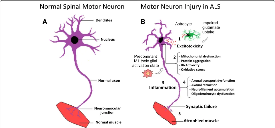
Several pathophysiological mechanisms have been pro-posed including: cytoplasmic protein mis-localization and aggregation [19], aberrant protein homeostasis [20], RNA toxicity [13], dysregulation of RNA processing [21], excitotoxicity mediated by excessive glutamate receptor activation [22], mitochondrial dysfunction [23], endoplas-mic reticulum stress response and endoplas-microglial activation [24], abnormal rearrangement of the cytoskeleton with impaired axonal transport [25], and oxidative stress [26]. Moreover, the contribution of microglial cells, oligoden-drocytes and astrocytes seems to be critical for the devel-opment of the disease influencing significantly the speed of disease progression after onset [27–31]. Indeed, ALS is considered as a non-cell autonomous disease, where the start and progression of motor neuron degeneration seems to be influenced by complex interaction among dif-ferent kinds of cells, together with the development of a sustained inflammatory milieu [32]. Figure 1 summarizes
the major pathological mechanisms contributing to motor neuron injury in ALS.
The complex heterogeneity of ALS, where several mo-lecular mechanisms contribute to the pathology, enables various opportunities for therapeutic intervention. How-ever, the complexity of the disease and clinical variability within patients inevitably makes the identification of a universal single drug or therapy capable of correcting the pathophysiology of ALS in its totality, very difficult. Various therapeutic strategies are being experimentally evaluated such as immunomodulation, approaches to improve mitochondrial function, induction of autophagy and anti-oxidant agents [16]. However, after over twenty years of encouraging results in preclinical studies, no efficacious treatment has been developed so far and riluzole, the only recognised neuroprotective agent in ALS, prolongs life expectancy by only approximately 3– 4 months [16].
During the last decade, progress in stem cell biology has paved the way for potential cellular based therapy in neurological diseases. Albeit still at an early stage and with several issues to be solved, stem cell therapy holds great promise for the treatment of ALS. Stem cells are a population of cells which are defined by functional char-acteristics. They are undifferentiated cells capable of self-renewal, able to form clones in vitro and capable of differentiation into mature cell lines of various tissues.
A
B
Fig. 1Molecular mechanisms in the pathology of amyotrophic lateral sclerosis.aSchematic representation of healthy spinal cord motor neuron.
[image:2.595.57.538.439.663.2]There are several potential advantages to the use of stem cells in ALS:
1) The complexity of ALS pathology may not allow the use of a single drug or targeted treatment;
2) The capability of stem cells to differentiate into neuron-like cells and potentially replace the neuronal population lost in ALS;
3) The degeneration of existing motor neurons could be prevented by the release of neuroprotective trophic factors and the immunomodulatory properties of trans-planted stem cells, thus modifying the toxic microenvir-onment in ALS.
This review will outline the recent progress relevant to stem cell-based therapies in general in ALS, and will focus on the therapeutic potential of mesenchymal stem cells by discussing the major technical issues, challenges and future directions.
Stem cell therapy in ALS
There are different types of stem cells which differ ac-cording to the source, clonogenic capacity, differenti-ation potential and availability.
Human embryonic stem cells (hESC)
Human embryonic stem cells (hESC) are derived from the inner cell mass of the blastocyst and can indefinitely propagate in vitro, preserving the capacity to differenti-ate into any cell type of the three embryonic germ layers (endoderm, mesoderm and ectoderm) [33]. For the first time in 2005, Shin and colleagues obtained motor neuron-like cells expressing markers such as islet1 and choline acetyltransferase from hESC using conditioned media containing basic fibroblast growth factor (bFGF), retinoic acid (RA) and sonic hedgehog (Shh) [34]. The survival, differentiation and beneficial neurotrophic sup-port of motor neuron progenitors (MNP) derived from hESC has also been demonstrated after lumbar intraspinal transplantation into SOD1G93A mice and other MND models [35, 36]. Wyatt et al., transplanted hESC derived MNPs directly into the spinal cord of immunosuppressed SOD1G93Amice, spinal muscular atrophy (SMA)Δ7SMN pups and rats with spinal cord injury (SCI), demonstrating the in vivo differentiation of the engrafted cells into a mixed population of mature and immature motor neuron cells [36]. The axons of the differentiated cells did not reach the periphery, and the authors did not prove the integration of the differentiated cells into the existing neural circuit. However, the transplanted cells were able to reduce motor neuron loss in proximity to the injection site by actively releasing neurotrophic factors such as neurotrophin-3 (NT-3) and nerve growth factor (NGF) [36]. In particular, in SOD1G93Amice that received MNPs, 43 ± 5 endogenous neurons cranial to the injection site survived until the end of the study (110 days old), in
comparison to the vehicle control group in which 27 ± 3 neurons were counted [36]. Yet, the use of hESCs in the clinic is hindered because of ethical concerns, po-tential tumorigenicity in vivo and the popo-tential for graft rejection [37].
Foetal neural progenitors (NSC)
Foetal neural progenitors (NSC) are multipotent stem cells derived from foetal spinal cord or brain, capable of in vitro self-renewal and able to differentiate into astrocytes, neurons and oligodendrocytes. Given their partial matur-ation state they have less propensity to form teratomas in vivo [38]. Several studies investigated the safety and therapeutic potential of spinal, intrathecal or intracra-nial transplantation of hNSC in ALS rodent models [39–41]. In particular, a well-characterized hNSC cell line (NSI-566RSC) derived from an 8-week human foetal spinal cord showed very promising results in transplanted SOD1G93Arodents [42, 43].
In 2006, Yan et al. performed spinal cord injections of NSI-566RSC cells in the ventral horn of 8-week-old SOD1G93A mice at the lumbar level L4-L5, under com-bined immunosuppression or CD4 antibodies [42]. Four separate injections were carried out per mouse, with a total of 8 × 10⁴cells. The authors showed that the graft survived for more than two months after transplantation, with most of the engrafted NSCs showing differentiation into TUJ1+ neurons, and evidence of synaptic contacts with host neurons [42]. Moreover, in mice injected with live NSCs cells, disease onset was delayed by 15 days and life span extended by 12 days in comparison to the control group that received injections of dead cells. A statistically significant later onset and a slowing of disease progres-sion, was also confirmed by analysis of motor performance [42]. The same group of authors, investigated the thera-peutic potential of the NSCs-566RSC cell line after injec-tion of around 8 × 10⁵cells into the lumbar spinal cord of SOD1G93Arats at a pre-symptomatic disease stage [43]. In this study, rats that received live NSCs showed an increase in survival of around 11 days and a delay in disease onset of 7 days when compared to the control placebo group. The beneficial effect could be associated with the release of neurotrophins such as glial-derived neurotrophic factor (GDNF) and brain-derived neurotrophic factor (BDNF), which in turn delayed the death of α-motor neurons in the lumbar region [43].
Despite these encouraging data, the restricted number of cells available for transplantation represents a potential limitation for obtaining therapeutic efficacy in humans.
Induced pluripotent stem cells (iPSCs)
factors that preserve pluripotency in ESCs (KLF4, SOX2, OCT4 and c-MYC) [44, 45]. The differentiation of human iPSCs into electrically active motor neurons has been accomplished by several groups both in vitro and in vivo [46, 47].
Interestingly, Popescu et al. demonstrated in vivo dif-ferentiation of human iPSC-derived neural progenitors (NPs) in presymptomatic SOD1G93Arats following stem cell injection into the ventral horns of the lumbar spinal cord [48]. At 30 days post-transplantation human mito-chondria positive cells displayed expression of the neur-onal precursor marker doublecortin (DCX), indicating the presence of undifferentiated progenitors. Substantial differentiation into mature motor neurons could be ob-served only after 60 days, with the majority of engrafted cells expressing the neuronal marker MAP2 [48]. This relatively long time is something to bear in mind, consid-ering that transplantation was performed before disease onset and NPs could survive and differentiate within a less toxic environment in comparison to the symptomatic stage. If cells were transplanted during disease progres-sion, as would occur in the clinic, NPs may not have a permissive environment and/or the necessary time to differentiate into mature motor neurons. The therapeutic potential of iPSC-derived NPs has also been investigated in the SOD1G93Amouse model of ALS [49]. iPSC-derived neural stem cells (NSCs), further selected for the ex-pression of the integrin VLA4+, were transplanted either by repeated (n= 3) intrathecal or weekly intravenous in-jections. Intrathecal injection of cells extended survival by 10 days, while systemic delivery increased survival by 23 days, compared to control PBS injected mice [49]. The molecular protective mechanism of iPSC-derived NSCs may be attributed to the capacity of these cells to secrete trophic factors such as GDNF, BDNF, NT-3 and TGF-α, which in turn protects resident motor neurons and re-duces astrogliosis [49].
The opportunity for reprogramming somatic cells into neural stem cells (NSCs) could overcome the immune rejection problem by autologous transplantation, and bypass the ethical problems related to the use of ESCs and foetal cells. However, several issues need to be ad-dressed, such as reprogramming efficiency, epigenetic memory and safety before translation of the use of iPSCs into clinical practice.
Mesenchymal stem cells (MSCs)
Adult mesenchymal stem cells (MSCs) are stromal mul-tipotent stem cells that can be derived from umbilical cord, bone marrow, adipose tissue and peripheral blood and have the capacity to differentiate into different components of mesodermal origin (cartilage, bone, fat, muscle and stroma) [50]. These cells can be expanded and maintained by several passages in plastic-adherent
tissue culture. MSCs show fibroblast-like morphology, they do not differentiate spontaneously and are character-ized by the expression of specific surface markers [50]. In addition to mesodermal commitment, several authors showed the potential of bone marrow-derived MSCs (BM-MSC) to differentiate into neuron-like cells, oligo-dendrocytes and astrocytes [51–59].
Several characteristics make the use of MSCs very at-tractive in ALS cell therapy. MSCs can be obtained and expanded from adults relatively easily, bypassing the ethical constraints related to the use of embryonic and human foetal derived stem cells. Also, they are less im-munogenic and can be harvested from ALS patients so allowing both allogenic and autologous transplantation [50]. Moreover, these cells are capable of homing to areas of insult, possess immunomodulation and anti-apoptotic properties, and are capable of secreting several cytokines, extracellular matrix proteins and growth factors relevant in neuroprotection and tissue repair [60]. Because of these properties, mesenchymal stem cells are receiving significant attention amongst researchers.
Proof-of-concept for MSCs therapy in the SOD1G93A
model of ALS
Several studies were performed in ALS rodent models in order to investigate the potential of either human (hMSC) or murine (mMSC) bone marrow-derived MSCs (BM-MSCs) for cell therapy in ALS. Different approaches have been tested by varying the delivery method, the amount of injected cells, the timing of intervention, and the differentiation state. Table 1 shows a summary of pre-clinical studies described in the literature with injection of BM-MSCs in ALS models.
Intravenous delivery
In 2007, Zhao and colleagues delivered 3 million hBM-MSCs into 60 day pre-irradiated SOD1G93A mice by intravenous infusion [61]. The recipient mice showed about 14 days delay in disease onset, and prolonged sur-vival of about 18 days in comparison to untreated mice. Moreover, the decrease in motor performance (rotarod test) was delayed by 3 weeks [61]. However, immunostain-ing experiments showed that only a few transplanted cells migrated and penetrated into the grey and white spinal cord matter, surviving for no more than 20 days [61].
capacity and survival of engrafted cells into the central nervous system (CNS) of SOD1G93A mice, resulting in enhanced and prolonged benefits [58]. The migration capacity might be explained by high expression of che-mokine receptors such as CCR2 and CXCR4, which were significantly more expressed after neurogenin1 in-duction. [58]. Interestingly, when MSCs were injected at a pre-symptomatic stage, disease onset in the treated group was delayed by 5 days with an increase in life span of only 3 days. Conversely, when injected close to dis-ease onset, Ng1-MSCs were able to incrdis-ease survival by about 7 days, suggesting the importance of the time of intervention for stem cell therapy in ALS [58].
Furthermore, the Uccelli group demonstrated that mouse BM-MSCs isolated from non-transgenic mice, were therapeutically effective when transplanted during the symptomatic stage of the disease in the SOD1G93A mice [62]. In this study, mBM-MSCs expanded ex-vivo for 8–15 passages, were transfected with the Luciferase gene reporter vector pL-Luc-HI for in vivo tracking and injected into the tail vein of SOD1G93A mice. The mice that received the MSC transplantation showed an ex-tended survival of 17 days, delayed decline in motor performance and decreased weight loss when compared to the control PBS-injected mice [62]. In addition, the transplantation of MSCs alleviated the pathology of the disease in the ALS spinal cord, by reducing astrogliosis and microglial activation, and by restoring antioxidant components such as glutathione-S-transferase and metallothioneins to their baseline level of expression and activity. However, the engraftment efficiency of intravenous delivered cells within the CNS was very low, with luciferase-positive cells almost completely ab-sent twenty days post- injection. This suggested that MSCs delivered by the intravenous route, can exert clinically positive effects during the disease course that do not correlate with the efficiency of long-term en-graftment in the host [62].
Intrathecal delivery
Using a different approach, several authors have injected a variable number of BM-MSCs directly into the cisterna magna of pre-symptomatic SOD1G93A mice [63–66]. Direct injection into the CSF allows the obstacle of the brain blood barrier (BBB) to be bypassed. Moreover, the injected cells may migrate along the spinal cord possibly reaching segments specifically affected by motor neuron degeneration.
Indeed, it has been demonstrated that the injected BM-MSCs were able to delay disease onset, improve motor performance, ameliorate motor neuron death and prolong survival in transplanted SOD1G93Amice [64–67]. These beneficial effects were enhanced by multiple intra-thecal injections or by using a considerable number of
MSCs (1 × 10⁶) [64, 65]. However, injected cells rarely mi-grated into the parenchyma, suggesting a neuroprotective effect of MSCs from the CSF [64, 65, 67]. In fact, it was shown that injection of MSCS into the CSF of SOD1G93A mice consistently inhibited microglial activation and the release of inflammatory molecules, possibly by diffusion of soluble factors where direct contact between cells is not a requirement [67].
In 2009, Boucherie and colleagues, investigated the thera-peutic potential of rat wild-type (WT) bromodeoxyuridine-labelled BM-MSCs (BrdU-MSC) by intrathecal injection in SOD1G93A transgenic rats, at disease onset [54]. Stem cell transplantation in SOD1G93Arats resulted in a reduction in the rate of disease progression, as the first signs of paralysis were detected 2 weeks later in in comparison to control mice. Moreover, treated mice showed reduced local inflam-matory response and an increase in life span of 16 days [54]. Interestingly, this is the only study to show a signifi-cant trans-differentiation of MSCs into astrocyte-like cells in vivo. Indeed, transplanted BrdU-MSCs were found to penetrate into the grey matter of the ventral horns, and stained positively for the astrocyte marker GFAP [54]. Re-markably, around 40% of the BrdU-MSCs that successfully migrated in proximity to motor neurons, co-localized with the astrocyte marker, and about 30% of the GFAP-positive cells were actually positive for the BrdU. This study demon-strated a considerable local chimerization of the astroglial population near the site of motor neuron injury in the lum-bar spinal cord of SOD1G93Arats [54]. However, cell fusion events with resident astrocytes cannot be excluded. Surpris-ingly, the overall level of astrogliosis was not changed upon MSCs treatment, and the loss of expression of the astrocyte glutamate reuptake transporter (GLT-1), typically observed in SOD1G93Arats, was not rescued [54].
The perineural net (PNN) is a specialized matrix struc-ture present at a high density around motor neurons and is fundamental in axon development as well as neuronal plasticity [66]. Modification and deterioration of the PNN has been observed in the spinal cord of SOD1G93A rats during neurodegeneration [66]. Interest-ingly, intrathecal delivery of hBM-MSCs into early post-symptomatic SOD1G93Arats, resulted in partial rescue of PNN structures suggesting a role of MSCs in reactivat-ing CNS plasticity [66]. In this study, stem cell injection increased survival of 14 days in comparison to the pla-cebo group.
transplanted cells were found mostly concentrated on lumbar, thoracic and cervical meninges, but consider-able numbers of bisbenzimide positive cells were found in proximity to motor neurons within the spinal cord [68]. It is noteworthy that the use of bisbenzimide as a marker for cell transplantation has been questioned since the dye may transfer from labelled cells to host cells [69].
Intraparenchymal delivery
Other authors have attempted to inject human or mouse MSCs directly into the dorsal horn of spinal cord of SOD1G93A mice [70, 71]. In the Vercelli group experi-ments, hMSCs were found to engraft, migrate to the ven-tral horn close toα-motor neurons and survive more than 10 weeks after surgery, although without signs of differen-tiation into neurons or astrocytes [70]. Also, male but not female recipient mice showed a consistent (38%) increased motor neuron survival as well as reduced gliosis and at-tenuated astrocyte activation in comparison to control mice [70]. Though the authors claimed an extended life-span in male treated mice, not all of the experimental groups were followed until the disease end-point, and a Kaplan-Meier survival curve was not shown.
Intramuscular delivery
Since early pathologic mechanisms of disease involve destruction of neuromuscular junctions before motor neuron death, intramuscular injection of MSCs at early stages of disease has also been proposed. hBM-MSCs, genetically engineered to express green fluorescent pro-tein (GFP) and to constitutively secrete GDNF, were injected into the forelimb triceps brachii muscles of pre-symptomatic SOD1G93Arats [72].
The intramuscular transplantation of MSCs did not delay disease onset, however, survival was prolonged by 18 days. Furthermore, a reduction of endplate denerv-ation in SOD1G93A rats was observed, when compared to the control vehicle group [72]. However, MSCs were not able to regenerate motor end-plates, nor to reduce neuroinflammation [72]. Moreover, to obtain significant survival and integration of MSCs in the host, the induc-tion of focal muscle injuries was necessary.
MSCs in ALS: Clinical trials
Mazzini trials: Intraparenchymal delivery of autologous BM-MSC
Despite the absence of any preclinical data, in 2001 Mazzini et al. embarked upon the first clinical trial in order to evaluate the safety and feasibility of MSC injec-tion into the spinal cord of sporadic ALS patients [73]. The study comprised the recruitment of 7 SALS patients with spinal onset and severe lower limb impairment without respiratory complications [73]. Autologous
mesenchymal bone marrow-derived stem cells were ex-panded for 3–4 weeks under good manufacturing practice (GMP) conditions and cytogenetic analysis, viability and cytofluorimetric analysis for characterization of antigens were carried out before infusion [73]. However, no de-tailed data was provided.
In this study, following three hours in serum-free medium, a variable number of cells ranging from 7 to 152 million were suspended in 1–2 ml of autologous CSF and transplanted directly into the parenchyma of spinal cord at thoracic level (T7-T9) [73, 74].
The patient follow-up was performed every 3 months for six years. After surgery, no signs of increased neuro-logical defects or toxicity were observed, indicating that implantation into the spinal cord of ex vivo expanded MSCs was safe and well tolerated by patients [73, 74].
To further validate the safety of the therapy, in 2010 a second Phase I clinical trial was conducted in another 10 patients following the same protocol as described above, with slight modifications [75]. Cells (11–122 mil-lion) were transplanted in the anterior horn at the thor-acic level (T4-T6) with different numbers of injection sites (2 to 5) [75]. Two years post-surgery, no tumour forma-tion, side effects or toxicity had been detected [75].
A long-term (9 years) safety study, concluded in 2012, was carried out by the same authors on the basis of 19 ALS patients who underwent autologous MSCs implant-ation in two separate phase I studies from 2001 to 2003 [76]. Importantly, magnetic resonance imaging (MRI) analysis demonstrated no tumour formation or abnor-mal cell growth, indicating that ex vivo expansion of ALS patient-derived MSCs does not affect karyotype or cellular senescence, making their use safe in the clinic [76]. No data describing stem cell characterization were shown and evidence of engraftment in post-mortem tis-sues have not yet been described.
Blanquer trial: Intraparenchymal delivery of autologous BMNC
Based on promising results in a mouse model of MND [77], Blanquer et al. tested the safety and tolerability of intraspinal transplantation of mononuclear cells derived from autologous bone marrow (BMNC) in ALS patients in an open label phase I clinical trial [78]. The study was performed on 11 sporadic ALS patients with spinal onset and no evidence of significant respiratory dysfunction [78].
After 1 year of follow up, no severe adverse effects or acceleration of the disease progression related to the treatment were observed, demonstrating the safety and feasibly of the described procedure in ALS patients [78]. Moreover, spinal cord pathology on post mortem ma-terial showed preservation of motor neurons at the in-jection sites [78]. Interestingly, preserved motor neurons were surrounded by spherical CD90+ cells negative for neuronal markers, CD45 (hematopoietic stem cells) and CD68 (monocytes/macrophages) markers [78]. These motor neurons did not show any sign of degeneration [77, 78].
Karussis trial: Intrathecal and intravenous delivery of autologous MSCs
In 2010, a phase 1/2 clinical trial was conducted in Israel in 15 patients with multiple sclerosis (MS) and 19 ALS patients [80]. In order to enhance the potential benefits of MSC transplantation, after 40–60 days in culture, a median of 60 million autologous MSCs were trans-planted intrathecally in combination with a mean of 20 million MSCs delivered intravenously [80]. Also, in 9 pa-tients MSCs were pre-labelled with ferumoxides in order to track their fate in vivo by MRI [80].
At the end of 25 months of follow-up, no severe ad-verse effects were registered in any of the patients, with signs of disease stabilization in some patients during the first 6 months after the intervention [80]. Furthermore, MRI screening 24 h, 48 h, 1 and 3 months after the infu-sion of cells, showed the presence of ferumoxides in nerve roots, meninges and the parenchyma of the spinal cord. However, these results are not conclusive of the presence of MSCs in the CNS, since the contrast agent can be ingested by phagocytes which have migrated to inflammatory lesions [80]. Also, flow cytometry analysis and subsequent proliferative response assay of peripheral blood monocytes obtained from ALS patients 4 h and 24 h after infusion of MSCs, showed a dramatic increase in CD4+ CD25+ regulatory T cells, coupled with a reduc-tion in activated dendritic cells and lymphocyte prolifera-tion, demonstrating the immediate immunomodulatory properties of MSCs following transplantation [80].
Oh trial: Repeated intrathecal infusion of autologous BM-MSCs
Very recently, a single open-label clinical trial was per-formed in order to evaluate the clinical feasibility and safety of two repeated infusions of autologous MSCs into the CSF by lumbar puncture in 8 ALS patients [81]. At intervals of 26 days, 1 × 10⁶ MSC cells per kg were injected diluted in autologous CSF [81].
After 1 year, no deaths were recorded and the proced-ure was considered safe, without long-term adverse ef-fects [81]. In addition, CSF samples of two patients were collected before both the first and the second injection
in order to evaluate cytokine levels. Il-10, TGF-β (I, II and III) and IL-6 levels were increased after MSC trans-plantation, while the level of the monocyte chemoattract-ant protein 1 (MCP-1) was decreased [81]. A phase II clinical trial in 64 ALS patients is on-going, with a placebo control group allowing proper evaluation of efficacy and safety (NCT01363401).
The same group performed repeated intrathecal MSC injections in another 37 patients between 2007 and 2010, with the aim of identifying MSC markers capable of predicting the response in ALS patients [82]. First, they measured the level of various trophic factors in MSC cultures from each patient by ELISA assay. They then transplanted MSCs from different patients in the SOD1G93Amouse model and analysed differences in on-set, MN loss and immunoreactivity [82]. The authors concluded that the beneficial effects (symptom improve-ment and slowing of decline) observed in a proportion of patients, were positively correlated with the increased capacity of MSC cells to secrete VEGF, ANG and TGF-β in vitro [82]. Moreover, the clinical efficacy observed in some patients was confirmed also in SOD1G93A mice which exhibited prolonged survival and lower levels of neuroinflammation [82].
Adipose tissue: A fat tissue source for mesenchymal stem cells
Mesenchymal stem cells represent an ideal source of adult stem cells for cell therapy given their immunosup-pressive nature, low potential for immunogenicity and trans-differentiation capacity. However, the collection of MSC from bone marrow is an invasive procedure which can be painful and anaesthesia is often required [83]. Moreover, the proportion of stem cells within the total cell population in bone marrow aspirate is usually about 0.001–0.002% which leads to extended culture times and increased expense in order to obtain sufficient GMP cells for clinical application [83].
Subcutaneous (buttocks and abdomen) and visceral (omentum) white adipose tissue (WAT) may represent an alternative source of stromal adult stem cells since they can be obtained by minimally invasive, simple pro-cedures such as liposuction or lipectomy and they are relatively abundant, representing 1% of WAT cells after processing [83–85]. Several names and abbreviations have been used to refer to adipose-derived mesenchymal stem cells. Here, the abbreviation ADSC will be used.
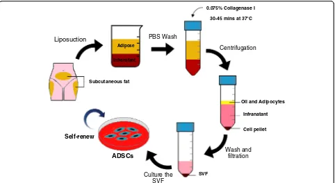
In the literature, the method for isolation of ADSCs from human fat is performed following almost always the same protocol described by Zuk and colleagues [83]. An illustration of the ADSC isolation method is showed in Fig. 2.
by extracellular matrix digestion with collagenase. After enzymatic neutralization, the homogenate is centrifuged to obtain the stromal-vascular fraction (SVF) pellet. The pellet is then suspended in culture medium and left overnight in a flask in the incubator at 37 °C with 5% CO2. After incubation, cells are washed with phosphate buffered saline (PBS) to remove residual non-adherent cells. The culture medium usually consists of Dulbecco’s modified eagle medium (DMEM) supplemented with foetal bovine serum (FBS), penicillin and streptomycin.
In spite of some differences in the expression of clus-ter of differentiation (CD) markers such as CD49d and CD106, ADSC cells are phenotypically similar to BM-MSC cells, showing fibroblast-like morphology, charac-teristic expression of mesenchymal stem cell markers (CD44+, CD105+, CD73+, CD90+, CD29+, CD45−, CD34 −, CD14− CD19−), lack of major histocompatibility
com-plex class II (MHC-II) and the capacity to differentiate into osteoblasts, chondrocytes and adipocytes in specific culture conditions [83]. Thus, hADSCs comply with all the minimum criteria for the characterization of hMSCs, established in 2005, by the International Society for Cel-lular Therapy (ISCT) [86].
Like MSCs obtained from bone marrow aspirates, hADSCs secrete several soluble factors such as BDNF, NGF, hepatocyte growth factor (HGF), vascular
endothelial growth factor (VEGF), insulin-like growth factor-1 (IGF-1) and basic fibroblast growth factor (bFGF) that may contribute to neurotropism, neuroprotection and tissue regeneration by paracrine mechanisms [87].
Even though the literature appears controversial, there is evidence supporting the potential of ADSCs to trans-differentiate into progenitors or mature cells of ecto-dermal origin, therefore opening the way to a future in-vestigation of the feasibility of cell replacement in neurodegenerative disorders (Safford et al., 2002; Kram-pera et al., 2007; Ahmadi et al., 2012; Feng et al., 2014).
In vitro neuronal differentiation capacity of ADSC
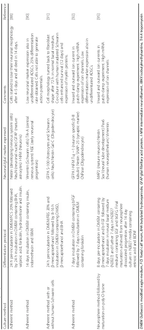
The capacity of ADSCs to differentiate in vitro into ma-ture, stable and functional neurons is an open field of debate among researchers. Table 2 summarises the pro-tocols for neuronal differentiation of ADSCs published in literature. Usually, the differentiation protocol consists of expanding ADSCs as adherent cells for several pas-sages, followed by induction with media containing differ-ent cocktails of chemical agdiffer-ents, cytokines and growth factors [88–92].
A different method to obtain neuron-like cells from ADSCs is based on neurosphere formation capacity, generally achieved by culturing cells in EGF and bFGF conditioned serum-free media [93, 94]. A neurosphere
Liposuction PBS Wash
Adipose
Infranatant
Oil and Adipocytes
Infranatant
Cell pellet
Centrifugation
0.075% Collagenase I
30-45 mins at 37°C
Wash and filtration
Subcutaneous fat
Culture the SVF
SVF ADSCs
Self-renew
[image:9.595.58.540.87.351.2]is a cluster of cells containing neural precursors able to proliferate and survive as floating and non-adherent structures. The neurosphere can be dissociated, seeded onto a poly-L-lysine feeder layer and differentiated by adding neuronal induction components [93–95]. Alter-natively, ADSCs are cultured in the presence of ESCs or Schwann cells either in the presence or absence of induction factors [91, 96]. In particular, when co-cultured with Schwann cells, ADSCs showed long-lasting (12 days) Schwann-like cell morphology and expression of myelin protein [91]. Pre-irradiation of Schwann cells excluded eventual cellular fusion artefacts [91]. Since the addition of supernatant from Schwann cell cultures failed to differ-entiate ADSCs, the authors speculated that the addition of specific chemical components or growth factors to the ADSC cultures, may promote the initial steps toward dif-ferentiation in vitro, which can be completed in the appro-priate microenvironment and by required cell-to-cell interaction with mature cells in vivo.
The trans-differentiation of ADSCs towards motor neuron-like cells has also been reported. In 2012, Abda-nipour and Tirahini [97] induced rat ADSCs to trans-differentiate into motor neuron-like cells by a two-step protocol. ADSCs were committed to neural progenitors expressing nestin, neurofilament 68 and neuro D by pre-induction for 24 h with selegiline. Selegiline is an inhibi-tor of monoamine oxidase B (MAO-B) which was used to trans-differentiate BM-MSCs into dopaminergic neural-like cells and proved to be a safer pre-inducer in comparison to the toxic β-mercaptoethanol and butyl-ated hydroxyanisole compounds, widely used for ecto-dermal differentiation of MSC [97]. The maturation of the pre-induced ADSCs into a motor-neuron phenotype was then achieved by incubation with Shh and RA, resulting in a differentiation efficiency of around 70%. The resultant motor neuron-like cells (MNLCs) were characterized by an initial and transient high expression of oligo-2 and islet-1 (markers for early motor neuron commitment during in vitro differentiation), followed by a decrease in these two markers accompanied by an in-crease in the expression of the motor neuron marker HLBX9 [97]. Mature MNLCs, but not high HLBX9 ex-pressing cells, were functionally tested for the capacity to release pre-synaptic vesicles by staining and destain-ing with the fluorescent probe FM1–43. Quantitative analysis of vesicle release after stimulation, showed a 4-fold increase in comparison to pre-induced ADSCs. MNLCs, also formed innervation-like contacts with myotubes in a co-culture in vitro system [97].
Very recently, the same authors proposed a different method to obtain MNLCs from rat ADSCs [98]. ADSCs were first converted into neurospheres by induction with NM medium consisting of DMEM, B27, EGF and bFGF for 7 days. Next, neural stem cells (NSC) were obtained
by neurosphere dissociation into single cells, and incuba-tion for 10 days with NM supplemented with 10% FBS. Maturation into motor neurons was achieved by incuba-tion with Shh and RA for 5 days, followed by addiincuba-tion in culture media of BDNF, GDNF, CTNF and NT-3 for an-other 7 days [98]. During the maturation process of ADSC-NSCs into motor neurons, an increase in the ex-pression of islet-1, HB9 and ChAT was observed, with MN-like cells positive for the neural marker MAP2 at day 14 of maturation. These findings were documented by both immunocytochemistry and RT-PCR. The func-tional activity of mature MNLCs was investigated by quantification of the release of synaptic vesicles by FM1– 43 loading and release experiments upon ion stimulation. The synaptic vesicle activity correlated with changes in intracellular calcium concentration and membrane depolarization, as demonstrated by further investigations utilizing calcium and voltage-sensitive dyes [98].
Is the trans-differentiation of ADSCs into neurons real? The differentiation into neurons is commonly described as morphological changes such as body retraction, bi-or multi-polar shape and branching extension, and by the expression of neuronal markers revealed by immu-nohistochemistry (IHC), western blotting (WB) and quantitative RT-PCR analysis. However, several studies showed that the neural-like phenotype obtained by chemical induction was a very fast but transient event which may be a result of cytoskeletal rearrangement due to the toxicity of compounds added into the induction media [99, 100]. Furthermore, several neuronal markers considered to confirm neuron maturation such as Nestin, NSE, trk-A and vimentin are already present at low levels in undifferentiated ADSCs [89, 90, 92, 95, 101].
Thus, morphological changes and expression of neur-onal markers on the cell surface should not be consid-ered as a definitive proof of neuronal differentiation, and functional characterization (i.e. electrophysiology) is ne-cessary. However, very few studies confirmed electrical activity of ADSC-derived neurons by patch-clamp exper-iments [92, 95].
In vivo studies with ADSCs
For the first time in 2013, the therapeutic potential of ADSCs was investigated in the SOD1G93A mouse model [104]. ADSCs were isolated from C57BL/6 GFP-expressing mice, and a total of two million cells were injected into the tail vein of B6SJL-SOD1G93Amice after the first clinical signs of disease. The control group re-ceived injection of PBS only as vehicle. mADSCs delayed the motor performance decline and transiently attenuated motor neuron death, in comparison to the controls. How-ever, no differences in the degree of astrogliosis, nor in survival were observed between the two groups [104]. The authors claimed that GFP-positive cells were found to mi-grate into damaged CNS areas, such as the grey matter in the spinal cord, however, low magnification images showing the entire spinal cord section or co-staining with neuronal specific markers were not shown and hist-ology on PBS-treated control spinal cord sections was not performed. Finally, increased levels of trophic factors such as GDNF and bFGF were found in spinal cord homoge-nates of ADSC injected mice. This was despite the fact that mADSC used in this study were not capable of producing GDNF in vitro. It is likely that, in addition to trophic factor production and secretion, ADSCs could have an indirect biological effect by stimulating the secretome of surrounding astrocytes, which are known to produce GDNF [104].
Recently, another group tested the therapeutic poten-tial of human ADSCs isolated from three healthy donors aged 64, 69 and 84 [105]. Female transgenic SOD1G93A mice before clinical evidence of disease received 1 × 10⁶ hADSCs by intravenous (IV) or 2 × 10⁵hADSCs by in-tracerebroventricular (ICV) injections. As a control, the sham group received PBS only.
Rotarod test and paw grip endurance were monitored and a reduction of 15% was considered as disease onset. The endpoint (survival) was defined by the lack of a righting reflex within 30 s after being placed on their side [105]. The authors found that disease onset in mice that received ICV injections was significantly delayed by 26 days, while in mice infused IV there was only an 11 day delay in disease onset. Remarkably, survival was prolonged by 24 days and 9 days respectively in ICV and IV mice when compared to the controls [105].
IHC evaluation of the spinal cord of ICV transplanted mice revealed that 6.8% of transplanted cells survived up to 4 weeks post-injection. However, only 0.7% migrated to the grey matter of the lumbar spinal cord and very few cells were positive for neuronal markers (MAP2 or I-NFM). In contrast, after IV infusion very few undiffer-entiated cells were found to engraft in the meninges of the spinal cord [105]. In addition, when the TUNEL assay was performed on spinal cord from ICV mice, re-duced levels of apoptosis were seen in the anterior grey
matter compared to IV and control recipients. Also, RT-PCR and ELISA assays on spinal cord homogenates showed a significantly higher concentration of neurotrophic factors in the ICV group in comparison to controls [105]. ELISA assay on the supernatant from hADSC cultures showed high levels of neurotrophic factors that are relevant in neuroprotection such as IGF-1 and VEGF [105]. The anti-apoptotic effect was further confirmed in vitro by cul-turing primary neural cells from normal mice with hADSC conditioned media [105].
To date, these are the only two published studies using ADSCs in an experimental model of ALS. However, the therapeutic potential of ADSCs has been tested in other experimental models of neurodegeneration. In particular, the use of ADSCs showed encouraging results in animal models of chronic stroke, Parkinson’s disease, Alzhei-mer’s disease and traumatic brain injury and ageing [106–109]. The use of ADSCs in ALS patients has not been tested so far. However, currently two different clin-ical trials are recruiting participants.
A clinical trial sponsored by The Royan Institute in Iran will test the safety of intravenous injections of ADSCs derived from healthy donors (2 million cells/kg) in 8 ALS patients (ClinicalTrial.gov identifier: NCT02492516). The Andalusian Initiative for Advanced Therapies is recruiting 40 ALS patients for a phase I/II, multicentre, randomized, placebo controlled clinical trial to evaluate the safety and efficacy of intravenous infusion of different concentrations of autologous ADSCs (ClinicalTrials.gov identifier: NCT02290886).
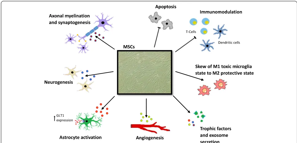
MSCs for ALS therapy: Proposed mechanisms of action The in vitro differentiation of mesenchymal stem cells into neuron-like cells brought great enthusiasm into the idea of cellular replacement as a therapeutic strategy in ALS. However, there are many practical issues to be solved. The real trans-differentiation of MSCs into neu-rons has been questioned and further investigation in order to study molecular pathways and optimized proto-cols enhancing the efficiency, stability and degree of maturation are needed. Also, only a few studies have re-ported MSCs or MSC-derived NSCs showing signs of in vivo maturation when transplanted into mice (detailed in the next section).
Moreover, different pathological mechanisms described in ALS such as glutamate excitotoxicity [22], oxidative stress [26] and loss of metabolic support [110] may affect the viability of transplanted cells and inhibit maturation. Finally, stem cell maturation and integration into the host neuronal circuits could last a relatively long time, which may not be compatible with the fast progression rate of the disease.
Besides the challenge of cell replacement in ALS, in recent years increased attention has been paid to the “bystander effect” mechanism through which MSCs could exert their therapeutic effect. Different mecha-nisms have been proposed to explain the role of MSCs in neuroprotection. Although the precise mechanisms are still unknown, the secretion of anti-inflammatory cy-tokines and growth factors by MSCs may influence the progression of ALS in multiple ways, including endogen-ous regenerative processes such as neuronal plasticity, angiogenesis and axonal re-myelination [66, 111].
The delivery of growth factors such as BDNF, insulin-like growth factor-1 (IGF-I), vascular endothelial growth factor (VEGF) and glial derived neurotrophic factor (GDNF) into ALS experimental models has been very promising since these factors were shown to be neuro-protective, improved motor function and prolonged motor neuron survival [112]. However, translation into the clinic failed to provide any beneficial effect in ALS patients [112]. This is thought to be related to the small amount of growth factor that effectively reached the central nervous system (CNS) either because of an in-ability to cross the blood brain barrier or because of the short half-life after intravenous injection [112]. Moover, intrathecal injections of growth factors did not re-sult in relevant benefits for ALS patient [112].
The use of MSCs as “carriers” for the uninterrupted supply of growth factors has been proposed, having shown positive results in an ALS rodent model [113, 114]. In-deed, MSCs transduced to overexpress specific growth factors (GDNF, VEGF) or neuroprotective agents (e.g. glucagon-like peptide 1) transplanted into SOD1G93A ro-dent models, significantly preserved neuromuscular junc-tions, attenuated motor neuron death, improved motor function, delayed symptom onset and prolonged survival [113, 114]. Recently, the Karussis group and Brainstorm-Cell therapeutics completed an open lab phase 1/2 clinical trial evaluating the safety and feasibility of intrathecal injections of MSCs induced to secrete neurotrophic fac-tors (BDNF, GDNF) (NurOwn™) in 12 ALS patients (NCT01051882). Although, no results have been pub-lished so far, the details of another two separate active clinical trials can be found at ClinicalTrials.gov. An open label phase 2a escalating-dose trial (NCT01777646) and a phase 2 randomized, double-blind trial (NCT02017912) in order to evaluate safety and efficacy of multiple
intramuscular and intrathecal combined injection of NurOwn™ cells into ALS patients are reported as in progress.
Through paracrine activity and cell-to-cell contacts, MSCs were able to induce astrocytes and glial cells to secrete enhanced levels of GDNF, VEGF and CNTF (cil-iary neurotrophic factor) both in vitro and in the SOD1G93A mice, resulting in anti-apoptotic effects and motor neuron protection [104, 115].
Another potential mechanism by which MSC could participate in tissue repair, is in the secretion of exosomes [116]. Exosomes are membrane vesicles originating from the inner cell membrane, which contain proteins, mRNAs and microRNAs. The content of exosomes, after their se-cretion from stem cells, could be transferred to neigh-bouring cells and mediate a plethora of biological pathways from free radical scavenging to the activation of self-regenerative programmes [117]. Interestingly, exo-somes derived from murine adipose derived stem cells (ADSCs), were able to protect both naïve and SOD1G93A transfected (overexpression of the human SOD1 gene with the G93A mutation) NSC34 motor neuron-like cells from oxidative stress in an in vitro culture system, with a sig-nificant reduction of apoptosis events and an increase in cell viability [118].
MSCs may act by modulating astrocyte dysfunction since they showed the ability to induce expression of glutamate re-uptake transporter 1 (GLT1) and inhibit caspase-3 cleavage in SOD1G93A mutated astrocytes, thus limiting excitotoxicity [119].
Several studies on experimental models of ALS dem-onstrated the capacity of MSCs to attenuate astrogliosis [67, 68, 70]. When transplanted into SOD1G93A mice, MSCs may exert immunomodulatory effects indirectly by stimulating host cells to secrete anti-inflammatory in-terleukins (IL) such as IL-10, IL-3 and IL-13 [68].
switch toward active CD4+CD25 Foxp3 regulatory T cells in vitro [123].
T regulatory cells are reduced in patients with aggressive ALS and reduced levels of these cells in early disease cor-relates with rapid progression of neurodegeneration [124]. Interestingly, peripheral blood monocytes obtained from ALS patients 4 h and 24 h after an intravenous infusion of MSCs, showed a dramatic increase in CD4+ CD25+ regu-latory T cells, coupled with a reduction in activated den-dritic cells and lymphocyte proliferation [80].
The establishment of a sustained pro-inflammatory milieu in ALS spinal cord has been demonstrated which is probably accompanied by the shift of microglia cells from an anti-inflammatory state (often referred to as M2 in the literature) to an active neurotoxic state (often re-ferred to as M1 in the literature) [125].
Of interest, MSC-conditioned media significantly inhib-ited the production and secretion of pro-inflammatory cy-tokines in microglia activated by lipopolysaccharide (LPS) [126] [127]. This, has been attributed to the capacity of MSCs to secrete TGF-β, which in turn inhibited the NFκB pathway and restored a protective microglial phenotype [127]. Thus, through paracrine effects, MSCs could modu-late the functional properties of microglia by switching the detrimental M1 activated microglial state to the beneficial M2 activated microglial state after LPS induction [127].
Therefore, the immunomodulatory properties of MSCs may play an important role in attenuating neuroinflam-matory processes during the progression of ALS and fur-ther work is needed in order to explore this specific mechanism.
An overview of the potential beneficial effects that MSCs could exert in modulating pathophysiology in neurodegenerative conditions is summarized in Fig. 3.
Delivery route for MSCs in ALS
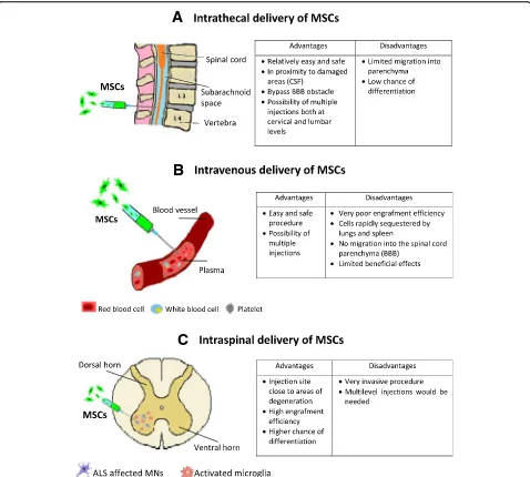
One of the major technical issues in ALS cellular therapy is to effectively deliver stem cells into the CNS. Several studies demonstrated that injecting human stem cells directly into the spinal cord of ALS rodents is feasible, safe and efficient [40, 70, 71, 128]. The surgical proced-ure for intraspinal infusion of hMSCs into the spinal cord of ALS patients by specifically designed micro-injection manipulators have also been shown to be safe in open-label clinical trials [75, 76, 78, 79, 129]. How-ever, direct CNS injection remains an invasive proced-ure which could cause serious clinical complications and permanent damage to CNS areas already affected by the disease. Thus, it might be a considerable ethical issue in clinical trials when investigating the efficacy of the therapy, especially where placebo controls are required.
[image:14.595.55.538.429.662.2]Since early pathologic mechanisms of disease involve destruction of neuromuscular junctions before motor neuron death, intramuscular injection of MSCs at an early stage of disease has also been proposed. Intramus-cular injections of hMSCs engineered to overexpress GDNF into SOD1G93A rats resulted in delayed disease onset and prolonged survival, but MSCs were not able to regenerate motor end-plates, nor to ameliorate motor neuron loss [72].
Given the immunomodulatory properties, together with the homing capacity in response to inflammatory signals, hMSCs showed beneficial effects when infused intravenously in ALS animal models [58, 61]. However, even though intravenous infusion could be the easiest and safest way for stem cell delivery, very few cells ap-pear to be able to migrate, penetrate and engraft into the spinal cord parenchyma. Thus, a large number of cells may need to be transplanted in order to obtain ef-fective results in human subjects.
The delivery of stem cells into the CSF surrounding the spinal cord by intrathecal injection may be a useful compromise, since stem cells would be placed in proxim-ity to damaged areas without direct delivery into the spinal cord parenchyma. Furthermore, it would be possible to perform multiple injections both at the cervical and lum-bar levels, allowing stem cells to migrate towards more af-fected areas of neurodegeneration/inflammation [67]. In some preclinical studies, stem cells were able to migrate from the CSF into the parenchyma, however, the mecha-nisms of migration remains unknown [64, 68]. Figure 4 is a representation of different proposed strategies to deliver MSCs into ALS patients with the relative advantages and disadvantages.
Strategies to enhance tissue penetration
The study of CNS inflammation models allowed the dis-covery of chemokines, receptors, adhesion molecules, and inhibitors that govern and regulate lymphocyte mi-gration, extravasation and penetration into the CNS par-enchyma. Even though the migration of lymphocytes from the blood to the CSF has been extensively studied, very little is known about the transmigration of lympho-cytes from the perivascular space to the CNS parenchyma, where astrocytes and the basal membrane constitute a second impermeable barrier [130].
Curiously, important chemokines released during in-flammation which stimulate lymphocyte migration through the BBB such as CXCL12 and CXCL10, were shown to inhibit infiltration into the parenchyma, indu-cing lymphocytes to persist in the perivascular space [131, 132]. Conversely, production of metalloproteases such as MP-2 and MP-9 seems to be necessary in order to break the dystroglycan basal membrane required for penetration into the CNS [130, 133].
MSCs express several chemokine receptors and hom-ing properties similar to that of lymphocytes which allow them to migrate into damaged or inflamed tissues. How-ever, donor age and the number of passages in culture were shown to negatively influence the stem cell’s ex-pression of homing factors [134].
Interestingly, Corti and colleagues have performed intrathecal and intravenous injection in SOD1G93A mice of hiPSC-derived neural cells after being sorted for expres-sion of the integrin VLA4 (CD49d) by FACS [49]. A con-siderable number of transplanted cells were able to migrate into the grey and white matter of spinal cord [49]. Engineering MSCs in order to induce overexpression of chemokine receptors or factors involved in homing and migration to sites of inflammation, may enhance the delivery into the spinal cord parenchyma. The characterization of chemokines and relative concentra-tion in spinal cord and CSF of ALS models during dif-ferent stages of disease would be of interest to identify the best candidate and also the peak of inflammatory chemokine production in order to define the ideal time of intervention for stem cell injection.
Future directions in clinical trials
During the past two decades, several clinical trials inves-tigated the use of stem cells in patients with ALS, mostly focusing on the safety and feasibility of the intervention. Nonetheless, as reported by Appel and Armon, stem cells have often been transplanted into ALS patients with lim-ited preclinical data, without providing details on adverse effects and using small numbers of participants [135]. Moreover, the long-term safety of stem cell transplant-ation (e.g. non accelertransplant-ation of disease progression asso-ciated with the cell implantation) has been evaluated by comparison with historical control groups. This is an important limitation since heterogeneity in the patient population and differences in clinical care between the treated group and historical controls may affect the readouts [135].
Although single-armed, small phase I/II clinical trials found that cell-based therapy for ALS is relatively safe and feasible, it is uncertain whether stem cell transplant-ation may be clinically beneficial leading to functional improvements and of the slowing of disease progression. The majority of the clinical trials were principally fo-cused on the safety and feasibility of the surgical proced-ure related to the stem cell implantation. Secondly, these clinical trials were not powered to demonstrate any clin-ical benefit [136].
quasi-RCT or cluster quasi-RCT involving the use of stem cells in ALS/MND patients. Thus, there is an absence of high-quality published evidence to assess the safety and efficacy of stem cell transplantation in ALS [136]. Moreover, when analyzing the single arm phase I/II clinical studies avail-able in the literature, the authors found substantial vari-ability between clinical trials in terms of selection criteria, intervention methods and objective outcomes, with the involvement of small number of participants and a short-term follow-up period [136] .
To investigate the long-term safety and efficacy of cellular therapy for ALS, well-designed prospective randomized-controlled trials with larger sample size,
long-term-follow up and standardization of cell prod-ucts, are urgently needed [136].
Recently, the ALS Clinical Trials Workshop (Airlie Conference, Virginia, 2016), driven by the ALS commu-nity, clinicians, researches, industry representatives, government representatives, ALS patients and family members, released and submitted to the FDA the draft version of the “Guidance for Industry on Drug Devel-opment for Amyotrophic Lateral Sclerosis”. The manu-script has been drafted with the intention of improving and accelerating the drug development process, includ-ing guidelines for a more effective clinical trial design in ALS [137]. In particular, the guideline highlighted
A
B
C
[image:16.595.59.537.87.517.2]the importance of reducing clinical and genetic hetero-geneity when establishing the patient selection criteria. With regards to clinical heterogeneity, inclusion and exclusion criteria for patient enrolment must be well justified for each study, which could vary depending on the phase of development [137].
Selection criteria should also depend on the specific trial goals, since some therapies may be more effective in early stage of disease or in specific sub-group of pa-tients. Thus, investigators should endeavor to incorpor-ate reliable predictive and prognostic biomarkers for clinical trial eligibility or stratification criteria [137].
Because of the multifactorial nature of ALS, post-hoc analysis of reliable biological markers and neurophysio-logical data is extremely important, since in specific sub-groups of patients a beneficial effect could be missed dur-ing the analysis. However, responder analyses must only be used as hypothesis generating, which will need to be confirmed and further investigated in future trials [137].
Importantly, although early phase trials are not meant to investigate efficacy, for cell-based therapies which em-brace potentially invasive delivery methods and lifelong biological effects, evaluation of efficacy should be in-cluded in the design of phase I trials. Furthermore, a long-term monitoring plan to evaluate tumorigenesis, stem cell engraftment and long-term efficacy should be included [137].
Finally, RCTs with placebo controls is considered as the gold standard for clinical investigation. However, in rare, rapidly progressive and untreatable diseases such as ALS, the requirement for a placebo could be revisited, especially if the intervention involves invasive methods. While in phase III clinical trials a randomized placebo group is indispensable, in earlier phase studies, the use of historical controls and predictive algorithms could be accepted. However, the development of target-specific biomarkers, standardization of outcome measures and validation of surrogate end-points is essential, especially in cell-based studies in which the mechanisms mediating the therapeutic effect are not well established.
Conclusions
During the last decade, the great advances achieved in regenerative medicine have created an unprecedented enthusiasm and new hope for amelioration of the devas-tating and until now incurable disease that is ALS. The initial exciting idea of replacing lost of motor neurons, with MSCs is unlikely to be achieved, although other strategies may provide some promise for stem cell derived motor neuron replacement [138]. Indeed, transplanted cells should differentiate and integrate with the host spinal motor circuits within the toxic and non-permissive environment that characterizes ALS. However, the neuro-protective and immunomodulatory potential of MSCSs
could match perfectly with the multifactorial nature of ALS.
Among different types of stem cells, MSCs represent a promising candidate for clinical application. However, several technical issues need to be addressed including: route of administration, optimal dose, and differentiation state and neuroprotective mechanisms of transplanted cells. In addition, in the majority of pre-clinical experi-ments stem cell injection was performed before disease onset. This is an important limitation for translation into the clinic where the diagnosis takes several months and early markers of disease are lacking [139]. Another limi-tation is the exclusive use of the SOD1G93A model, which may reflect the pathological features of only a very small proportion of patients. Although these mice repre-sent a robust model of ALS, only a small proportion of ALS cases are caused by SOD1 mutations [140]. Thus, the use of other ALS models such those driven by mutant TDP-43 and C9ORF72 expansions would be of interest [141].
Also, despite the excellent utility of the SOD1G93A mice in revealing pathological ALS features, translation of promising therapies from this model has failed in hu-man clinical trials [142]. Apart from considerable differ-ences between rodents and humans, the answer to this problem could be attributed to the existence of a high rate of intra- and inter-laboratory variability [142]. Such variability could be improved by a more meticulous pre-clinical design, use of defined inbred mouse strains, trans-gene copy number analysis and trying to standardize preclinical investigation methods [142, 143].
In general, the presence of injected stem cells within the host nervous system of treated rodents was evaluated only a few days or weeks post-transplantation. Thus, the design of experiments looking at the long-term survival and integration of cells is necessary.
Evaluation of the presence of injected cells in the host has been carried out by the exclusive use of immunohis-tochemistry. Adopting advanced microscopy techniques such as confocal and two-photon microscopy would be of great interest to confirm stem cell engraftment, which could also allow tracking in vivo the fate of transplanted cells [144, 145].
greater cellular engraftment after transplantation in animals [146–148].
Thus, the regenerative potential of stem cells could be considerably improved by adopting 3D culture methods. Transplantation of bio-scaffolds or encapsulating MSCs to sustain favorable conditions for stem cell survival, growth, migration and maturation is showing promising results in experimental models of spinal cord injury, traumatic brain injury and nerve regeneration, and must be considered in ALS models where the presence of a hostile microenvironment is one of the main factors that negatively affects stem cell engraftment and therefore therapeutic potential [147, 149, 150].
The optimal maturation level for the transplantation of stem cells to obtain the greatest therapeutic benefit is also unclear. If undifferentiated cells may represent the best way to obtain immunomodulation and trophic fac-tor production, induced neural progenifac-tors may over-come the possibility of tumour formation, along with the possibility of in vivo maturation and integration into existing neuronal circuits.
A combination strategy could be interesting, where intravenous infusion of immature cells may generate permissive environmental conditions for the successive implantation of neuronal committed cells within the CSF. This would require an in-depth study of the matur-ation process of MSCs into neurons, trying to under-stand, optimize and standardize differentiation protocols that promote the generation of stable precursors.
Last but not least, although autologous transplantation can reduce the probability of immune rejection, it has been reported that patient-derived MSCs may have im-paired or reduced therapeutic effects [151, 152]. Other evidence indicates no functional alteration or accelerated cellular senescence in BM-MSCs derived from sporadic cases of ALS [153]. Moreover, it was shown that there was the possibility to functionally restore defective ALS-derived MSCs by correcting alterations in DNA methylation [153, 154]. However, the identification of MSC biological markers predictive of a positive/negative therapeutic response in ALS patients would be of great value [82]. In relation to the use of autologous MSCs in patients carrying ALS causative genetic mutations, stem cells could be genetically corrected by adopting CRISPR-Cas9-mediated gene editing technology [155].
In conclusion, several strategies have been tested in the SOD1G93Amouse model, but with different hurdles. All the discussed parameters should be reconsidered and optimized before the translation of stem cell therapy from mice to humans in order to avoid undesirable de-lays or therapy failure as has happened for most of the promising results derived from SOD1G93A transgenic mouse models, with failure of translation into clinical benefits for ALS patients.
Abbreviations
ADSC:Adipose derived stem cells; ALS: Amyotrophic lateral sclerosis;
BBB: Blood brain barrier; BDNF: Brain derived neurotrophicfFactor; bFGF: basic fibroblast growth factor; BM: Bone marrow; BMNC: Bone marrow
mononuclear cells; CD: Cluster of differentiation; CNS: Central nervous system; CNTF: Ciliary neurotrophic factor; CSF: Cerebrospinal fluid; DCX: Doublecortin; DMEM: Dulbecco’s modified eagle medium; FALS: Familial amyotrophic lateral sclerosis; FBS: Foetal bovine serum; FUS: Fused in sarcoma protein; GDNF: Glial derived neurotrophic factor; GFAP: Glial fibrillary acidic protein; GFP: Green fluorescent protein; GLT1: Glutamate re-uptake transporter 1; GMP: Good manufacturing practice; hESC: human embryonic stem cells; HGF: Hepatocyte growth factor; ICV: Intracerebroventricular; IGF-I: Insulin-like growth factor-1;
IHC: Immunohistochemistry; IL: Interleukin; iPSC: induced pluripotent stem cells; ISCT: International Society for Cellular Therapy; IV: Intravenous; MAO-B: Monoamine oxidase B; MCP-1: Monocyte chemoattractant protein 1; MHC-II: Major histocompatibility complex class II; MND: Motor neuron disease; MNLCs: Motor neuron-like cells; MNPs: Motor neuron progenitors; MNs: Motor neurons; MS: Multiple sclerosis; MSCs: Mesenchymal stem cells; NGF: Nerve growth factor; Ngn1: Neurogenin1; NPs: Neural progenitors; NSC: Neural stem cells; NT-3: Neurotrophin 3; OPTN: Optineurin; PBMC: Peripheral blood mononuclear cell; PBS: Phosphate buffered saline; PNN: Perineural net; RA: Retinoic acid; RCT: Randomized controlled trial; SALS: Sporadic amyotrophic lateral sclerosis; SCI: Spinal cord injury; Shh: Sonic hedgehog; SMA: Spinal muscular atrophy; SOD1: Superoxide dismutase 1; SVF: Stromal vascular fraction; TARDBP: TAR DNA-binding pro-tein 43; TGFα: Transforming growth factor-alpha; TGFβ: Transforming growth factor-beta; VCP: Valosin containing protein; VEGF: Vascular endothelial growth factor; WAT: White adipose tissue; WB: Western blotting; WT: Wild-type
Acknowledgements
University of Sheffield-Tongji University Joint Doctoral Researcher Programme (YC, PJS, RM), the NIHR Sheffield Biomedical Research Centre for Translational Neuroscience (PJS). Medical Research Council (MRC) Award Number: MR/M010864/1 (KN). Medical Research Council (MRC) award Number: MR/M010864/1 (PJS). MND Association Award Number: 983-797 (RJM).
Funding
This work was funded by a University of Sheffield-Tongji University collaborative PhD scholarship awarded to YC. PJS is an NIHR Senior Investigator and this work was also supported by the NIHR Sheffield Biomedical Research Centre for Translational Neuroscience.
Availability of data and materials
Not applicable.
Authors’contributions
All authors assisted in the planning and conception of the review article, YC drafted the manuscript and figures, RJM, PJS, KN and XJ edited the manuscript. All authors read and approved the final manuscript.
Ethics approval and consent to participate
Not applicable.
Consent for publication
Not applicable.
Competing interests
The authors declare that they have no competing interests.
Publisher’s Note
Springer Nature remains neutral with regard to jurisdictional claims in published maps and institutional affiliations.
Author details
1
Sheffield Institute for Translational Neuroscience (SITraN), Department of Neuroscience, Faculty of Medicine, Dentistry and Health, University of
Sheffield, 385a Glossop Rd S10 2HQ, Sheffield, UK.2Tongji University School