replication of the virus and are the site of specific interactions with unidentified factors present in a host cell nuclear extract (P. Tam and C. R. Astell, Virology 193:812–824, 1993; P. Tam and C. R. Astell, J. Virology 68:2840–2848, 1994). In order to examine this region in finer detail, a comprehensive library of linker-scanning mutants spanning the region was tested for the ability to support replication of minigenome constructs and for the ability to interact with host cell factors. Three short discrete sequence elements critical for replication competence were observed. Binding of host cell nuclear factors was localized to four sites, with two major complexes each appearing to have two binding sites within the region. All factor binding sites were found to be directly adjacent to or overlapping with sequence elements contributing to replication competence, and evidence suggesting a correlation between factor binding and minigenome replication is presented. A possible model is proposed for function of a viral origin within the region of the internal replication sequence which addresses the still-unresolved problem of how parvoviruses overcome the thermodynamic energy barrier involved in the rearrangement of the 5*-terminal palindrome from an extended form to a hairpin conformation.
Minute virus of mice (MVM) is a member of the parvovirus family of nondefective parvoviruses. It has a single-stranded negative-sense DNA genome of 5,149 nucleotides (nt), with unique palindromic sequences at either terminus which can fold to form the characteristic hairpin telomeres common to this virus family. Current models for parvoviral DNA replica-tion (1, 2, 5, 7, 22) postulate that these hairpin termini serve as essential primers for DNA synthesis, with the stably
base-paired 39end being elongated to convert the incoming genome
to a double-stranded monomer replicative form (mRF). The 59
terminus, now in an extended double-stranded form, is pre-sumed to undergo a rearrangement to form two hairpin struc-tures, thereby affording a primer for extension back along the
newly synthesized strand and displacement of the 39 hairpin
structure to generate a dimeric double-stranded replicative form (dRF). The dimer bridge can be resolved asymmetrically (14) (through a process which requires the viral NS-1 protein; the precise mechanism is as yet unknown) to generate progeny mRF molecules, or a further round of terminal rearrangement can occur, followed by extension to lead to higher-order con-catemers (Fig. 1).
Sequences inboard of the 59palindrome have been shown to
contribute to viral minigenome replication competence; gross deletions within this region (the internal replication sequence [IRS]) reduce or abolish the replication of MVM minigenomes (19). Gel retardation and footprinting analyses of sequences in this region with an LA9 cell nuclear extract indicate that un-known host cell nuclear factors bind specifically within the IRS (20). In this report, we extend these observations by identifying three short discrete sequence elements which contribute to the replication competence of MVM minigenomes. Additionally, we present data to identify where the binding sites for host cell
factors occur within the IRS and, finally, data to suggest a relationship between binding of these factors and viral repli-cation efficiency.
MATERIALS AND METHODS
Oligonucleotides.The synthetic oligonucleotides employed in this study are presented in Table 1. All oligonucleotides were prepared on an ABI 371 or 374 DNA synthesizer.
Plasmid constructs. Details of the construction of pCA4.0, pPTLR, and pCMVNS-1 have been published previously (19). For conciseness, for some of the following clones only the outline of the construction is presented; complete details of all construction schemes are available on request. Construct pJB2.0 was made by cloning a PCR product corresponding to viral nt 4484 through 4777 between EcoRI and BamHI tags (obtained by using primers MVMA5 and MVMB3 on a pCA4.0 template) between the EcoRI and BamHI sites of pUC19. Oligonucleotide MVMA5 simultaneously introduced a single T3C silent nucle-otide substitution at viral nt 4486 to introduce a unique BstEII site. The mini-genome used in these studies, pJBLR4.3, was constructed from pPTLR by digestion, filling in, and religation of the BamHI linkers flanking the viral mini-genome to convert them to ClaI sites; introduction of a single T3C silent nucleotide substitution at viral nt 4486 to introduce a unique BstEII site in the minigenome; and substitution of the viral sequence TAGGTTAAT at nt 4780 with CCCTAGGC, thereby introducing a unique BamHI site. No significant change in replication levels of this construct relative to that of the original minigenome was noted (data not shown).
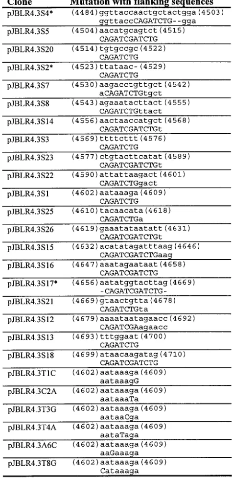
Construction of a linker-scanner library. Digestion of pJB2.0 with either EcoRI or BamHI followed by limited digestion with Bal 31 nuclease and religa-tion in the presence of excess synthetic double-stranded BglII linker (pCAG ATCTG) created pools of clones with partial deletions of the cloned viral sequences, known asDEco orDBam clones, respectively. Clones were sequenced (Sequenase 2.0; U.S. Biochemicals) to determine deletion endpoints, andDEco andDBam clones which matched to recreate the original cloned viral sequence with a central gap of 8 nt filled by the BglII linker were joined together at the linker. Clones which were found to together reconstitute the original cloned viral sequence with a gap of exactly 12 bp were digested with BglII, the recessed 39end was filled in by using Klenow fragment in the presence of all four deoxynucleo-side triphosphates, and the clones were ligated together to recreate the original viral sequence with an internal 12 bases replaced with a ClaI linker (CAGATC GATCTG). Clones produced in either fashion are referred to as pJB2.0Sx, where x is a number specific to the particular linker-scanning mutation.
In some cases where a matching set ofDEco andDBam clones could not be found, PCR was used to create a suitable match to an existing deletion;
specif-* Corresponding author. Phone: (604) 2142. Fax: (604) 822-5227. E-mail: [email protected].
9087
on November 9, 2019 by guest
ically, clones S20, S21, S22, and S23 were created this way, with the matching clones made by PCR between primer pairs S20E-MVMB3, S21B-MVMA5, S22B-MVMA5, and S23B-MVMA5, respectively, with pJB2.0 used as a tem-plate. These products were cloned into pUC19 vector linearized with SmaI and then subjected to digestion with BglII (followed by filling in with Klenow frag-ment for S23) and either EcoRI (S21, S22, and S23) or BamHI (S20) and used as were the nativeDEco andDBam clones.
Clones S25 and S26 were made totally by PCR. Mutant S25 was made by PCR using oligonucleotides S25-2 (phosphorylated with T4 polynucleotide kinase) and MVMA5 on a pJB2.0 template; the product was ligated to the small SspI fragment of pJB2.0, and the resulting mixture of products was used as a template for PCR with primers MVMA5 and MVMB3. The resulting product was di-gested with EcoRI and BamHI and cloned into pJBLR4.3 vector didi-gested with the same enzymes, directly yielding pJBLR4.3S25. Mutant S26 was constructed by PCR using primer pairs S26C-MVMA5 and S26D-MVMB3 on a pJB2.0 template. The products were isolated and ligated together, and the ligation product was used as a template for a PCR with primers MVMA5 and MVMB3. The resulting product was digested with EcoRI and BamHI and cloned into pJBLR4.3 vector digested with the same enzymes, to yield pJBLR4.3S26.
Dual mutant S8/22 was made by using pBLR4.3S8 as a template for PCR with
primers MVMA5 and S822C and using pJBLR4.3S22 as a template for PCR with primers S822D and MVMB3 and ligating the two products. The ligation product was used as a template for PCR with MVMA5 and MVMB3, and the resulting product was digested with BamHI and BstEII prior to being cloned into pJBLR4.3 vector digested with the same enzymes. Scanner mutant clones in the context of pJB2.0 were introduced into the minigenome by replacing the equiv-alent BstEII-to-BamHI fragment of pJBLR4.3 with that of pJB2.0Sx to yield pJBLR4.3Sx.
All clones were sequenced to ensure their integrity.
Point mutants.T1C, C2A, T3G, T4A, A6C, and T8G mutants were made by PCR with the respective oligonucleotides and MVMA5 on a pJB2.0 template. Gel-isolated products were ligated to isolated PCR product obtained with prim-ers S1P and MVMB3 on a pJB2.0 template. The resulting ligation products were used as templates for PCR with primers MVMA5 and MVMB3, and the prod-ucts were gel isolated, digested with BstEII and BamHI, and cloned into pJBLR4.3 digested with the same enzymes to yield the respective minigenomes containing single point mutations.
[image:2.612.126.472.69.248.2]Cell lines and virus.COS-7 cells (8) were grown in Dulbecco’s modified Eagle’s medium supplemented with 10% fetal bovine serum, 10 mM HEPES (pH 7.3), and 20mg of gentamicin per ml. A9 oubrL (LA9) cells (13) were grown
[image:2.612.48.570.522.726.2]FIG. 1. MVM replication cycle. Monomeric genome (step 1) is extended from the base-paired 39terminus (step 2), displacing and extending the 59hairpin structure (mRF) (step 3). The extended hairpin termini undergo rearrangement to self-associate (step 4), affording a primer for further extension to a dimer-length molecule (dRF) (step 5). This may then undergo further rounds of terminal rearrangement as in step 4 and extension to higher-order concatemers, or asymmetric resolution by site-specific nicking by NS-1 (shaded arrows) and ligation to generate the forms in steps 6a and 6b. The step 6a form is identical to the form in step 3 and may be used to continue the cycle, while the step 6b form serves as a template to generate viral progeny by site-specific nicking by NS-1 (shaded arrow) and hairpin transfer (step 7). Encapsidation of the upper progeny strand (step 8) as a mature genome (step 1) allows the lower strand to regenerate (step 6b), thereby continuing the cycle.
TABLE 1. Oligonucleotides used in this study
Oligonucleotide Sequence
MVMA5...GGAATTCCGGTTACCAACTGCTACTGGAA MVMB3 ...CGGGATCCCGAACCACCCTTCCACCCTTTTA S20E...CAGATCTGTTATAACAAGACC
S21B...CAGATGTGTAAGTACCATATTA S22B...CAGATCTGATATGAAGTACAG S23B...CAGATGTGAAAGAAAAAGCATG
S25-2 ...ATTATATTTCTCAGATCTGTCTTTATTAGTCTTAATAATATATG S26C...TATGTTGTATCTTTATTAGTCTTAATAATATATG
S26D ...CAGATCGATCTGTACATATAGATTTAAGAAATAG S822C...AAGCATGGTTAGTTAG
S822D ...pTTTCTTTCTGTACTTC
T1C ...CCTTTATTAGTCTTAATAATATATG C2A...TATTTATTAGTCTTAATAATATATG T3G...TCGTTATTAGTCTTAATAATATATG T4A...TCTATATTAGTCTTAATAATATATG A6C...TCTTTCTTAGTCTTAATAATATATG T8G...TCTTTATGAGTCTTAATAATATATG S1P...pTACAACATAGAAATATAATATTAC Vecpro...CGCGTTTCGGTGATGAC
JAVA...TTTTGGTCCTTAACATCAAG
on November 9, 2019 by guest
http://jvi.asm.org/
(SDS)–53Denhardt’s reagent–10mg of fragmented, denatured salmon sperm DNA per ml at 42°C for 4 h and then hybridized in 63SSPE–1% SDS with 5 ng of32P-59-end-labeled Vecpro oligonucleotide at 39°C for 10 to 15 h. The blots
were washed twice at room temperature in 63SSPE–1% SDS for 3 to 5 min and once in 23SSPE at 39°C for 3 min prior to visualization with a phosphorimager (Molecular Dynamics PhosphorImager SI; IP Lab Gel Version H [Signal Ana-lytics] was used in image analysis). Results of this exposure provide data on input DNA present in each sample, as Vecpro hybridizes uniquely to the vector backbone. The blots were then stripped (washed in 63SSPE–50% formamide at 65°C for 30 min and then in 23SSPE at 65°C for 30 min) and again visualized to verify removal of the probe. Prehybridization, hybridization, washing, and exposure were then carried out exactly as before, but with 59-end-labeled oligo-nucleotide probe JAVA, which allows for determination of the amount of viral sequence present in each sample by hybridizing uniquely to sequences present in the minigenome.
Replication data were analyzed by preparing a standard curve from the amount of signal obtained from the range of linearized pJBLR4.3 standard applied to each blot and using this standard curve to determine the quantity of DNA present in each of the samples corresponding to each of the transfections. Replication efficiency (RE) was calculated as RE5(viral sequence2input DNA)/input DNA. Samples consisting of viral minigenome alone were taken as having no replication, and a scaled replication efficiency (SRE) was calculated for each sample (n) as SRE(n)5RE(n)2RE(pJBLR4.3). Samples consisting of wild-type viral minigenome and pCMVNS-1 were taken as having 100% replication, and a scaled relative replication efficiency (SRRE) was calculated for each sample (n) as SRRE(n)5SRE(n) / SRE(pJBLR4.31pCMVNS-1). In-dividual data sets thus normalized for 0 and 100% replication were pooled for statistical analysis.
Competitive band shift assays.Gel retardation assays were carried out with gel-purified PCR products or restriction fragments prepared from the wild-type or scanner mutant template or clone. Scanner mutants S4, S5, S20, S2, and S7 were analyzed with the BstEII-SspI restriction fragment (viral nt 4484 to 4626; BS fragment); mutants S8, S14, S3, S23, S22, S1, and S15 were analyzed with a PCR product obtained with primers S20E and S21B (viral nt 4524 to 4668, flanked on either side by CAGATCTG; 2021 fragment); and mutants S16, S17, S21, S12, S13, and S18 were analyzed with a PCR product obtained with primers S1P and MVMB3 (viral nt 4610 to 4781; SB fragment). All purified fragments were quantitated by binding of Hoechst 33258 dye and fluorometry on a Hoeffer TKO-100 fluorometer. Assays were performed by incubating 1 ng of end-labeled wild-type fragment in 50 mM NaCl–10 mM Tris–1 mM dithiothreitol–5% glyc-erol (pH 7.1) containing approximately 3.75mg of LA9 nuclear extract (20) for 30 min. Competition by a given mutant was assayed by including 40 ng of unlabeled mutant-derived fragment in the binding reaction mixture and allowing it a 10-min preincubation with the nuclear extract before addition of the labeled wild-type probe. The reaction products were analyzed on 4% polyacrylamide–1% glycerol vertical gels run at 15 V/cm in 0.53Tris-borate-EDTA–1% glycerol buffer at 4°C. The gels were dried and visualized by phosphorimaging.
RESULTS
Three distinct short sequence elements within the IRS con-tribute to the replication competence of minigenomes. Previ-ous studies of the IRS had used gross deletions to map the locations of two elements (element A, between nt 4489 and 4636, and element B, between nt 4637 and 4695) which con-tribute to replication of viral minigenomes. In order to exam-ine the IRS in greater detail for the specific sequence elements contributing to replication competence, a series of
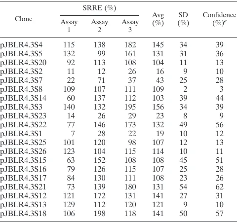
[image:3.612.308.545.78.566.2]linker-scan-ning mutations (S1 through S26 [Fig. 2]) were engineered into a minigenome construct (pJBLR4.3). The minigenome con-structs were assayed for replication competence (Fig. 3) in three independent trials. The results of these assays are shown in Table 2 and displayed graphically in Fig. 4. Three regions (those replaced by scanner mutations S2 and S7, mutation S23,
FIG. 2. Twenty-six linker-scanning and point mutations presented in order of sequence position. For each clone, the associated wild-type sequence is pre-sented with the nucleotide positions relative to full-length MVM of the first and last nucleotide shown in parentheses (top line). The same sequence region from the mutant is then presented in alignment, with the mutation indicated in up-percase type (bottom line). Three mutations (*) do not maintain exact spacing; placeholders in the sequence to maintain wild-type spacing are indicated (dash-es). The gap indicated in the wild-type sequence associated with mutation S2 indicates that this inserts a single nucleotide relative to the wild-type sequence.
on November 9, 2019 by guest
and mutation S1) which when replaced with linker sequences resulted in a statistically significant reduction in replication competence relative to that of the wild-type minigenome at the 95% confidence level were found. Mutations S2, S23, and S1 all reduce minigenome replication efficiencies to below 25% of the wild-type level, while S7 reduces replication efficiency to less than 40% of wild-type levels. That the sum of these rep-lication defects is greater than 100% suggests some degree of interdependence of the function(s) of these sequence ele-ments.
Host cell factor binding sites are adjacent to sequences important in conferring replication competence. In order to assess the effect of the linker-scanning mutations on the bind-ing of factors present in an LA9 nuclear extract, a series of electrophoretic mobility shift (band shift) assays were per-formed. Since the entire IRS was found to be too large to be
conveniently examined with a single probe, the region was divided into three overlapping DNA fragments of approxi-mately equal lengths: BS fragment (143 bp), 2021 fragment (145 bp), and SB fragment (172 bp) (Fig. 5). Each linker-scanning mutation was analyzed in the context of only one of these probes so that none of the mutations were tested near the ends of the probe fragment (see Materials and Methods). Competition gel retardation assays carried out as outlined in Materials and Methods revealed the presence of two distinct complexes (A and B) formed by factors present in the nuclear extract. A third shifted species (nonspecific [ns]) was observed with all three probes only when competitor fragment was present. No difference between the abilities of specific and nonspecific competitors to titrate the formation of this species was observed across a range of concentrations up to 400-fold excess, indicating that this species is due to a nonspecific in-teraction (data not shown).
None of the mutations examined with either the BS or the SB probe resulted in a loss of competition activity (Fig. 6A and C). For mutations examined with the 2021 probe, both com-plexes were effectively competed for by a 40-fold excess of several competitors. However, competitor 2021-S8 failed to compete for the binding of complex B; competitors 2021-S22 and 2021-S26 lost the ability to compete for the binding of complex 2021-A, with the S22 mutation having the least com-petition activity; and adjacent-mutation competitors 2021-S1 and 2021-S25 had a reduced ability to compete for binding of complex 2021-B (Fig. 6B). It is of note that in each case there appears to be a weaker loss of competition for the other of the two complexes as well; the implications of this observation will be considered below (see Discussion).
[image:4.612.118.482.70.235.2]The results of these studies with mutant competitors suggest that there are four binding sites for factors present in a host cell nuclear extract, a major and minor one each for the com-plexes forming 2021-B (major site under S8, minor site under S1 and S25) and 2021-A (major site under S22, minor site under S26). The appearance of a number of other weak shifted species at approximately the same mobility as the two major species, all of which appear to be due to specific interactions, suggests that each of the major complexes may consist of a number of subunits whose presence or absence in a small proportion of the complexes gives rise to heterogeneity in the samples. In all cases, the observed binding sites for host factors
FIG. 3. Replication assay as described in the text. The first column of slots (samples 1 through 5) is a pJBLR4.3 standard curve, the second column is a pCMVNS-1 standard curve as a control for hybridization specificity, and subsequent columns (samples 6 through 29) are Hirt extracts. Samples 6 and 28 are from cells transfected with pJBLR4.3 alone (negative replication controls), samples 7 and 29 are from cells cotransfected with pJBLR4.3 and pCMVNS-1 (wild-type replication controls), and the other samples are from cells cotransfected with a mutant minigenome and pCMVNS-1. Replication efficiency is calculated as described in Materials and Methods.
TABLE 2. SRREs of linker-scanning mutants
Clone
SRRE (%)
Avg
(%) (%)SD Confidence(%)a Assay
1 Assay2 Assay3
pJBLR4.3S4 115 138 182 145 34 39
pJBLR4.3S5 132 99 161 131 31 36
pJBLR4.3S20 92 113 108 104 11 13
pJBLR4.3S2 11 12 26 16 9 10
pJBLR4.3S7 22 71 37 43 25 28
pJBLR4.3S8 109 107 111 109 2 3
pJBLR4.3S14 60 137 112 103 39 44
pJBLR4.3S3 140 132 195 156 34 39
pJBLR4.3S23 14 26 29 23 8 9
pJBLR4.3S22 77 146 173 132 49 56
pJBLR4.3S1 7 28 22 19 10 12
pJBLR4.3S25 101 120 98 107 12 13
pJBLR4.3S26 123 104 115 114 10 11
pJBLR4.3S15 63 152 108 108 45 51
pJBLR4.3S16 79 126 115 107 25 28
pJBLR4.3S17 84 130 111 108 23 26
pJBLR4.3S21 73 139 180 131 54 62
pJBLR4.3S12 121 172 131 141 27 31
pJBLR4.3S13 129 112 120 121 9 10
pJBLR4.3S18 106 198 118 141 50 57
aConfidence calculated at the 95% level.
on November 9, 2019 by guest
http://jvi.asm.org/
[image:4.612.50.290.492.717.2]are directly adjacent to (or, in the case of mutation S1, coin-cident with) sequences contributing to replication competence, suggesting a probable correlation between these functions.
Simultaneous deletion of host factor binding sites sup-presses replication competence.To try to address whether the coincidence of sequences important in minigenome replication (S2 and S7, S23, and S1) with those binding host cell factors (especially S8 and S22) is biologically relevant, a dual-site scanner mutant in which the major binding sites for complexes 2021-A (under scanner mutation S22) and 2021-B (under scan-ner mutation S8) were simultaneously replaced was con-structed. Replication assays carried out with this construct are
summarized in Table 3. While neither individual mutation had an apparent effect on replication competence (Table 2), the dual mutant replicates at only 20% of wild-type levels (Table 3), suggesting that bound factors do play a direct role in rep-lication.
[image:5.612.129.468.70.273.2]Effects of point mutations within the region defined by scan-ner mutation S1.As scanner mutation S1 was shown to have an effect on minigenome replication competence in addition to being the site of binding of a host cell factor, it was desirable to attempt to identify which nucleotides within this 8-bp seg-ment contribute to these properties. To examine this, single-nucleotide point mutations were made at each of the six bases
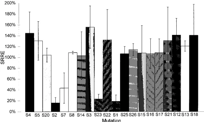
FIG. 4. SRREs for each of the linker-scanner mutations in the minigenome context. Mutations are arranged in order of location, and each is designated to indicate the corresponding linker-scanning mutation as listed in Fig. 2. The values plotted are the averages of triplicate assays, with error bars indicating a 95% confidence interval.
FIG. 5. Schematic representation of the IRS region and location of sequence elements observed in this study. The top line represents the IRS (element A is nt 4489 to 4636, and element B is nt 4637 to 4695) plus nearby flanking regions. Locations of RsaI sites delimiting RsaI A (nt 4431 to 4579) and B (nt 4580 to 4662) fragments are indicated. Linker-scanning mutations causing a loss of minigenome replication competence and their relative positions are shown on the next line (shaded boxes). The third line indicates the names and locations of linker-scanning mutations interfering with the binding of host cell factors (shaded according to whether they interfere with the binding of complex 2021-A [Complex A] or 2021-B [Complex B]). At the bottom, regions used as band shift probes are shown (see text).
on November 9, 2019 by guest
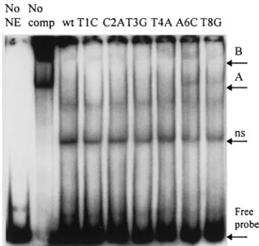
altered in the scanner mutant, converting the wild-type base to that present in the scanner mutant individually. These mutants were then assayed both for replication competence in a mini-genome-based assay (summarized in Table 3) and for the abil-ity to interact with host cell nuclear factors by band shift assay (Fig. 7). While two of the point mutants (A6C and T8G) appear to lose some ability to inhibit the formation of both the A and the B complexes, none of the point mutants display a detectable replication defect.
DISCUSSION
[image:6.612.59.277.73.663.2]Existing genetic evidence indicates that the MVM genome has origins of DNA replication at either terminus, with that at the right genomic end containing the stronger signals. Previ-ously published data indicate that functional elements of this origin include sequences between viral nt 4489 and 4695, the IRS (19). Data presented in this study identify three specific short sequence elements which contribute to the activity of this IRS. When these elements are replaced with unrelated DNA sequences, a dramatic loss of minigenomic replication effi-ciency is observed. Together, these elements appear to form a multipartite origin of DNA replication reminiscent of that
[image:6.612.308.549.89.207.2]FIG. 6. Competition band shift assays performed with probes representing the entire library of linker-scanner mutations. Lanes No NE, no nuclear extract; lanes No comp, no competitor fragment; lanes wt, 40-fold excess of unlabeled wild-type competitor; other lanes, 40-fold excess of the respective linker-scan-ning mutation-derived competitor fragment. Results with the BS probe (A), the 2021 probe (B), and the SB probe (C) are shown. ns, nonspecific band; A and B, complexes A and B.
[image:6.612.332.521.493.673.2]FIG. 7. Competition band shift assays performed with competitors represent-ing each of the S1 site srepresent-ingle-nucleotide substitutions. Lane No NE, no nuclear extract; lane No comp, no competitor fragment; lane wt, 40-fold excess of unla-beled wild-type competitor; and the other lanes, 40-fold excess of the indicated point mutation-derived competitor fragment. All DNAs are the respective 2021 PCR products. ns, nonspecific band; A and B, complexes A and B.
TABLE 3. SRREs of point mutants and the dual-site scanner mutant
Clone
SRRE (%)
Avg
(%) (%)SD Confidence(%)a Assay
1 Assay2 Assay3
pJBLR4.3T1C 100 92 94 95 4 5
pJBLR4.3C2A 166 123 135 141 22 25
pJBLR4.3T3G 105 125 129 120 13 14
pJBLR4.3T4A 97 109 109 105 7 8
pJBLR4.3A6C 112 87 166 122 41 46
pJBLR4.3T8G 136 137 131 135 3 3
pJBLR4.3S8/22 18 26 18 20 4 5
aConfidence calculated at the 95% level.
on November 9, 2019 by guest
http://jvi.asm.org/
however, data presented here allow an upper limit to be placed on the relative strength of the left-hand origin at approximately 20% that of the right-hand origin. (This conclusion assumes that if any of the linker-scanning mutations examined in this assay completely abolished activity of the right-hand origin, the residual replication activity of the minigenome must arise from the left-hand origin.) All of the replication-defective mutations (with the exception of S7, which may very well only touch on the edge of a replication element) reduce minigenome repli-cation efficiency to approximately the same level (20%), sug-gesting the possibility that each of these mutations (S2, S23, S1, and the S8/S22 dual mutation) completely abrogates the func-tion of the right-hand origin. If this is the case, the simulta-neous requirement of all of these discrete sequence elements for origin function would indicate that there is a functional interaction between the elements.
While the S1 mutation was able to reduce minigenome rep-lication efficiency to 23% of wild-type levels, individual re-placement of each of the 6 nt varied from the wild-type se-quence in this mutation did not result in any detectable replication defect (Table 3). This observation suggests that no single nucleotide within this element is particularly responsible for the replication activity associated with the element. The small but definite replication hypercompetence exhibited by two of these point mutations (C2A and T8G) is puzzling, as is the weak hypercompetence observed with several of the scan-ner mutations (e.g., S3 [Fig. 4]). While no wholly satisfactory explanation for these observations can be put forward at this time, it is interesting that similar observations have been made in studies of other viral origins (18).
Earlier studies conducted with two RsaI restriction frag-ments on the interaction of host cell nuclear factors with se-quences from the region of the IRS indicated the formation of several specific complexes in this region (20). Data presented in this study extend these observations by indicating that there are two distinct complexes formed within the IRS: a larger complex (2021-B) which binds under the sequence element defined by the S8 scanner mutation and more weakly to se-quences under the S22, S1, and S25 mutations, and a smaller complex (2021-A) which binds to sequences replaced in the S22 mutation and less strongly to sequences replaced by the S26 mutation. The appearance of a number of faint, smaller shifted forms in the presence of competitors S8, S26, and S15 is most likely due to partial formation of the disrupted species. In each case where a mutation-containing band shift compet-itor fragment disrupts one of these binding sites and allows retention of the complex by the labeled wild-type probe, a weak retention of the other complex is observed. This suggests that binding of the complexes responsible for these two distinct shifted species is cooperative, a hypothesis which is supported
ancy between these findings and the data presented here is clearly explained if one assumes that the binding of the 2021-A and 2021-B complexes is cooperative; as the boundary between the RsaI fragments lies at viral nt 4579, the major binding site for complex 2021-B is on one fragment while the binding sites for complex 2021-A are on the other (Fig. 5). Studies with the
RsaI fragments individually would then be expected to detect
some shared complexes through protein-protein interactions. The lack of the major site for the binding of the other complex on each probe could readily be expected to result in a reduc-tion of complex stability and its potential partial dissociareduc-tion into constituent subunits, resulting in a number of smaller shifted forms.
Another line of evidence for cooperative interaction comes from consideration of the dual-site mutation and is discussed below.
In each case, the factor binding sites observed are adjacent to (or, in the case of sequences replaced in the S1 mutant, overlying) sequence elements required for efficient minige-nome replication. This suggests a possible functional relation-ship between factor binding and viral origin function, which is strongly supported by the results obtained with the S8/22 dual mutation. Neither of these mutations individually resulted in an apparent loss of minigenome replication competence; how-ever, the dual mutation reduces minigenome replication to the same basal level observed with the S2 and S1 mutations. The simplest interpretation of these observations would be that the factors responsible for formation of the 2021-A and 2021-B complexes are required for activity of the origin within the IRS. The already postulated cooperative interaction between the elements of these two complexes could account for the lack of a replication deficit in either of the individual mutations; bind-ing of either complex and subsequent localization of the sec-ond complex through protein-protein interaction may be suf-ficient to activate replication to wild-type levels observed in the minigenome assay employed.
The identities of the constituents of these complexes cannot be stated at present. Evidence from other origins suggests that transcription factors, helicases, and single-stranded binding proteins are all potential candidates, and sequence analysis has indicated that the observed 2021-A binding site (under muta-tion S22) is similar to a binding site for a poorly characterized transcription factor found in mouse (9). Genetic approaches to identify members of these complexes which are under way should help to clarify our understanding of the apparently complex interactions taking place at this origin of replication. When the model for viral replication as outlined in Fig. 1 is considered, an obvious question arises regarding what the function of sequences within the IRS is. A possible answer is
forthcoming when the 59-terminal rearrangement from an
on November 9, 2019 by guest
tended linear duplex to a dual-hairpin form is examined (Fig.
1, step 4). Given that the 59 palindrome is 206 nt long, the
spontaneous interconversion between these forms faces a pro-hibitively high thermodynamic barrier under physiological con-ditions (15) and argues for an enzymatic path if the virus is to replicate within a reasonable time frame. A model for replica-tion of the closely related H1 parvovirus proposed by Rhode and Klaassen (17) is of interest, as it postulates a canonical bidirectional eukaryotic origin in the region of the IRS which becomes active in the context of the mRF form. The leading strand originating from this origin along the upper template strand results in the stepwise displacement of the lower strand by the advancing replication machinery in an ATP-driven en-ergetically favored process. Full displacement of the lower strand allows it to spontaneously assume a hairpin
conforma-tion in which a free 39hydroxyl is able to act as a primer for
extension synthesis back along the mRF template to dRF form, as in Fig. 1. Careful consideration of this model, however, reveals (Fig. 8b) that as postulated by Rhode and Klaassen, with both leading and lagging strands originating from the IRS, nascent Okazaki fragments will result along the lower strand, with disastrous consequences for the virus: these Okazaki frag-ments will stop the palindrome from annealing back along its
full length and effectively block replication of the extreme 39
end of the mRF form, a fatal result from the point of viral replication.
If instead a leading-strand-only origin in the region of the IRS is postulated (Fig. 8a), an energetically favorable pathway for the terminal rearrangement required by all current models for parvovirus replication is afforded, while simultaneously the trap inherent in the Rhodes and Klaassen model is avoided (Fig. 8a). The idea of an exclusively leading-strand origin brings to mind mitochondrial DNA replication, which
pro-ceeds in a polymerase gamma (Pol g)-dependent
leading-strand-only fashion. Studies by Kollek et al. (11, 12) employing polymerase inhibitors in the course of native H1 infections led
to an identification of an early Pola–primase-dependent step
(initiation) followed by a longer Polg-dependent process.
Two recent reports in the literature have bearing on this
model. Cossons et al. (6) observed rearrangement of the 59
extended palindrome of MVM from a linear extended form to a hairpin-containing structure in vitro and concluded on the basis of selective sensitivity of this process to polymerase
in-hibitors that Pol d was required for the rearrangement. Of
particular interest is their observation that while hairpin
for-mation was observed, extension synthesis to form the dRF did
not occur. Current models for the action of Poldindicate that
it acts to synthesize leading and lagging strands in a coordinate manner. The replication model presented here would suggest
that an in vitro system in which Poldis acting at this viral origin
would be able to form an incomplete hairpin structure at the 59
terminus but not be able to prime synthesis to the dRF repli-cative intermediate. It is conceivable that the apparent
involve-ment of Poldin the in vitro studies by Cossons et al. may not
reflect the in vivo mechanism.
Other in vitro studies by Baldauf et al. (3) demonstrated that an LA9 cellular extract was capable of supporting the conver-sion of viral genomes into a covalently closed replicative-form (cRF) monomer, and only when the extract was supplemented with purified NS-1 were terminal resolution of the cRF, exten-sion to the mRF, and the appearance of dRF species observed. The ability of the supplemented extract to support replication to the level of dRF was suggested by the authors to result entirely from site-specific nicking by NS-1 as a requirement to open the cRF species and allow subsequent generation of the
linear 59 termini, a prerequisite for terminal rearrangement
and elongation to the dimeric species (3). This explanation is somewhat unsatisfactory, given that the system without NS-1 in reference 6 achieved both the linear mRF and some form of terminal rearrangement. An intriguing possibility not previ-ously considered is that NS-1 may play an obligate role in suppressing lagging-strand synthesis from the IRS origin, ei-ther by influencing the choice of polymerase used or through a more direct mechanism.
Preliminary studies in progress in our laboratory with the IRS elements isolated on a plasmid system indicate that the IRS may act as a leading-strand-only origin which is capable of driving the replication of plasmid sequences under the proper circumstances.
ACKNOWLEDGMENTS
These studies were supported by a grant from the Medical Research Council (Canada) to C.R.A. J.B. is the recipient of Natural Science and Engineering Research Council PGS-A and PGS-B scholarships and University of British Columbia University Graduate Fellowships.
REFERENCES
1. Astell, C. R., M. B. Chow, and D. C. Ward. 1985. Sequence analysis of the termini of virion and replicative forms of minute virus of mice DNA suggests a modified rolling hairpin model for autonomous parvovirus DNA replica-tion. J. Virol. 54:171–177.
2. Astell, C. R., Q. Liu, C. E. Harris, J. Brunstein, H. K. Jindal, and P. Tam. 1996. Minute virus of mice cis-acting sequences required for genome repli-cation and the role of the trans-acting viral protein, NS-1. Prog. Nucleic Acid Res. Mol. Biol. 55:245–285.
3. Baldauf, A., K. Willwand, E. Mumtsidu, J. Nu¨esch, and J. Rommelaere. 1997. Specific initiation of replication at the right-end telomere of the closed species of minute virus of mice replicative-form DNA. J. Virol. 71:971–980. 4. Bohenzky, R., R. LeFebvre, and K. Berns. 1988. Sequence and symmetry requirements within the internal palindromic sequences of the adeno-asso-ciated virus terminal repeat. Virology 166:316–327.
5. Chen, K. C., J. T. Tyson, M. Lederman, E. R. Stout, and R. C. Bates. 1989. A kinetic hairpin transfer model for parvoviral DNA replication. J. Mol. Biol. 208:283–296.
6. Cossons, N., E. Faust, and M. Zannis-Hadjopoulos. 1996. DNA polymerase
d-dependent formation of a hairpin structure at the 59terminal palindrome of the minute virus of mice genome. Virology 216:258–264.
7. Cotmore, S. F., and P. Tattersall. 1987. The autonomously replicating par-voviruses of vertebrates. Adv. Virus Res. 33:91–174.
8. Gluzman, Y. 1981. SV40-transformed simian cells support the replication of early SV40 mutants. Cell 23:175–182.
9. Hirsch, M.-R., L. Gaugler, H. Deagostini-Bazin, L. Bally-Cuif, and C. Gor-dis.1990. Identification of positive and negative regulatory elements gov-erning cell-type-specific expression of the neural cell adhesion molecule gene. Mol. Cell. Biol. 10:1959–1968.
10. Hirt, B. 1967. Selective extraction of polyoma DNA from infected mouse cell cultures. J. Mol. Biol. 26:365–369.
FIG. 8. Leading-strand-only initiation model for action of the IRS to allow for rearrangement of viral 59termini from extended to hairpin conformation. (a) Model as described in the text; (b) diagram of how the presence of a lagging strand will interfere with hairpin formation and lead to incomplete replication of template. Nascent leading strands (solid arrows) and nascent lagging strands (dashed arrows) are indicated.