0022-538X/95/$04.0010
Copyrightq1995, American Society for Microbiology
Enhancer-Mediated Role for Polyomavirus Middle T/Small T
in DNA Replication
MING CHU CHEN, DIANE REDENIUS, FARZANEH OSATI-ASHTIANI,
ANDMICHELE M. FLUCK*
Department of Microbiology, Interdepartmental Program in Cell and Molecular Biology, Michigan State University, East Lansing, Michigan 48823-1101
Received 3 June 1994/Accepted 11 October 1994
A major role for polyomavirus middle T/small T antigens in viral DNA synthesis was uncovered by examining the replication of middle T/small T-deficient mutants (hr-t mutants). hr-t mutants in the A2 genetic back-ground showed a 16- to 100-fold defect in genome accumulation relative to the wild type when infections were carried out in exponentially growing NIH 3T3 cells in medium supplemented with low levels of serum (<2.0%). A proportional decrease in the level of viral early transcripts was also seen. The replication defect of the hr-t mutants was partially overcome in the presence of the phorbol ester 12-O-tetradecanoylphorbol-13-acetate. The defect was also alleviated by a duplication encompassing the alpha core enhancer domain that contains binding sites for the transcriptional activators PEA1/AP-1 and PEA3/c-ets. Such a duplication is present in all naturally occurring hr-t mutants and absent in the A2 strain. The effects of 12-O-tetradecanoylphorbol-13-acetate and alpha core duplication were additive but did not fully complement the absence of middle T/small T. In mixed infection competition experiments with two hr-t mutants, a genome that carried an alpha core duplication had a replication advantage (up to 17-fold) over a genome without duplication. This result demonstrates that one effect of the duplication is exerted directly at the level of DNA replication. The advantage of the duplication-bearing genome was established during the earliest stages of replication and was not further amplified in later rounds of replication. In the presence of middle T/small T, both genomes replicated to high levels and the advantage of the duplication-bearing genome was eliminated. On the basis of these results, we propose that factors that bind the alpha core domain (presumably PEA1 and PEA3) are present in limiting amounts in exponentially growing NIH 3T3 cells and play a crucial role in polyomavirus DNA replication. We further suggest that middle T and/or small T stimulates viral DNA replication by activating these factors. The fact that all middle T-/small T-defective hr-t mutants have evolved to contain enhancer duplications that encompass the PEA1 and PEA3 binding sites in the alpha core domain and partially restore their replication defect (A. Amalfitano, M. C. Chen, and M. Fluck, unpublished data) provides an adequate explanation for the fact that the importance of the role of the middle T and/or small T function in DNA replication has not been recognized previously. Much evidence is available in support of separate elements of this model. The important new aspect of the present experiments lies both in linking these elements together, demonstrating for the first time a major effect of middle T on viral DNA replication, and in anticipating a similar effect on host DNA replication.
The discovery of the polyomavirus transforming function, and subsequently of the middle T oncogene, ensued from the isolation of the so-called hr-t mutants: natural variants that fail to grow on normal mouse fibroblastic cell lines but that retain the ability to grow on polyomavirus-transformed cells (3). Concomitant with the loss of some lytic cycle function(s), all hr-t mutants also incurred a complete loss of transformation potential (3, 50). Although a variable but mostly minor defect in DNA replication was occasionally noted, the major growth defect of these mutants was attributed to a defect in matura-tion, related to phosphorylation of the major capsid protein (19, 52).
The mutations responsible for the hr-t phenotype map within the large T antigen intron and affect the overlapping middle and small T antigens (middle T/small T) (14). The majority (17 of 20) are out-of-frame deletions in the amino-terminal domain which abolish all known middle T (and probably small T) functions (7, 23). The amino terminus of middle T interacts with c-src (11, 34) and other members of the c-src tyrosine kinase family (8, 29) and thereby activates a
mitogenic pathway akin to that triggered by the binding of ligand to those cognate receptors that possess tyrosine kinase activity. This response is pleiotropic, involving a series of protein kinases, and results in gene activation. The major known mediators of the nuclear response are transcriptional activators such as AP-1 that are activated by phosphorylation and dephosphorylation events (see references 22 and 26 for reviews). In middle T-induced signalling, the transcription of reporter genes containing binding sites for PEA1 (the murine homolog of AP-1) as well as PEA3 (the murine homolog of c-ets) is induced (58, 60). Since the viral genome contains both PEA1 and PEA3 binding sites which control early gene expression (Fig. 1), it seems possible that middle T participates in an autogenous control of the expression of the viral genome. Interestingly, extensive studies of the viral enhancer suggest that the alpha core subdomain, which contains binding sites for PEA1 and PEA3 (Fig. 1), is the most important enhancer domain, at least for expression in common mouse cell lines (39, 51).
Recent evidence for the involvement of transcriptional activators in DNA replication raises a possibility for a new role for middle T in the lytic cycle. Most relevant is the demonstra-tion that the replicademonstra-tion of a plasmid that contains the minimal polyomavirus origin is stimulated by a v-Jun/LexA hybrid
* Corresponding author. Phone: (517) 5014. Fax: (517) 353-8957. Electronic mail address: [email protected].
326
on November 9, 2019 by guest
http://jvi.asm.org/
protein when the origin is located downstream of LexA binding sites (59) or by overexpression of the c-jun and c-fos proto-oncogenes when the origin is positioned downstream of an AP-1 site (42). These observations are congruent with previous demonstrations of a strong correlation between the polyoma-virus enhancer sequences required for transcription and those required for DNA replication (12, 40, 51, 53, 55). In particular, the sequences corresponding to the PEA1 and PEA3 binding sites stimulate both processes (39, 51). Together, these obser-vations suggest that middle T may have a role in DNA replication mediated by the activation of transcriptional acti-vators. In the present experiments, we have explored this hypothesis and shown that middle T-/small T-defective mu-tants have a profound defect in DNA replication and that the major target for middle T/small T is the enhancer domain that contains binding sites for PEA1 and PEA3. We have also demonstrated that one effect of the PEA1 and PEA3 sites is exerted directly at the level of DNA replication and has the greatest impact during the initial rounds of replication.
MATERIALS AND METHODS
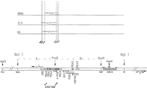
Virus and cell strains.The wild type, A2 (18), was used as a reference standard. The enhancer of this viral strain does not contain any duplication (Fig. 1). Mutant A185, a gift from M. Fried, was constructed from A2 by site-directed mutagenesis and contains a deletion of approximately 50 bp around nucleotide 676, affecting both middle T and small T (30). hr-t mutants B2 and II-5, a gift from T. Benjamin, were isolated as natural variants (3, 50) and contain deletions of 241 and 101 bp, respectively, also affecting both middle T and small T (7, 23). 808A, also from T. Benjamin, harbors a point mutation in the acceptor site for the middle T mRNA, resulting in no middle T synthesis (33). In addition, the B2,
II-5, and 808A genomes carry duplications of approximately 40 bp overlapping the alpha core domain as shown in Fig. 1 (1a, 7, 47). Strains that carry a duplication encompassing the alpha core domain are referred to as Dup1in the text, in contrast to Dup2strains derived in the A2 background. Dup1strain A2(eB2) was constructed by replacing the enhancer of A2 (BglI-BclI fragment) with that of hr-t mutant B2.
Mouse cell lines NIH 3T3 and polyomavirus-transformed Py-3T3 (Py-6) were cultured in Dulbecco’s modified Eagle’s medium supplemented with 10% newborn calf serum. Py-6, a gift from T. Benjamin, was derived from polyoma-virus infection of Swiss-3T3 cells (3), contains the middle T/small T coding region (17, 25), and displays a transformed phenotype (3).
Infections.Cells were kept in exponential growth in Dulbecco’s modified Eagle’s medium supplemented with 10% heat-inactivated newborn calf serum (Gibco) and were replated at 20% confluence in medium containing 5 to 10% serum prior to infection. Monolayers were infected 4 to 6 h after replating and were cultured in Dulbecco’s modified Eagle’s medium containing 0.5 to 5% serum. Culturing the infected monolayers in medium without serum led to cell death; a 0.5% serum condition usually resulted in very low yield of genomes in wild-type infection, a condition that was remediated considerably by the addition of 12-O-tetradecanoylphorbol-13-acetate (TPA). A 1 to 2% serum condition was adopted for most experiments, except where indicated. Where mentioned, TPA (Sigma) was added at the beginning of the infection at the concentrations shown.
[image:2.612.61.554.69.365.2]mRNA and DNA analysis.Total RNA was isolated in guanidine thiocyanate, separated on CsCl gradients, and deposited on nylon membranes for hybridiza-tion analysis (dot blots) with probes for viral and cellular gene transcripts. Counts in the dots were obtained by direct counting of the membranes. Low-molecular-weight or total DNA was extracted and purified by standard procedures, digested with the appropriate enzymes, separated by electrophoresis, and transferred to nylon membranes. Blots were hybridized with the appropriate viral probes, and the level of viral genomes was quantitated by scanning the blots with a beta scanner (AMBIS). Digestion with MspI was used to separate two parental strains in competition experiments. The enhancer duplication in Dup1 strains is contained in MspI fragment 3 and is easily separated from the A2 fragment by electrophoresis in 2% agarose in Tris-borate buffer. Similarly, the middle T/small T deletions are contained in MspI fragment 4 and can be used to identify and FIG. 1. The polyomavirus enhancer. The following elements are shown: the enhancer A domain (BclI-PvuII), B domain (PvuII-PvuII) and alpha core domain (40); the sequences that make the minimum origin (ORI) region (hatched) (39); the early (E) and late (L) cap sites; the binding sites of various transcriptional activators, notably the PEA1 and PEA3 sites, the mouse homologs of AP-1 and c-ets, respectively (36, 45) (note two additional PEA3 sites outside of the alpha core domain); and the positions of relevant restriction endonuclease sites. The duplications of the B2, II-5, and 808A strains are shown. The genomes that carry a duplication of the alpha
core domain are referred to as Dup1, in contrast to the Dup2genomes without such a duplication. The nucleotide numbering is as described in reference 48.
on November 9, 2019 by guest
http://jvi.asm.org/
quantitate two competing parental genomes. Hybridization probes for MspI fragments 3 and 4 were carried out as indicated in the figure legends.
RESULTS
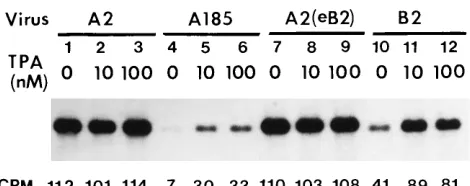
(i) A defect in genome accumulation in infections with middle T-/small T-defective mutants. The accumulation of viral genomes was compared in NIH 3T3 cells cultured in medium supplemented with a low concentration of serum and infected with the wild type, A2, or the mutant derived from A2, A185, and containing a deletion in the middle T/small T coding region. The results are shown in Fig. 2, lanes 1 and 4. A reduction of approximately 16-fold was observed in the level of mutant relative to wild-type genomes at 59 h postinfection (hpi). This defect could be alleviated partially by treatment with an activator of protein kinase C, TPA, which resulted in a fourfold increase in the level of mutant genomes. A maximal effect was obtained at a TPA concentration of 10 nM (lanes 4 to 6), and the relief was only partial. The same treatment had no effect on wild-type infection (lanes 2 and 3). An effect of TPA on polyomavirus DNA replication has been previously demonstrated in the absence of middle T and small T by using a plasmid that contains the polyomavirus origin downstream of the alpha core domain (nucleotides 5107 to 5130 [Fig. 1]) (41). The presence of a higher concentration (5%) of serum also alleviated the replication defect of A185, but to a lesser extent than TPA (data not shown). The effect of a naturally derived duplication (Dup) of 41 bp encompassing the alpha core domain was studied. This duplication also led to a partial compensation (approximately sixfold increase), as shown in Fig. 2 for the Dup1, middle T-/small T-deficient hr-t mutant B2 (compare lanes 4 and 10). This difference in replication is not specific to the A185 and B2 pair of mutants, since all other hr-t mutants tested, which all harbor alpha core duplications, replicate better than A185. Furthermore, the advantage of the B2 strain was mapped to the enhancer, because the replace-ment of the B2 enhancer by that of A2 converted B2 into a poorly growing strain (7a). Replication in infections with the Dup1 B2 mutant was still sensitive to TPA enhancement (approximately twofold increase [compare lanes 10 and 11]). Although the effects of TPA and enhancer duplication were additive (compare lanes 4, 10, and 11), these conditions alone or in combination did not fully restore replication. In multiple repeat experiments, the yield of genomes with duplications reached 30 to 80% of wild-type levels in the presence of TPA
(80% in the experiment shown). In contrast to its potent effect in the absence of middle T/small T, the alpha core duplication had no effect in the wild-type background [compare strain A2 (lanes 1 to 3) with strain A2(eB2) (lanes 7 to 9), i.e., a wild type that harbors the alpha core duplication of mutant B2].
(ii) Comparative analysis of middle T/small T effects in replication and in transcription. The compensatory effect of the alpha core duplication in the absence of hr-t function suggests that the proteins that bind these sequences mediate at least part of the enhancement. These sequences include bind-ing sites for transcriptional activators, such as PEA1 and PEA3 (36, 40, 45), that are usually assumed to have a function in transcription. To compare the effect of middle T/small T deficiency on DNA replication with that on transcription, a kinetic study was undertaken with mutant A185 and the wild type, A2, used at a multiplicity of infection of 5 each. (The dosage of viral genomes was confirmed by extraction from the infected cells prior to DNA synthesis and showed a 1.7-fold excess of the wild-type genome relative to the mutant genome [Table 1]).
The accumulation of viral genomes was examined up to 120 hpi. Clear cytopathic effects were observed in the wild-type-infected cells from 60 hpi, in contrast to mutant-wild-type-infected cells, which did not show obvious signs of lysis to the end of the experiment. The results are presented in Table 1 as the average counts hybridized for duplicate samples determined by count-ing bands in Southern blots. The reduction in viral genomes at 60 h was 100-fold in mutant-infected cells, and despite the absence of lysis, very little increase in the level of genomes was obtained by further incubating the infected cells to 120 h.
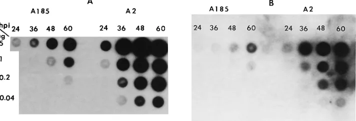
Results for viral early message, shown in Fig. 3A and Table 1, demonstrate that, at 48 hpi (before cytopathic effects became noticeable in wild-type-infected cells), the accumula-tion of viral early transcripts was reduced by a factor of 18 in mutant relative to wild-type infection (after standardization with counts bound to the cellular glyceraldehyde phosphate dehydrogenase [GAPDH] transcripts), compared with a 41-fold reduction in genome levels. Thus, it appears that, on a per genome basis, transcription is slightly more efficient (2.3-fold) in mutant- than in wild-type-infected cells. However, this figure is not likely to accurately represent the situation, because genomes are removed from the replication pool by maturation, and hence, presumably, from the transcription pool as well, especially late in infection (i.e., when the genome/transcript ratio is high in the wild-type-infected cells) (46). The appar-ently higher transcript/genome ratio in mutant-infected cells may also reflect differences in encapsidation between the wild type and hr-t mutants (19) as well as a compensatory effect of the large T-mediated autogenous control of early mRNA synthesis (9). A decrease in the level of large T antigen was also observed but not quantitated.
[image:3.612.61.296.68.161.2]To probe the putative correlation between middle T-medi-ated effects in transcription and the activation of transcrip-tional activators PEA1/AP-1 and PEA3/c-ets, the induction of the cellular transin gene, known to be controlled by AP-1 and c-ets binding sites (21) and to be induced by middle T and TPA (37), was examined. As shown in Fig. 3B and Table 1, the level of transin transcripts was elevated in wild-type-infected cells (.50-fold induction at 48 hpi) compared with that in mock-infected cells. In contrast, infection with the middle T-/small T-defective mutant A185 resulted in a low level of transin induction (approximately fourfold). Rehybridization of the blots with a probe for GAPDH demonstrated that the low level of viral early and cellular transin message was not due to a problem in RNA loading in the samples from mutant-infected cells. (The numbers discussed in the text above for the
FIG. 2. Comparison of replication of the wild type and middle T-/small T-defective mutants and the effect of phorbol ester and enhancer duplication. NIH 3T3 cells were infected with the strains indicated (each used at a multiplicity of infection of 5), as described in Materials and Methods, in medium supple-mented with 2% serum. Mutant A185 lacks middle T and small T; B2 is an hr-t mutant lacking middle T and small T but with a duplication of the alpha core domain; A2(eB2) is the wild type, A2, into which the alpha core duplication of mutant B2 has been introduced. TPA was added at the time of infection at the concentrations shown. Low-molecular-weight DNA was extracted at 59 hpi, linearized with EcoRI, and quantitated by Southern blotting as described in Materials and Methods. Counts per minute in the various bands are shown and were obtained by scanning the blot with a beta detector (AMBIS).
on November 9, 2019 by guest
http://jvi.asm.org/
induction of viral and cellular transcripts are normalized to equal levels of GAPDH transcripts.) To our knowledge, the effect of polyomavirus large T antigen on transin induction has not been studied; the reduction in large T antigen levels in A185 infection compared with those in wild-type infection might also contribute to the absence of induction in transin expression in A185-infected cells.
Thus, the absence of middle T/small T resulted in multiple effects: a 12-fold reduction in the induction of a cellular message presumably controlled by AP-1 and c-ets, an 18-fold reduction in the accumulation of viral early message, a perhaps proportional reduction in large T antigen level, and a severe reduction in the level of viral genome (100-fold reduction).
(iii) The reduction in the accumulation of viral genomes involves an alpha core-mediated effect exerted at the level of DNA replication.Two general models might explain the pro-found decrease in genome replication in infections with middle T-/small T-deficient mutants. Reduced viral transcription lead-ing to decreased levels of large T antigen could result in decreased replication. Alternatively, the transcription factors activated by middle T/small T may participate directly in DNA replication. To distinguish between these two alternatives,
[image:4.612.59.555.83.219.2]competition experiments were undertaken with pairs of strains that differ in the copy number of alpha core sequences: Dup2 genomes derived from the wild type, A2, that carry a single copy of the alpha core domain (e.g., A2 and A185) and Dup1 genomes, naturally evolved or constructed to carry two copies [e.g., B2 and A2(eB2)]. These experiments were carried out as mixed infections of exponentially growing NIH 3T3 cells in 1% serum at a 1:1 input genome ratio, a condition in which the two genomes compete for replication factors (including large T antigen) from a common pool. (The multiplicity of infection [3 each] was kept low to avoid or minimize capsid-mediated activation of the c-fos gene and hence AP-1/PEA1 [61].) The results of competition in the absence of middle T/small T (Dup2 mutant A185 competing with Dup1 mutant B2) are shown in Fig. 4. Each parent in the infection can be traced by the restriction endonuclease fragment that contains the dele-tion (in this case, MspI fragment 4). These fragments are 461 bp in the case of B2 and approximately 651 bp for A185. Genome levels at 4 hpi (i.e., unreplicated input) show that the ratio of Dup1B2 to Dup2A185 in the infection mix was 1:1.4 (corresponding to a ratio of counts per minute of 1:2). As expected, the genome ratio remained constant prior to DNA
[image:4.612.129.485.560.681.2]FIG. 3. Comparison of viral and cellular transcripts in wild-type and middle T/small T mutant infections. NIH 3T3 cells were infected in parallel with the wild type, A2, and mutant A185 at a multiplicity of 5 and cultivated in medium supplemented with 1% serum. Total RNA was isolated at the times shown, deposited on membrane in the amounts shown, and hybridized with the appropriate probes. (A) Polyomavirus probe. MspI fragment 4 corresponds to the early transcripts. The exposure time was 4 h. (B) Transin probe. The blot shown in panel A was stripped and rehybridized with a probe for the cellular transin gene (a gift of Lynn Matrisian). The exposure time was 4 days.
TABLE 1. Kinetics of viral and cellular gene expression and viral DNA synthesis in the presence or absence of middle T/small T
Time of DNA and RNA isolation (hpi)
Kinetics of gene expression and DNA synthesis (cpm) ina :
A2 A185
DNA, Py
RNA
DNA, Py
RNA
Py Transin GAPDH Py Transin GAPDH
4 2 1.2
24 42 22 0.2 18 3.4 5.6 0 7.5
36 466 258 1.3 24 8.2 9.6 0.1 19
48 497 751 4.7 34 12 31 0.3 27
60 1,407 386b 2.7 38 14 70 0.3 24
72 2,280 15
96 1,262 77
120 1,445 80
a
NIH 3T3 cells were infected with the wild type, A2, and middle T-/small T-deficient mutant A185 at a multiplicity of infection of 5 and cultivated in medium supplemented with 1% serum. Total RNA and total DNA were isolated at the times shown. (Note that by 96 hpi, cells infected with the wild type had lysed and DNA was collected in the lysate. No lysis was seen in the mutant infection, even by 120 h.) DNA was digested with EcoRI, separated by gel electrophoresis, blotted, and hybridized with a polyomavirus-specific probe, and the level of hybridized probe was determined with a beta scanner, as described in Materials and Methods and the legend to Fig. 2. RNA was analyzed by dot blots (shown in Fig. 3), which were processed for hybridization followed by stripping in succession with three different probes: (i) polyomavirus early genes (MspI fragment 4) (Fig. 3A); (ii) cellular transin (Fig. 3B), and (iii) cellular GAPDH (not shown). Counts in dots were determined with a beta scanner.
b
This number was obtained using 0.2 and 0.04mg of RNA. (The numbers obtained using 5 and 1mg of RNA were aberrant. In all other cases, the data obtained using the four amounts of RNA shown in Fig. 3 were consistent with each other.)
on November 9, 2019 by guest
http://jvi.asm.org/
replication, which started between 15 and 24 hpi. By 24 h, a 42-fold total genome amplification had taken place, represent-ing a 6.5-fold amplification of the A185 genome and a 110-fold amplification of the B2 genome. Hence, the Dup1B2 genome had a 17-fold replication advantage over the Dup2 A185 genome in this initial phase of genome amplification. This value is compatible with the effect seen in single infections (a 5.9-fold increase in genome level in infection with B2 com-pared with that with A185 [Fig. 2]).
In the competition experiment, a further 45-fold overall genome amplification took place between 24 and 72 h (Fig. 4), representing a 5-fold reduction in the rate of genome accumu-lation in this late phase compared with the rate in the initial phase of replication. This shift in replication rate is intriguing but difficult to interpret because the cells were not synchro-nized. Interestingly, the ratio of Dup1 to Dup2 genomes remained constant through the late phase of amplification. This finding may have various explanations, including contin-ued replication on a master template with an absence of reentry of progeny genomes in the replication pool at those times. Rolling circle-type replication would be compatible with this observation and has in fact been documented for late stages of polyomavirus DNA replication (5). Models with theta-form replication can be envisaged as well. That pro-longed treatment with the phorbol ester TPA, which is ex-pected to downregulate protein kinase C (16), does not result in a lower yield of genomes (results from Fig. 2) is also compatible with this hypothesis. Other explanations are clearly possible at present.
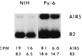
[image:5.612.59.299.69.158.2]The replication advantage of genomes containing a duplica-tion of the alpha core region in competiduplica-tion with viruses without duplication establishes that at least part of the effect of the duplication is exerted directly at the level of DNA repli-cation. This phenomenon has been observed in multiple cases, in conditions with higher multiplicities of infection (10 for each parent), involving pairs of genomes in different backgrounds (various hr-t mutants), and with duplications that differ slightly in their end points. For example, mutant Dup1II-5 shown in Fig. 1 replicates better than Dup2A185 (data not shown and reference 7b). Finally, no competition between two Dup1 mutants, such as II-5 and B2, was observed (data not shown). When the same pair of mutants (A185 plus B2) used in the experiment shown in Fig. 4 were used to infect Py-3T3 (Py-6)
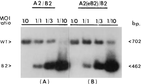
cells which express middle T/small T, the A185 genome was able to compete better against the exogenous B2 and the endogenous defective viral genome integrated into Py-6 cells. (The Py-6 genome also harbors a duplicated alpha core domain and also replicates during exogenous infections [17] [Fig. 5].) The A185/B2 ratio of counts per minute increased from 1.8:7.4 to 7.1:16.7, representing a 1.8 decrease in the degree of competition of B2 against the A185 genome. Fur-thermore, when the infection was carried out in NIH 3T3 cells in the presence of middle T/small T [mixed infection with the Dup2 wild type, A2, and Dup1 reconstructed variant, A2(eB2)], the Dup1genome replicated at the same level as the Dup2 A2 genome (Fig. 6), while higher levels of overall replication were obtained (confirming the results shown in Fig. 2). Finally, when the B2 genome was used in competition with the wild type, A2, competition against the Dup2wild type was not observed at a low B2/A2 ratio (1:1 or 3:3) but took place only when a high ratio (10:1) was used, which presumably corresponds to a very low concentration of middle T/small T (Fig. 7A). In contrast, when the same competition was set up with the Dup1wild type [A2(eB2)], the yield of the Dup1wild type was unaffected even at a high mutant/wild-type ratio (10:1) (Fig. 7B). The obviation of duplication-mediated over-replication in the presence of middle T/small T (in single infections and in competitions) suggests that the duplication of alpha core sequences is only advantageous when factors that recognize these sequences are limiting but that the binding of more factors does not per se lead to an increased rate of replication.
[image:5.612.365.508.71.170.2]To distinguish between middle T and small T effects, experiments were carried out with strain 808A (Dup1), pre-sumed to produce small T but not middle T. The results were
FIG. 4. Replication competition for factors that bind the alpha core domain. NIH 3T3 cells were infected as described in Materials and Methods with a mixture of mutants A185 and B2 at a multiplicity of infection of 3 each and incubated in medium supplemented with 0.5% serum. Low-molecular-weight DNA was isolated in duplicate at the times shown (HPI [hpi]), digested with
MspI, and subjected to electrophoresis in 2% agarose. For the 4- and 15-hpi time
points, the amount of sample loaded was twice that for other time points. The gel was blotted onto nylon membrane and hybridized with a probe for polyomavirus
MspI fragment 4, which can be used to identify the two competing genomes as
shown and as described in Materials and Methods. Samples from 4 to 24 hpi were hybridized separately from samples from 36 to 72 hpi under identical conditions (same probe). A sixfold longer exposure is shown for the 4- to 24-hpi samples. The counts in each band were obtained by direct counting of the membrane: top numbers, A185; bottom numbers, B2.
FIG. 5. Relief of competition between middle T/small T mutants with and without alpha core duplications in Py-3T3 cells that express middle T/small T. A mixture of A185 and B2 middle T-/small T-deficient mutants was used to infect NIH 3T3 cells (NIH) and Py-6 cells in parallel. Conditions were identical to those described in the legend to Fig. 4. Low-molecular-weight DNA was isolated at 48 hpi. Hybridization was to MspI fragment 4. The wild-type size band correspond-ing to the Py-6 genome has been left out. The counts in the A185 and B2 fragment 4 bands are given.
FIG. 6. Competition for alpha core binding factors between wild types. Wild-type strains that differ in the alpha core region [Dup1A2(eB2) and Dup2 A2] were used to coinfect NIH 3T3 cells. The conditions and treatment were as described in the legends to Fig. 4 and 5, except that a probe for MspI fragment 3 was used for hybridization. The amount of sample for the 4- and 15-hpi points was twice that for other time points. These samples were electrophoresed on a separate gel and also hybridized separately, but with the same probe. The exposure times were 2 weeks for the 4- and 15-hpi points, 48 h for the 24-, 36-, and 48-hpi points, and 12 h for the 72-hpi point.
on November 9, 2019 by guest
http://jvi.asm.org/
[image:5.612.325.548.603.650.2]similar to those with strains that produce neither protein. In single infections, the level of 808A genomes was similar to that obtained with B2. In competition experiments, the 808A genome, which does carry an enhancer duplication, replicated more efficiently than the A185 genome and replicated equally as well as the B2 genome (data not shown). However, these experiments are inconclusive, because 808A did not produce detectable levels of small T at the multiplicity of infection used in the present experiments.
DISCUSSION
The following major conclusions can be drawn from the results presented above. (i) The absence of middle T/small T resulted in a severe (16- to 100-fold) reduction in the accumu-lation of viral genomes. This decrease in replication could not be accounted for solely by a decrease in the level of early viral transcripts. (ii) The DNA replication defect of middle T/small T mutants could be alleviated by a duplication of the alpha core enhancer domain that contains binding sites for the transcription factors PEA1/AP-1 and PEA3/c-ets. A substantial fraction of this effect was exerted directly at the level of DNA replication. (iii) TPA (and to a lesser extent serum) also partially restored the replication defect of hr-t mutants, but none of these agents was as potent as middle T/small T. These results suggest that middle T/small T activates a mitogenic pathway that has a direct effect on DNA synthesis. We propose that a major event in this pathway is affected by the activation of PEA1/AP-1 and PEA3/c-ets. We further suggest that the level of active factors is limiting in exponentially growing cells and that middle T/small T, TPA, and serum activate these factors, which then bind to their cognate sites in the polyoma-virus enhancer and stimulate DNA replication.
Much evidence is available in support of three aspects of this model: (i) that middle T and TPA activate PEA1/AP-1 and PEA3/c-ets, (ii) that the PEA1 plus PEA3 sites encompassing the alpha core domain play a crucial role in the polyomavirus life cycle with a coupled effect in both gene expression and DNA replication, and (iii) that some (if not all) transcriptional activators play a role in DNA replication. The important new aspect of the present experiments is to link these three observations together and to demonstrate for the first time a major effect of middle T on DNA replication. The evidence for the first point, i.e., the connection between middle T and
PEA1/AP-1 and PEA3/c-ets, is compelling: as reviewed in the introduction, AP-1 and c-ets are downstream targets of middle T signalling (58, 60), and the activation of these two transcrip-tional activators has a synergistic effect, at least at the tran-scriptional level (57). The same is true in the case of the connection between TPA and PEA1 plus PEA3 (2, 31, 57, 59). The second point, concerning the importance of the PEA1 plus PEA3 sites in the alpha core domain, in polyomavirus gene expression and replication, is also well established and was also reviewed in the introduction (39, 51). The compensatory effect of the alpha core containing the 41-bp enhancer duplication in the absence of middle T/small T supports these data. As for the third point, the role of transcriptional activators in DNA replication is gaining recognition. Most relevant in the case of polyomavirus is the parallel effect, on transcription and DNA replication, of mutations in the PEA1 and PEA3 binding sites. Studies of the mechanisms by which transcriptional activators participate in DNA replication have just begun, and a role for replication protein A is emerging in experiments carried out with the polyomavirus origin (24, 32). Replication protein A interacts with large T antigen, at least in the case of the simian virus 40 protein (38), and plays a role in the initiation of DNA replication (10, 13, 27, 28, 38), perhaps by stimulating replica-tion protein A-dependent unwinding of DNA by T antigen (24). Similarly, an interaction between replication protein A and the PEA1/AP-1 and PEA3/c-ets factors may be envisioned. The fact that the v-jun oncogenic component of AP-1 activates DNA replication from a polyomavirus origin supports a role for AP-1 and c-ets (42, 59). Whatever the mechanism, the role of transcriptional activators in polyomavirus DNA replication is profound, as judged from the effect seen in the present experiments.
The advantage in replication of Dup1over Dup2genomes in single and in mixed infections in the absence of middle T/small T may reflect the fact that Dup1genomes are better able to compete for the limiting amount of active PEA1 and PEA3 present in exponentially growing NIH 3T3 cells. The kinetics of competition shows that the advantage of the Dup1 genome is established during the very first rounds of DNA replication (between 15 and 24 hpi). This result is compatible with an effect at the initiation stage. The advantage of the Dup1genome is extensive (17-fold in the experiment shown in Fig. 4). This may reflect a synergistic effect of AP-1 plus c-ets in DNA replication as was documented for transcription (57), the increase in the possible number of pairs of binding sites for these two factors (a sixfold increase if only PEA1/AP-1 and PEA3/c-ets sites are involved), and/or the cumulative effect of multiple rounds of genome replication between 15 and 24 hpi. (For example, two rounds of replication with a 4.2-fold advan-tage of the Dup1over the Dup2genome would account for the 17-fold overrepresentation of the Dup1 genome in the final genome population.) The replication advantage of the Dup1 mutants is probably of physiological significance, be-cause naturally occurring middle T/small T (hr-t) mutants have almost uniformly evolved to contain such duplications (1a). Furthermore, the present observations suggest that competi-tion for alpha core binding factors is the basis of the previously described dominant lethal effect of hr-t mutants (15).
A role for small T in DNA replication and no role for middle T have been previously reported in experiments on the repli-cation of plasmids containing the polyomavirus origin and enhancer that are not directly comparable to the present conditions (4, 35). The present experiments do not allow the distinguishing of a role for middle versus small T. Experiments with strain 808A, presumed to produce small T but no middle T, gave the same results as those with strains that produce
FIG. 7. Competition for alpha core binding factors at various levels of middle T/small T. A Dup2(A) or Dup1(B) wild type (WT) [A2 or A2(eB2)] was used along with the Dup1middle T-defective B2 mutant to coinfect NIH 3T3 cells. Conditions and treatments were as described in the legends to Fig. 4 and 5. The dosage of the wild type was kept constant, while that of the mutant was varied to reach the ratios shown. MOI, multiplicity of infection.
on November 9, 2019 by guest
http://jvi.asm.org/
[image:6.612.61.298.71.211.2]neither protein, suggesting that middle T is the key protein; however, in our hands, 808A did not produce detectable levels of small T. Experiments to establish this point and the roles of various domains of middle T require the reconstruction of all middle T and small T mutants in the same genetic A2 back-ground and are in progress. However, the established connec-tion between middle T and AP-1 and c-ets strongly suggests middle T rather than small T as the major mediator of this effect. Very little is known about small T except for its ability to bind phosphatase 2A (PP2A), an activity that it shares with middle T (43, 44, 54, 56). A connection between PP2A and AP-1 binding elements has been established in various ex-periments (1, 6, 49), but the results are contradictory. For example, a case for a positive effect of removal of PP2A by binding to simian virus 40 small T in the activation of the MAP kinase pathway has been made (49), but a previous experiment suggested that microinjection of PP2A stimulates AP-1-medi-ated transcription (1). Thus, it is not clear at present how the binding of PP2A by middle T and/or small T impacts upon the function of AP-1 in polyomavirus infections. In any case, because both middle T and small T bind PP2A, this activity is not expected to differentiate the two proteins, should it have a role in DNA synthesis.
Interestingly, the DNA replication defect of middle T/small T mutants has not come to light as a major defect in numerous previous physiological studies (19), with perhaps one exception (52). We believe that this lack of recognition reflects the conditions and specific viral strains used, which partially mask the defect. These include conditions that may activate AP-1 and c-ets independently of middle T (plus small T), such as use of higher serum concentrations; use of unusual batches of serum (that perhaps contain hormones); use of cells recently released from G0 (7c); and use of high multiplicities of infection (7b), in which case capsid binding to the polyomavi-rus receptor may activate the same signal transduction pathway as middle T (61). Most importantly, these conditions also include the use of Dup1mutants. Indeed, most of the physi-ological studies of middle T-/small T-deficient mutants were carried out with Dup1hr-t mutants. Altogether, these consid-erations also offer an explanation for the variability in the permissiveness for the growth of hr-t mutants that has been observed over the years (15, 20).
In summary, we have shown that middle T and/or small T plays a major role in viral DNA replication and that this role is mediated by enhancer sequences that include AP-1 and c-ets binding sites. It has been traditionally assumed that the activation of the tyrosine kinase receptor signal transduction pathway by growth factors and oncogenes such as middle T results in the induction of cellular genes, e.g., transin, whose products contribute to cellular growth. The present experi-ments open the possibility that a function of the transcription factors activated by oncogenes or growth factors may be affected in the direct stimulation of cellular DNA replication.
ACKNOWLEDGMENTS
These experiments were supported by grant CA29270 from the National Cancer Institute.
We thank Susan Conrad for helpful comments on the manuscript.
REFERENCES
1. Alberts, A. S., T. Deng, A. Lin, J. L. Meinkoth, A. Scho¨nthal, M. C. Mumby, M. Karin, and J. R. Feramisco.1993. Protein phosphatase 2A potentiates activity of promoters containing AP-1-binding elements. Mol. Cell. Biol. 13: 2104–2112.
1a.Amalfitano, A., M. C. Chen, and M. M. Fluck. Unpublished data.
2. Angel, P., M. Imagawa, R. Chiu, B. Stein, R. J. Imbra, H. J. Rahmsdorf, C.
Jonat, P. Herrlich, and M. Karin. 1987. Phorbol ester-inducible genes contain a common cis element recognized by a TPA-modulated trans-acting factor. Cell 49:729–739.
3. Benjamin, T. L. 1970. Host range mutants of polyoma virus. Proc. Natl. Acad. Sci. USA 67:394–399.
4. Berger, H., and E. Wintersberger. 1986. Polyomavirus small T antigen enhances replication of viral genomes in 3T6 mouse fibroblasts. J. Virol.
60:768–770.
5. Bjursell, G. 1978. Effects of 29-deoxy-29-azidocytidine on polyoma virus DNA replication: evidence for rolling circle-type mechanism. J. Virol. 26: 136–142.
6. Black, E. J., A. J. Street, and D. A. F. Gillespie. 1991. Protein phosphatase 2A reverses phosphorylation of c-Jun specified by the delta domain in vitro: correlation with oncogenic activation and deregulated transactivation activ-ity of v-Jun. Oncogene 6:1948–1958.
7. Carmichael, G., and T. L. Benjamin. 1980. Identification of DNA sequence changes leading to loss of transforming ability in polyoma virus. J. Biol. Chem. 255:230–235.
7a.Chen, M. C., A. Amalfitano, and M. M. Fluck. Unpublished data. 7b.Chen, M. C., and M. M. Fluck. Unpublished data.
7c.Chen, M. C., D. Redenius, and M. M. Fluck. Unpublished data. 8. Cheng, S. H., R. Harvey, P. C. Espino, K. Semba, T. Yamamoto, K.
Toyoshima, and A. E. Smith.1988. Peptide antibodies to the human c-fyn gene product demonstrate pp59c-fyn is capable of complex formation with the middle-T antigen of polyomavirus. EMBO J. 7:3845–3855.
9. Cogen, B. 1978. Virus-specific early RNA in 3T6 cells infected by tsA mutant of polyoma virus. Virology 85:222–230.
10. Collins, K. L., and T. J. Kelly. 1991. Effects of T antigen and replication protein A on the initiation of DNA synthesis by DNA polymerasea-primase. Mol. Cell. Biol. 11:2108–2115.
11. Courtneidge, S., and A. Smith. 1984. The complex of polyoma virus middle-T antigen and pp60c-src. EMBO J. 3:585–591.
12. de Villiers, J., W. Schaffner, C. Tyndall, S. Lupton, and R. Kamen. 1984. Polyoma virus DNA replication requires an enhancer. Nature (London)
312:242–246.
13. Dornreiter, I., L. F. Erdile, I. U. Gilbert, D. Von Winkler, T. J. Kelly, and E.
Fanning.1992. Interaction of DNA polymerase alpha-primase with cellular replication protein A and SV40 T antigen. EMBO J. 11:769–776. 14. Feunteun, J., L. Sompayrac, M. Fluck, and T. Benjamin. 1976. Localization
of gene functions in polyoma virus DNA. Proc. Natl. Acad. Sci. USA
73:4169–4173.
15. Fluck, M. M., R. J. Staneloni, and T. L. Benjamin. 1977. Hr-t and ts-a: two early gene functions of polyoma virus. Virology 77:610–624.
16. Fournier, A., and A. W. Murray. 1987. Application of phorbol ester to mouse skin causes a rapid and sustained loss of protein kinase C. Nature (London)
330:767–769.
17. Friderici, K., C. Priehs, and M. M. Fluck. 1986. Superinfection rescue of an integrated defective polyomavirus genome. J. Virol. 57:205–210. 18. Fried, M., B. E. Griffin, E. Lund, and L. Robberson. 1974. Polyomavirus: a
study of wild-type, mutant and defective DNAs. Cold Spring Harbor Symp. Quant. Biol. 39:45–52.
19. Garcea, R. L., and T. L. Benjamin. 1983. Host range transforming gene of polyoma virus plays a role in virus assembly. Proc. Natl. Acad. Sci. USA
80:3413–3417.
20. Goldman, E., and T. L. Benjamin. 1975. Analysis of host range of nontrans-forming polyoma virus mutants. Virology 66:372–384.
21. Gutman, A., and B. Wasylyk. 1990. The collagenase gene promoter contains a TPA and oncogene-responsive unit encompassing the PEA3 and AP-1 binding sites. EMBO J. 7:2241–2246.
22. Gutman, A., and B. Wasylyk. 1991. Nuclear targets for transcription regu-lation by oncogenes. Trends Genet. 7:49–54.
23. Hattori, J., G. G. Carmichael, and T. L. Benjamin. 1979. DNA sequence alterations in Hr-t deletion mutants of polyoma virus. Cell 16:505–513. 24. He, Z., B. T. Brinton, J. Greenblatt, J. A. Hassell, and C. J. Ingles. 1993. The
transactivator proteins VP16 and GAL4 bind replication factor A. Cell 73: 1223–1232.
25. Kamen, R., D. M. Lindstrom, H. Shure, and R. W. Old. 1974. Virus-specific RNA in cells productively infected or transformed by polyomavirus. Cold Spring Harbor Symp. Quant. Biol. 39:187–198.
26. Karin, M., and T. Smeal. 1992. Control of transcription factors by signal transduction pathways: the beginning and the end. Trends Biochem. Sci. 17: 418–422.
27. Kenny, M. K., S.-H. Lee, and J. Hurwitz. 1989. Multiple functions of human single-strand-DNA binding protein in simian virus 40, DNA replication: single strand stabilization and stimulation of DNA polymerase alpha and delta. Proc. Natl. Acad. Sci. USA 86:9757–9761.
28. Kenny, M. K., U. Schlegel, H. Furneaux, and J. Hurwitz. 1990. The role of human single-stranded DNA binding protein and its individual subunits in simian virus 40 DNA replication. J. Biol. Chem. 265:7693–7700. 29. Kornbluth, S., and H. Hanafusa. 1987. Association of the polyomavirus
middle-T antigen with c-yes protein. Nature (London) 325:171–173.
on November 9, 2019 by guest
http://jvi.asm.org/
30. Lania, L., M. Griffiths, B. Cooke, Y. Ito, and M. Fried. 1979. Untransformed rat cells containing free and integrated DNA of a polyoma nontransforming (Hr-t) mutant. Cell 18:793–802.
31. Lee, W., P. Mitchell, and R. Tjian. 1987. Purified transcription factor AP-1 interacts with TPA-inducible enhancer elements. Cell 49:741–752. 32. Li, R., and M. Botchan. 1993. The acidic transcriptional activation domains
of VP16 and p53 bind the cellular replication protein A and stimulate in vitro BPV-1 DNA replication. Cell 73:1207–1221.
33. Liang, T. J., G. G. Carmichael, and T. L. Benjamin. 1984. A polyoma mutant that encodes small T antigen but not middle T antigen demonstrates uncoupling of cell surface and cytoskeletal changes associated with cell transformation. Mol. Cell. Biol. 4:2774–2783.
34. Markland, W., and A. E. Smith. 1987. Mapping of the amino-terminal half of polyomavirus middle-T antigen indicates that this region is the binding domain for pp60c-src
. J. Virol. 61:285–292.
35. Martens, I., S. A. Nilsson, S. Linder, and G. Magnusson. 1989. Mutational analysis of polyomavirus small-T-antigen functions in productive infection and in transformation. J. Virol. 63:2126–2133.
36. Martin, M. E., J. Piette, M. Yaniv, W. J. Tang, and W. Folk. 1988. Activation of the polyomavirus enhancer by a murine activator protein 1 (AP1) homolog and two contiguous proteins. Proc. Natl. Acad. Sci. USA 85:5839– 5843.
37. Matrisian, L. M., N. Glaichenhaus, M. C. Gesnel, and R. Breathnach. 1985. Epidermal growth factor and oncogenes induce transcription of the same cellular mRNA in rat fibroblasts. EMBO J. 4:1435–1440.
38. Melendy, T., and B. Stillman. 1993. An interaction between replication protein A and SV40 T antigen appears essential for primosome assembly during SV40 DNA replication. J. Biol. Chem. 268:3389–3395.
39. Mueller, C. R., W. J. Muller, and J. A. Hassell. 1988. The polyoma-virus enhancer comprises multiple functional elements. J. Virol. 62:1667– 1678.
40. Muller, W. J., D. Dufort, and J. A. Hassell. 1988. Multiple subelements within the polyomavirus enhancer function synergistically to activate DNA replication. Mol. Cell. Biol. 8:5000–5015.
41. Murakami, Y., M. Asano, M. Satake, and Y. Ito. 1990. A tumor promoting phorbol ester, TPA, enhances polyomavirus DNA replication by activating the function of the viral enhancer. Oncogene 5:5–13.
42. Murakami, Y., M. Satake, Y. Yamaguchi-Iwai, M. Sakai, M. Muramatsu,
and Y. Ito.1991. The nuclear protooncogenes c-jun and c-fos as regulators of DNA replication. Proc. Natl. Acad. Sci. USA 88:3947–3951.
43. Pallas, D. C., V. Cherington, W. Morgan, J. DeAnda, D. Kaplan, B.
Schaffhausen, and T. M. Roberts.1988. Cellular proteins that associate with the middle and small T antigens of polyomavirus. J. Virol. 62:3934– 3940.
44. Pallas, D. C., L. K. Shahrik, B. L. Martin, S. Jaspers, T. B. Miller, D. L.
Brautigan, and T. M. Roberts.1990. Polyoma small and middle T antigens and SV40 small t antigen form stable complexes with protein phosphatase 2A. Cell 60:167–176.
45. Piette, J., and M. Yaniv. 1987. Two different factors bind to the A-domain of
the polyoma virus enhancer, one of which also interacts with the SV40 and c-fos enhancers. EMBO J. 6:1331–1337.
46. Roman, A. 1985. Kinetics of reentry of polyoma progeny form I DNA into replication as a function of time postinfection. Virology 96:660–663. 47. Ruley, H. E., and M. Fried. 1983. Sequence repeats in a polyoma virus DNA
region important for gene expression. J. Virol. 47:233–237.
48. Salzman, N. P. 1986. The papovaviridae, vol. I. The polyomaviruses. Plenum Press, New York.
49. Sontag, E., S. Fedorov, C. Kamibayashi, D. Robbins, M. Cobb, and M.
Mumby.1993. The interaction of SV40 small tumor antigen with protein phosphatase 2A stimulates the map kinase pathway and induces cell proliferation. Cell 75:887–897.
50. Staneloni, R. J., M. M. Fluck, and T. L. Benjamin. 1977. Host range selection of transformation-defective hr-t mutants of polyoma virus. Virol-ogy 77:598–609.
51. Tang, W. J., S. L. Berger, S. J. Triezenberg, and W. R. Folk. 1987. Nucleotides in the polyomavirus enhancer that control viral transcription and DNA replication. Mol. Cell. Biol. 7:1681–1690.
52. Tu¨rler, H., and C. Salomon.1985. Small and middle T antigens contribute to lytic and abortive polyomavirus infection. J. Virol. 53:579–586.
53. Tyndall, C., G. La Mantia, C. M. Thacker, J. Favaloro, and R. Kamen. 1981. A region of the polyoma virus genome between the replication origin and late protein coding sequences is required in cis for both early gene expres-sion and viral DNA replication. Nucleic Acids Res. 9:6231–6250. 54. Ulug, E. T., A. J. Cartwright, and S. A. Courtneidge. 1992. Characterization
of the interaction of polyomavirus middle T antigen with type 2A protein phosphatase. J. Virol. 66:1458–1467.
55. Veldman, G. M., S. Lupton, and R. Kamen. 1985. Polyomavirus enhancer contains multiple redundant sequence elements that activate both DNA replication and gene expression. Mol. Cell. Biol. 5:649–658.
56. Walter, G., R. Ruediger, C. Slaughter, and M. Mumby. 1990. Association of protein phosphatase 2A with polyoma virus medium tumor antigen. Proc. Natl. Acad. Sci. USA 87:2521–2525.
57. Wasylyk, B., C. Wasylyk, P. Flores, A. Begue, D. Leprince, and D. Stehelin. 1990. The c-ets proto-oncogenes encode transcription factors that cooperate with c-Fos and c-Jun for transcriptional activation. Nature (London) 346: 191–193.
58. Wasylyk, C., J. L. Imler, and B. Wasylyk. 1988. Transforming but not immortalizing oncogenes activate the transcription factor PEA1. EMBO J.
7:2475–2483.
59. Wasylyk, C., J. Schneikert, and B. Wasylyk. 1990. Oncogene v-jun modulates DNA replication. Oncogene 5:1055–1058.
60. Wasylyk, W., P. Flores, A. Gutman, and B. Wasylyk. 1989. PEA3 is a nuclear target for transcription activity by non-nuclear oncogenes. EMBO J. 8:3371– 3378.
61. Zullo, J., C. D. Stiles, and R. L. Garcea. 1987. Regulation of c-myc and c-fos mRNA levels by polyomavirus: distinct roles for the capsid protein VP1 and the viral early proteins. Proc. Natl. Acad. Sci. USA 84:1210–1214.