0022-538X/96/$04.0010
Copyrightq1996, American Society for Microbiology
Possible Origin of Murine AIDS (MAIDS) Virus: Conversion of
an Endogenous Retroviral p12
gag
Sequence to a
MAIDS-Inducing Sequence by Frameshift Mutations
YOSHINAO KUBO,1KAZUHIRO KAKIMI,2KYOKO HIGO,1HIROHIKO KOBAYASHI,1
TAKESHI ONO,1YUKO IWAMA,1KAGEMASA KURIBAYASHI,2HIROSHI HIAI,3
AKIO ADACHI,1ANDAKINORI ISHIMOTO1*
Laboratory of Gene Analysis, Department of Viral Oncology, Institute for Virus Research,1and
First Department of Pathology, Faculty of Medicine,3Kyoto University, Kyoto, and
Department of Bioregulation, Mie University School of Medicine, Mie,3Japan
Received 2 February 1996/Accepted 20 May 1996
The murine AIDS (MAIDS) virus has a unique sequence in its p12gag
region, which is responsible for MAIDS development. A transcript hybridizing with this sequence is expressed in normal C57BL/6 mice. The transcript,
designatedEdv, has been previously cloned and sequenced (Y. Kubo, Y. Nakagawa, K. Kakimi, H. Matsui, K.
Higo, L. Wang, H. Kobayashi, T. Hirama, and A. Ishimoto, J. Gen. Virol. 75:881–888, 1994). Compared with the nucleotide sequence of the helper LP-BM5 ecotropic virus, the pathogenic replication-defective MAIDS
virus has a 16-bp deletion and a 1-bp insertion in the 5*and 3*regions of the p12gag
sequence, respectively, and
theEdvtranscript contains only a 3-bp deletion. Therefore, the amino acid sequence of the defective MAIDS
virus p12gag
region is not homologous to that of the helper virus and the Edv transcript because of the
frameshift. To determine whether the amino acid sequence resulting from the frameshift is critical for MAIDS
development, we constructed chimeric viruses that contained the p12gag
regions of the helper virus and theEdv
transcript, respectively, with and without the same frame as the defective MAIDS virus by the artificial frameshift mutations. The mutant viruses with the frameshift mutations induced MAIDS in inoculated mice, but the viruses without the mutations did not. These results suggested that the MAIDS virus was generated by
frameshift mutations in the p12gag
region ofEdvor a related sequence.
Murine AIDS (MAIDS) is induced by infection of C57BL/6 mice with the Duplan strain of murine leukemia virus (MuLV) (2, 11, 21). The disease has many similarities to human AIDS and is used as a mouse model of human AIDS (12, 20, 22, 23). The MAIDS virus complex contains a pathogenic replication-defective virus and nonpathogenic helper MuLVs (4). The pol and env genes are deleted, but the gag gene is almost conserved in the pathogenic MAIDS virus. However, the MAIDS virus
has unique sequences in the p15gag and p12gagregions (1, 5).
Thus, it has been suggested that the unique sequences are responsible for MAIDS induction. Pozsgay et al. reported that
the p15gagand p12gagregions are sufficient for MAIDS
devel-opment (24), and we have shown that these unique sequences are both required for MAIDS induction (14).
Normal C57BL/6 mice express a transcript which hybridizes
with the MAIDS virus p12gagsequence (5, 6), and previously
we cloned and sequenced this transcript (Edv) (15). There are some other lines of evidence indicating that normal mice con-tain the MAIDS virus-related sequence (3, 7, 10). Since the MAIDS virus was originally isolated from radiation-induced leukemic C57BL/6 mice (18), the Edv transcript expressed in C57BL/6 mice might represent the origin of the MAIDS virus.
In this study, we examined the pathogenicity of p12gagregions
of the Edv transcript and the helper virus by constructing the chimeric viruses including these regions. The nucleotide
se-quence of the p12gagregion of the endogenous Edv transcript
is homologous to that of the MAIDS virus, but the amino acid sequence is not due to frameshift mutations (Fig. 1).
There-fore, we constructed mutant viruses containing the p12gag
re-gion of the endogenous Edv, sequence or the helper virus with or without frameshift mutations resulting in amino acid ho-mology to the pathogenic MAIDS virus to know whether the amino acid sequence of the MAIDS virus resulting from the frameshift mutations is responsible for MAIDS induction.
Clones of the helper LP-BM5 ecotropic MuLV (BM5eco) (5), pathogenic MAIDS virus (G1B) (14), and MAIDS virus-related endogenous sequence (pEDV-2) (15) were used as parental clones for construction of recombinant viruses. The
p15gag region of the G1B DNA clone was amplified by PCR
with the H-1 and H-2 primers (Fig. 2). The p12gagregion of the
pEDV-2 or BM5eco DNA clone was amplified by the H-3 and H-4 or H-5 primers (Fig. 2). The G1B DNA clone, but not the
pEDV-2 or BM5eco clone, contains a SmaI site in its p12gag
region. To construct chimeric DNA clones between the G1B clone and pEDV-2 or BM5eco clones, the SmaI site was in-troduced to the pEDV-2 and BM5eco clones by PCR ampli-fication with the H-2 and H-3 primers, which contain SmaI
sites. The p15gag and p12gag PCR products were ligated at
the SmaI site, resulting in a BstPI-NcoI fragment containing
the p15gag and p12gagregions. The XbaI-SalI fragment of the
BM5eco DNA clone, which contains the 39region of the env
gene, 59long terminal repeat (LTR), whole gag gene, and 59
region of the pol gene, was subcloned (5). The BstPI-NcoI frag-ment in this plasmid DNA was replaced by the corresponding fragment from the PCR products described above. The BstPI and NcoI sites are unique in the XbaI-SalI fragment. The
SalI-SpeI fragment of the BM5eco clone, which contains the 39
region of the pol gene, whole env gene, 39LTR, and 59region
of the gag gene, was subcloned, and the SpeI site was changed to a HindIII site. The SalI-HindIII fragment was then inserted into the SalI-HindIII site of the plasmid containing the
XbaI-* Corresponding author.
6405
on November 9, 2019 by guest
http://jvi.asm.org/
SalI fragment. Finally, the fragment containing the 59
LTR-gag-pol-env-39LTR was constructed (5, 14).
As shown in Fig. 1, the p12gagregion of the defective
patho-genic MAIDS virus (G1B DNA clone) (14) has a 16-bp dele-tion and a 1-bp inserdele-tion, and the MAIDS virus-related endog-enous sequence, Edv (pEDV-2 DNA clone) (15), isolated from normal C57BL/6 mice has only a 3-bp deletion, compared with that of the helper ecotropic LP-BM5 virus (BM5eco DNA clone) (5). Therefore, the amino acid sequence of the MAIDS
virus p12gag region (positions 89 to 184 in Fig. 1) is less
ho-mologous to either the BM5eco virus or Edv sequence because of the frameshift in the region. To determine whether the frameshift in the MAIDS virus is essential for the development
of MAIDS, chimeric viruses containing the p12gag region of
Edv or the BM5eco virus with or without a 1-bp deletion
(position 89 in Fig. 1) and a 1-bp insertion (position 184 in Fig. 1) were constructed (Fig. 2B). The insertion mutation was prepared by PCR-mediated mutagenesis with the H-3 and H-4 primers for the helper BM5eco virus and the E-1 and E-2 primers for the Edv sequence (Fig. 2B and C). The H-4 and E-2 primers contained the 1-base insertion (Fig. 2C). The deletion mutation was prepared by site-directed mutagenesis with the H-M and E-M primers (Fig. 2B and C) for the helper BM5eco virus and Edv sequences, respectively. Site-directed mutagenesis was performed with a Transformer Site-Directed Mutagenesis Kit (Clontech Co., Ltd.) according to the method of Kunkel (9, 17). The nucleotide sequence of a selection primer, which exchanges the XhoI site of the pBluescript
vec-tor for a PstI site, was 59-GATACCGTCGACCTGCAGGGG
GGGCCCGGTACC-39. The mutations were confirmed by
se-quencing of the resulting DNA clones. The frameshift muta-tions resulted in no termination codon.
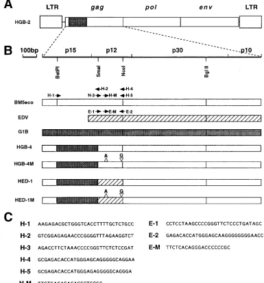
A chimeric virus, HGB-2, was constructed by replacing the
p15gagand p12gagregions (BstPI-NcoI fragment) of the helper
virus with the corresponding region of the pathogenic G1B DNA (Fig. 2A) (14). The HGB-2 chimeric virus is defective and requires a helper virus to produce virions. Chimeric vi-ruses constructed in this study contained structures with re-placement of the SmaI-NcoI region of the HGB-2 virus by the corresponding region of BM5eco or Edv with or without the frameshift mutations. HGB-4 and HED-1 chimeric viruses
contained the p12gag regions of the helper BM5eco and Edv
sequences without the mutations, respectively. HGB-4M and
HED-1M carried the modified p12gag regions from BM5eco
and Edv, respectively. The resulting amino acid sequences of
the p12gag region of the mutant viruses are shown in Fig. 3.
Chimeric viruses were constructed so as to contain the p15gag
region of G1B and the p12gag region with and without the
frameshift mutation (Fig. 2B), since the p15gagand p12gag
re-gions of the MAIDS virus are both required for MAIDS de-velopment (14).
We reported previously that the chimeric viruses containing
either the p15gag or p12gag region of the pathogenic MAIDS
virus are replication defective (14). The amphotropic
packag-ing cells GP1envAm12 (19) were cotransfected with the
chi-meric viral DNA and pSV2neo, and neomycin-resistant
colo-nies were selected in the presence of G418, since the chimeric
viruses constructed in this study containing the p15gagregion of
the defective MAIDS virus were also expected to be replica-tion defective. The best producer was selected by its ability to express the highest level of viral RNA determined by Northern (RNA) hybridization of virion RNA with the representative probe, which also provided us with data about the rough struc-ture of the generated virus. The producers for the HGB-4, HGB-4M, HED-1, HED-1M, and G1B viruses expressed al-most the same levels of viral RNA. One-month-old C57BL/6
mice were inoculated with the transfected cells (106cells per
mouse), since the level of pathogenicity of cell supernatant harvested from the packaging cells transfected with the patho-genic G1B DNA was very low (data not shown). However, only 3 of 11 C57BL/6 mice inoculated with the helper-free
G1B-transfected GP1envAm12 packaging cells developed MAIDS.
Therefore, C57BL/6 mice were inoculated with the mixture of
G1B virus-expressing packaging cells (106cells per mouse) and
BM5eco-infected SC-1 cells (106cells per mouse) to help the
further replication of the defective G1B virus. All inoculated mice (five of five) developed MAIDS this way (Table 1). In the following experiments, C57BL/6 mice were inoculated with a mixture of the chimeric virus-expressing packaging cells and the BM5eco-infected SC-1 cells.
Sixteen of 21 mice inoculated with cells producing the HED-1M virus and 5 of 21 mice inoculated with cells produc-ing the HGB-4M virus developed splenomegaly and lymphad-enopathy within the 10-month observation period (Table 1). A functional assay for the T-cell response to concanavalin A showed that in the spleen cells of diseased mice inoculated
with the mutant viruses, the level of [3H]thymidine
incorpora-tion was lower than that in the spleen cells of HED-1- and HGB-4-infected mice and control mice, which were inoculated
with the mixture of nontransfected GP1envAm12 cells and
BM5eco-infected SC-1 cells (Table 1). Histological examina-tion of the enlarged spleen revealed that the pathogenicity of the mutant viruses was biologically indistinguishable from that of the wild-type G1B virus (data not shown). We concluded that the mice inoculated with the mutant viruses developed MAIDS. The spleen weight in HGB-4M-infected mice was lower than that in HED-1M-infected mice, and HGB-4M-inoculated mice developed the disease at a lower rate (5 of 21) than the HED-1M-inoculated mice (16 of 21) (Table 1).
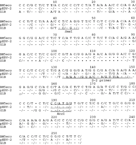
His-FIG. 1. Nucleotide sequence comparison of the p12gagregions between the
BM5eco, pEDV-2, and G1B DNA clones. Dashes indicate identity within the pEDV-2 and G1B sequences for the residue in BM5eco. Dots indicate the absence of the corresponding nucleotide. Three nucleotides between slashes correspond to one amino acid codon. The SmaI and NcoI sites and the location of the D-2 primer are underlined.
on November 9, 2019 by guest
http://jvi.asm.org/
[image:2.612.60.297.70.323.2]tological examination indicated that the diseased mice infected with the HGB-4M virus were in a less-advanced stage of MAIDS than those inoculated with the G1B or HED-1M virus according to the stage classification by Hartley et al. (11) (data not shown). These results suggested that the HGB-4M mutant virus was less pathogenic than the HED-1M mutant and G1B viruses.
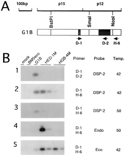
To determine whether the inoculated mutant viruses actu-ally induced MAIDS, we examined the proliferation of mutant virus in the inoculated mice by PCR. To avoid PCR amplifi-cation of endogenous MuLV-related sequences present in C57BL/6 mice, SC-1 cells in which the endogenous sequences were hardly detected were cocultured with spleen cells isolat-ed from the diseasisolat-ed mice inoculatisolat-ed with mutant viruses. Genomic DNA was then prepared from the infected SC-1 cells after five passages, among which cells from C57BL/6 mice were rare. We then performed PCR of the genomic DNAs with D-1, D-2, and H-6 primers (Fig. 4A); the H-6 primer can hybridize
with both MAIDS and helper viruses. Because the D-1 and
D-2 primers correspond to parts of the p15gag and p12gag
unique sequences of the MAIDS virus, respectively, the prim-ers cannot hybridize with helper sequences. Furthermore, the D-2 primer cannot hybridize with Edv, because only 12 among 20 bp of the D-2 primer are conserved (60%) (Fig. 1).
When PCR was performed with the D-1 and D-2 primers,
FIG. 3. Amino acid sequences of the p12gagSmaI-NcoI regions of the G1B,
[image:3.612.111.496.74.488.2]HGB-4M, and HED-1M mutant viruses. Bars indicate identical amino acid residues. Colons indicate one of the following equivalents: basic amino acids (K, R, and H), acidic amino acids (D and E), uncharged polar amino acids (N, Q, S, T, and Y), or nonpolar amino acids (G, A, V, L, I, P, F, M, W, and C). FIG. 2. Structure of the HGB-2 chimeric virus (24) (A), the gag structure of the mutant virus constructed in this study and locations of the primers used for mutagenesis (B), and nucleotide sequences of the primers (C). The fragments derived from the BM5eco, EDV, and G1B clones are shown as open, hatched, and shaded boxes, respectively.
on November 9, 2019 by guest
http://jvi.asm.org/
the fragment hybridizing with the MAIDS virus-specific probe (DSP-2) was detected only in the diseased mice inoculated with the G1B virus (Fig. 4B-1). The sizes of the PCR products
were confirmed by the lStyI size marker (data not shown).
The DSP-2 probe was derived from the 130-bp SmaI-NcoI
fragment of the p12gag region of the defective virus genome
(16). This result excluded the possibility of contamination with the wild-type MAIDS virus in the diseased mice inoculated with the mutant viruses. When PCR was performed with the D-1 and H-6 primers, fragments which hybridized with the DSP-2 probe were detected in the diseased mice inoculated with the HED-1M virus, as in the G1B-infected mice. Further-more, in one of two animals infected with the HGB-4M mutant virus, the hybridizing fragment was detected (Fig. 4B-2, left
lane, HGB-4M) when hybridization was performed at 428C.
However, when hybridization was performed at 508C for high
specificity, fragments which hybridized with the DSP-2 probe were detected only in the G1B virus. No hybridizing fragments were detected in the HGB-4M- and HED-1M-infected mice
(Fig. 4B-3). Because the p12gagregion of the BM5eco virus has
about 74% homology to that of the MAIDS virus (15), that of the HGB-4M mutant virus hybridized with the DSP-2 probe at
low stringency (428C).
When the PCR products were hybridized with the
SmaI-NcoI segment of the pEDV-2 DNA clone (endo probe) (15) at
508C, the fragments were detected in both of the
HED-1M-infected mice (Fig. 4B-4). On rehybridization with the
SmaI-NcoI segment of the BM5eco virus (eco probe) (15), the
hy-bridizing fragments were detected not only in the diseased mice inoculated with the HGB-4M virus but also in those inoculated with the G1B or HED-1M virus (Fig. 4B-5). This result suggested the emergence of recombinant viruses
con-taining the p15gag unique sequence of the MAIDS virus and
the p12gag sequence of the BM5eco virus in mice inoculated
with G1B or HED-1M. Finally, these results showed that the inoculated mutant virus proliferated in the diseased mice. These results indicated that the amino acid sequence resulting from the frameshift mutations is essential for MAIDS devel-opment.
It was shown here that virus containing the MAIDS virus
p15gag and the BM5eco p12gag sequences appeared in
G1B-and in HED-1M-infected mice (Fig. 4B-5). This suggested that recombination occurred between the G1B or HED-1M and helper BM5eco viruses. Such recombination has been de-scribed previously (8). Although we tried to determine the nucleotide sequences of several PCR products recovered from the infected mice, no such recombinant virus was detected (data not shown). Therefore, the recombinant virus may be less abundant than the parental viruses.
Recombination between the defective MAIDS virus and an endogenous sequence has been reported, suggesting that the creation of virus variants by the recombination event may play an important role in the pathogenesis and escape from host immune attack (10, 13). However, we observed no such event,
although the various changes of nucleotide sequences in p12gag
regions from the inoculated mutant viruses were determined by PCR amplification (data not shown).
Recently, Cho et al. (7) reported the presence of a provirus structurally related to MAIDS virus in BXH-2 mice. Although
the sequence of the p12gagof the provirus is different from that
of MAIDS virus, it may be one of the candidates for the origin of MAIDS virus (7). Some of the mice inoculated with the
HGB-4M mutant virus, which contained the BM5eco p12gag
[image:4.612.58.561.82.172.2]region with the frameshift mutations, developed MAIDS. Thus, it was also possible that the MAIDS virus was generated
TABLE 1. Development of MAIDS by infection of C57BL/6 mice with mutant viruses
Virus No. of diseased mice/no.
of inoculated mice
Avg latency (days)a
Avg spleen wt (mg)a
[3H]thymidine uptake (cpm [104])b
Nonc
Concanavalin A
Control 0/5 2.0 10.8
G1B 5/5 104–142 (118) 270–700 (526) 3.4 2.6
HGB-4 0/16 2.6 13.7
HGB-4M 5/21 142–212 (161) 290–570 (424) 3.9 2.8
HED-1 0/17 2.5 15.9
HED-1M 16/21 129–222 (167) 480–2,510 (1,039) 3.5 2.8
aValues in parentheses represent the means.
bAverage value of three mice.
cNon, without concanavalin A.
FIG. 4. Southern hybridization of PCR products. Locations of the primers used in this PCR experiment are indicated (A). PCR products with D-1 and D-2
primers were hybridized with the MAIDS virus-specific probe (DSP-2) at 428C
(B-1). Those with D-1 and H-6 primers were hybridized with the DSP-2 probe at 428C (B-2) or 508C (B-3), with the endo probe at 508C (B-4), or with the eco probe at 428C (B-5). The nucleotide sequences of the primers were as follows: D-1, CCTTTTCCTTTATCGACACT; D-2, CTTCTTAACTGGTCCCTTTG; and H-6, TTTGCCCCGCAGGCGAGACACCATGG.
on November 9, 2019 by guest
http://jvi.asm.org/
[image:4.612.71.284.394.657.2]by frameshift mutations in the p12gag region of the BM5eco
viral genome. However, it was shown here that the HGB-4M mutant virus was less pathogenic than the HED-1M virus.
Furthermore, the nucleotide sequence of the p12gagregion of
the MAIDS virus is more homologous to that of Edv than that of BM5eco. Therefore, it was likely that the MAIDS virus was generated by frameshift mutations in the p12 sequence of Edv or related sequences.
We thank K. Tokunaga and M. Kawamura for valuable technical suggestions and N. Aoyama and T. Ishikawa for technical assistance. This work was supported by a grant-in-aid to A. Ishimoto for AIDS research from the Ministry of Health and Welfare of Japan and by Special Coordination Funds of the Science and Technology Agency of the Japanese Government.
REFERENCES
1. Aziz, D. C., Z. Hanna, and P. Jolicoeur. 1989. Severe immunodeficiency disease induced by a defective murine leukaemia virus. Nature (London) 338:505–508.
2. Buller, R. M. L., R. A. Yetter, T. N. Fredrickson, and H. C. Morse III. 1987. Abrogation of resistance to severe mousepox in C57BL/6 mice infected with LP-BM5 murine leukemia virus. J. Virol. 61:383–387.
3. Casabianca, A., and M. Magnani. 1994. A p12 gag gene homologue is present in the mouse genome. Biochem. Mol. Biol. Int. 32:691–696. 4. Chattopadhyay, S. K., H. C. Morse III, M. Makino, S. K. Ruscetti, and J. W.
Hartley.1989. Defective virus is associated with induction of murine retro-virus-induced immunodeficiency syndrome. Proc. Natl. Acad. Sci. USA 86: 3862–3866.
5. Chattopadhyay, S. K., D. N. Sengupta, T. N. Fredrickson, H. C. Morse III, and J. W. Hartley.1991. Characteristics and contributions of defective, ecotropic, and mink cell focus-inducing viruses involved in a retrovirus-induced immunodeficiency syndrome of mice. J. Virol. 65:4232–4241. 6. Cheung, S. C., S. K. Chattopadhyay, H. C. Morse III, and P. M. Pitha. 1991.
Expression of defective virus and cytokine genes in murine AIDS. J. Virol. 65:823–828.
7. Cho, B. C., J. D. Shaughnessy, Jr., D. A. Largaespada, H. G. Bedigian, A. M. Buchberg, N. A. Jenkins, and N. G. Copeland.1995. Frequent disruption of the Nf1 gene by a novel murine AIDS virus-related provirus in BXH-2 murine myeloid lymphomas. J. Virol. 69:7138–7146.
8. Coffin, J. M. 1992. Genetic diversity and evolution of retroviruses. Curr. Top. Microbiol. Immunol. 176:143–164.
9. Deng, W. P., and J. A. Nickoloff. 1992. Site-directed mutagenesis of virtually any plasmid by eliminating a unique site. Anal. Biochem. 200:81–86. 10. Gayama, S., B. A. Vaupel, and O. Kanagawa. 1995. Sequence heterogeneity
of murine acquired immunodeficiency syndrome: the role of endogenous
virus. Int. Immunol. 7:861–868.
11. Hartley, J. W., T. N. Fredrickson, R. A. Yetter, M. Makino, and H. C. Morse
III.1989. Retrovirus-induced murine acquired immunodeficiency syndrome:
natural history of infection and differing susceptibility of inbred mouse strains. J. Virol. 63:1223–1231.
12. Jolicoeur, P. 1991. Murine acquired immunodeficiency syndrome (MAIDS): an animal model to study the AIDS pathogenesis. FASEB J. 5:2398–2405. 13. Kanagawa, O., B. A. Vaupel, S. J. Korsmeyer, and J. H. Russell. 1995. Apoptotic death of lymphocytes in murine acquired immunodeficiency syn-drome: involvement of Fas-Fas ligand interaction. Eur. J. Immunol. 25:2421– 2427.
14. Kubo, Y., K. Kakimi, K. Higo, L. Wang, H. Kobayashi, K. Kuribayashi, T. Masuda, T. Hirama, and A. Ishimoto.1994. The p15gagand p12gagregions
are both necessary for the pathogenicity of the murine AIDS virus. J. Virol. 68:5532–5537.
15. Kubo, Y., Y. Nakagawa, K. Kakimi, H. Matsui, K. Higo, L. Wang, H. Koba-yashi, T. Hirama, and A. Ishimoto.1994. Molecular cloning and character-ization of a murine AIDS virus-related endogenous transcript expressed in C57BL/6 mice. J. Gen. Virol. 75:881–888.
16. Kubo, Y., Y. Nakagawa, K. Kakimi, H. Matsui, M. Iwashiro, K. Kuribayashi, T. Masuda, H. Hiai, T. Hirama, S.-I. Yanagawa, and A. Ishimoto.1992. Presence of transplantable T-lymphoid cells in C57BL/6 mice infected with murine AIDS virus. J. Virol. 66:5691–5695.
17. Kunkel, T. A. 1985. The use of phosphorothioate-modified DNA in restric-tion enzyme reacrestric-tions to prepare nicked DNA. Proc. Natl. Acad. Sci. USA 82:488–492.
18. Latarjet, R., and J.-F. Duplan. 1962. Experiment and discussion on leuke-mogenesis by cell-free extracts of radiation-induced leukemia in mice. Int. J. Radiat. Biol. 5:339–344.
19. Markowitz, D., S. Goff, and A. Bank. 1988. Construction and use of a safe and efficient amphotropic packaging cell line. Virology 167:400–406. 20. Morse, H. C., III, S. K. Chattopadhyay, M. Makino, T. N. Fredrickson, A. W.
Hugin, and J. W. Hartley.1992. Retrovirus-induced immunodeficiency in the mouse: MAIDS as a model for AIDS. AIDS 6:607–621.
21. Mosier, D. E., R. A. Yetter, and H. C. Morse III. 1985. Retroviral induction of acute lymphoproliferative disease and profound immunosuppression in adult C57BL/6 mice. J. Exp. Med. 161:766–784.
22. Nakagawa, Y., K. Kakimi, W. Ling, Y. Kubo, K. Higo, T. Masuda, K. Kuribayashi, M. Iwashiro, Y. Komatz, T. Hirama, A. Adachi, and A. Ishi-moto.1994. Inhibition of murine AIDS (MAIDS) development by the trans-plantation of bone marrow cells carrying the Fv-4 resistance gene to MAIDS virus-infected mice. J. Virol. 68:1438–1441.
23. Portnoi, D., A. M. Stall, D. Schwartz, T. C. Merigan, L. A. Herzenberg, and T. Basham.1990. Zidovudine inhibits characteristic early alterations of lym-phoid cell populations in retrovirus-induced murine AIDS. J. Immunol. 144: 1705–1710.
24. Pozsgay, J. M., M. W. Beilharz, B. D. Wines, A. D. Hess, and P. M. Pitha. 1993. The MA (p15) and p12 regions of the gag gene are sufficient for the pathogenicity of the murine AIDS virus. J. Virol. 67:5989–5999.