A Dissertation on
A STUDY ON RETROPERITONEAL SOFT TISSUE SARCOMA
Submitted to
The Tamilnadu Dr.M.G.R.Medical University in partial fulfillment of the requirement
for the award of degree of
M.S. (GENERAL SURGERY)
BRANCH I
KILPAUK MEDICAL COLLEGE
THE TAMILNADU Dr.M.G.R.MEDICAL UNIVERSITY CHENNAI, TAMILNADU
BONAFIDE CERTIFICATE
Certified that the dissertation titled “A STUDY ON RETROPERITONEAL
SOFT TISSUE SARCOMA” is a bonafide work of the Candidate
Dr.RM.PALANIAPPAN, carried under my supervision. Certified further that to
the best of my knowledge the work reported herein does not form part of any other
thesis or dissertation on the basis of which a degree or award was conferred on an
earlier occasion on this or any other candidate.
Prof. M.Dhanapal M.D.,D.M. Prof.G.Gunaseelan, M.S.,
Dean Prof. & Head
Kilpauk Medical College Department of General Surgery
ETHICAL COMMITTEE OF
GOVERNMENT KILPAUK MEDICAL COLLEGE HOSPITAL KILPAUK, CHENNAI-10.
Venue: Dean Chamber, Date: 3.1.2008
Chair person
Prof. Dr. M. Dhanapal, M.D, D.M.
The Director of Medical Education (OSD) &
The Dean
Govt. Kilpauk Medical College & Hospital, Chennai - 600010.
TO WHOMSOEVER IT MAY CONCERN
Dear Sir / Madam
Sub: General Surgery – MS PG’s Dissertation Ethical Committee – Reg. Ref: Requisition from H.O.D. General Surgery
Sl.No Name of the Post Graduate
Dissertation Topic
1 Dr.Valarmathi Clinical study on Gastric outlet obstruction in 50 cases
2 Dr.RM.Palaniappan A study on Retroperitoneal Sarcoma 3 Dr.Martin Paniraj Study on Secondary neck nodes from
squamous cell carcinoma
4 Dr.Krishna Kumar A study on clinicopathological analysis of solitary nodule thyroid
5 Dr.Arul Kumar Analytical study on ventral hernias presenting as surgical emergency 6 Dr.Emmanuel Thas An analysis of obstructive jaundice 7 Dr.Skanda A discussion of gut and genitourinary
anomalies encountered in emergency surgery.
8 Dr.Sathish Role of emergency gastrointestinal ostomies in present scenario
9 Dr.Madhu Sudhanan Splenic abscess – an audit 10 Dr.Senthil Kumar Abdominal tuberculosis
11 Dr.Suja Profile of diagnosis and management of anorectal fistulae
12 Dr.Saravanan Sigmoid volvulus presentation and management
13 Dr.ShivShankar Analysis of scrotal swelling in Government Royapettah Hospital
We are glad to inform you that at the EC meeting held on 3.1.08 on the above topics were discussed and Ethically approved.
Chair person
Prof. Dr. M. Dhanapal, M.D, D.M.
The Director of Medical Education (OSD) &
Dean,
Govt. Kilpauk Medical College & Hospital, Chennai - 600 010.
Chairman & Members of the Ethical Committee:
Chairman
1.Prof. Dr. M. Dhanapal M.D,D.M.,
The Director of Medical Education(OSD).,
& The Dean, Govt. Kilpauk Medical College & Hospital, Chennai-600 010.
2. Dr.G.Gunaseelan M.S. 3. Dr.C.R.Anand Moses M.D.
Prof. & HOD, Prof. & HOD,
Dept. of General Surgery Dept. of Diabetology
4. Dr.Selvaraj M.S.,M.Ch 5. Dr.M.D.Selvam M.D. Prof. & Chief, Prof. & HOD,
Dept. of Urology Dept. of General Medicine
6. Dr.Kamalakannan M.S.,M.Ch 7. Dr.Vijayalakshmi, M.D.
Dept. of Vascular Surgery Dept. of Pathology
8. Mr. Thangaraj 9. Mrs. Vijaya Lakshmi Social Worker Nursing Superintendent
We confirm that no member of the study team is on the Ethics Committee and no member of the study team voted.
The trial will also follow the Ethics Guidelines for Bio-Medical Research On Human subjects issued by ICMR, New Delhi and will not involve any expense to the Government and will not be detrimental to the normal functioning of the Institution.
ACKNOWLEDGEMENTS
I am most pleased to acknowledge the Dean Prof..Dr.M.DHANAPAL,M.D.,D.M.
of Kilpauk Medical College and Hospital for the opportunity to conduct this study
in the Department of Surgery, Kilpauk Medical College.
My deepest gratitude to my guide and mentor,PROF.Dr.G.GUNASEELAN,M.S.
Head of the Department of General Surgery and Chief of Surgery Unit I who has
inspired me immeasurably during my training as a postgraduate student.
I acknowledge the invaluable advise and counseling received and also wish to
express my personal appreciation to PROF.Dr.R.RAJARAMAN,M.S.,M.Ch.,
Head of the Department of Surgical Oncology, Kilpauk Medical College and also
extremely grateful to Dr. S. Jegadesh Chandra Bose M.S., M.Ch, Assistant
Professor,Department of Surgical Oncology for his constant support, valuable
comments and suggestions in every phase of the study.
This study would have not been possible without the support of my Unit Assistant
Professors Dr.SURESH,D.A.,M.S. ,Dr.B.SATHYAPRIYA, M.S.,
Dr.VARADHARAJAN,M.S. and Dr.KOPERUNDEVI, D.G.O.,M.S. to whom I
I shall be failing in my duty if I do not thank my fellow Post graduates and
Technical staff and Para Medical staff for their generous assistance throughout this
study.
Lastly, I thank MY PATIENTS not only for their consent and co-operation
towards the preparation of this study but also for the privilege of practicing our
CONTENTS
1. INTRODUCTION
2. AIM OF STUDY
3. REVIEW OF LITERATURE
4. MATERIALS AND METHODS
5. OBSERVATION AND ANALYSIS
6. CONCLUSIONS
7. BIBLIOGRAPHY
INTRODUCTION
A STUDY ON RETROPERITONEAL SOFT TISSUE SARCOMA
1. INTRODUCTION
Soft tissue sarcomas are the most frequent sarcomas. They are a rare and
heterogeneous group of tumors that arise from the supporting extra skeletal tissues
(i.e., muscle, fascia, nerve, connective, fibrous, and fatty tissues. Although soft
tissues comprise 75% of the average body weight, these neoplasms represent less
than 1% of all adult and 15% of pediatric malignancies. Soft tissue sarcomas are a
disease of adulthood, occurring most commonly in persons between 30 and 60
years of age. The sole exception is rhabdomyosarcoma, which occurs in young
children.
Each of the various soft tissue sarcomas has a unique morphology, biological
behavior, and prognosis. However, like bone sarcomas, they all share certain
biological and behavioral characteristics. The clinical, radiographic, and surgical
management of most soft tissue sarcomas is identical, regardless of histogenesis.
The treatment of soft tissue sarcoma has become multidisciplinary, as advances
in biology, imaging, surgery, chemotherapy and radiotherapy have improved the
outlook for these patients who have these malignancies.
Fifteen percent of adult soft tissue sarcomas occur in the
retroperitoneum. Most retroperitoneal tumors are malignant, and about one-third
are soft tissue sarcomas. The most common sarcomas occurring in the
retroperitoneum are liposarcomas, malignant fibrous histiocytomas, and
leiomyosarcomas. In contrast to extremity sarcomas, local recurrence and
intra-abdominal spread are frequent patterns of relapse for retroperitoneal tumors.
The size at presentation depends on the location.
Tumors in the proximal extremities and retroperitoneum are often quite
large, whereas distal extremity tumors are often small. The anatomic
site of the primary disease represents an important variable that
influences treatment and outcome. Soft tissue sarcomas of the
extremities account for about 50% of all sarcomas, gastrointestinal (GI)
sarcomas for 25%, retroperitoneal sarcomas for 15-20%, and head and
neck for 9%. The most common subtypes of soft tissue sarcomas are
malignant fibrous histiocytoma, liposarcoma, leiomyosarcoma,
nerve sheath tumors; however, more than 50 different histologic
subtypes of soft tissue sarcoma have been identified. Soft tissue
sarcomas most commonly metastasize to the lungs; tumors arising in
the abdominal cavity more commonly metastasize to the liver and
2. AIM OF STUDY
1. To study the incidence of retroperitoneal sarcoma in our institution.
2. To study age distribution and sex incidence.
3. To study stage of the disease at presentation.
4. To study the incidence of various pathological types.
5. To study the completion of resection of tumour and adjacent organs removed.
3. REVIEW OF LITERATURE
Embryogenesis of Retroperitoneum
Normal Development
The peritoneum develops around the third week of embryonic life.
Differentiation to mesothelial cells by the primitive mesodermal lining of the early
fetal coelomic cavity produces the parietal and visceral layers.
The development of the retroperitoneal fasciae is enigmatic and obscure.
The dorsal myotomes are responsible for the development of the psoas major and
the quadratus lumborum muscles. The ventral myotomes are responsible for the
genesis of the transversus abdominis muscle. Perhaps both myotomes are
responsible for the genesis of these periparietic fasciae, which are united at the
lateral border of the psoas major muscle.
The transversalis fascia and other fasciae related to the lumbar
musculature are of mesodermal origin. The muscles of the trunk are derived from
dorsal myotomes of truncal somites and characteristically maintain their
innervation from the segmental spinal nerves at the levels of the origin of the
Surgical Anatomy
The retroperitoneal space is the area of the posterior abdominal
wall that is located between the parietal peritoneum and the deep or internal
surface of the transversalis fascia. Within this space are embryologically related
organs which are referred to as the retroperitoneal viscera. These include the
adrenals, kidneys, and ureters. There are also numerous vascular and neural
structures, including the aorta and its branches, the inferior vena cava and its
tributaries, the lymphatics and the lymph nodes, the lumbar plexus with its
branches, and the sympathetic trunks.
In addition to the organs and tissues that develop in the
retroperitoneum, several other organs attain a secondarily retroperitoneal position
in later embryologic development. These include most of the duodenum, the
pancreas, and major portions of the ascending and descending colon.
Within the greater retroperitoneal space, there are also
several small spaces, or subcompartments. Loose connective tissue and fat
surround the anatomic entities, and, to a variable degree, occupy the smaller
spaces. The parietal peritoneum is in continuity with the visceral peritoneum, and
Fig ure
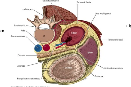
[image:16.612.79.532.51.351.2]Compartments of the Retroperitoneal Space
Three compartments of the retroperitoneal space are related to the kidney:
1.Anterior pararenal compartment
2.Posterior pararenal compartment
3.Perirenal compartment
The renal fascia, a collagenous connective tissue of mesodermal origin enveloping
the kidney, is responsible for this compartmentalization.
The fascial layers and the spaces related to the kidney are as follows, from anterior
to posterior:
• Peritoneum
• Anterior pararenal space (with a variable quantity of loose connective tissue and fat)
• Anterior lamina of Gerota's fascia
• Perirenal space (the kidney and the ureter; the adrenal in a separate subcompartment; fat)
• Posterior pararenal space (usually with a large content of more compact fat)
• Thoracolumbar (lumbodorsal) fascia and the fascia of the psoas muscle To generalize, the muscle fascia lining the abdomen is referred to
as the transversalis fascia. More specifically, however, the transversalis fascia,
which is the fascial lining of the transversus abdominis muscle, is continuous with
the subdiaphragmatic fascia above. Medially, it is continuous with the psoas fascia
and the thoracolumbar (or lumbodorsal) fascial investment (anterior lamina) of the
quadratus lumborum muscle. Below, it is continuous with the fascia of the iliacus
muscle and the parietal muscular fascia of the true pelvis.
Retroperitoneal Lymphatics
From an anatomic standpoint, the retroperitoneal lymph nodes can be
rather difficult to classify. The retroperitoneal lymphatics form a very rich and
extensive chain from the inguinal ligament and pelvis to the respiratory diaphragm
and posterior mediastinal nodes. Usually, these lymph nodes are located close to
the aorta and IVC. The right paraaortic lymph nodes are in very close relationship
with the left paracaval lymph nodes. The number of abdominal and pelvic lymph
nodes is approximately 230.
• Aortic Group
– Preaortic nodes
– Retroaortic
nodes
– Paraaortic nodes
• Caval Group
– Precaval (prevenous) nodes
– Retrocaval (retrovenous) nodes
– Paracaval (laterovenous) nodes
• Pelvic Group
– Common iliac nodes
– External iliac nodes
– Internal iliac (hypogastric) nodes
– Obturator nodes
Retroperitoneal Innervation
Six nerves and the lumbar sympathetic chains are present in
the retroperitoneal space. The six nerves are branches of the lumbar plexus, which
is formed by a branch of T12 as well as by the anterior primary rami of the first
four lumbar nerves. Most of the branches of the plexus are related to the psoas
major muscle, passing through it or behind it or being formed within it.
The nerves formed by the plexus are:
• Iliohypogastric • Ilioinguinal • Genitofemoral
• Lateral femoral cutaneous • Obturator
• Femoral
The retroperitoneum can be approached and explored by several
routes, including the transperitoneal route and the extraperitoneal route. There are
two accepted procedures used for diagnosis of retroperitoneal injuries and for
exploration of clinicopathological entities. The Cattell maneuver exposes
Cattell maneuver
Step 1. Incise the lateral peritoneum along the cecum, ascending colon, and
hepatic flexure
Step 2. Divide the white line of Toldt (peritoneal reflection at the area of the
lateral wall of the cecum and ascending colon)
Step 3. Perform duodenal mobilization (Kocherization)
Step 4. Mobilize all right-sided anatomic entities anteromedially
Mattox maneuver
Step 1. Incise the lateral peritoneum along the splenic flexure, descending
colon, and upper sigmoid
Step 2. Divide the white line of Toldt
Step 3. Carefully mobilize the spleen, including the pancreatic tail, stomach,
Step 4. Gently push all left-sided anatomic entities anteromedially
History
The word sarcoma dates to Galen and the Greek term describing a fleshy
growth. The idea of a sarcoma as a distinct type of cancer was not formalized until
the mid 1800s by Virchow.
Incidence
Soft tissue sarcoma represents less than 1% of all adult and 15% of pediatric
malignancies. Retroperitoneal sarcomas are rare tumors accounting for only 1%–
2% of all solid malignancies. Of all sarcomas, the majority occur outside of the
retroperitoneum. Only 10%–20% of sarcomas are retroperitoneal sarcomas, and the
overall incidence is 0.3%–0.4% per 100000 of the population . The peak incidence
is in the 5th decade of life, although they can occur in any age group.
Distribution
Soft tissue sarcoma can occur in any site throughout the body. Forty three percent are in extremities with two thirds of extremity lesions occurring in the
retroperitoneal (15%) lesions. Trunk sarcoma occurs in 10% of individuals and
others in 10% of patients.
Etiology
Most soft tissue sarcomas have no clearly defined cause, although multiple associated or predisposing factors have been identified.
Various genetic syndrome which are predisposing to soft tissue
sarcoma are neurofibromatosis type 1, retinoblastoma, LI-Fraumeni syndrome,
Gardner’s syndrome, Werner’s syndrome, Goblin’s syndrome, Carney’s triad and
tuberous sclerosis.
Radiation therapy is the known cause of soft tissue sarcoma. They are most
often seen in diseases that are commonly treated with radiotherapy and in those in
which a long survival period is expected. The prime candidate diseases are breast
cancer, lymphoma, and cervical cancer. The children are at risk due to time latency
Classification
It is based on line of differentiation i.e. the type of tissue formed rather than
from the type of origin. WHO’s classification is used widely.
• Fibrous tumors
• Fibrohistiocytic tumors • Lipomatous tumors • Smooth muscle tumors • Skeletal muscle tumors
• Tumors of Blood vessels & lymphatics • Perivascular tumors
• Synovial tumors • Mesothelial tumors
• Peripheral N. sheath tumors
The basis cell appearance on smears, they are classified clinically as follows
• Myxoid tumors • Spindle cell tumors • Pleomorphic tumors • Polygonal tumors • Round cell tumors • Miscellaneous
Pathology
The most common types of retroperitoneal soft tissue sarcomas in adults vary from
study to study. However, in most studies, the most frequently encountered cell
types are liposarcomas, leiomyosarcomas and malignant fibrous histiocytomas
(MFH) . Recently, the frequent diagnosis of MFH in the retroperitoneum has
been-disputed. With the use of immunohistochemistry, many of these fibrous tumors
have now been shown to represent other sarcoma types such as leiomyosarcomas
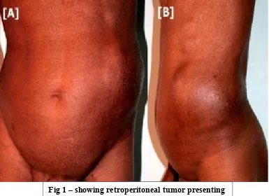
Retroperitoneal sarcomas generally present as large masses; nearly 50% are
larger than 20 cm at the time of diagnosis. They typically do not produce
symptoms until they grow large enough to compress or invade contiguous
structures. The differential diagnosis of a retroperitoneal tumor includes
lymphoma, germ-cell tumors, and undifferentiated carcinomas. The overall
prognosis for patients with retroperitoneal tumors is worse than that for patients
Fig 1 – showing retroperitoneal tumor presenting as abdominal mass (A) AP view ; (B) Lateral view
Grading of sarcoma
After establishing the diagnosis of sarcoma, the most critical piece of
information the pathologist can provide to the clinician is histologic grade. This
remains the most important prognostic factor for determining disease-free and
overall survival rate.
The pathologic features that define grade include cellularity, differentiation,
pleomorphism, necrosis, and number of mitoses.
Unfortunately, the criteria for grading are neither specific nor standardized.
Several grading scales and systems are used: a four-grade system (Broder’s), a
three-grade system (low, intermediate, high) such as National Cancer Institute
(NCI) grading system and that of the French Federation of Cancer Centers
Sarcoma group, and a binary system (low, high) as is used at Memorial Hospital.
Many pathologists consider mitotic activity and degree of necrosis to be the
most important pathologic features. To define a practical grading system, the
European Organization for Research and treatment of cancer (EORTC) conducted
3, 3 to 20, and more than 20 mitoses per 10 consecutive high-power fields), the
presence or absence of necrosis, and tumor size predicted survival.
Several tumors that are considered sarcomas have no recognizable normal
tissue counterpart ( e.g., alveolar soft part tumor, Ewing’s sarcoma, Epithelioid
sarcoma). These tumors often have unique clinical features and usually are not
graded. In 2002 AJCC/TNM staging system of sarcoma, only two grades, low
versus high are used to stage soft tissue sarcomas. To accurately determine tumor
grade, an adequate tissue sample must be well fixed, well stained, and reviewed by
an experienced sarcoma pathologist.
Staging
Staging has an important role in determining the most effective treatment of soft tissue sarcomas. The stage is determined by the size of the tumor, the
histologic grade, and whether it has spread to lymph nodes or distant sites. For
complete staging, a thorough physical examination, x-rays, laboratory studies, and
careful review of all biopsy specimens (including those from the primary tumor,
lymph nodes, or other suspicious lesions) are essential. CT chest done to find out
lung metastases. CT abdomen and pelvis to find out the extension of tumour and
The staging system applies to all soft tissue sarcomas except Kaposi’s
sarcoma,
dermatofibrosarcoma, infantile fibrosarcoma, and angiosarcoma. In addition,
sarcomas arising within the confines of the dura mater, including the brain, and
sarcomas arising in parenchymatous organs and from hollow viscera are not
optimally staged by this system.
Data to support this staging system are based on current analyses from multiple
institutions and represent the recommendations of an AJCC task force on soft
tissue sarcoma. In the era of cytoreductive neoadjuvant treatments, clinical and
pathologic staging may be altered in the future. Because pathologic staging drives
adjuvant therapy decisions, patients should be restaged after neoadjuvant therapies
have been administered.
Histologic type, grade, and tumor size and depth are essential for staging.
Histologic grade of sarcoma is one of the most important parameters of the staging
system. Grade is based on analysis of various pathologic features of a tumor, such
as histologic subtype, degree of differentiation, mitotic activity, and necrosis.
Accurate grading is not always possible on the basis of needle biopsies or in
tumors that have been previously irradiated or treated with chemotherapy.
Inclusions
The present staging system applies to soft tissue sarcomas. Primary sarcomas
can arise from a variety of soft tissues. These tissues include fibrous connective
tissue, fat, smooth or striated muscle, vascular tissue, peripheral neural tissue, and
visceral tissue.
Regional Lymph Nodes
Involvement of regional lymph nodes by soft tissue sarcomas is uncommon in
adults. When present, regional nodal disease has prognostic significance similar to
that of visceral metastatic disease.
Metastatic Sites
Metastatic sites for soft tissue sarcoma are often dependent on the original site
of the primary lesion. For example, the most common site of metastatic disease for
patients with extremity sarcomas is the lung, whereas retroperitoneal and
TNM / The American Joint Committee on Cancer (AJCC) has designated
staging by the four criteria of tumor size, nodal status, grade, and metastasis
(TNGM).
Grade and TNM Definitions
Tumor grade (G)
• GX: Grade cannot be assessed
• G1: Well differentiated
• G2: Moderately differentiated
• G3: Poorly differentiated
• G4: Poorly differentiated or undifferentiated
Primary tumor (T)
• TX: Primary tumor cannot be assessed
• T0: No evidence of primary tumor
• T1: Tumor 5 cm or less in greatest dimension
o T1a: Superficial tumor
o T1b: Deep tumor
• T2: Tumor 5 cm or larger in greatest dimension
o T2b: Deep tumor
Regional lymph nodes (N)
• NX: Regional lymph nodes cannot be assessed
• N0: No regional lymph node metastasis
• N1: Regional lymph node metastasis [Note: Presence of positive nodes (N1)
is considered stage IV.]
Distant metastasis (M)
• MX: Distant metastasis cannot be assessed
• M0: No distant metastasis
• M1: Distant metastasis
NOTES
1. Superficial tumor is located exclusively above the superficial fascia without
invasion
of the fascia; deep tumor is located either exclusively beneath the superficial
fascia, superficial to the fascia with invasion of or through the fascia, or both
superficial yet beneath the fascia. Retroperitoneal, mediastinal, and pelvic
sarcomas are classified as deep tumors.
STAGE GROUPING
IA T1a N0 NX M0 G1–2 G1 Low
T1b N0 NX M0 G1–2 G1 Low
IB T2a N0 NX M0 G1–2 G1 Low
T2b N0 NX M0 G1–2 G1 Low
IIA T1a N0 NX M0 G3–4 G2–3 High
T1b N0 NX M0 G3–4 G2–3 High
IIB T2a N0 NX M0 G3–4 G2–3 High
III T2b N0 NX M0 G3–4 G2–3 High
IV Any T N1 M0 Any G Any G High or Low
Any T Any N M1 Any G Any G High or Low
Evaluation and Workup
The initial evaluation and workup for retroperitoneal abdominal soft
tissue sarcomas are similar to that for the extremity sarcomas. This
workup involves a thorough H&P and appropriate imaging studies,
including an abdominal and pelvic CT with contrast with or without an
MRI. Chest imaging with a plain radiograph or CT should be done,
postoperative chemotherapy. If possible, the patient should be
reviewed by a multidisciplinary sarcoma panel. Note that for staging, all
retroperitoneal lesions are considered to be deep lesions. MRI is preferred for
extremity sarcomas, whereas CT is preferred for retroperitoneal sarcomas.
Radiologic assessment should include CT of the abdomen and pelvis to
define the extent of the tumor and its relationship to surrounding structures,
particularly vascular structures.Imaging should also encompass the liver for the
presence of metastases, the abdomen for discontiguous disease, and the kidneys
bilaterally for function. Thoracic CT is indicated to detect lung metastases. For
patients presenting with an equivocal history, an unusual appearance of the mass,
an unresectable tumor, or distant metastasis, CT-guided core-needle or
18Fluorodeoxyglucose-positron emission tomography (18FDG-PET) scan
may be useful for prognostication, grading and to assess response to chemotherapy.
Tumor metabolism data acquired by FDG-PET will be useful in accurate grading
and prognostication in sarcoma. Recent reports in literature have demonstrated the
value of FDG-PET scan in evaluating response to neoadjuvant chemotherapy in
patients with high-grade extremity soft tissue sarcomas, prediction of outcome in
liposarcoma.
BIOPSY
The differential diagnosis of retroperitoneal abdominal soft tissue mass
includes malignant lesions (such as other sarcomas, GIST, lymphomas,
or germ cell tumors), desmoids, and benign lesions. The need for a
biopsy remains somewhat controversial, and this decision should be
based on the clinician’s degree of suspicion that another malignancy is
possible. Proof of the histologic subtype by biopsy is necessary for
patients before receiving preoperative chemotherapy or RT; a
CT-guided core biopsy is preferred. The goal of this strategy is to avoid
inappropriate major resection of another tumor, such as an
intra-abdominal lymphoma or germ cell tumor. If a retroperitoneal
sarcoma is encountered unexpectedly at the time of laparotomy
establish the diagnosis as well as the histopathologic type and grade of
tumor. Then, the optimal subsequent resection could be performed.
Value of Trucut Biopsy
In general, the important issue is the adequacy of the sample. Sufficient viable
tissue is required that is both representative of the lesion and available for
histopathologic evaluation, immunohistochemistry, and, when necessary, electron
microscopy. As molecular markers become a factor in diagnosis, meticulous
attention to the adequacy of biopsy, tissue preservation, and evaluation will be
paramount.
Histopathologic interpretation varies from center to center and may be a major
variable in decision making. As with other relatively rare lesions, it is essential that
review of the histopathologic findings be made by an experienced group. More
recent studies show improved diagnostic accuracy and confluence of opinion, at
least for malignancy and grade.
Fine-Needle Aspiration Cytology
Fine-needle aspiration (FNA) cytology has been examined by a number of
authors. Some authors have argued that biopsy itself is not justified if FNA is
available. But it is usually confined to the confirmation of recurrence rather than
The use of FNA in patients with large sarcomas who are candidates for
neoadjuvant therapy to improve survival is also problematic due to difficulty in
grading and subtyping these tumors accurately from such small samples.
Frozen Section
In some institutions, frozen section is relied on as the diagnostic tool of choice.
For diagnosis of malignancy, frozen section is accurate, but for histopathologic
subtypes and grade, it is inferior to permanent sections of either Trucut or
incisional biopsy.
Frozen section can guide retrieval of adequate diagnostic material and,
depending on the initial evaluation, can be an important triage mechanism to direct
further pathologic workup. However, open biopsy with the help of
frozen-sectioning support may be indicated when the Trucut biopsy result is equivocal or
for other clinical reasons.
Fatty lesions are not suitable for frozen-section evaluation, because of a loss
of diagnostic material during frozen sectioning and other technical difficulties. In
addition, freezing compromises the final interpretation on permanent sections.
Important application of frozen section is assessment of margin of resection.
Negative margins of resection can be obtained by using this technique
Immunohistochemistry
As an ancillary technique, immunohistochemistry is an invaluable tool that
provides excellent information in assisting the surgical pathologist in establishing a
precise diagnosis, as well as providing relevant prognostic and therapeutic
information.
One of its major utilities is to correctly identify a tumor as being of
mesenchymal or nonmesenchymal origin. Once mesenchymal origin has been
established, histologic subtyping according to specific cell lineage may be
achieved with the use of lineage-specific markers. Tumors of uncertain cell lineage
and tumors with primitive small round cell morphology are often characterized by
a unique immunohistochemical phenotype. In this group of tumors,
immunohistochemistry is most widely applied and is of greatest value. By
diagnosing the small round cell tumors with aid of immunohistochemistry, line of
management is differed from spindle cell soft tissue tumors.
Despite the rapid development of molecular genetic techniques,
immunohistochemistry still remains the most important diagnostic tool in the
diagnosis of soft tissue tumors aside from recognition of morphologic features and
Primary Treatment
Surgery is the standard treatment for retroperitoneal abdominal
sarcomas. Complete surgical resection or macroscopic surgical
resection is only achieved in less than 70% of patients with primary
retroperitoneal sarcomas, because they often are near vital structures.
Local recurrence occurs in approximately half of the patients who have
undergone complete resection. Multimodality treatment is usually
favored for retroperitoneal sarcomas due to the inability to obtain
negative margin resections and high local recurrence rates.
Preoperative RT is often preferred, because the volume of abdominal
organs in the RT fields is smaller and it may render unresectable
tumors more amenable to resection. Preoperative chemotherapy may
have advantages over postoperative chemotherapy. However, the role
of adjuvant RT or preoperative chemotherapy vs. postoperative
chemotherapy has not yet been evaluated in randomized clinical trials.
Primary treatment depends on the resectability of the sarcoma. Biopsy is
performed only if preoperative therapy is considered. CT-guided core biopsy is
Once the diagnosis is made, the surgical team needs to determine if
the retroperitoneal sarcoma can be resected. Therefore one of the first
determinations to be made is whether the tumor is localized, its local extent, and
also if there is evidence of intra- or extra-abdominal metastatic spread of tumor.
The location and size of the tumor, its relationship to adjacent organs, presence or
absence of local extension, relationship to and/or involvement of major vascular
structures, as well as the presence of normal anatomic variants and anomalies of
major abdominal arteries and veins, are all crucial pieces of information that need
to be provided. Since resection of one kidney is not uncommon, any radiographic
evidence of unilateral renal dysfunction involving the kidney that is not adjacent to
the tumor should be relayed to the surgical team. While it may be unavoidable that
the patient will be left with a single poorly functioning kidney, the surgeon must be
provided with all relevant information prior to attempted tumor resection.
Preoperative RT or preoperative chemotherapy (for chemo sensitive
histologies) could be considered. Although most patients with
retroperitoneal sarcomas (which are often liposarcomas) could be
managed with surgical resection with or without intraoperative RT
(IORT), the options for other therapy should be discussed, especially if
two prospective trials showed favorable 5-year local recurrence-free
(60%), disease-free (46%) and overall survival rates (61%) among
patients who had R0 or R1 resection after preoperative RT for
intermediate or high grade retroperitoneal sarcoma.86 Postoperative RT
could be considered in patients with pathologic findings
of high grade disease following negative margin resection or for
microscopic positive margins (R1 resection). Macroscopic positive
margins (R2 resection) should be managed as unresectable disease.
Unresectable retroperitoneal sarcomas are defined as tumors that
involve unresectable vital structures or tumors whose removal would
cause unacceptable morbidity. Biopsy is recommended before any
treatment for a patient with unresectable or metastatic retroperitoneal
sarcoma . Patients with unresectable or metastatic
disease have several options for primary treatment after biopsy
including chemotherapy or RT to downstage tumors prior to
resection. In asymptomatic patients, palliative surgery for symptom
control, best supportive care, or observation are additional options.
Unresectable tumors that become resectable following primary
chemotherapy or RT should be managed as described under
Following primary treatment, if patients have progressive disease or
remain unresectable with no downstaging of tumor, management
decisions depend on whether patients are symptomatic or
asymptomatic. Observation is considered for asymptomatic patients,
whereas for symptomatic patients, treatment options are similar to
those listed under primary treatment for unresectable or metastases.
Guidelines for Radiation Therapy
Sophisticated treatments with intensity-modulated radiation therapy
(IMRT) and proton-beam should be considered to improve therapeutic
effect.If resections with microscopically positive or grossly positive margins are
anticipated, surgical clips should be left in place to identify
high risk areas for recurrence, particularly for retroperitoneal or
intra-abdominal sarcomas . Total doses of RT should be
determined by normal tissue tolerance.
Preoperative RT
The usual dose of preoperative RT is 50 Gy. An intraoperative boost or
a postoperative boost with brachytherapy or an external-beam RT is
recommended for positive or close margins. Preoperative RT has
several advantages. First, the treatment volume is smaller, because the
radiation may reduce seeding during surgical manipulation of the tumor.
The tumor may or may not regress with preoperative RT, but the
pseudocapsule may thicken and become acellular, easing resection
and decreasing the risk of recurrence. However, the main disadvantage
of preoperative RT is its effect on wound healing.A higher
complication rate has been observed when primary closure is used.
Therefore, involvement of a plastic surgeon in the team may be
necessary to reduce wound complications when preoperative radiation
is contemplated. After preoperative radiation, 3-6 weeks interval before
resection is necessary to decrease the risk of wound complications.
Very long intervals between resection and postoperative radiation are
not recommended.
If wide margins are obtained, additional radiation may not be needed.
Often, margins are close because of the proximity of many of these
tumors to major neurovascular bundles or bone. At the time of
resection, surgical clips should outline the area of recurrence risk.
Brachytherapy boosts should be delivered several days after surgery,
through catheters placed at operation, with doses of 12-20 Gy based on
margin status. Alternatively, a single intraoperative dose to the tumor
after resection with exposure of the area at risk, avoiding uninvolved
organs. External-beam RT boosts may be an alternative to
brachytherapy or intraoperative radiation: recommended doses are
10-14 Gy for close margins, 16-20 Gy for microscopically positive
margins, and 20-26 Gy for grossly positive margins. Many institutions
are no longer giving a boost after preoperative radiation to patients who
have widely negative margins, based on local control rates that
approach 95% with preoperative radiation at 50 Gy and negative
margins.
Postoperative RT
Postoperative RT has been to improve local control in patients with
high-grade extremity soft tissue sarcomas with positive surgical
margins.When surgical resection is the initial therapy, postoperative
RT choices include intraoperative radiation therapy (IORT),
brachytherapy or external beam RT. RT is not a substitute for
suboptimal surgical resection, and re-resection may be necessary. If
the patient has not previously received RT, one could attempt to control
microscopic residual disease with postoperative RT if re-resection is not
External-beam RT is delivered to large fields after surgical healing is
complete (at 3-8 weeks) to doses of 50 Gy. Most institutions include the
entire operative bed within that radiation field. Total doses of RT should
always be determined by normal tissue tolerance. For intraabdominal or
retroperitoneal tumors, this dose may be decreased to 45 Gy. An
intraoperative boost may not be possible if radiation morbidity is high.
If no intraoperative radiation or brachytherapy was used in the
immediate operative or postoperative period, an external-beam RT
boost should be added. For negative margins, an additional 10-16 Gy is
recommended to a reduced field that includes the original tumor bed,
based on grade and width of margins. For microscopically positive margins, an
additional 16-20 Gy is recommended; for grossly positive
margins, an additional 20-26 Gy is suggested.
Brachytherapy alone has been used as an adjuvant in patients with
negative margins. 45-50 Gy to the tumor bed has been shown to
reduce recurrence without a significant effect on wound healing.
However, brachytherapy-alone techniques require special expertise
and significant experience. If brachytherapy is used as a boost, doses
of 10-20 Gy based on margin are recommended; a boost dose of 10-16
Recurrent Disease
For patients with resectable, unresectable or disseminated recurrences,
the guidelines recommend the same management after biopsy, as
outlined for primary disease. Preoperative RT and/or
chemotherapy should be considered for recurrent disease, if not
administered previously. Palliative treatment for symptom control (RT,
chemotherapy or surgery) and best supportive care are potential
options that oncologists should discuss with symptomatic patients.
Enrollment in a clinical trial should be considered if an appropriate trial
is available.
Ewing's sarcoma and rhabdomyosarcoma are typically much more sensitive
to chemotherapy than are other adult soft tissue sarcomas. Adjuvant (or
neoadjuvant) chemotherapy is the standard of care for adults with a diagnosis of
rhabdomyosarcoma or Ewing's sarcoma. Typical regimens for small-cell pediatric
sarcomas, specifically rhabdomyosarcoma and Ewing's sarcoma, include the
combination of vincristine, doxorubicin, and cyclophosphamide (dactinomycin, in
particular, for rhabdomyosarcoma) and the combination of ifosfamide and
Surveillance
Patients with low-grade tumors that have been successfully resected
should have a follow-up physical examination with imaging
(chest/abdominal/pelvic CT) every 3-6 months for 2-3 years and then
annually. Patients with high-grade tumors that have been successfully
resected need more frequent surveillance. They should have a
follow-up physical examination with imaging (chest/abdominal/pelvic
CT) every 3-6 months for 2-3 years, then every 6 months for the next 2
years, and then annually. Chest imaging should be considered in both
4. MATERIALS AND METHODS
Patients admitted for retroperitoneal sarcoma between June 2004 to
November 2008 in the Department of Surgical Oncology, Government Royapettah
Hospital, Chennai were taken for study.
Data were collected in all patients. Patient’s age and sex were noted. Histories
like abdominal mass, its duration, presence of pain and its duration, other
symptoms and family history were recorded. Previous history of surgery, biopsy if
any and treatment were taken. Physical examination was done to note site, size of
swelling and presence or absence of metastases. Chest x-ray , CT abdomen and CT
chest were taken in all patients. Histopathology, grade, and margin status were
noted. Histopathology is compared with previous reports. Follow up was done
once in every 3-4 months in 1-2 year and once in 6 months in 3-5 yrs and then
thereafter annually once. Follow up was done with CT abdomen and pelvis and
5. OBSERVATION AND ANALYSIS
Retroperitoneal sarcoma consisted of 0.16% of all cancers admitted from June
2004 to November 2008. Retroperitoneal sarcoma forms 10 – 20% of the soft
tissue sarcomas according to Mettlin, C.Prior et al , but in this study it accounts to
9.85% of soft tissue sarcomas.
Cancers Number Percentage Retroperitoneal sarcoma 21 0.16
Others 12990 99.83
Total 13011 100
Cancers Number Percentage Retroperitoneal sarcoma 21 9.85 Other Soft tissue sarcoma 192 90.14
Total 213 100
In this study incidence of retroperitoneal sarcoma was seen more in males than in
females in a ratio of males:females=2.5:1
Sex Frequency Percentage
Male 15 71.42
Female 6 28.57
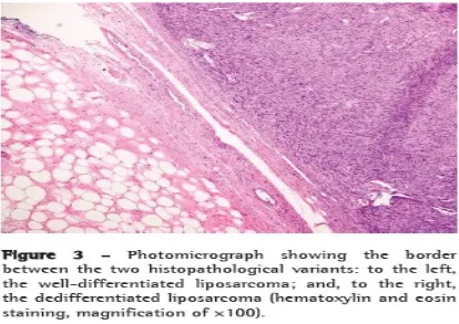
According to literature, the most common histopathologic
types in the retroperitoneum are liposarcoma (40%), leiomyosarcoma (25%),
MPNST, and fibrosarcoma. Approximately 55% of retroperitoneal liposarcomas
are well differentiated and low grade, with tumors in roughly 40% of patients
showing dedifferentiated, high-grade histologic features at primary presentation.In
this study , the results are the same as that of literature with liposarcoma (52.38%)
being the most common histopathology.
Histopathology Frequency Percentage
Liposarcoma 11 52.38
Leiomyosarcoma 4 19
PNET 3 14.3
Rhabdomyosarcoma 1 4.7
MPNST 2 9.5
Total 21 100
As per literature, the peak incidence is in the
5th decade of life, although they can occur in any age group. Study findings bides
with literature results with peak incidence in 5th decade, accounting about 33.33%
of the all age groups .
Age groups(yrs) Frequency Percentage
<20 2 9.5
21-30 4 19
31-40 1 4.7
41-50 7 33.33
51-60 3 14.3
61-70 3 14.3
>70 1 4.7
Male 71% Female 29%
SEX INCIDENCE
Lipos arco ma iomyo sarcoma PNET
Most of the retroperitoneal sarcoma are large in size at the time
of presentation and are considered as deep tumors according to the AJCC staging.
All belong to T2b. In this study , the average size of the tumor at presentation is
15.19 cm. The median size of retroperitoneal sarcoma is 25cm.
As per literature , most of the retroperitoneal sarcoma are seen in Stage III
accounting to 62%.
Stage Frequency Percentage
IA -
-IB 7 33.33
IIA -
-IIB -
-III 13 62
IV 1 4.7
Only one case was found to be in Stage IV in this study.
At the time of presentation, majority were primary tumors but few of them were
Stage I 33%
Stage III 62%
Stage IV 5%
STAGES
2 4 6 8 10 12
10
6
1 1
8
SYMPTOMS
Presentation Frequency Percentage
Primary 19 90.47
Recurrence 2 9.52
Total 21 100
Out of the two recurrent cases , one of them was a case of second time recurrence
and the other 3rd time recurrence. Management and work up plan of the recurrences
were managed like as for primary tumors.
Most patients present with an asymptomatic abdominal mass. On
occasion pain is present, and less common symptoms include gastrointestinal
bleeding, incomplete obstruction, and neurologic symptoms related to
retroperitoneal invasion or pressure on neurovascular structures. Weight loss is
uncommon, and incidental diagnosis is the norm. In one report, neurologic
symptoms related primarily to an expanding retroperitoneal mass were identified in
27% of patients.
Symptoms Frequency Percentage Abdominal pain 10 47.6
Abdominal mass 6 28.5
GI Bleed 1 4.7
Some of the patients came with more than one complaint. Abdominal pain was the
major complaint most of the patients in this study accounting about 47.6% .
Neurologic symptoms was found to be high 42.85% when compared to the
literature values 27%.
According to the literature it is difficult to give adequate clearance in
case of retroperitoneal sarcoma because of adjacent vital structures.
Frequency Percentage Complete resection 8 35 Incomplete resection 1 4.7
Inoperable 6 28.5
Metastasis 1 4.7
Chemotherapy without
surgery
50%
6% 38%
6%
OPERABILITY OF CASES
Complete Resection Incomplete Resection Inoperable
Majority of the cases were resectable , but less when compared to literature results.
In this study only one case was incompletely resected and in literature incomplete
resection is acceptable only in case of a retroperitoneal sarcoma with well
differentiated liposarcoma as the histopathology. In such cases the long term
survival is significantly increased , whereas in other cases incomplete resection has
the same survival rates as those without surgery.
Out of the 8 cases which had complete resection , 3 cases were
positive for margins . These cases were subjected to post operative radiotherapy.
Margin Frequency Percentage
Positive 3 37.5
Negative 5 62.5
Total 8 100
In a study of 28 patients with liposarcomas, adjacent organ
resection was carried out in more than half the cases, with partial or total resection
of the kidneys in 60%, colon in 50% and adrenal glands in 35% .Although
nephrectomy was performed in 60% of cases, the kidney itself was rarely involved.
Nevertheless, the encompassment of the kidney and the involvement of the hilar
renal vasculature make the resection of the kidney often necessary.
Adjacent organs removed Frequency Percentage
Kidney 6 66.66
Bowel loops 5 55.55
In this study, 9 cases went in for adjacent organ resection.
Nephrectomy was the most common procedure done along with resection of the
tumor. Left kidney removal (55.55%) was more common than right kidney
removal (11.11%). Most common bowel loops to be resected was descending
colon. In most of the cases , descending colon was completely removed and distal
2/3 rd transverse colon was anastomosed to either sigmoid colon or rectum.
Majority of cases which are inoperable are due to vascular
involvement(19%). Vascular structures commonly involved are inferior venacava
and common iliac veins.
Blood Vessel Frequency Percentage
IVC 2 50
Common iliac vein 2 50
Total 4 100
In this study no vascular resection and reconstruction was done. Such cases
were given post operative radiotherapy but overall survival was the same.
Radiotherapy helped in local control only in few patients and tumor was
progressive in some cases.
Out of the 21 cases , 12 cases (57.14%) had previous biopsy done.
According to literature pre operative biopsy is only essential in case of soft tissue
Pre operative biopsy is required in case of high suspicion when there is chance of
the retroperitoneal tumor being a lymphoma or primitive neuroectodermal tumor
where such radical resection is not at all required as they are curable by
chemotherapy alone.
The grading of the post resection tumor was compared with
previous biopsy results and were the same. In this study high grade tumors
accounted to 66.66%.
Grade Frequency Percentage
High 14 66.66
Low 7 33.33
Total 21 100
According to literature most of the retroperitoneal sarcoma are
low grade well differentiated tumors unlike the reports of this study.
In this study 5 cases had adjuvant and neoadjuvant chemotherapy . Especially Primitive neuroectodermal tumour(PNET) , Malignant peripheral nerve sheath tumour (MPNST) and Rhabdomyosarcoma responded well to chemotherapy.
Radiotherapy and chemotherapy was also tried in inoperable and marginally positive tumors. The outcome was poor to both therapies. The chemotherapy regimen commonly used in our institute was PVCE regimen consisting of Cisplatin, Vincristine,
6. Conclusions
In this study, retroperitoneal sarcoma is a rare sarcoma
accounting to 0.16% incidence of all cancers and forms 9.85% of all soft tissue
sarcoma .Most of it occurs in males than in females in a ratio of 2.5:1. As with
other series the age incidence is mainly in the 5th decade.
The major histopathology of the retroperitoneal sarcoma is
liposarcoma followed by leiomyosarcoma. Majority of the cases at the time of
presentation were about 15cm in diameter and most belonged to Stage III. (62%)
Abdominal pain, discomfort and neurologic pain were
the most common presenting complaints. Majority were primary tumors and only
9.52% being recurrent tumors.35% of tumors were completely resectable and
28.5% were inoperable due to involvement or proximity to vascular structures.
Only one case of metastasis was reported.
62.5% of operated cases were margin negative. Most of the tumors were
high grade tumors (66.66%). Most of the resections involved adjacent organ
removal with bowel loops and kidney being the common adjacent organs removed.
Kidney (66.66%)removal was slightly more common than bowel removal
Only primitive neuroectodermal tumor and rhabdomyosarcoma
showed good results with chemotherapy. Radiotherapy had no significant role in
BIBLIOGRAPHY:
1.Devita, Hellman & Rosenberg's Cancer: Principles & Practice of Oncology, 8th
Edition
2. Fletcher CD, Unni KK, Mertens F: Pathology and genetics of tumors of soft
tissue and bone. In: Kleihues P, Sobin LH, ed. World Health Organization
Classification of Tumors, vol 1. Lyon, France: IARC Press; 2002.
3. NCCN Practice Guidelines in Oncology – v.3.2008
4.Surg Clin N Am 88 (2008) 451-680
5.Skandalakis Surgical Anatomy : John E. Skandalakis , Gene L. Colborn, Thomas
A. Weidman, Roger S. Foster, Jr., Andrew N. Kingsnorth, Lee J. Skandalakis,
Panajiotis N. Skandalakis, Petros S. Mirilas
6. Jaques D, Coit D, Hajdu S, et al. Management of primary and recurrent soft
tissue sarcoma of the retroperitoneum. Ann Surg 1990;212:51.
7. Bevilacqua R, Rogatko A, Hajdu S, et al. Prognostic factors in primary
retroperitoneal soft tissue sarcoma. Arch Surg 1991;126:328.
8. Santoro A, Tursz T, Mouridsen H, et al. Doxorubicin versus CyVADIC versus
doxorubicin plus ifosfamide in first-line treatment of advanced soft tissue
sarcomas: a randomized study of the European Organization for Research and
Treatment of Cancer Soft Tissue and Bone Sarcoma Group. J Clin Oncol
9. Grier HE. The Ewing family of tumors. Ewing's sarcoma and primitive
neuroectodermal tumors. Pediatr Clin North Am 1997;44(4):991
10. Catton CN, O'Sullivan B, Kotwall C, et al: Outcome and prognosis in
retroperitoneal soft tissue sarcoma. Int J Radiat Oncol Biol Phys 29:1005, 1994.
[PMID: 8083069
11. Lewis JJ, Leung D, Woodruff JM, et al: Retroperitoneal soft-tissue sarcoma:
Analysis of 500 patients treated and followed at a single institution. Ann Surg
228:355, 1998. [PMID: 9742918]
12. Singer S, Corson JM, Demetri GD, et al: Prognostic factors predictive of
survival for truncal and retroperitoneal soft-tissue sarcoma. Ann Surg 221:185,
1995.
13. Retroperitoneal sarcomas
Isaac R Francis, Richard H Cohan, Datla G K Varma,and Vernon K Sondak.
Cancer Imaging. 2005; 5(1): 89–94 (PMID1665230)
14. Papanicolaou N, Yoder IC, Lee MJ. Primary retroperitoneal neoplasms: How
close can we come in making the correct diagnosis. Urol Radiol. 1992;14:221–8
15. van Dalus T, van Geel AN, van Coevorden F, et al. Dutch soft tissue sarcoma
group. Soft tissue carcinoma in the retroperitoneum:an often-neglected diagnosis.
16. Arca MJ, Sondak VK, Chang AE. Diagnostic procedures and pretreatment
evaluation of soft tissue sarcomas. Semin Surg Oncol. 1994;10:323–31.
17. Eilber FC, Eilber KS, Eilber FR. Retroperitoneal sarcomas. Curr Treat Opin
Oncol. 2000;1:274–8.
18. Varma DG. Imaging of soft tissue sarcomas. Curr Oncol Rep. 2000;2:487–90.
19. Stoeckle E, Coinbdre JM, Bonvalot S, et al. French Federation of Cancer
Centers Sarcoma Group. Prognostic factors in retroperitoneal sarcoma: a
multivariate analysis of a series of 165 patients of the French Cancer Center
Federation Sarcoma Group. Cancer. 2001;92:359–68.
20. Mahajan A. The contemporary role of the use of radiation therapy in the
management of sarcoma. Surg Clin Oncol N Am. 2000;9:503–24.
21. Pisters PWT, Ballo MT, Patel SR. Preoperative chemoradiation treatment
strategies for localized sarcoma. Ann Surg Oncol. 2002;9:535–42.
22. Alektiar KM, Hu K, Anderson L, Brennan MF, Harrison LB. High-dose rate
intraoperative radiation therapy (HD-IORT) for retroperitoneal sarcomas. Int J
Radiat Oncol Biol Phys. 2000;9:61–5.
23. Gupta AK, Cohan RH, Francis IR, et al. Patterns of recurrent retroperitoneal
PROFORMA
Name: Occupation: DOA:
Age/Sex: IP/CD No: DOS:
Phone No: DOD:
DIAGNOSIS:
Staging:
Complaints: Abdominal pain –
Abdominal mass –
GI Bleed –
Obstruction –
Neurological symptoms –
Previous H/O malignancy:
Previous H/O biopsy:
Comorbid illness:
Treatment H/o:
H/O Recurrences:
Family H/O Cancer:
USG Abdomen –
Chest X-ray –
CT abdomen –
CT chest –
Biopsy - histology =
Grade =
Immunohistochemistry=
Surgery: Complete / incomplete resection:
Margin negative / not :
Adjacent organs removed :
Vascular resection :
Post-op complications:
Post-op HPE: