1
ASSOCIATION OF OSTEOPONTIN GENE
SINGLE NUCLEOTIDE POLYMORPHISM
WITH SYSTEMIC LUPUS
ERYTHEMATOSUS
Dissertation submitted for
M.D. BIOCHEMISTRY BRANCH – XIII
DEGREE EXAMINATION
THE TAMILNADU
DR. M.G.R. MEDICAL UNIVERSITY
CHENNAI – 600 032
TAMIL NADU
2
BONAFIDE CERTIFICATE
This is to certify that this dissertation work entitled "ASSOCIATION
OF OSTEOPONTIN GENE SINGLE NUCLEOTIDE POLYMORPHISM
WITH SYSTEMIC LUPUS ERYTHEMATOSUS" is the original bonafide
work done by Dr.V.YOGESWARI, Post Graduate Student, Institute of
Biochemistry, Madras Medical College, Chennai under our direct supervision
and guidance.
Dr.R.CHITRAA., M.D., ( Guide ), Professor, Institute of Biochemistry,
Madras Medical College, Chennai – 600 003.
Dr.M.SHYAMRAJ., M.D., Director and Professor,
Institute of Biochemistry, Madras Medical College, Chennai – 600 003.
Dr.V.KANAGASABAI., M.D., Dean,
Government General Hospital Madras Medical College,
3
SPECIAL ACKNOWLEDGEMENT
The author gratefully acknowledges and sincerely thanks Professor
Dr.V.KANAGASABAI.,M.D., Dean, Madras Medical College and Rajiv Gandhi Government General Hospital, Chennai for granting his
4
ACKNOWLEDGEMENT
The author expresses her warm respects and profound gratitude to
Dr.M.SHYAMRAJ,M.D., Director and Professor, Institute of Biochemistry, Madras Medical College, Chennai, for his able guidance,
constant encouragement and support which made this dissertation
possible.
With extreme gratitude, the author acknowledges Dr.R.Chitraa, M.D, Additional Professor, Institute of Biochemistry, Madras Medical College for her constant guidance and keen interest and encouragement during the course of the study.
The author in particular, is extremely thankful to
Dr.Rukmangatharajan, Professor and Head of the Department, Department of Rheumatology, Rajiv Gandhi Government General
Hospital, Chennai, for granting permission to obtain blood samples from
the patients.
The author expresses her warm regards to former Director
5
Dr. Mythili and Dr.Siva of the Institute of Biochemistry, Madras Medical college for their guidance and constant encouragement.
The author highly appreciates the all long cooperation and genuine
support given by her colleagues and is very thankful to them. The author
gratefully acknowledges the help rendered by Mr.Boopathy,Statistician, during the statistical analysis of the study. The author is indebted to the
patients and the persons from whom blood samples were collected for
conducting the study.
Finally the author expresses her sincere thanks to her family
members especially her beloved parents, for the moral support and
encouragement extended by them which gave fulfillment to the
6
ABBREVIATIONS
AP-1 - Activator protein-1
OPN - Osteopontin
SLE - Systemic Lupus Erythematosus
ICs - Immune Complexes
Mφs - Macrophages
TLR - Toll Like Receptor
DCs - Dendritic Cells
BAFF - B cell Activation Factor
TIM - T cell Immunoglobulin Mucin
TBMφs - Tingible Body Macrophages
MFG-E8 - Milk Fat Globule Epidermal Growth Factor 8
HMGB1 - High Mobility Group Box 1protein
SIBLING - Small Integrin-Binding Ligand N-linked
7
AER - Albumin Excretion rate
ACR - Albumin Creatinine Ratio
IL - Interleukin
TGF-β - Transforming growth factor-β PCR - Polymerase Chain Reaction
PKC - Protein Kinase C
EDTA - Ethylene Diamine Tetra Acetic Acid
DNA - Deoxyribonucleic acid
RFLP - Restriction Fragment Length Polymorphism
LN - Lupus Nephritis
FcγRIIb - Fc Receptor 2 b(CD 32) IFNα - Interferron alpha
TNF - Tumour necrosis factor
MLR/lpr - Murine model of Murphy Roths
Large/lymphoproliferative strain
IRF-5 - Interferon regulatory factor-5
SHP-1 - Src homology region 2 domain-containing
8
CONTENTS
Sl.No. Title Page No.
1. INTRODUCTION 1
2. REVIEW OF LITERATURE 3
3. AIM OF THE STUDY 51
4. MATERIALS AND METHODS 52
5. STATISTICAL ANALYSIS 66
6. RESULTS 67
7. DISCUSSION 72
8. CONCLUSION 76
9. FUTURE PROSPECTS OF THE STUDY 78
BIBLIOGRAPHY
ANNEXURES
PROFORMA
9
INTRODUCTION
Systemic lupus erythematosus (SLE) is a prototype autoimmune
disease and is characterized by the presence of autoantibodies which
target self antigens. SLE is commoner in women than men by nearly ten
times, and is typically seen in women of child- bearing age1.Genome
screening studies done in twins have shown the significance of genetic
factors and multiple loci of interest 2. A number of candidate genes
susceptibility to SLE have been identified. The occurence of the disease
in monozygotic twins is approximately 25–50%3. This clearly suggests
that genetic factors do play a key role in the pathogenesis of the disease.
Environmental factors like sunlight, certain drugs, infections may
precipitate the disease in susceptible individuals. Abnormalities in the
activation and maturation of T and B lymphocyte leads to the production
of autoantibodies, which cause multiple organ damage. Lupus nephritis
is the most frequent and potentially serious complication of SLE.
Osteopontin(OPN) or early T lymphocyte activation 1(Eta-1) a
member of T helper 1 cytokine plays a key role in the pathogenesis of
SLE4 and Lupus nephritis. Osteopontin gene is located on the long arm of
chromosome 4, consisting of 7 exons. OPN mediates its
immunoregulatory effects by enhancing the proinflammatory T-helper1
10
stimulates B lymphocytes to express multiclone antibodies. Increased
serum levels of Osteopontin is detected in patients with SLE 7and
increased OPN expression is found in SLE patients with lupus
nephritis8,9. A number of haplotype analysis shows the association of
osteopontin gene polymorphisms and SLE. Similarly, Wong et al10 has
found significantly higher plasma OPN levels compared to healthy
controls.
In this study, the association of osteopontin gene in exon 7 at
position 9250 C T single nucleotide polymorphism (SNP) with SLE
11
REVIEW OF LITERATURE
Systemic lupus erythematosus (SLE) is a prototype autoimmune
disease affecting almost all organs and tissues11.
The term ‘lupus’was derived from Latin meaning ‘wolf’ which was
initially used to describe the classical skin lesions evincive of a ‘wolf’s
bite’. The term ‘lupus erythematosus’ was coined by a Viennese
physician named Ferdinand von Hebra in 1856 12. Although the term was
coined in the 19thcentury, nearly a 100 years lapsed to conclude that SLE
is a systemic disease of autoimmune etiology. SLE is characterized by
hyperactive T and B cells, auto-antibody production, and immune
complex(IC) deposition 13in multiple organs causing end organ damage.
Lupus nephritis is one of the most common clinical manifestation
affecting nearly 50%14of SLE patients.
Epidemiology :
SLE is 9 times commoner in women of child-bearing age group(15
to 35) than in men, and more common in non-European descent15,16. A
study done in Delhi, India showed a point prevalence of 3 per 100,00017
which is much lower when compared to 12.5 per100,000 adults in
England18, 39 per 100,000 in Finland19 and 124 per 100,000 in USA20.
The median age of onset of SLE in India is 24.5 years and sex ratio (F:M)
12
Etiology :
The etiology of SLE is complex and multitude involving genetic factors,
environmental factors, female gender, socioeconomic status, ethnicity and
immunological factors. Rhodes et al22has proposed that number of these
factors occurs either simultaneously or step by step over a time period for
the disease to develop, which occurs when a threshold of genetic and
environmental susceptibility effects is reached.This is shown in Figure 1.
DISEASE MODEL OF A AUTOIMMUNE DISEASE
For people with numerous susceptible genes a minor
13
end even strong environmental trigger might not predispose to the disease
in people with minimal genetic risk.
Genetic factors:
The fact that SLE has strong familial predisposition is supported by
studies done in twins which show that the occurrence of the disease was
about 25-50% in monozygotics and nearly 5 % in dizygotics23. A number
of candidate genes susceptible to the development of SLE have been
identified .Figure:2 shows the susceptible genes in SLE. Various studies
have shown that Human Leucocyte Antigens -A1, B8, and DR3 and
Early Complement Component deficiencies increases the risk of
developing SLE. At least 35 candidate genes have been detected in
patients with SLE24. Single nucleotide polymorphisms (SNP) in the
various immune regulatory substances are associated with increased risk
of SLE . These include SNPs in the interferon pathwayIRF5, IRF7, and
IRF8resulting in increased levels ofIRF5, IRF7, andIRF8transcript and
protein expressions25.Dysregulation in the expression of genes in the IFN
pathway is seen in more than 50% of SLE patients26.Others include
polymorphisms in the following genes, PTPN22,FCGR2A, FCGR2B, and
14
FIGURE-2
GENE DISCOVERIES IN SLE
15
Smoking and exposure to sun light (UVlight) are important factors
implicated in the disease29(Figure:3).
NATURAL HISTORY OF SYSTEMIC LUPUS ERYTHEMATOSUS.
Drug induced SLE occurs due to DNA hypomethylation 30.Some patients
with Ebstein Barr virus infection are prone to develop SLE due to
molecular mimicry between EBV nuclear antigen 1 and the common
lupus autoantigen Ro31.The Antigen presenting cells take up
Environmental and Self antigens, process them into smaller peptides and
present them to T cells. This activates the T cells which in turn stimulates
16
FIGURE 4
IMMUNE CELLS IN THE PATHOGENESIS OF SLE.
Hormonal factors:
SLE is predominantly a disease affecting women, the reason
attributed been the gene CD40 located in X chromosome 32 . Increased
serum levels of oestrogen proliferates B cells and hence increase the
production of antibodies.33,34,35,36Oestrogen is also found to increase the
cell surface expression of CD40 ligand in cultured T cells from SLE
[image:16.612.119.498.185.425.2]17
be distinctive to patients with SLE, signifying that T cells in SLE patients
are more responsive to oestrogens. On the other hand serum levels of
androgens were found to be inversely related to SLE disease activity39.
Women with SLE usually tolerate pregnancy without disease
flares. However, a small proportion develops severe flares requiring
aggressive glucocorticoid therapy or early delivery and a positive link
between pregnancy outcome and the status of disease at conception has
been noted40. Treating patients with dehydroepiandrosterone has shown
some clinical benefit41.
Pathogenesis:
The pathogenesis of SLE is linked to autoimmunity directed
against native cellular components. The three important causes of
pathogenesis include
i) impaired removal of apoptotic bodies &dysregulation of apoptosis.
ii) dysregulation of T and B cells.
iii) dysregulation of expression of certain cytokines.
Multiple susceptible genes have been discovered. The major
histocompatibility complexes 42 present antigens to T-cells,which elicits
an immune response against self-antigens. This results in deposition of
18
Autoantibodies are the prime cause of tissue injury in SLE patients.
The role of the immune system in the SLE development was studied
using mouse strains in the last few years.Based on these
findings,dysregulation of immune system has been divided into two
events :
(I) systemic production of autoantibodies which leads on to
formation of antinuclear antibodies and anti-double stranded
DNA antibodies in serum.
(II) local production of autoantibodies against target organs
resulting in tissue damage. Defective cellular as well as
humoral immunity play an important role in the causation
and development of lupus.
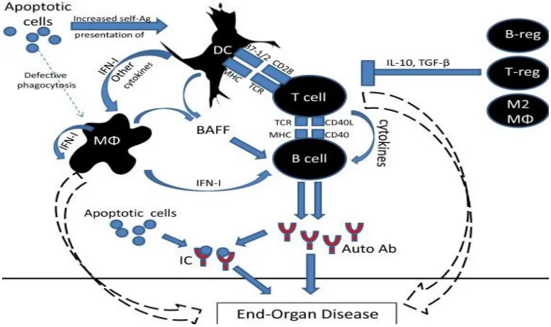
Elicting an immune responses against self antigens is characteristic
of SLE. The self antigens present on the surface of apoptotic blebs are
engulfed by dendritic cells which present them to T cells resulting in their
activation. These T cells secrete interleukin 10 (IL10) and interleukin
23(IL23),which stimulate B cells to produce autoantibodies. There is an
another way of production of autoantibodies not depending on T cells
through the both B cell antigen receptor (BCR) and TLR signalling.
Defects in the apoptosis leads to persistence of endogenous nucleic
19
autoimmune process by destroying self-tolerance (Figure:5). The
immune complexes following their deposition amplify and maintain the
inflammatory events. The antibody complexes damage the critical
vessels, such as the glomeruli of the kidney resulting in lupus nephritis.
Researchers have identified the individual genes and their corresponding
inflammatory mediators produced.One such protein is osteopontin
(OPN), overexpressed in SLE. Each protein is linked to autoimmune
disease and research is being conducted to discover drugs against those
links43,44.
FIGURE 5
[image:19.612.122.511.482.713.2]20
Autoantibodies:
The core of the disease is immune dysregulation with subsequent
formation of autoantibodies. B lymphocytes produce antibodies against
self antigens which are present in the nucleus, cytoplasm, and soluble
molecules like immunoglobulins and clotting factors. The presence of
antinuclear autoantibodies (ANA) is the immunological hallmark of SLE.
About 98% of SLE patients show positive for antinuclear antibodies
(ANA) which is used as a screening test. Approximately 60% of patients
show positive for anti-DNA antibodies 45 which is used as a specific test.
The Sm antigen i.e., small nuclear ribonucleoprotein (snRNP) and
anti-dsDNA antibodies are specific to SLE46. Anti-Sm antibodies directed
against core proteins is constantly produced, whereas anti-DNA
antibodies produced against DNA found over the basement membrane
of glomerulus, antigens like C1q, nucleosomes, heparan sulfate, and
laminin47varies.
Anti-DNA antibodies reflect disease activity48, being pathogenic in
some people . Hence, these are used as disease marker. These antibodies
cause glomerulonephritis both in healthy mice and severe combined
immunodeficient mice49,50. Not all anti-DNA antibodies cause nephritis
as shown by the previous studies where they are found to be negative in
21
impairment.The differences in anti-DNA antibodies pathogenicity can be
due to different isotypes, varying ability of fixing complement. Studies in
SLE have shown that certain clinical features in SLE is linked with
certain antibodies in serum. For instance the anti-ribosomal P antibodies
are associated with psychosis and anti-Ro antibodies with congenital
heart block. Deposition of immune complex with subsequent activation
of complement system is a postulated mechanism of autoantibodies
mediated tissue damage. This is supported by the association finding of
decreased complement levels with vasculitis in SLE. The autoantibodies
also damage the tissues directly through cytotoxic reactions.
Disturbed apoptotic cell clearance :
Casicala-Rosen et al. demonstrated that target autoantigens are
found in the blebs of apoptotic cells51. Apoptosis may be increased by
exposure to UV light, certain infections and drugs. Various studies have
revealed that few T cells in SLE patients increase the production of the
oncogene bcl-2, thereby increase the survival of the dying cells by
attenuating apoptosis. This results in persistence of autoreactive T cells
and progression of the autoimmune disease.
Inefficient removal of apoptotic bodies can be one of the postulated
22
work conducted in mice strains in which the genes coding for DNase I,
serum amyloid protein P (SAP), serum immunoglobulin M,and tyrosine
kinase c-mer,normally involved in the removal of apoptotic bodies are
found to be defective and associated with increased production of
anti-nuclear antibodies52.
The C1q-deficient mice was found to have defective engulfment of
apoptotic bodies53 by peritoneal macrophages and they develop
proliferative glomerulonephritis. This finding reveals that the
complement system and FcR are involved in the clearance of apoptotic
substances.
Apoptosis regulators defects:
Apoptotic cells in the earlier stage express signals such as
phosphatidylserine (PS), to the outer leaflet. Phosphatidylserine (PS) is
the efficient signal to mediate apoptotis by specialized phagocytes called
tingible body macrophages54(TBMφs). Chemo-attractant factors or "find me signals" such as ribosomal protein S19 are secreted in situ for
phagocytosis. "Find me signals" such as Tyro-3, Axl, and Mertk (TAM)
receptor tyrosine kinases are capable of binding to apoptotic cells by
vitamin K-dependent factors, growth arrest specific protein 6 and protein
23
that those lacking Mertk was found to have autoimmune response due to
defective removal of apoptotic bodies. Tisch and colleagues found the
important of Mertk on T-cell central tolerance where they increase the
negative selection of autoreactive T cells.
MFG-E8 (milk fat globule epidermal growth factor 8)57 acts as the
connecting molecule between dying cells and phagocytic cells. Low
levels of MFG-E8 has been reported in some pediatric and adult SLE
patients 58. Subsequently the another molecule involved in the clearance
was identified which is the T-cell immunoglobulin mucin (TIM) gene. Its
function include the regulation of tolerance and removal of apoptotic
bodies by binding to phosphatidylserine (PS) found on the surface of
dying cells59. Mice deficient in TIM was found to have autoantibodies
directed against dsDNA with increased activation of T and B cells60.
High mobility group box 1 (HMGB1) protein was found to be
increased in SLE patients61.It is made of two DNA strapping domains
(i.e,) HMG boxes A and B. HMGB1 is formed during late apoptosis and
necrosis62 and function of this protein is to prevent the double stranded
DNA, single stranded DNA, distorted DNA from undergoing apoptosis
by interfering in the binding of "find me signals" to phosphatidylserine.
24
Microparticles (MPs) are vesicles derived from plasma membrane
which engulf cytokines, growth factors, acute phase proteins, DNA and
RNA. Plasma MPs was found to be increased in SLE patients 63,64.
Microparticles has the capacity to induce B cells by binding to their
immunoglobulin receptors on the surface of B cells and increases the
longivity of DNA and RNA-specific autoreactive B cells. Normally
these microparticles behave like autoadjuvants in the control of both
central tolerance and activation of peripheral B-cells. They express
increased level of phosphatidylserine on their outer leaflet and restrain the
phagocytosis of normal apoptotic cells resulting in accumulation of
apoptotic bodies and aggravate the autoimmune response.
Neutrophil extracellular traps (NETs) consisting of chromatin
networks function to detain and exterminate invading microorganisms.
SLE patients were found to have antibodies directed against NETs,
myeloperoxidase and proteinase-3. Type I interferons 65 in SLE induces
the expression of neutrophils and the formation of NETs. Immediately
after the function of NETs is completed it is removed to avoid the
presentation of self-antigens.
The function of the enzyme DNase I is to break the phosphodiester
25
apoptosis.Some studies have shown significant low level of DNase I in
serum and urine of mice with lupus when compared to normal66.Further
the mice lacking the enzyme presented with lupus like syndrome and
clinical manifestations like nephritis. The delayed clearance in SLE was
found due to lack of this enzyme67.This leads on to use DNase I as a
enzyme replacement therapy in those SLE patients lacking the
enzyme68,69.
Dysregulation of B cell and T cell:
SLE is characterised by a abnormal B cells, T cells and monocytic
lineage cells leading to activation of polyclonal B cells, increased
antibody producing cells, increased immunoglobulins, autoantibody
production, and formation of immune complexes in serum as well as in
tissues. These immune cells release diverse cytokines and various
inflammatory mediators, thereby causing progression of the disease. This
results in increased activation of the leukocytes as well as the
autoantibodies production which finally mediate end organ damage,most
26
Systemic autoimmunity; Adaptive immune cells: The role of B
cells in SLE pathogenesis was studied by Shlomchik and colleagues70.In
one of their study they found that the mice deficient in B cells do not
have autoantibodies and showed an absence of disease concluding the
role of B cell in the production of autoantibodies in the pathogenesis of
lupus . In a different study, Shlomchik and his associates71showed that B
cells produce disease independent of autoantibodies. B cell also
reciprocally activate T-cell which in turn stimulate B cell to produce
autoantibodies .Activated T cells secrete cytokines which causes
inflammatory response and organ damage. The most common cytokine
produced by the T helper1 cells is IFN-γ, which augments the self antigen presentation with production of antinuclear autoantibodies.
B cells secrete cytokines which mediate inflammation like
interleukin-6 (IL-6), interferon-gamma (IFN-γ) as well as anti-inflammatory cytokine IL-10. Kumar and colleagues72 found that the
defect in the negative selection of autoreactive B cells in their immature
stage is due to polymorphism of Ly108 gene, located at Sle1 locus,
resulting in loss of B-cell tolerance .B cell autoimmunity in SLE is also
the result of increased B-cell signaling by CD19 particularly of mature
27
Toll-like receptors (TLRs) participate in the additive stimulation of
B cells by both BCR and TLR signaling. Activation of both BCR and
TLR signaling pathways74 is due to simultaneous involvement of BCRs
which are specific for DNA in apoptotic material and TLR9 on B cells .
In the study conducted in mouse, binding of a synthetic DNA to TLR9
results in progression of nephritis and elevated levels of antibodies to
ds-DNA 75. TLR9-deficient autoimmune mouse models are associated with
low level of antibodies which are directed against DNA and chromatin.
B-cell activation factor (BAFF) is essential for the survival of B-cell.
Transgenic mice with BAFF was found to contract a disease akin to SLE
with increased number of peripheral B-cell producing autoantibodies76.
B-cell activation factor promote the continued existence of B cells in the
germinal center and in the periphery by removing the checkpoint at the
level of the T1 transitional stage. These findings leads on to development
of a soluble receptor specific for BAFF, TACI-Ig, which can be used in
the treatment of lupus in murine77.
Cell mediated immune cells:
The number of monocytes and macrophages are found to be
decreased in the exudates arising from inflammation of SLE patients and
28
defective phagocytosis contribute to defective removal of apoptotic
debris, resulting in autoimmunity and also cause elaboration of
proinflammatory cytokines and chemokines. Kilmon and colleague78
studies reveal the presence of regulatory molecules such as IL-6 and
CD40L produced by the macrophages which keeps check on the
proliferation of B-cell was found to be decreased in mice with lupus.
Dentritic cells (DCs) contribute to pathogenesis of SLE by producing
proinflammatory cytokine, IFN-α. IFN- α stimulate B cells to produce immunoglobulin G targetting soluble autoantigens 79 and also upregulate
production of B cell activation factor leading to increased longivity of
autoreactive B cells and plasmablasts80. This results in a pathogenic
cycle, resulting in increased production of autoantibodies.
Tissue autoimmunity:
This is mediated by (i) ICs and infiltrating cells
(ii) resident cells
Immune complexes (ICs) produced by antibodies against dsDNA
get deposited in the kidney, which initiates the inflammatory response in
the target organ by Fc gamma receptor and complement dependent
mechanisms .There is an antigen similarity between DNA and antigens
29
which results in cross reactivity of antibodies against DNA to renal
antigens. Heymann and colleagues 81found the role of both cytotoxic T
(Tc) cells and T helper cells in the causation of glomerulonephritis. This
results in local release of cytokines, IFN-γ and IL-12 82 and chemokines with recruitment of inflammatory cells depicting type IV
30
Cytokines:
The function of cytokines is to regulate systemic inflammation and
immunomodulation and hence they play an important role in SLE
pathogenesis. IL-6, TNFα, IFNα, and BLyS are candidate biomarkers for SLE and are been evaluated for potential target oriented therapies. Serum
Interleukin-6 and TNF α levels are found to correlate with the disease activity and response to therapy in SLE 83. Autoreactive T-cell clones in
SLE generate huge amounts of IL-6 and TNF α, and thereby triggering B-cell activation and autoantibody production84.Increased expression of
IL-6 was detected in renal biopsy specimens in lupus nephritis and its
urinary levels was also found to be raised85, 86,87. About 52% of SLE
nephritis patients88was found to have upregulated TNFα gene expression with local production of TNFα.
Interferons prevent apoptosis and increase the proliferation of
B-cells89. The primary gene implicated in IFNα secretion which is to be associated with the propensity to develop SLE was transcription factor
IRF5 gene 90. The risk loci of IRF5 is associated with the generation of
autoantibodies to dsDNA and RNA-binding proteins91. Increased levels
31
activity and also found to be associated with the development of
nephritis92,93.
B-lymphocyte stimulator (BLyS) levels are high in nearly half of
SLE patients and is also found to parallel disease activity. Recently,
novel therapeutic drugs targeting BLyS are been developed.
IL-10,a T helper 2 cytokine acts as a powerful activator of B cell
production and differentiation and hence polyclonal B cell activation in
SLE. IL-10 transcript expression and serum levels were significantly
raised in SLE patients compared with controls and further paralleled
disease activity94,95,96,97.
Mechanism of tissue injury:
Accumulation of immune complexes and activation of
complement pathways cause organ damage through cytokines production.
Normally these accumulated complexes are removed by Fc gamma
receptors and complement receptors.Defective removal of complexes
results in their accumulation in the target organs resulting in recruitment
of inflammatory cells, generation of free radicals, secretion of
proinflammatory cytokines, and initiation of the coagulation cascade .The
32
Neuropsychiatric SLE (NPSLE)was one of the manifestation of
autoantibody dependent tissue injury . Mutations in the DNA repair
enzyme 3’ exonuclease 1 (TREX1) is associated with the defective
breakdown of single stranded DNA which results in augmentation of
immune mediated response causing tissue injury.
Lupus nephritis:
Nephritis is one of the common and lethal presentation present in
nearly half of the SLE patients. The International Society of Nephrology
revised the World Health Organization classification of lupus nephritis98.
International Society of Nephrology/Renal Pathology Society 2003 classification of systemic lupus erythematosus (SLE) nephritis
Class I
Minimal mesangial
Normal histology with mesangial deposits
Class II
Mesangial proliferation
Mesangial hypercellularity with expansion of the mesangial matrix
Class III
Focal nephritis
Focal endocapillary ± extracapillary proliferation with central
33
Class IV
Diffuse nephritis
Diffuse endocapillary ± extracapillary proliferation with
disseminated subendothelial immune deposits and mesangial
alterations
Class V
Membranous nephritis
Thickened basement membranes with diffuse subepithelial immune
deposits, may occur with Class III or IV lesions and is otherwise
called as mixed membranous and proliferative nephritis
Class VI
Sclerotic nephritis
Global sclerosis of almost all glomerular capillaries
In situ production of inflammatory cytokines like interferon alpha
(IFNα) and tumour necrosis factor (TNF) mediate the inflammation and injury in the renal cells. Three established theories that lead to the
placement of immune complexes over the basement membrane of
glomeruli and result in the heterogeneity of disease99.These includes,
34
b) increase affinity of autoantibodies for the native antigens
present in the glomerulus.Native antigens include heparin,
laminin and annexin II.
c) antibodies to double stranded DNA and to chromatin have
affinity towards nucleosomes and the DNA found in the
matrix of glomerulus101.
The nucleosomes present in the injury site can arise both from the
plasma and from necrotic debris of the glomerular cells. The nucleosomes
and DNA present in the plasma gets attracted towards the glomerular
basement membrane because of charge to charge interactions and act as
source of antigen for autoantibodies. All these leads to repression of the
gene for the enzyme, DNAse I which normally degrades DNA with
accumulation of nucleosomes in the glomerulus102. These accumulated
materials further activate immune mediated response with simultaneous
induction of FcγRs and Toll-like receptors (TLRs) and complement cascade103.
Figure 6:
Shows an overview of multiple factors involved in the causation of SLE
35
apoptotic materials leads on to presentation to B cells which produces
autoantibodies against self-antigens such as heparin ,laminin in the
kidney. This leads to accumulation of immune complexes (ICs) in the
glomerulus which induces the activation of complement cascade and
proliferation of native mesangial cells (MC) and endothelial cells (EC).
Activated renal cells (MC and podocytes), and inflammatory cells such as
macrophages and dendritic cells (DCs) form free radicals resulting in
additive tissue damage affecting both the tubular and the glomerular
cells.FIGURE 6
36
Clinical features:
SLE may involve one or several organ systems; over time,
additional manifestations may occur. Systemic symptoms, particularly
fatigue and myalgias/arthralgias, are most common and are present most
of the time. Most patients experience relapses and remissions, but
permanent complete remissions are rare. The clinical features of SLE are
diverse involving the following systems,
1.Mucocutaneous features
Lupus dermatitis can be classified as discoid lupus erythematosus
(DLE), systemic rash, subacute cutaneous lupus erythematosus (SCLE).
The classical SLE rash is a photosensitive, slightly raised erythema,
which is sometimes scaly,mostly over the face .It predominantly involves
the cheeks and nose depicting the "butterfly" rash. It may also involve the
ears, chin, V region of the neck, upper back, and extensor surfaces of the
arms. Clinical exacerbation of the rashes might be associated with flares
of the disease.
2.Musculoskeletal involvement
Most SLE patients have discontinuous polyarthritis, varying from
37
joints, commonly involving the hands, wrists, and knees. Joint
deformities (hands and feet) develop in only 10% of patients
3. Nephritis
Nephritis is the most grave manifestation of SLE,since it is one of
the leading causes of death in SLE. Since nephritis is asymptomatic in
most lupus patients, urinalysis should be ordered in any person suspected
of having SLE. Lupus nephritis is currently defined as the presence of
more than 3+ or 500mg/24 hrs proteinuria or presence of cellular casts of
any type. The classification of lupus nephritis is primarily histologic and
renal biopsy is the Gold Standard investigation.104.
4. Neurological
The most common manifestation of diffuse CNS lupus is cognitive
dysfunction. Headache is also a common symptom among patients, but
when severe, it is considered to indicate a flare in SLE. Any type of
seizure may be caused by lupus. Rarely the principal clinical
manifestation of SLE can be psychotic behaviour 105. The ACR
recommends the term neuropsychiatric SLE (NPSLE).
38
The most frequent hematologic manifestation of SLE is anemia,
generally normochromic and normocytic. Sometimes intravascular
hemolysis can occur, which might be acute and life threatening.
Platelets
Two forms of thrombocytopenia (platelet count <100×109/l) are
found in SLE: (1) a chronic form generally associated with mild disease
and (2) an acute form similar to idiopathic autoimmune
thrombocytopenic purpura.
Platelet destruction appears to be mediated by antiplatelet
antibodies and antiphospholipid antibodies.
White blood cells
Leukopenia is also common and almost always consists of a
39
6. Gastrointestinal problems
Oral ulcers, dyspepsia and peptic ulcers are common. Vasculitis
involving the intestine may be grave occasionally by causing bowel
perforations, ischemic necrosis of bowel, bleeding and sepsis. The
incidence of hepatomegaly is 12–25%.
7.Cardiovascular features
Pericarditis is the most frequent cardiac manifestation; usually
cured with anti-inflammatory drugs and infrequently causes cardiac
tamponade. Other serious cardiac manifestations are myocarditis and
Libman-Sacks endocarditis. The endocardial involvement can lead to
valvular insufficiencies, most commonly of the mitral or aortic valves, or
to embolic events106. Patients with SLE are at increased risk for
myocardial infarction, usually due to accelerated atherosclerosis, which
probably results from chronic inflammation107.
8.Lungs
SLE patients most frequently develop pleuritis with or without
pleural effusion as a pulmonary disease. Rarely grave manifestations
include interstitial pneumonitis leading to fibrosis, shrinking lung
40
9.Eye
The commonest ocular involvement is Sicca syndrome and
nonspecific conjunctivitis. Infrequently retinal vasculitis and optic
neuritis are serious manifestations blindness can develop over days to
weeks.
Diagnosis of SLE:
The American College of Rheumatology has a criteria for the
classification of patients as having SLE. Criteria for SLE classification
was developed in 1971, revised in 1982, and revised again in 1997108. If a
patient has, at any time in his or her medical history, 4 of the 11 criteria
documented, the diagnosis of SLE can be made with about 95%
specificity and 85% sensitivity.
Criterias,
1. Malar Rash Fixed erythema, flat or raised, over the
malar eminences, tending to spare the
nasolabial folds
2. Discoid rash Erythematous raised patches with
adherent keratotic scaling and follicular
plugging; atrophic scarring may occur in
older lesions
3. Photosensitivity Skin rash as a result of unusual reaction to
sunlight, by patient history or physician
41
4. Oral ulcers Oral or nasopharyngeal ulceration, usually
painless, observed by physician
5. Nonerosive Arthritis Involving 2 or more peripheral joints,
characterized by tenderness, swelling, or
effusion
6. Pleuritis or Pericarditis 1. Pleuritis--convincing history of
pleuritic pain or rubbing heard by a
physician or evidence of pleural effusion
1. OR
2. Pericarditis--documented by
electrocardigram or rub or evidence of
pericardial effusion
7. Renal Disorder
1. Persistent proteinuria > 0.5 grams per
day or > than 3+ if quantitation not
performed
1. OR
2. Cellular casts--may be red cell,
hemoglobin, granular, tubular, or mixed
8. Neurologic Disorder
1. Seizures--in the absence of offending
drugs or known metabolic derangements;
e.g., uremia, ketoacidosis, or electrolyte
imbalance
1. OR
2. Psychosis--in the absence of offending
drugs or known metabolic derangements,
e.g., uremia, ketoacidosis, or electrolyte
42
9. Hematologic Disorder 1. Hemolytic anemia with reticulocytosis
OR
2. Leukopenia< 4,000/mm3 on≥2 occasions
OR
3. Lyphopenia--< 1,500/ mm3 on≥2 occasions
OR
4. Thrombocytopenia--<100,000/ mm3
in the absence of offending drugs
10. Immunologic Disorder 1. Anti-DNA: antibody to native DNA in
abnormal titer
OR
2. Anti-Sm: presence of antibody to Sm
nuclear antigen
OR
3. Positive finding of antiphospholipid
antibodies on:
a) an abnormal serum level of IgG or
IgM anticardiolipin antibodies,
b) a positive test result for lupus
icoagulant using a standard method,
OR
c) a false-positive test result for at least 6
months confirmed by Treponema
pallidum immobilization or fluorescent
43
11. Positive Antinuclear
Antibody
An abnormal titer of antinuclear antibody
by immunofluorescence or an equivalent
assay at any point in time and in the
absence of drugs .
Serology in SLE109 :
1. Antinuclear antibody (ANA)
ANAs are antibodies which are produced against any one of the
following auto-nuclear antigens:
1. Double stranded-DNA
2. Extractable nuclear antigens (ENA)
3. Histones
4. Nuclear RNA
ANA testing by direct immunofluorescence method is the gold
standard modality. ANA testing is highly sensitive as nearly 95% patients
show a high titre and hence a negative result commends testing or
re-evaluation.
Anti-double stranded DNA antibody (anti-dsDNA)
This antibody is highly specific for SLE. The positivity of
anti-dsDNA by radioimmunoassay and Crithidia lucilae method in SLE is in
44
entire period of the disease is nearly 90%. Thus it is not a good screening
test. The anti-dsDNA titres correlate with the severity of the disease.
Antibodies to extractable nuclear antigens (anti-ENA)
These comprise anti-Sm, anti-UIRNP, anti-Ro and anti-La
antibodies.They are detected in only a half of the patients tested positive
for ANA. The most specific antibody for SLE is Anti-Sm antibody but it
is detected in only one-third of SLE patients. Anti-Ro is antibody is found
in patients with ANA negative SLE, neonatal SLE and subacute
cutaneous lupus erythematosus. Anti-La is positive in cases with SLE and
Sjogren’s syndrome. Antihistone antibodies are coupled with
drug-induced SLE.
Complement levels (C3 and C4)
Serum levels of C3 and C4 are useful to follow up patients with
SLE as they are negatively correlated with lupus activity.
Management189: Patient Education
Avoidance of sun-exposure – by using protective clothing and sun
45
Pharmacotherapy in SLE Mild SLE
The drugs prescribed in order are NSAIDs and analgesics,
anti-malarials (chloroquine, hydroxychloroquine). These are predominantly
useful for the dermatological lesions in SLE.
Moderate SLE
Steroids are the mainstay of treatment, prednisolone 1 mg/kg per
day, taken orally, is the drug of choice. Along with steroids anti-malarials
are also prescribed.
Calcium supplements and vitamin D are prescribed along with
steroids to retard osteoporosis.
Severe SLE
Both steroids and cytotoxics/ immunosuppressants are used to treat
severe flares. The various immunosuppressants used are
cyclophosphamide, mycophenolate mofetil, azathioprine, chlorambucil,
methotrexate and leflunomide.The pulse therapies are given once a
month for 6 months and then a maintenance pulse is administered every 3
46
Osteopontin (OPN)
Osteopontin (Eta-1), secreted phosphoprotein110 is a glycoprotein secreted in bone, inflamed renal tissues, and T cells. Osteopontin (OPN)
is an important cytokine found to have key roles in inflammation and
immunity. OPN carry out a variety of functions in the body which
includes T lymphocyte activation, increases T-helper 1 cell population
and decreases T-helper 2 cells, contribute to cell-mediated immunologic
response and induce B lymphocyte to produce multi-clone antibodies111
.
Increased production of OPN was found to be associated with the
progression of the autoimmune diseases .
OPN is constitutively expressed in bone and epithelial tissues
mediating bone remodeling, repair of tissue and migration112 of cells.
OPN is expressed upon activation during inflammation in endothelial
cells, macrophages, and smooth muscle cells113. OPN is pleiotropic
cytokine secreted by natural killer (NK) cells and activated T cells. OPN
is also called as Eta-1 (early T-cell activation-1) as it is produced soon
after the activation of cells and enhance the T-cell helper 1 (TH1) and
inhibit the T helper 2 responses114,115.The function of the innate immunity
is to protect the organs from various intracellular pathogenic microbes.
Autoimmunity against specific organs results due to increase response of
47
confirmed by a study conducted in mice with absence of Eta-1 gene
expression. Mice shows defective cell mediated(innate) immunity against
certain viral , bacterial infections and failure to develop sarcoid like
granulomas. Deficiency of osteopontin leads to decreased production of
Interleukin-12 (IL-12), interferon-gamma and increases the IL-10
production.OPN acts over the target cells through a
phosphorylation-dependent interaction between the amino-terminal portion of Eta-1 and its
integrin receptor and increases IL-12 production.In other way OPN
interacts with CD44 receptor through phosphorylation-independent
mechanism where it decreases IL-10 production .These findings led to
hypothesis that Eta-1 as a T helper1cytokine is essential for mounting
type-1 immune responses through its variable control of production of
macrophages ,IL-12 and IL-10 cytokines.
Osteopontin receptors and receptor-binding motifs:
The osteopontin mediates its function by binding to the receptors
on the cell surface.This binding is carried out by the presence of specific
receptor-binding motifs in the osteopontin gene sequence.Most
commonly OPN interacts with integrins and CD44.
48
they can bind to a different type of ligands. OPN binding will cause
clustering and activation of the focal adhesion complex, consisting of a
number of regulatory and structural proteins such as like focal adhesion
kinase, Src, cytoskeletal proteins. This results in activation of a number of
diverse signal transduction pathways ultimately affecting the proliferation
and survival of cells116.
Osteopontin was found to interact with a number of heterodimeric
integrins αvβ3, αvβ1 and αvβ5 117,118 through the RGD sequence of arginine,glycine,aspartate aminoacids. Additional integrins by which
osteopontin interacts areα4β1, α9β1 , andα8β1.
Through the RGD sequence of arginine,glycine,aspartate
aminoacids osteopontin interacts with αvβ3 integrin receptor in osteoclasts , smooth muscle cells and tumor cells. Thrombin cleavage site
is present close to the RGD sequence.So the osteopontin is liable to
cleavage by thrombin which is formed during the blood coagulation
cascade .The osteopontin cleaved by thrombin was found to exist in
side-by-side with the full-length of protein(OPN-FL) 119.Thrombin cleaved
osteopontin shows an increase effects in the cell survival when compared
49
Thrombin-cleaved osteopontin binds mostly to a active αvβ3 integrin receptor. The cleavage of osteopontin induce the conformation
change of the molecule, around the RGD motif, and thereby promote the
binding to the αvβ3 integrin.The RGD motif is present in the amino terminal region of thrombin-cleaved osteopontin which induces an
amplified response120.
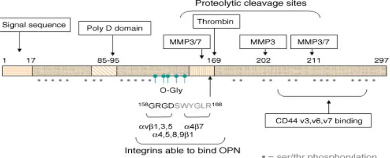
Figure 7:
Thrombin cleavage of human osteopontin. Thrombin cleave the
osteopontin between Arg152 and Ser153 aminoacids producing two
fragments an amino terminal fragment consisting of RGD sequence and
carboxyl-terminal fragment consisting of SVVYGLR sequences
(OPN-R). The carboxyl-terminal fragment can be further acted upon by
Carboxypeptidase B (CPB) which removes C-terminal arginine
converting OPN-R to OPN-L. Both OPN-R and OPN-L are able to bind
integrin receptors. Thrombin-cleaved OPN (OPN-R) exposes an epitope
for the integrins α4β1,α9β1,and α9β4.These integrin receptors are also found on the surface of immune cells such as mast cells,neutrophils,and T
cells which use diverse signal transduction pathways to elicit immune
50
FIGURE 7
THROMBIN CLEAVAGE OF HUMAN OSTEOPONTIN
The CD44 family encoded by a single gene produces different protein
isoforms, because of alternative splicing of sequences.Osteopontin
interaction with CD44 on the surface of macrophages results in decrease
production of interleukin-10 by macrophages and are implicated in the
development of metastases 121. Osteopontin mediate the chemotaxis and
attachment of cells mostly by binding to CD 44 receptor.
The recruitment of inflammatory cells by OPN is carried out by
interaction of arginine-glycine-aspartate (RGD) sequence with integrin
receptors. OPN is associated with a number of pathologic conditions such
as autoimmune disease, cardiovascular disease, cancer, aging, diabetes,
obesity and metabolic syndrome.
[image:50.612.158.544.155.312.2]51
The human OPN gene (OPN) is located on chromosome
4q22.1-figure 8.
CHROMOSOMAL LOCALISATION OF OPN
Osteopontin belongs to the group of SIBLING proteins which consists of
five structurally similar proteins. The human protein consists of 314
amino acid residues122. OPN undergo posttranslational modifications like
glycosylation,phosphorylation with a molecular mass of 44 kiloDalton123.
The OPN gene consists of seven exons. Of the seven exons, six code for
the expression of osteopontin.The 5' untranslated region (5' UTR)
consists of the first two exons . Exon 2 codes for 17 aminoacids,exon 3
codes for 13 aminoacids,exon 4 codes for 27 aminoacids,exon 5 codes for
14 aminoacids, exon 6 codes for 108 aminoacids and exon 7 codes for
134 aminoacids. Casein kinase II as well as cAMP-dependent protein
[image:51.612.156.544.200.376.2]52
shows the secondary structure of OPN consisting of eight α-helices and six β-sheets125.Correlation studies have shown the association of certain OPN genotypes and the increased production of OPN level126.
[image:52.612.159.373.251.389.2]SECONDARY STRUCTURE OF OSTEOPONTIN(OPN).
Figure:8-Shows the coding regions of osteopontin. Exon 1 is present in the 5'-untranslated region consisting of a transcription initiation site (AGC).
- Half of the exon 7 is present in the 3'-untranslated region having
53
- The leader Sequence that directs the protein to the endoplasmic
reticulum and the first two amino acids in the protein is coded by
exon 2
- Exon 3 and 5, the two typical Ser-Ser-Glu-Glu phosphorylation
sequences.
- Exon 4, the two transglutaminase-reactive glutamine residues.
- Exon 6, the aspartic-rich sequence.
- Exon 7 is the major exon encoding roughly half of the protein
together with the RGD motif and the central thrombin cleavage
site.
The activity of osteopontin is regulated by the presence of the
numerous cell interacting domains in addition to multiple protease
cleavage sites.
[image:53.612.121.522.555.658.2]54
* RGD sequence comprising Arg159-Asp159 amino acid
sequence is important for binding to multiple forms of
integrins such as alphaVbeta3, alphaVbeta1, alphaVbeta5
and alpha5beta1.
* Aspartate domain consisting of amino acid sequence
Asp86-Asp89 which is essential for binding hydroxyapatite in the
bone.
* Amino acid sequence Ser162-Arg168 present in the
SVVYGLR sequence binds alpha9beta1 and alpha1beta1
integrins.
* Calcium binding domain consisting of amino acid sequence
Asp216-Ser228 is for binding calcium.
* Amino acid sequence Arg168-Ser169 – with RGD sequence
is the thrombin cleavage site.
* Heparin binding domain - amino acid sequence
Asp290-Ile305- mediates CD44binding.
OPN undergoes post-translational modifications including
55
* O- and N-glycosylation
Isoforms:
The three splice variants of osteopontin transcripts are OPN-a,
OPN-b and OPN-c. These varients are due to alternative splicing of OPN
gene.Exon 5 is absent in the OPN-b varient and OPN-c is lacking exon
4.The regulation of transcription of OPN is composite and it includes
different pathways of Wnt/b-catenin/APC/GSK-3b/Tcf-4,AP-1, Myc,
v-Src, RunX/CBF and TGF-B/BMPS/Smad/Hox .
Intracellular OPN (i-OPN) is produced with an different translation
initiation site on the similar mRNA species that codes for the
extracellular isoform 127.This different translation initiation site is present
in the lower part of the amino-terminal leader sequence.The leader
sequence directs the mRNA to lumen of the endoplasmic reticulum
thereby permitting the translation of OPN in the cytoplasm.
The secretion of osteopontin is stimulated by many factors and is
controlled at the level of transcription 128.It acts through multiple
signaling pathways on specific cell types.Analyses of the promoter region
of osteopontin revealed the possible sites for interactions 129,130 of
transcription factors such as progesterone, glucocorticoids, vitamin D3
56
protein-1. Activator protein-1 binds with a highly preserved enhancer-like
element found in many viral and cellular genes, such as osteopontin gene.
Biological functions of Osteopontin,Figure-11:
OPN in bone remodeling
Osteopontin (OPN) is a major noncollagenous bone matrix protein.
The GRGDS sequence consisting of glycine-arginine-glycine-aspartic
acid-serine amino acids mediate cell attachment through cellular αvb3 integrin131.Normally osteoclasts expresses OPN during the course of
57
and is considered to play important roles in bone formation, resorption,
and remodeling.OPN increases the segregation and production of
osteoblast cells thereby increases ALP activity132. OPN also cause bone
resorption by acting on osteoclasts and change them into ruffled
borders.OPN presentin the urine inhibits formation of the kidney stone.
Immunological functions
Osteopontin is necessary for the development of cell mediated
immunity. It acts by increasing the expression of interferon-gamma and
interleukin-12 and decreases the production of interleukin-10.
Osteopontin (OPN) is produced by a number of immune cells such as
macrophages, neutrophils, dendritic cells,T cells and B cells in different
concentrations. OPN interacts with multiple integrin receptors α4β1, α9β1, and α9β4 present on the surface of leukocytes. OPN is an immune modulator133. It has multiple functions including recruitment of immune
cells to inflammatory sites, an adhesion protein participating in cell
attachment ,wound healing and augments the survival of cells by
58
Chemotaxis
The chemotactic function of OPN was shown by a number of
studies.OPN plays an significant role in recruitment of neutrophils in
alcoholic liver disease.Another study reveal the function of recruitment of
inflammatory cells to arthritic joints in the development of rheumatoid
arthritis134 .In vitro study conducted in 2008135 inferred that OPN
knock-out mast cells showed a reduced level of chemotaxis when compared to
normal mast cells.
Activation of cells
Osteopontin induces the secretion of cytokine IL12 which causes
T cells activation and make them to differentiate towards the T helper
1cells.The T helper1cells expresses cytokines such as IL-12 and IFN-γ. At the same time OPN attenuate the production of the T helper2 cytokine
IL-10,ultimately leading to increased T helper 1cells response. OPN
induces cell-mediated immunity and T helper 1cells comprises cytokine
functions. OPN stimulate B cells to produce multiple clone of
immunoglobulins and their proliferation. The researchers found that
IgE-mediated anaphylaxis was considerably low in mice without OPN when
compared to mice with OPN. Activation of macrophages by osteopontin
59
unable to cause activation of macrophages compared to tumors cells
producing osteopontin.
OPN in Apoptosis
OPN is an significant anti-apoptotic cytokine.OPN prevents the
activation-induced apoptosis of macrophages,T cells ,fibroblasts and
endothelial cells when exposed to detrimental stimulating factors137. OPN
was found to inhibit the non-programmed loss of cells in inflammatory
colitis. Charles describes research findings that apoptotic dysfunction in
the development of autoimmunity138. Defective removal of apoptotic
bodies was one of the mechanisms that leads to the occurance of SLE.
OPN in autoimmune diseases
OPN was found to be involved in the development of rheumatoid
arthritis.Study workers have found the increased level of OPN-R, the
thrombin-cleaved form of OPN in the synovial fluid of rheumatoid
arthritis joint.But the exact pathogenic events in rheumatoid arthritis is
not studied in detail.One study group found that OPN knock-out mice do
not develop arthritis. OPN has also been involved in the pathogenesis of
other autoimmune diseases such as autoimmune hepatitis, allergic airway
60
OPN in Malignancy and inflammatory diseases
The expression of osteopontin in several human carcinomas was
shown by Brown and co-workers 139. High level of osteopontin mRNA
was seen in the screening of tumors of colon, breast, lung, stomach,
endometrium and thyroid when compared to normal tissues. Osteopontin
promote the development of cancer through numerous and composite
mechanisms such as binding with cell surface receptors, regulation of
growth factor and receptor pathways and proteases.Proto-oncogeneras ,a
GTPase protein increases the transcription of osteopontin which is
implicated in the transformation, metastasis and progression 140of
neoplastic cells. Normally osteopontin is expressed at low concentrations
in tissues but in case of premalignant and malignant cells it is increased.
Osteopontin increases the survival of endothelial cells by increasing the
expression of pro-survival genes and decreasing the expression of
anti-survival genes through the activation of nuclear factor κB 141
Figure;12.Further by interacting with integrin αvβ3 , it induces the expression of osteoprotegerin, a tumor necrosis factor receptor which has
been found to protect endothelial cells from programmed cell death.
Antiosteopontin therapeutic strategies are being in research to target OPN
61
ROLE OF OSTEOPONTIN IN APOPTOSIS
OPN in allergy and asthma
Osteopontin was found recently to be associated with asthma and
diseases. From the study conducted in mice it was established that the
secreted form of OPN (OPN-s) exhibit opposing effects by increasing the
T helper2 responses when compared to other forms of OPN which
increases the T helper 1 cells.This results in allergic disease like asthma
with primary systemic sensitization through pro-inflammation and
62
antigens through the regulation of different dendritic cell population. The
absence of OPN was seen to protect from remodeling and asthma142.
OPN in muscle disorders
Osteopontin is involved in a number of pathways that lead to
development of skeletal muscle diseases, such as Duchenne muscular
dystrophy. Osteopontin mediates inflammation of dystrophic and injured
muscles and cause high scarring of diaphragm muscles in the aged
dystrophic mice.A latest study revealed that a mutation in the promoter
region of osteopontin gene causes decreased levels of osteopontin
expression.This seems to be associated with a decrease in the severity of
clinical manifestations in patients with Duchenne muscular dystrophy143.
Therefore, handling of plasma OPN levels can be helpful in the
treatment of autoimmune diseases, malignancies, osteoporosis and
63
OSTEOPONTIN IN SLE
Systemic lupus erythematosus (SLE ) is a typical autoimmune
disease characterized by abnormal immunologic response.T lymphocyte
and B lymphocyte activation results in production of multifarious
autoantibodies with several tissue damage.Lupus nephritis (LN) is the
most common and lethal manifestation of SLE. Lupus nephritis is
frequently associated with abnormal production of cytokines. OPN
induces B lymphocyte to produce polyclonal antibodies 144. OPN carry
out a variety of functions in the body which includes early T lymphocyte
activation that’s why it is called as early T lymphocyte activation 1
(Eta-1), increases T-helper 1 cell population and decreases T-helper 2 cells and
thus contribute to cell-mediated immunologic response 145,146.Several
studies have found that OPN is an essential component in the
autoimmune mediated pathogenesis of SLE147,148.There is a promising
association of OPN gene polymorphism with systemic lupus
erythematosus. A single nucleotide polymorphism (SNP) at position 9250
with replacement of C by T in exon 7 of the OPN gene (OPN gene 9250)
is newly detected in humans149.
Humans with SLE overexpress osteopontin suggesting a role of
OPN150 in the pathogenesis of the disease . Enhanced expression of
64
confirmed by the study in MRL/lpr –murine model of Murphy roths
large/lymphocyte proliferation which shows increased production of
osteopontin compared to controls and increased production in the
proximal tubule appears to mediate infiltration of
macrophage.Osteopontin is upregulated in different types of renal
damage151.Particularly in humans, crescentic 152glomerulonephritis is
associated with enhanced production of osteopontin .Increased production
of osteopontin is found to produce certain clinical features of lupus in
mice and humans with SLE show overexpression of osteopontin in
plasma and at confined sites of renal inflammation. This provoked the
present study of an osteopontin gene single nucleotide polymorphism in
the region of exon 7 in SLE patients.Multiple polymorphisms in the
coding gene of the human OPN was identified in diverse populations,that
in Japanese population some polymorphisms have been located in the 5′ flanking region, Chinese population was found to have polymorphisms in
65
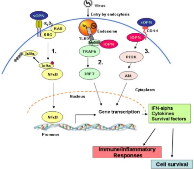
FIGURE:13
OSTEOPONTIN SIGNAL PATHWAYS
By binding to these receptors OPN stimulate multiple signaling
pathways to initiate immune responses154. (a) The circular form of
osteopontin binds to integrin αvβ3 receptor and the intracellular signals are carried out through the Src and FAK tyrosine kinases activating
66
factor and nuclear translocation of NFκB which mediates the transcription of a number of pro-inflammatory cytokines. (b) In dendritic
cellsDCs,intracellular OPN (iOPN in red) interacts with MyD88 during
TLR9 engagement with viral DNA of endosome, causing TLR9 signaling
in the direction of IRF7 instead of IRF-5/NFkB leading to vigorous
production of IFN-γ.(c) Interaction of OPN with CD44 increases the intracellular concentration of a second messenger phosphatidyl inositol
3phosphate which is acted upon by kinase cascade of PI3k /Akt signaling
mediating cell survival. OPN promoted the survival of activated T cells
by decreasing the transcription factor Foxo3a through the translocation of
the NF- B and by varying expression of the pro-apoptotic proteins Bim,
Bak and Bax. These events together decrease the death of lupus-reactive
T cells, connecting OPN to the development of SLE.
The susceptible genes in SLE which secrete cytokines are of
important topic in research. Increased concentration of OPN have been
documented in biopsies of injured tissues in SLE and also in other
autoimmune diseases .Numerous studies have found that raised plasma
OPN level is associated with progression of disease activity in SLE.
In mouse models,OPN is important for the expression of
interferon-alpha (IFN-α) . IFN-α levels are found to be raised in more
67
SLE .SLE risk-related allele of OPN rs9138C was linked with elevated
levels of serum OPN and IFN-α in adolescent female and men with SLE .
This observable fact in which a number of SLE-risk loci associated with
the cytokine profiles has been confirmed as well .
Also identified the association of SLE-risk loci with the specific
clinical manifestations of SLE .For example a study has found a
relationship between rs7687316 in the promoter region and
lymphadenopathy in European descent individuals . Another study
comprising of 81 SLE patients of European American descent established
an connection between a identical change in exon 7 with avascular
necrosis and renal damage. The rs11730582 C and rs9138 C alleles of the
osteopontin (OPN) gene was found to be separately related with increased
possibility of lupus. The cytokine actions of OPN include the activation
of macrophage and T-cell migration.OPN is found to protect against
herpes viruses and bacterial infections through the activation of the T
helper1 activity and stimulation of T helper1 - cell-mediated
68
AIMS AND OBJECTIVES
The aim of the study is
1. to determine the association of single nucleotide polymorphism at 9250 C→T in exon 7 of Osteopontin(OPN) gene among systemic lupus erythematosus patients, with and
without nephritis and in healthy controls.
69
MATERIALS AND METHODS
This is a case-control study and was conducted after obtaining
ethical committee clearance. The study was carried out during the period
April 2011- September 2012 at Madras medical college and Rajiv Gandhi
government general hospital.
Study population:
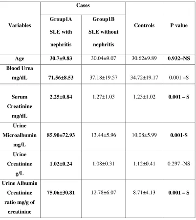
CASES:
100 SLE cases attending rheumatology outpatient department of
our hospital were included in the study after obtaining consent and were
categorised into
Group 1A: 50 SLE patients with lupus nephritis
Group 1B: 50 SLE patients without lupus nephritis
Nephritis cases are included based on renal biopsy findings.Cases
with clinical or laboratory evidence suggestive of mixed connective tissue
disorders were excluded from the study.
CONTROLS:
70
Sample collection:
Blood samples:
About 5 mL of blood was drawn from the cubital vein of the
subjects and collected in EDTA tube.The samples were centrifuged and
plasma was separated and transferred into 2 mL eppendorf. Plasma was
stored at -20°C for estimation of osteopontin.
Urine samples:
Early morning urine samples were collected in sterile plastic
containers and Albumin Creatinine ratio was estimated.
BUFFY COAT SEPARATION
Buffy coat was obtained by centrifugation of EDTA tubes at 2000
revolutions for 20 minutes.Buffy coat was transferred to 2 ml eppendorf
and was used for DNA extraction. DNA extraction was done on the same
day and extracted DNA stored at -20°C.
DNA EXTRACTION BY MODIFIED HIGH SALT METHOD155 RBC Lysis:
• 400µL of buffy coat in a 2mL eppendorf is mixed with 1.6mL of
0.17M ammonium chloride and mixed by inversion until red cells
71
• The cells are centrifuged at 4000rpm for 10minutes.
• The white cell pellet is washed with 800µL of 0.17M ammonium
chloride solution. The procedure is repeated till a clear white cell
pellet is obtained.
WBC Lysis
• To the pellet 500 µL of TKM I solution is added. It is centrifuged
at 10,000rpm for 10minutes.
Nuclear Lysis
• Discard the supernatant. To the pellet add 500 µL of TKM II
solution. To that add 300 µL of 6M NaCl and 50 µL of 10% SDS.
• Mix well (vortex), Centrifuge at 10,000 rpm for 10 minutes.
• Save the supernatant. Transfer it to 1.5mL eppendorf.
DNA Precipitation
• To the supernatant double the volume of 100% ethanol is added.
• The sample is stored at -20◦C for 1 hour.
72
• The supernatant is discarded. To this 500 µL of 70% ethanol is
added. The pellet is mixed and centrifuged at 10,000 rpm for
10minutes at 4◦C and later air dried.
Storage
• To the pellet 30 µL of LTE buffer is added and the extracted DNA
is stored at -20◦C for future use.
Identification
• Extracted DNA was identified by 0.8% agarose gel electrophoresis
with a constant voltage of 7V/ cm and comparison with a known