RESEARCH NOTE
Considerations for skin carcinogenesis
experiments using inducible transgenic mouse
models
Martyna C. Popis
1, Rebecca E. Wagner
2, Fernando Constantino‑Casas
3, Sandra Blanco
4and Michaela Frye
1*Abstract
Objective: This study was designed to estimate the percentage of non‑malignant skin tumours (papillomas) pro‑ gressing to malignant squamous cell carcinomas (SCCs) in a carcinogenesis study using established transgenic mouse models. In our skin cancer model, we conditionally induced oncogenic point mutant alleles of p53 and k‑ras in undif‑ ferentiated, basal cells of the epidermis.
Results: Upon activation of the transgenes through administration of tamoxifen, the vast majority of mice (> 80%) developed skin papillomas, yet primarily around the mouth. Since these tumours hindered the mice eating, they rapidly lost weight and needed to be culled before the papillomas progressed to SCCs. The mouth papillomas formed regardless of the route of application, including intraperitoneal injections, local application to the back skin, or sub‑ cutaneous insertion of a tamoxifen pellet. Implantation of a slow releasing tamoxifen pellet into 18 mice consistently led to papilloma formation, of which only one progressed to a malignant SCC. Thus, the challenges for skin carcino‑ genesis studies using this particular cancer mouse model are low conversion rates of papillomas to SCCs and high frequencies of mouth papilloma formation.
Keywords: Skin carcinogenesis, Mouse model, Papilloma, SCC, p53, k‑ras
© The Author(s) 2018. This article is distributed under the terms of the Creative Commons Attribution 4.0 International License (http://creativecommons.org/licenses/by/4.0/), which permits unrestricted use, distribution, and reproduction in any medium, provided you give appropriate credit to the original author(s) and the source, provide a link to the Creative Commons license, and indicate if changes were made. The Creative Commons Public Domain Dedication waiver (http://creativecommons.org/ publicdomain/zero/1.0/) applies to the data made available in this article, unless otherwise stated.
Introduction
The most common human cancers arise from epithe-lia including skin, colon, breast, prostate, or lung, and together cause several million deaths per year [1]. Squa-mous cell carcinoma (SCC) is the second most common form of skin cancer and predominantly occurs in sun-exposed regions of skin [2]. In other epithelia such as lung and oesophagus, SCCs are often induced by muta-gens including tobacco and alcohol or the human papil-lomavirus (HPV) [3, 4].
Genetic alterations in the RAS and P53 genes are com-monly identified as driver genes in aggressive SCCs [5]. To model the human disease, many transgenic mouse cancer models have been generated that accurately reca-pitulate the genetic alterations found in human tumours.
Due to the high frequency of RAS mutations in human epithelial cancers, investigations into the role of onco-genes in tumourionco-genesis commonly induce endogenous
ras mutations in mice [6]. Endogenous oncogenic ras
is sufficient to initiate transformation by stimulating proliferation, yet further genetic lesions are required to progress to a malignant tumour [6]. Inactivation of the p53 tumour suppressor is an additional frequent event in tumorigenesis [7]. Activation of both cancer-causing genetic mutations in an inducible fashion in particular cell types of the epidermis is often used to study the cel-lular and molecular origins of SCCs [8–13].
By applying cancer-inducing genetic alterations to mice, they provide valuable in vivo tumour models to study skin cancer origin, progression, metastasis and chemotherapy resistance. Here, we discuss practical con-siderations for skin carcinogenesis experiments using inducible transgenic mouse models.
Open Access
*Correspondence: [email protected]
1 Department of Genetics, University of Cambridge, Downing Street, Cambridge CB2 3EH, UK
Main text Methods
All transgenic mouse lines in this study are routinely used in carcinogenesis studies and have been described previ-ously: K14-CreER [14], K-rasLSL-G12D [6], and p53LSL-R172H [15]. The lines were obtained from The Jackson Labora-tory (https://www.jax.org/). All mice were on a mixed genetic background and age-matched males and females were used. All mice were housed in individually venti-lated cages (IVC). The experiments were not performed blinded as the genotype was known to the investigators. However, all mice for breeding and the experiments were chosen randomly.
Tamoxifen was either applied as solution or in form of tamoxifen (free base) pellets (5 or 7.5 mg/pellet) (Innova-tive Research of America, cat. nr. E-361) were implanted subcutaneously into the neck area of mice. 13 mice were implanted with 5 mg pellets (5 males, 8 females), and 5 mice were implanted with 7.5 mg pellets (4 males, 1 female). The mice were anaesthetised with isofluorane. Caprofen (Caprieve Small Animal Solution for Injection, Norbrook, National Veterinary Services, Code 219129) was administered pre-operatively at 8.3 mg/kg via sub-cutaneous injection as analgesic. The wound at the pel-let insertion site was closed with GLUture topical tissue adhesive (Zoetis, National Veterinary Services, Code 288615). Tumour development and appearance were monitored and recorded daily.
For histological analyses, the tumours were fixed over-night with 4% paraformaldehyde (Santa Cruz, cat nr sc-281692), transferred to 70% EtOH (Ethanol absolute, Sigma cat nr 32205 diluted to 70% in water) and embed-ded in paraffin. Samples were then cut at 8 µm. Hema-toxylin and Eosin staining of paraffin-embedded tumours were performed as described previously [16].
Results
K14CreER driven activation of K‑rasG12D and p53LSL‑R172H
in the epidermis
To trigger the formation of SCCs in the mouse back skin we used a Cre-recombinase inducible transgenic mouse line that carried oncogenic point mutations in the alleles of p53 (p53LSL-R172H) and K-ras (K-rasLSL-G12D) (Fig. 1a, b) [6, 15]. Skin tumour formation is initiated by activa-tion of the endogenous K-rasLSL-G12D and p53R172H alleles (Fig. 1a) [13]. We activated the oncogenic alleles in the epidermis by conditionally inducing Cre-recombinase under the control of the keratin 14 (K14) promoter by administering tamoxifen (Fig. 1b) [14]. K14 (together with K5) forms the main keratin in keratinocytes in the basal, undifferentiated layer of stratified squamous epi-thelia that includes the skin and the inner lining of the mouth and the esophagus [17–19]. K14-driven activation
of the oncogenes lead to the development of skin papil-lomas, a proportion of which were expected to undergo malignant conversion into invasive SCCs (Fig. 1c, d) [20].
To maintain the transgenic lines and generate experi-mental cohorts, the mice have to be kept as heterozygous for both K-rasLSL-G12D and p53LSL-R172H alleles. K-Ras LSL-G12D homozygous animals lack functional K-Ras and show early embryonic lethality, and no embryos survive past E11.5 [6]. Mice homozygous for the p53LSL-R172H alleles are prone to develop a variety of internal tumours within 5–6 months [21]. Thus, only K14-CreER+::K-rasLSL-G12D/ wt::p53LSL-R172H/wt were used for this study. Untreated control animals with this genotype showed very low lev-els of leakiness for the transgenes. Around 13% (2 out of 15 animals) of untreated CreER+::K-rasLSL-G12D/wt::p53 LSL-R172H/wt mice developed spontaneous papillomas after 16 weeks of age.
Intraperitoneal injections and topical application of tamoxifen to activate K‑rasG12D and p53LSL‑R172H in the epidermis
In pilot experiments, we first administered tamoxifen via intraperitoneal (IP) injection or topical application directly to a shaved area of the back skin in two small cohorts of mice (4 mice for IP and 17 mice for topical). Both methods are standard methods to apply tamox-ifen and as expected, resulted in the development of skin papillomas. However, in response to both methods of tamoxifen application, all mice developed papillomas around the mouth area, which hindered them from eat-ing. Due to weight loss, all mice in our experiments had to be culled before they developed malignant carcinomas (data not shown).
Using tamoxifen pellets to activate K‑rasG12D and p53LSL‑R172H in the epidermis
In order to minimise mouth papilloma formation, we aimed to induce the transgenes more locally and used tamoxifen pellets that can be subcutaneously inserted using a trochar (Fig. 1e). Subcutaneous pellet insertion has the advantage that it continuously releases tamoxifen over a prolonged period of time. Here, we used pellets that release a total dose of 5 or 7.5 mg of the drug over 21 days.
Mouth papilloma formation is the bottleneck of carcinogenesis experiments
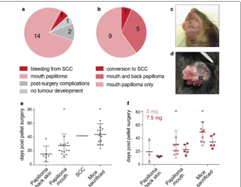
15 out of the 18 experimental animals, which underwent pellet insertion surgery developed tumours (Fig. 2a). With the exception of one animal, 14 developed mouth papillomas (Fig. 2b, c). Only 6 out of the 15 animals also
developed papillomas on the back skin. The back papil-loma of only one mouse progressed to a SCC (Fig. 2d).
Over the course of our experiment, back skin pap-illomas formed first, on average on day 16 post pellet implantation (Fig. 2e). Mouth papillomas formed later, on average on day 27 post pellet implantation. Mice
Fig. 1 a Schematic representation of the keratin‑14 (K14)‑driven inducible Cre‑recominase (Cre) and the KrasG12D and p53R172H transgenes prior
the excision of lox‑STOP‑lox cassette. ßg int: Beta‑globin 5’ untranslated region (UTR) and an intronic sequence. ERtam: tamoxifen inducible estrogen
[image:3.595.60.538.87.533.2]were maintained on average for 17 days from the first appearance of the papillomas. Then, they lost weight and needed to be culled. A standard measurement in UK home office licences is that a mouse that lost more than 15% of body weight compared to wild-type lit-termates must be culled. This leads to the termination of the experiment in average on day 44 post-surgery (Fig. 2e; see ‘mice sacrificed’). The single SCC conver-sion occurred in our model on day 42 post-surgery. Thus, the development of mouth papillomas is the bot-tleneck for the experimental protocol. Our experiment illustrates that due to the development of mouth papil-lomas, it is very challenging to maintain this particular mouse model long enough to allow for conversion to malignant SCCs.
The high incidence of mouth papilloma formation might be due small wounds caused by abrasion inside the mouth induced by solid food pellets [22]. To refine the procedure, we fed the mice with a non-solid diet. While the use of non-solid food failed to reduce mouth papil-loma formation, it improved the well-being of the mice by for instance reducing bleeding from the papillomas.
Finally, we tested whether higher doses of tamox-ifen enhanced formation of SCC. We used two types of tamoxifen pellets: a total dose of 5 mg or 7.5 mg released over 21 days. The increase of tamoxifen dose from 5 to 7.5 mg slightly sped up the formation of all types of tumours, yet the difference was not statistically signifi-cant. The higher dose of tamoxifen did not affect the for-mation of mouth papillomas (Fig. 2f).
[image:4.595.61.539.86.456.2]Conversion rate of papillomas to SCCs is low
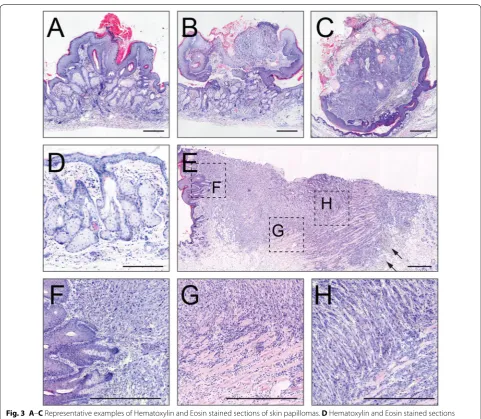
Histological section of typical papillomas and the SCC
are shown in Fig. 3A–C, E–H. A common phenotype
in response to the oncogenic activation in mouse epi-dermis is hyperplasia of cells in the stratum basale and the sebaceous glands (Fig. 3D), which can be explained by the activation of a single mutant KrasG12D allele [23]. The carcinoma showed neoplastic regions with an invasive front towards the deeper tissues, which are common characteristics of a SCC (Fig. 3E–H). In addi-tion, we detected inflammation and dermal fibroblast proliferation (desmoplasia) (Fig. 3E; arrows). Neoplas-tic keratinocytes are polygonal, lack polarity and show anisocytosis and anisokaryosis (Fig. 3H). Together, one
out of 15 (7%) papillomas progressed to a malignant SCC.
Limitations
In this study we highlight some limitations when using the K14-CreER::K-rasLSL-G12D::p53LSL-R172H genetic mouse model to study the progression from benign papillomas to malignant carcinomas in skin.
Generating the mice by crossing requires in average 1 year, given that experimental mice carry at least the fol-lowing alleles: p53LSL-R172H, K-rasLSL-G12D, an inducible Cre-recombinase, and often an additional transgene of interest. Breeding the mice to generate the experimental cohorts required a large number of mice. Here, we bred
[image:5.595.57.539.283.702.2]144 animals to generate 18 experimental mice carry-ing the required transgenes. Due to the genetic crosscarry-ing strategy and low tumour progression frequencies, less than 1% of mice originally set up for the experiment were used for further studies.
Combining the point mutant alleles p53LSL-R172H and K-rasLSL-G12D has been shown to increase skin tumour formation, accelerate tumour progression, and induce metastasis when compared with single deletion of p53 or over-expression of K-ras [13]. Given that both p53LSL-R172H and K-rasLSL-G12D alleles need to be hete-rozygous, the yield of experimental animals with correct genotype is low. A possible solution might be using dif-ferent conditional alleles of oncogenic H-Ras or N-Ras and p53 that can be maintained as homozygous [24, 25]. Using these alleles would increase the number of study animals and the number of malignant SCCs. However, whether the use of different alleles enhances the progres-sion rate to malignant SCCs after activation with tamox-ifen remains to be determined.
The main limitation for our study was however, that K14 promoter-driven expression of oncogenic K-Ras
and p53 leads to the formation of mouth papillomas in the vast majority of experimental animals. These benign tumours interfere with food intake and animal health, resulting in culling the animals before the onset of SCC formation on the back skin. The use of alternative pro-moters to drive oncogenic driver mutations in different cell lineages might be a solution to induce the tumour formation at a different location.
Abbreviations
SCC: squamous cell carcinomas; K14: keratin 14; Cre: Cre recombinase; ER: estrogen receptor; K5: keratin 5; wt: wild‑type; IP: intraperitoneal.
Authors’ contributions
MCP designed experiments, generated and analysed the data, and drafted the manuscript. REW designed experiments, generated and analysed data. FCC analysed tumour sections and provided interpretation of the data. SB designed experiments and performed experiments. MF wrote the manuscript. All authors read and approved the final manuscript.
Author details
1 Department of Genetics, University of Cambridge, Downing Street, Cam‑ bridge CB2 3EH, UK. 2 Wellcome Trust‑Medical Research Council Cambridge Stem Cell Institute, Tennis Court Road, Cambridge CB2 1QR, UK. 3 Department of Veterinary Medicine, Queen’s Veterinary School Hospital, University of Cam‑ bridge, Madingley Road, Cambridge CB3 0ES, UK. 4 CIC bioGUNE, Bizkaia Technology Park, 801 Building, 48160 Derio, Spain.
Acknowledgements
We gratefully acknowledge the support of the Wellcome Trust‑Medical Research Council Cambridge Stem Cell Institute core facility managers, in particular for this work Gemini Chu and all staff members of the University Biomedical Services (UBS).
Competing interests
The authors declare that they have no competing interests.
Availability of data and materials
All data is provided and all material e.g. mouse lines are publically available.
Consent for publication
Not applicable.
Ethics approval and consent to participate
This research has been regulated under the Animals (Scientific Procedures) Act 1986 Amendment Regulations 2012 following ethical review by the University of Cambridge Animal Welfare and Ethical Review Body (AWERB) under the terms and conditions of the Home Office Licence PPL80/2619.
Funding
This work was funded by Cancer Research UK (C10701/A15181), World‑ wide Cancer Research (15‑0168), and the Medical Research Council (MR/ M01939X/1).
Publisher’s Note
Springer Nature remains neutral with regard to jurisdictional claims in pub‑ lished maps and institutional affiliations.
Received: 28 November 2017 Accepted: 16 January 2018
References
1. Ferlay J, et al. Estimates of worldwide burden of cancer in 2008: GLOBO‑ CAN 2008. Int J Cancer. 2010;127(12):2893–917.
2. Alam M, Ratner D. Cutaneous squamous‑cell carcinoma. N Engl J Med. 2001;344(13):975–83.
3. Poirier MC. Chemical‑induced DNA damage and human cancer risk. Discov Med. 2012;14(77):283–8.
4. Syrjanen KJ. HPV infections and oesophageal cancer. J Clin Pathol. 2002;55(10):721–8.
5. Pickering CR, et al. Mutational landscape of aggressive cutaneous squa‑ mous cell carcinoma. Clin Cancer Res. 2014;20(24):6582–92.
6. Tuveson DA, et al. Endogenous oncogenic K‑ras(G12D) stimulates prolif‑ eration and widespread neoplastic and developmental defects. Cancer Cell. 2004;5(4):375–87.
7. Rivlin N, et al. Mutations in the p53 tumor suppressor gene: impor‑ tant milestones at the various steps of tumorigenesis. Genes Cancer. 2011;2(4):466–74.
8. Nassar D, et al. Genomic landscape of carcinogen‑induced and genetically induced mouse skin squamous cell carcinoma. Nat Med. 2015;21(8):946–54.
9. Lapouge G, et al. Identifying the cellular origin of squamous skin tumors. Proc Natl Acad Sci USA. 2011;108(18):7431–6.
10. White AC, et al. Defining the origins of Ras/p53‑mediated squamous cell carcinoma. Proc Natl Acad Sci USA. 2011;108(18):7425–30.
11. Martinez‑Cruz AB, et al. Spontaneous squamous cell carcinoma induced by the somatic inactivation of retinoblastoma and Trp53 tumor suppres‑ sors. Cancer Res. 2008;68(3):683–92.
12. Lowry WE, Flores A, White AC. Exploiting mouse models to study Ras‑induced cutaneous squamous cell carcinoma. J Invest Dermatol. 2016;136(8):1543–8.
13. Caulin C, et al. An inducible mouse model for skin cancer reveals distinct roles for gain‑ and loss‑of‑function p53 mutations. J Clin Invest. 2007;117(7):1893–901.
14. Vasioukhin V, et al. The magical touch: genome targeting in epidermal stem cells induced by tamoxifen application to mouse skin. Proc Natl Acad Sci USA. 1999;96(15):8551–6.
15. Olive KP, et al. Mutant p53 gain of function in two mouse models of Li‑ Fraumeni syndrome. Cell. 2004;119(6):847–60.
16. Blanco S, et al. The RNA‑methyltransferase Misu (NSun2) poises epidermal stem cells to differentiate. PLoS Genet. 2011;7(12):e1002403.
• We accept pre-submission inquiries
• Our selector tool helps you to find the most relevant journal
• We provide round the clock customer support
• Convenient online submission
• Thorough peer review
• Inclusion in PubMed and all major indexing services
• Maximum visibility for your research
Submit your manuscript at www.biomedcentral.com/submit
Submit your next manuscript to BioMed Central
and we will help you at every step:
18. Nelson WG, Sun TT. The 50‑ and 58‑kdalton keratin classes as molecular markers for stratified squamous epithelia: cell culture studies. J Cell Biol. 1983;97(1):244–51.
19. Moll R, et al. The catalog of human cytokeratins: patterns of expression in normal epithelia, tumors and cultured cells. Cell. 1982;31(1):11–24. 20. Ratushny V, et al. From keratinocyte to cancer: the pathogenesis
and modeling of cutaneous squamous cell carcinoma. J Clin Invest. 2012;122(2):464–72.
21. Lang GA, et al. Gain of function of a p53 hot spot mutation in a mouse model of Li‑Fraumeni syndrome. Cell. 2004;119(6):861–72.
22. Bailleul B, et al. Skin hyperkeratosis and papilloma formation in transgenic mice expressing a ras oncogene from a suprabasal keratin promoter. Cell. 1990;62(4):697–708.
23. Mukhopadhyay A, Krishnaswami SR, Yu BD. Activated Kras alters epider‑ mal homeostasis of mouse skin, resulting in redundant skin and defective hair cycling. J Invest Dermatol. 2011;131(2):311–9.
24. Ise K, et al. Targeted deletion of the H‑ras gene decreases tumor forma‑ tion in mouse skin carcinogenesis. Oncogene. 2000;19(26):2951–6. 25. To MD, et al. Interactions between wild‑type and mutant Ras genes in