Copyright © 2004, American Society for Microbiology. All Rights Reserved.
Role of Human Immunodeficiency Virus Type 1 Matrix Phosphorylation
in an Early Postentry Step of Virus Replication
Rajnish Kaushik and Lee Ratner*
Department of Medicine, Pathology, and Molecular Microbiology, Washington University School of Medicine, St. Louis, Missouri 63110
Received 6 August 2003/Accepted 17 November 2003
The matrix domain (MA) is important for targeting of human immunodeficiency virus type 1 Gag assembly to the plasma membrane, envelope incorporation into virions, preintegration complex import into the nucleus, and nuclear export of viral RNA. Myristylation and phosphorylation are key regulatory events for MA function. Previous studies have indicated that MA phosphorylation at serine (Ser) residues is important for viral replication. This study defines the molecular mechanisms of virus particle assembly and infectivity through a detailed study of the role of MA serine phosphorylation. We show that the combined mutation of Ser residues at positions 9, 67, 72, and 77 impairs viral infectivity in dividing and nondividing cells, although the assembly of these Ser mutant viruses is comparable to that of wild-type virus. This defect can be rescued by pseudotyping these mutant viruses with vesicular stomatitis virus G protein, suggesting that these serine residues are critical in an early postentry step of viral infection. The phosphorylation level of MA in defective mutant viruses was severely reduced compared to that of the wild type, suggesting that phosphorylation of Ser-9, -67, -72, and -77 is important for an early postentry step during virus infection.
The matrix protein (MA) of human immunodeficiency virus type 1 (HIV-1) is derived by proteolytic cleavage of the
N-ter-minal part of the Gag precursor Pr55Gag(10). Virus assembly
requires plasma membrane association of Pr55Gag, Gag
oligo-merization, raft association, and the release of virus particles mediated by components of the vacuolar protein-sorting path-way (6, 11, 19, 26, 28, 29). The viral envelope glycoprotein and
genomic RNA accumulate with Pr55Gagand the Gag-Pol
pre-cursor proteins at the site of assembly and bud from infected cells. Particle maturation occurs in newly budded virions when a viral protease cleaves Gag into MA, capsid (CA), nucleocap-sid (NC), and p6 proteins as well as two small peptides. HIV-1, like other lentiviruses, infects both dividing and nondividing cells. Upon entry into the host cell, uncoating of the virus core is followed by reverse transcription of the viral RNA into cDNA, active transport of the viral cDNA into the nucleus, and integration of the cDNA into the host genome. The MA pro-tein plays a major role in all of these steps of the virus life cycle. MA is a 132-amino acid (aa) structural protein that is myri-stylated at the N terminus. The three-dimensional structure of MA has been determined by nuclear magnetic resonance as well as X-ray crystallography (22, 27), and it consists mainly of
five␣-helices, of which helices 1 to 4 form a compact globular
domain while the C-terminal helix (helix 5) projects away from the membrane. The N-terminal myristyl moiety facilitates bind-ing of MA to the membrane. Membrane bindbind-ing also depends on an electrostatic interaction between the basic domains of MA, which cluster about an extruded cationic loop that
con-nects-strands 1 and 2, and acidic phospholipids in the inner
phase of the lipid bilayer.
MA is required for the incorporation of the envelope glyco-protein into the virion (5, 7, 36). Mutation at MA residue Leu-12 or Leu-30 blocks envelope incorporation in the virion (15). However, this defect is rescued by truncation of the cy-toplasmic tail of the Env transmembrane protein gp41 (14, 15). In addition, the highly basic domain of MA (aa 17 to 31) and residues 84 to 88 contain a major determinant for HIV-1 Gag plasma membrane targeting (30). MA is also important in early postentry events of the virus life cycle. Mutation of a highly conserved Leu at MA aa 20 or a deletion at the C terminus causes a significant defect in an early step in the virus life cycle (24, 35). As a component of the preintegration complex (PIC), some studies have suggested that MA is required for PIC transport to the nucleus. Although two nuclear localization sig-nals have been identified in MA (3, 20), it remains controver-sial whether they are able to carry out this function alone (9, 13). In addition to myristylation, phosphorylation has been shown to be a critical regulator of MA functions. It was initially pro-posed that MA was phosphorylated at Tyr-132 and regulated the nuclear localization of the PIC (17, 18). However, other groups were unable to confirm this finding (2, 12). MA is also phosphorylated at serine residues, as protein kinase C was identified as one of the kinases for MA phosphorylation and Ser-111 was identified as a putative phosphoacceptor residue (4). However, the role of this modification in virus replication has not been defined. Bukrinskaya and colleagues reported that at least five different serines are phosphorylated during HIV-1 entry into susceptible cells (2). By using kinase inhibi-tors, they observed that MA phosphorylation at serine or tyro-sine residues regulates nuclear targeting of virus nucleic acids. In addition, Nef has been shown to enhance MA phosphory-lation through a Nef-associated serine-threonine kinase (34). However,thesestudiesfailedtoidentifythephosphoacceptorse-rines and the functions of the modifications of each residue.
The present study examines the contribution of each serine
* Corresponding author. Mailing address: Department of Medicine, Pathology, and Molecular Microbiology, Washington University School of Medicine, Box 8069, 660 South Euclid Ave., St. Louis, MO 63110. Phone: (314) 362-8836. Fax: (314) 747-2120. E-mail: [email protected] .edu.
2319
on November 8, 2019 by guest
http://jvi.asm.org/
moter, were cultured in the same medium supplemented with puromycin, hy-gromycin, and G148 (31). PM1 cells were grown in RPMI 1640 (Invitrogen) medium supplemented with 10% fetal bovine serum. Human monocyte-derived macrophages, isolated from whole human blood, were prepared by adherence to plastic followed by culturing in RPMI 1640 medium with 10% human serum and 500 U of macrophage colony-stimulating factor (R&D Laboratories)/ml.
Wild-type (WT) and mutant virus stocks were generated by transfection of 293T cells with proviral clones by use of TransIT-LTI (Mirus Corp.), harvested after 48 h, and normalized for p24Gagcontent by use of a commercially available
p24 antigen enzyme-linked immunosorbent assay (ELISA) kit (Beckman Coulter). Plasmid construction.Proviral clones were derived from the HIV-1 NL4-3 strain with a substitution of the YU-2 envelope to generate a macrophage-tropic virus. Serine residues in theBssHII-SphI fragment (nucleotides [nt] 711 to 1446) of the MA region of the proviral clone were mutated to alanine singly or in different combinations by use of a PCR-based site-directed mutagenesis system (23). The mutant fragments were inserted back into the pNL4-3 clone and confirmed by sequence analysis. The primers used for the mutagenesis are available upon request.
Immunoprecipitation.293T cells transfected with proviral clones were labeled for 16 h with Trans35S-labeling mix (⬎1,000 Ci/mmol) (ICN Radiochemicals) in
Cys- and Met-free DMEM supplemented with 10% dialyzed fetal bovine serum and penicillin-streptomycin. The supernatants were clarified by centrifugation (500⫻g, 5 min). Cell lysates and supernatants were prepared in 1⫻lysis buffer (50 mM Tris-HCl at pH 7.5, 150 mM NaCl, and 1% Triton X-100), and the samples were incubated overnight with AIDS patient sera or rabbit polyclonal sera against the MA protein. The antigen-antibody complexes were precipitated with protein A-agarose beads (Repligen Corp.) for 1 h at 4°C and were washed three times with RIPA buffer (50 mM Tris at pH 7.5, 150 mM NaCl, 1% NP-40, 0.5% sodium deoxycholate, and 0.1% sodium dodecyl sulfate [SDS]). The anti-gen-antibody complexes were resuspended in SDS-polyacrylamide gel electro-phoresis (PAGE) loading buffer (50 mM Tris-Cl [pH 6.8], 100 mM dithiothreitol, 2% SDS, 0.1% bromophenol blue, and 10% glucose), heated to 100°C for 3 min, electrophoresed in a 12 to 17.5% gel, and visualized by autoradiography.
Infectivity assay.Single-cycle infections were carried out with MAGI-5 cells as described previously (31). Briefly, MAGI-5 cells were seeded at 104cells/well in
96-well plates and infected for 3 h in triplicate with each of three different dilutions of virus. At 24 to 48 h postinfection, the cells were fixed and stained with X-Gal (5-bromo-4-chloro-3-indolyl--D-galactopyranoside) for 45 min. The substrate was washed off with phosphate-buffered saline and the number of blue cells was counted in each well. Standard errors were calculated for five indepen-dent experiments. The virus titers were normalized to the amount of p24. For macrophage infections, the cells were incubated with 20 ng of a p24 equivalent of virus for 24 h, washed, and returned to fresh medium. Supernatants were collected every 3 to 4 days for 6 weeks, and reverse transcriptase (RT) activity was measured as described by Poiesz et al. (32).
Envelope incorporation assay.After transfection of HeLa cells with proviral clones and labeling of cells with a Trans35S-label as described above, the
super-natants were cleared of cell debris by centrifugation (500⫻g, 5 min) and the virus particles were pelleted from the supernatant by centrifugation through a 20% sucrose cushion in an SW55Ti rotor (Beckman Coulter) at 45,000 rpm for 45 min at 4°C. Virus particles were resuspended in 1⫻lysis buffer and immu-noprecipitated with AIDS patient sera as described above.
VSV-G and MLV Env pseudotyping assays.Vesicular stomatitis virus G pro-tein (VSV-G) pseudotyped virions were produced by cotransfection of 293T cells with 500 ng of VSV-G expressed with plasmid pHCMVg along with 2g of the proviral clone. Similarly, WT and mutant virions were pseudotyped by expression
of the amphotropic murine leukemia virus (A-MLV) Env from pMLVenv (25). The cleared supernatants were used as the source of virus for the MAGI-5 assay described above.
PCR analysis of viral cDNA synthesis.Newly synthesized viral cDNAs in infected cells were analyzed as described previously (2). Briefly, virus superna-tants were treated with DNase I (Worthington Biochemical Corp.) prior to infection to remove residual proviral plasmid DNA. PM1 cells (5⫻106) were
infected with 50 ng of p24 from the viral supernatant. Cells (106) were collected
at different time points, and the total cellular DNA was extracted by use of a blood DNA mini kit (Qiagen, Valencia, Calif.). Late reverse transcription prod-ucts were amplified by using the specific primers R (5⬘G485GGAGCTCT CTG
GCTAACT) and gag (5⬘G912GATTAACTGCGAATCGTTC). The PCR
con-ditions were as follows: first denaturation at 94°C for 3 min; 35 cycles of 94°C for 30 s, 66°C for 30 s, and 73°C for 30 s; and a final extension at 73°C for 5 min. PCRs were performed in the presence of [␣-32P]dCTP (6,000 Ci/mmol) (ICN
Radiochemicals). Samples were electrophoresed in 6% nondenaturing polyacryl-amide gels, and autoradiography was performed.
Phosphorylation studies.For phosphorylation studies, 293T cells were trans-fected with proviral clones containing WT or mutant MA sequences. At 24 h posttransfection, the cells were labeled for 16 h with 0.5 mCi of [32P]phosphoric
acid (ICN Radiochemicals)/ml in phosphate-free DMEM containing Ser-Thr phosphatase inhibitors (20 nM okadaic acid, 50 nM cantharidic acid, and 100 nM calyculin A [Sigma, St. Louis, Mo.]). Cellular supernatants were cleared of debris by centrifugation (500⫻g, 5 min) and then were centrifuged through a 20% sucrose cushion to pellet virus particles. The virus samples were lysed in 1⫻lysis buffer containing phenylmethylsulfonyl fluoride, aprotinin (Sigma), and phos-phatase inhibitors and were immunoprecipitated with rabbit polyclonal MA antibodies. After three washes with RIPA buffer, the samples were electropho-resed by SDS–17.5% PAGE and transferred to a polyvinylidene difluoride mem-brane. The membrane was autoradiographed and subsequently subjected to Western blotting with a rabbit polyclonal MA antibody to determine the amount of MA present in each sample.
RESULTS
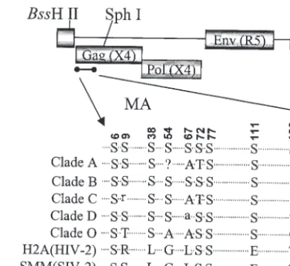
[image:2.603.314.525.68.261.2]Construction of mutants. There are 10 serine residues present in the MA protein of strain pNL4-3. Most of these serines are conserved among different strains of HIV-1 (Fig. 1). The serines at positions 6, 9, 38, 72, 77, 111, 126, and 129 are more conserved than those at residues 54 and 67. The serines at positions 6, 72, 77, and 129 are also present in HIV-2 and simian immunodeficiency virus MA proteins. The serine at
FIG. 1. Alignment of MA amino acid sequences. Locations of serine residues in the MA region of HIV-1 Gag polyproteins and their alignment among different clades as well as with HIV-2 and simian immunodeficiency virus are depicted.
on November 8, 2019 by guest
position 6 is important for MA myristylation and therefore for the plasma membrane association of Gag (33). Mutants were generated with the proviral clone of pNL4-3 with a substitution of the YU-2 envelope gene, resulting in a macrophage-tropic strain. Serine residues were changed to alanines as single mu-tations or in different combinations.
Mutations at serine residues other than Ser-6 have no del-eterious effects on viral assembly and maturation.The pro-duction of viruses containing serine mutations was analyzed by transfection of 293T cells with the proviral clones and
estima-tion of the amount of p24Gagin the cell supernatants by p24
antigen ELISA (Fig. 2). The particle production of all the mutants was comparable to that of the WT, except for
combi-natorial mutants 2S (S126/129A) and 3Sa (S111/126/129A). For
assessment of Gag processing, transfected 293T cells were met-abolically labeled overnight and cell lysates and supernatants were immunoprecipitated with AIDS patient sera (Fig. 3). No defect in the assembly or processing of Gag was observed for any of the mutants, as was evident by the similar amounts of viral proteins and cleaved Gag polyproteins. It was previously reported that mutation of Ser-6 inhibits the production of virus particles (16); thus, the combinatorial mutant 10S produced no detectable virus (Fig. 2 and 3). The efficiency of virus assembly was assessed by comparing the ratio of immunoprecipitated p24 Gag in the supernatant to Gag protein synthesized in transfected cells. The ratios were equivalent for all the mutants and for WT NL4-3, with the exception of mutant 10S (data not shown). This indicates that the mutation of any serine residue other than residue 6 does not deleteriously affect virus assem-bly or Gag processing.
Serines 9, 67, 72, and 77 are critical for infection of dividing and nondividing cells.To test the effects of serine mutations
on virus replication in dividing cells, we infected MAGI-5 cells with similar amounts of p24 Gag virus in single-cycle virus replication assays. All mutant viruses with single serine mutations had infectivity titers of at least 70% that of WT virus (Fig. 4A). Viruses with mutations at multiple Ser
res-idues were also tested (Fig. 4B). Viruses 2S (S126/129A), 3Sa
(S111/126/129A), and 5S (S38/54/111/126/129A) exhibited
simi-lar levels of infectivity as the WT virus. However,
vi-ruses 3Sb (S67/72/77A), 6S (S67/72/77/111/126/129A), and 8S
(S38/54/67/72/77/111/126/129A) exhibited infectivities of
approxi-mately 20% that of WT virus. Together, these results indicate that Ser-67, -72, and -77 are critical for virus infection.
More-over, virus 9S (S9/38/54/67/72/77/111/126/129A) exhibited an
infectiv-ity of ⬍5% that of WT virus, suggesting that Ser-9 is also
critical for infection in vitro. Viruses 4S (S9/67/72/77A) and 3Sc
(S9/72/77A) were constructed subsequently, altering only those
serine residues which might be deleterious for virus replica-tion. The assembly and Gag processing of both of these viruses were similar to those of WT virus (data not shown). Both of
these viruses showed an infectivity of⬍5% that of WT virus,
similar to virus 9S (see Fig. 7A). Virus 10S, with mutations at all 10 Ser residues, produced very low levels of virus particles (Fig. 2 to 3) and no detectable infection of MAGI-5 cells.
Human macrophages were used to examine the replication of mutant viruses in nondividing primary cells (Fig. 5). The pattern of infection of the mutant viruses in macrophages was
similar to that in MAGI-5 cells. Virus 3Sa (S111/126/129A) showed
replication in macrophages that was similar to that of WT virus.
Viruses 6S (S67/72/77/111/126/129A) and 8S (S38/54/67/72/77/111/126/129A)
showed impaired replication in macrophages compared to WT
virus. Virus 9S (S9/38/54/67/72/77/111/126/129A) exhibited the most
impaired replication in macrophages. No detectable
replica-FIG. 2. Relative production of virus particles of WT and serine mutants. 293T cells were transfected with the WT or with mutant proviral clones, and the supernatants were harvested at 48 h posttransfection. The amount of virus made from each mutant was measured by determining the p24 antigen level from the culture supernatant by quantitative ELISA. The bars represent standard errors for four different experiments. Locations of Ser-Ala substitutions for each mutant are indicated.
on November 8, 2019 by guest
http://jvi.asm.org/
[image:3.603.85.503.70.312.2]tion in macrophages was observed with virus 10S. Together, these results suggest that serines at positions 9, 67, 72, and 77 are important for virus replication in both dividing and nondi-viding cells.
Envelope incorporation is unaffected in viruses with MA Ser mu-tations.Viruses 4S (S9/67/72/77A) and 9S (S9/38/54/67/72/77/111/126/129A)
exhibited ⬎90% decreases in infection of MAGI-5 cells and
macrophages. This result could be due to a defect in an entry, uncoating, or nuclear import process that depends upon MA
functioning. To determine if mutations in MA affected entry, possibly through effects on MA interactions with the envelope, we examined envelope glycoprotein incorporation into mutant 4S and 9S virus particles. Although the results shown in Fig. 3B demonstrated that the gp120 envelope was released into the cell supernatant, these results do not differentiate free gp120 from virus-associated gp120. Indeed, the detection of gp120 in the supernatant from cells transfected with mutant clone 10S, despite the lack of detectable released Gag protein, indicates
FIG. 3. Radioimmunoprecipitation analysis of virion-associated proteins. 293T cells were transfected with the indicated molecular clones and metabolically labeled with a Trans35S-label. Supernatants containing virus particles were immunoprecipitated with AIDS patient sera. The posi-tions of Env glycoprotein (gp120), p41 (partially cleaved Gag), p24 (CA), and p17 (MA) are indicated. (A) Single mutaposi-tions. (B) Multiple mutaposi-tions.
FIG. 4. Single-cycle infection assays. MAGI-5 cells were infected with WT or mutant viruses with equivalent amounts of p24 in three different dilutions. At 40 h postinfection, cells were fixed and incubated with-galactosidase substrate. Blue colonies were counted for each mutant and plotted on theyaxis. The error bars represent values for triplicate samples from five independent experiments. (A) Single mutations. (B) Multiple mutations.
on November 8, 2019 by guest
that free gp120 is released from transfected cells. For exami-nation of the level of virus-associated gp120 envelope protein, culture supernatants from transfected and metabolically la-beled HeLa cells were pelleted through a 20% sucrose cushion. The virus pellet was resuspended, immunoprecipitated with AIDS patient sera, and electrophoresed, and gp120 was visu-alized by autoradiography. The Env incorporation in 4S and 9S particles was comparable to that in the WT virus (Fig. 6). A
⌬envvirus, with the luciferase gene in place of HIV-1env, was
used as a negative control. The absence of detectable gp120 in the 10S lane rules out contamination with free gp120 derived from the supernatant. Therefore, these results suggest that the defect observed does not result from the failure of Env incor-poration in virions.
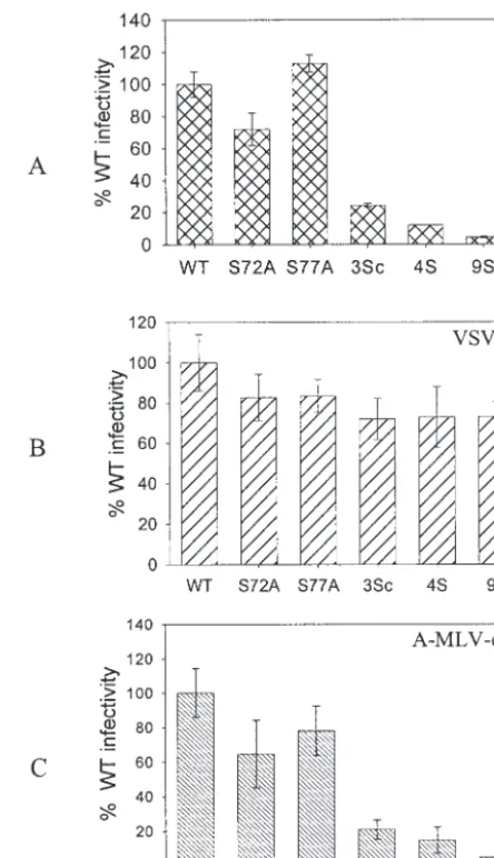
VSV-G pseudotypes of defective serine mutant viruses over-come the infection defect.It has been shown that Nef-defective mutants can be rescued when pseudotyped with VSV-G (1). For testing of similar effects, the defective serine mutant vi-ruses were pseudotyped with VSV-G by cotransfection of
pro-viral clones with a plasmid expressing VSV-G. All of the pseu-dotyped mutant viruses were as infective as pseupseu-dotyped WT virus in a single-round infection assay with MAGI-5 cells (Fig. 7). VSV-G-pseudotyped viruses were several times more in-fectious than nonpseudotyped viruses, and hence smaller amounts of VSV-G-pseudotyped viruses were used to measure the infection in order to remain within the linear range of the assay. As a control, these viruses were also pseudotyped with the A-MLV envelope. The pseudotyped viruses were two to three times more infectious than the corresponding nonpseu-dotyped viruses. However, the relative levels of infection of the A-MLV-pseudotyped MA mutants or WT viruses were similar to those of the nonpseudotyped viruses. This was expected, as A-MLV-pseudotyped viruses resulted in pH-independent fu-sion, similar to that utilized by the HIV envelope glycoprotein. These results indicate that the VSV-G-mediated entry path-way, which employs pH-dependent endocytosis, rescues
[image:5.603.48.279.67.231.2]defec-FIG. 5. Relative replication of WT and serine mutant viruses in macrophages. Human macrophages were infected with WT or mutant viruses with equivalent amounts of p24. The culture supernatants were collected every week, and the virus replication was determined by measuring the RT activity in those supernatants.Pvalues were deter-mined by a two-sidedt test comparing RT values for mutants with those from infection with WT virus.
FIG. 6. Env incorporation in WT and mutant virions. HeLa cells were transfected with WT and mutant proviral constructs and labeled with a Trans35S-label, and viruses in the culture supernatants were pelleted through a 20% sucrose cushion. The pelleted viruses were lysed, immunoprecipitated with AIDS patient sera, and separated by SDS-PAGE.
FIG. 7. Relative infection of pseudotyped MA mutant viruses. Pro-viral clones were cotransfected with either a VSV-G or A-MLV Env expression plasmid to make pseudotyped particles. MAGI-5 cells were used to determine the relative levels of infection. (A) Nonpseudotyped viruses. (B) Viruses pseudotyped with VSV-G. (C) Viruses pseudo-typed with A-MLV Env. 3Sc virus, S9/72/77A. The other mutants are described in the legend for Fig. 3.
on November 8, 2019 by guest
http://jvi.asm.org/
[image:5.603.309.531.68.454.2] [image:5.603.53.274.555.667.2]tive mutant viruses. These observations suggest that the viruses with serine mutations are defective in an early postentry step of the HIV-1 life cycle.
MA serine mutant viruses have a defect in viral cDNA syn-thesis.For examination of the early events in HIV entry, the synthesis of viral cDNAs of MA Ser mutant viruses was mea-sured by radiolabeled PCR using long terminal repeat-Gag-specific primers (Fig. 8) (21). Mutant viruses 4S and 9S were found to be defective in viral cDNA production in comparison to WT virus, for which a 427-bp PCR product was evident as early as 6 h after infection (Fig. 8). This demonstrates that the MA Ser mutant viruses 4S and 9S have a defect in an early postentry step of virus replication.
Mutant viruses exhibit hypophosphorylation of MA. For determination of the cause of defects associated with mutation of serine residues 9, 67, 72, and 77, the phosphorylation status of the MA mutants was determined. Cells transfected with
proviral clones were labeled with [32P]phosphoric acid, and
virus particles pelleted from the supernatants were analyzed for MA phosphorylation. The amount of MA protein in each lane was quantitated by Western blotting with a polyclonal MA
antiserum. As shown in Fig. 9, mutant virus 3Sb (S67/72/77A),
3Sc (S9/72/77A), or 4S (S9/67/72/77A) exhibited markedly reduced
levels of MA phosphorylation compared to WT virus or virus
3Sa (S111/126/129A). There was no significant difference in the
levels of MA protein (Fig. 9B). The alteration of single serine residues did not affect the level of phosphorylation (data not shown). The extent of phosphorylation was calculated by com-paring band intensities of different samples and normalizing them with the amount of MA in each lane. Virus 4S MA was 5% phosphorylated compared to the WT, whereas the percent phosphorylation for the 3Sb and 3Sc MA proteins was 20 and 10%, respectively. These findings suggest that Ser-9, -67, -72, and -77 are phosphoacceptor residues. The explanation for the
presence of two 32P-labeled bands for MA is unclear, but
similar results were observed in another study (2).
tant 9S, with mutations at all serine residues but Ser-6, was similar to that of the WT (data not shown). No alteration in Gag or Env packaging into particles or in processing was de-tected. However, mutation of the serines at positions 9, 67, 72, and 77 resulted in a profound defect in virus infection of MAGI-5 cells and macrophages. These serine residues are among the most conserved residues in HIV MA (Fig. 1). Nev-ertheless, single serine-to-alanine mutations at these positions do not significantly affect virus infectivity, suggesting an addi-tive effect of these residues.
Several key steps of the viral life cycle were examined in order to identify the defect in viral infection of the serine mutants. Env incorporation into virus particles is mediated by MA, but the MA serine mutant viruses had comparable amounts of virion-associated envelope glycoprotein compared to WT virus. Although envelope oligomerization, receptor and
[image:6.603.57.270.68.237.2]core-FIG. 8. PCR amplification of viral DNA at early times postinfec-tion. PM1 cells were infected with WT or mutant viruses, and equal numbers of cells were harvested at the indicated times. Total DNA was purified from the samples, and PCR was performed with the R and gag specific primers (sequence positions are described in Materials and Methods) in the presence of [␣-32P]dCTP. The PCR products were electrophoresed in 6% polyacrylamide gels and autoradiographed.
FIG. 9. Phosphorylation of HIV-1 MA. 293T cells were transfected with WT or mutant proviral clones. Transfected cells were labeled with [32P]phosphoric acid overnight in the presence of Ser-Thr phosphatase inhibitors. The labeled culture supernatants were spun through a 20% sucrose cushion to pellet virus particles. The samples were lysed in RIPA buffer, immunoprecipitated with anti-MA antibodies, separated by SDS–17.5% PAGE, and electroblotted onto a polyvinylidene diflu-oride membrane. Phosphorylation was analyzed by autoradiography (A and C), and the amount of MA present in each sample was deter-mined by Western blotting with an anti-MA antibody (B).
on November 8, 2019 by guest
[image:6.603.308.533.417.628.2]ceptor binding, conformational changes, and activation of the fusion process were not examined and cannot be ruled out, there is no precedent for a role of MA in these activities. Pseudotyping of the mutant virions with VSV-G rescued the defect of serine mutants in MAGI-5 cells, suggesting that MA acts at an early step of the HIV-1 life cycle that is either circumvented or facilitated by targeting of virus entry to the endocytic pathway. Mutants pseudotyped with A-MLV Env remained defective, confirming the earlier observations, as the MLV Env-mediated entry pathway is similar to that of HIV-1. Viral cDNA made by RT after uncoating of the virus core was not observed for the defective MA mutant viruses, sug-gesting that the defect lies in or prior to the uncoating event. Earlier studies by Bukrinskaya and colleagues suggested that the nuclear transport of viral nucleic acids was defective in the presence of protein kinase inhibitors (2). However, their stud-ies were carried out with a hypophosphorylated virus deficient in phosphorylation of MA and other viral proteins.
MA phosphorylation in the defective serine mutant viruses 3Sb, 3Sc, and 4S was found to be severely reduced compared to that in the WT virus, suggesting a phosphorylation-dependent role of MA during infection. The phosphorylation of single serine mutants was found to be at a level similar to that of the WT virus (data not shown), suggesting that there is an additive effect of multiple serine mutations. A three-dimensional struc-ture was generated (Fig. 10) for monomeric MA with coordi-nates available from its crystal structure (22). The presence of basic residues, essential for membrane binding, and serines 9, 67, 72, and 77 on the putative membrane-binding surface sug-gests that phosphorylation at multiple sites might change the overall charge balance on this surface of MA, which may be a prerequisite for its release from the plasma membrane. Our observations confirm and extend the hypothesis that phosphor-ylation of MA might disrupt the electrostatic interaction be-tween positively charged residues of MA and anionic phospho-lipid head groups in the membrane that is necessary for successful uncoating of the virus core (2). Although these de-fects indicate a role of MA phosphorylation in the postentry step, changes in the MA conformation or alterations in pro-tein-protein interactions have not been ruled out. A direct correlation between MA phosphorylation and a replication defect needs to be further confirmed by studying mutants in which the serines at positions 9, 67, 72, and 77 are altered to either threonine or aspartic acid. Changing serine residues to threonine may not alter the phosphorylation pattern, whereas altering these residues to aspartate would add negative charges required for membrane dissociation in the postentry event. Further efforts will characterize the phosphorylation of these residues in infected cells as well as in producer cells. Never-theless, this is the first study to determine the role of phos-phorylation of specific residues in HIV-1 MA and to define the role of these residues in virus replication.
Similar observations have been made with Nef-defective mu-tants, suggesting its role in early postentry steps, although these steps are not well understood (1). Nef has been shown to enhance MA phosphorylation by interacting with virion-associated cellular kinases (34), suggesting that MA may be a functional target of Nef, but Nef has also been shown to enhance virus infectivity in the absence of MA (8). However, the functional relationship between MA and Nef can be further examined by using
phos-phorylation-defective mutants of MA with a Nef deletion. Studies of the effects of different mutations at residues 9, 67, 72, and 77 may provide additional insights into their individual roles in virus infection. Moreover, the effects of MA phosphor-ylation on cellular protein and lipid interactions may provide a biochemical explanation for the biological effects described in this study. A further understanding of MA phosphorylation, its role in virus replication, and the responsible cellular kinases may uncover new targets for therapeutic interventions.
ACKNOWLEDGMENTS
We thank Nancy Vander Heyden, Christopher Brooke, Lingling Jin and Robert Horton for technical help. We thank Michael Belshan for critical review of the manuscript.
This work was supported by PHS grants. R.K. is supported by an American Foundation for AIDS Research (AMFAR) grant.
REFERENCES
1. Aiken, C.1997. Pseudotyping human immunodeficiency virus type 1 (HIV-1) by the glycoprotein of vesicular stomatitis virus targets HIV-1 entry to an endocytic pathway and suppresses both the requirement for Nef and the sensitivity to cyclosporin A. J. Virol.71:5871–5877.
2. Bukrinskaya, A. G., A. Ghorpade, N. K. Heinzinger, T. E. Smithgall, R. E. Lewis, and M. Stevenson.1996. Phosphorylation-dependent human immu-nodeficiency virus type 1 infection and nuclear targeting of viral DNA. Proc. Natl. Acad. Sci. USA93:367–371.
[image:7.603.311.531.70.373.2]3. Bukrinsky, M. I., S. Haggerty, M. P. Dempsey, N. Sharova, A. Adzhubel, L. Spitz, P. Lewis, D. Goldfarb, M. Emerman, and M. Stevenson.1993. A
FIG. 10. Schematic model for MA showing a view of the putative membrane-binding surface as seen from above. Serine residues are shown in red, with their positions depicted, and basic residues, essen-tial for membrane binding, are shown in yellow. The coordinates for this structure were taken from the work of Hill et al. (22), and the model was generated with DeepView/Swiss-PdbViewer software (http: //www.expasy.org/spdbv/).
on November 8, 2019 by guest
http://jvi.asm.org/
10. Freed, E. O.1998. HIV-1 Gag proteins: diverse functions in the virus life cycle. Virology251:1–15.
11. Freed, E. O.2003. The HIV-TSG101 interface: recent advances in a budding field. Trends Microbiol.11:56–59.
12. Freed, E. O., G. Englund, F. Maldarelli, and M. A. Martin.1997. Phosphor-ylation of residue 131 of HIV-1 matrix is not required for macrophage infection. Cell88:171–173.
13. Freed, E. O., G. Englund, and M. A. Martin.1995. Role of the basic domain of human immunodeficiency virus type 1 matrix in macrophage infection. J. Virol.69:3949–3954.
14. Freed, E. O., and M. A. Martin.1996. Domains of the human immunodefi-ciency virus type 1 matrix and gp41 cytoplasmic tail required for envelope incorporation into virions. J. Virol.70:341–351.
15. Freed, E. O., and M. A. Martin.1995. Virion incorporation of envelope glycoproteins with long but not short cytoplasmic tails is blocked by specific, single amino acid substitutions in the human immunodeficiency virus type 1 matrix. J. Virol.69:1984–1989.
16. Freed, E. O., J. M. Orenstein, A. J. Buckler-White, and M. A. Martin.1994. Single amino acid changes in the human immunodeficiency virus type 1 matrix protein block virus particle production. J. Virol.68:5311–5320. 17. Gallay, P., S. Swingler, C. Aiken, and D. Trono.1995. HIV-1 infection of
nondividing cells: C-terminal tyrosine phosphorylation of the viral matrix protein is a key regulator. Cell80:379–388.
18. Gallay, P., S. Swingler, J. Song, F. Bushman, and D. Trono.1995. HIV nuclear import is governed by the phosphotyrosine-mediated binding of matrix to the core domain of integrase. Cell83:569–576.
19. Garrus, J. E., U. K. von Schwedler, O. W. Pornillos, S. G. Morham, K. H. Zavitz, H. E. Wang, D. A. Wettstein, K. M. Stray, M. Cote, R. L. Rich, D. G. Myszka, and W. I. Sundquist.2001. Tsg101 and the vacuolar protein sorting pathway are essential for HIV-1 budding. Cell107:55–65.
20. Haffar, O. K., S. Popov, L. Dubrovsky, I. Agostini, H. Tang, T. Pushkarsky, S. G. Nadler, and M. Bukrinsky.2000. Two nuclear localization signals in the HIV-1 matrix protein regulate nuclear import of the HIV-1 pre-integration complex. J. Mol. Biol.299:359–368.
virus encode small peptide motifs that recruit Tsg101 to sites of particle assembly to facilitate egress. Nat. Med.7:1313–1319.
27. Massiah, M. A., M. R. Starich, C. Paschall, M. F. Summers, A. M. Chris-tensen, and W. I. Sundquist.1994. Three-dimensional structure of the hu-man immunodeficiency virus type 1 matrix protein. J. Mol. Biol.244:198– 223.
28. Nguyen, D. H., and J. E. Hildreth.2000. Evidence for budding of human immunodeficiency virus type 1 selectively from glycolipid-enriched mem-brane lipid rafts. J. Virol.74:3264–3272.
29. Ono, A., and E. O. Freed.2001. Plasma membrane rafts play a critical role in HIV-1 assembly and release. Proc. Natl. Acad. Sci. USA98:13925–13930. 30. Ono, A., J. M. Orenstein, and E. O. Freed.2000. Role of the Gag matrix
domain in targeting human immunodeficiency virus type 1 assembly. J. Virol. 74:2855–2866.
31. Pirounaki, M., N. A. Heyden, M. Arens, and L. Ratner.2000. Rapid pheno-typic drug susceptibility assay for HIV-1 with a CCR5 expressing indicator cell line. J. Virol. Methods85:151–162.
32. Poiesz, B. J., F. W. Ruscetti, A. F. Gazdar, B. P. A., J. D. Minna, and R. C. Gallo.1980. Detection and isolation of type C retrovirus particles from fresh and cultured lymphocytes of a patient with cutaneous T-cell lymphoma. Proc. Natl. Acad. Sci. USA77:7415–7419.
33. Spearman, P., J. J. Wang, N. Vander Heyden, and L. Ratner.1994. Identi-fication of human immunodeficiency virus type 1 Gag protein domains es-sential to membrane binding and particle assembly. J. Virol.68:3232–3242. 34. Swingler, S., P. Gallay, D. Camaur, J. Song, A. Abo, and D. Trono.1997. The Nef protein of human immunodeficiency virus type 1 enhances serine phos-phorylation of the viral matrix. J. Virol.71:4372–4377.
35. Yu, X., Q. C. Yu, T. H. Lee, and M. Essex.1992. The C terminus of human immunodeficiency virus type 1 matrix protein is involved in early steps of the virus life cycle. J. Virol.66:5667–5670.
36. Yu, X., X. Yuan, Z. Matsuda, T. H. Lee, and M. Essex.1992. The matrix protein of human immunodeficiency virus type 1 is required for incorpora-tion of viral envelope protein into mature virions. J. Virol.66:4966–4971.