Analysis of Binary Interactions between OTUB1
and E2 Ubiquitin-Conjugating Enzymes
Thesis submitted in accordance with the requirements of the University of Liverpool for the degree of Doctor in Philosophy
Nurulisa Zulkifle
ii
Untuk Ezra yang disayangi selama-lamanya
Anak unta anak kuda,
Kejar mengejar keliling batu,
iii
Analysis Of Binary Interactions Between OTUB1 And
E2 Ubiquitin-Conjugating Enzymes
Nurulisa Zulkifle
Abstract
Post-translational modification of proteins via ubiquitination is mediated by three enzyme families; E1 activating enzymes, E2 conjugating enzymes and E3 ligases, all of which work in a hierarchical manner to facilitate different forms of protein ubiquitin ranging from mono-ubiquitination to the formation of different forms of ubiquitin chains (Ciechanover et al., 2000). Deubiquitinating enzymes (DUBs) act to remove ubiquitin from modified substrates. Apart from the classic interactions within the E1-E2-E3 enzymatic cascade, an unusual non-hierarchical interaction has been observed between some E2 enzymes and a DUB called OTUB1 (Markson et al., 2009). This observation raises interesting questions concerning the molecular mechanisms and specificity of this unusual E2:DUB partnership. In this study, systematic yeast two-hybrid (Y2H) screens were performed between all human E2 and DUB proteins to analyse the extent of E2:DUB interactions. Putative partnerships between OTUB1 and UBE2D1, UBE2D2, UBE2D3, UBE2D4, UBE2E1, UBE2E2, UBE2E3 and UBE2N were identified. These data correlate well with data from other independent studies, including HTP Y2H screens (Markson et al., 2009) and mass spectrometry (Sowa et al., 2009). An N-terminal truncated form of OTUB1 (ΔNOTUB1) was generated by removing a predicted 39aa N-terminal disordered region (Edelmann et al., 2009). Using this construct in combination with wild type (WT) OTUB1, complementary biophysical studies were performed to investigate the formation of complexes with UBE2D2 and UBE2E1 as these represented the strongest interactions detected in preliminary Y2H studies. Gel filtration chromatography showed convincing complex formation for both ΔNOTUB1:UBE2D2 and ΔNOTUB1:UBE2E1 in 1:1 stoichiometry. The thermodynamic profile of each complex was
iv
OTUB1. Point mutants corresponding to predicted contact residues in UBE2D2 were generated and tested in Y2H studies to determine their role in facilitating the formation of both E2:OTUB1 and E2:E3-RING complexes. This data suggests that in some, but not all cases, OTUB1 and E3-RINGs bind competitively to the same interface on E2 proteins. Preliminary immunofluorescence studies show that partner proteins predominantly co-localise in the cytoplasm, except UBE2E1 which is predominantly nuclear. Data from this study allowed us to propose a model of how OTUB1:UBE2D2 complex may forms and functions. Significantly, many of these predictions have now been verified by independent structural studies and subsequent live cell microscopy studies in our lab.
v
Table of Contents
TITLE PAGE i
ABSTRACT iii
TABLE OF CONTENTS v
LIST OF FIGURES xi
LIST OF TABLES xiv
LIST OF ABBREVIATIONS xv
ACKNOWLEDGEMENTS xviii
Chapter One: Introduction
1.1 Overview of the human ubiquitin system 1
1.2 Ubiquitin and ubiquitin-like molecules 2
1.3 Roles of ubiquitination
1.3.1 Intracellular protein degradation by ubiquitin- proteasome pathway: The classical ubiquitin role
5
1.3.2 The diversity of ubiquitination chain configuration 8 1.4 Ubiquitination conjugation components
1.4.1 Human E1 ubiquitin-activating enzyme 10
1.4.2 Human E2 ubiquitin-conjugating enzyme 10
1.4.2.1 E2 structure 11
1.4.2.2 E2 family diversity 15
1.4.3 Human E3 ubiquitin-protein ligases 17
1.4.3.1 HECT domain E3s 17
1.4.3.2 RING domain E3s 17
1.5 Hierarchical structure and specificity of the ubiquitin system 19 1.6 The deubiquitinating enzyme
1.6.1 Classification of deubiquitinating enzyme 20
1.6.1.1 USP domain 21
1.6.1.2 UCH domain 21
1.6.1.3 OTU domain 21
1.6.1.4 Josephin domain 21
vi
1.6.2 General function of DUB 24
1.7 Protein-protein interaction 25
1.7.1 Genetic in vivo methods
1.7.1.1 Yeast two-hybrid screen 26
1.7.2 Biophysical and theoretical methods
1.7.2.1 Isothermal titration calorimetry (ITC) 29
1.7.2.2 Nuclear magnetic resonance (NMR) 31
1.7.2.3 X-ray crystallography 33
1.8 Thesis overview and aims 35
Chapter Two: Materials and Methods
2.1 Introduction 36
2.2 Preparation of clones for Y2H screening
2.2.1 Reagents 36
2.2.2 Proofreading PCR-amplification of protein coding inserts from pDONR223 entry clones
37
2.2.3 Agarose gel electrophoresis 38
2.2.4 Purification of PCR product by gel extraction 39
2.2.5 Yeast media preparation 39
2.2.6 Yeast transformation / gap repair 42
2.2.7 Diagnostic yeast colony PCR (YC-PCR) 43
2.2.8 Autoactivation assay 44
2.2.9 Glycerol stocks 45
2.2.10 Y2H matrix mating 45
2.3 Molecular biology
2.3.1 Reagents 46
2.3.2 Cloning
vii
2.3.2.2.1 Target DNA amplification and restriction digest
47
2.3.2.2.2 Ligations of double digested DNA inserts and vector
47
2.3.2.3 Directional TOPO® Cloning 48
2.3.3 QuikChange® site-directed mutagenesis 48
2.3.4 Transformation of cloning reactions into chemically competent cells.
50
2.3.5 Diagnostic bacterial colony PCR (BC-PCR) 51
2.3.6 DNA amplification, purification and glycerol stock 52
2.3.6.1 Miniprep 52
2.3.6.2 Midi- and maxiprep 52
2.3.7 Sequencing 54
2.4 Protein expression methods
2.4.1 Reagents 55
2.4.2 Protein expression and purification
2.4.2.1 Small-scale expression test 55
2.4.2.2 Large-scale protein production 56
2.4.2.3 (1H15N)-labelled protein expression 58 2.4.3 SDS polyacrylamide gel electrophoresis (SDS-PAGE) 58 2.5 Biophysical procedures
2.5.1 Reagents 60
2.5.2 Gel filtration chromatography 60
2.5.3 Isothermal titration calorimetry 60
2.5.4 Nuclear magnetic resonance 60
2.5.5 Crystallisation trial 61
2.6 Cell biology
2.6.1 Reagents 62
2.6.2 Cell culture 62
2.6.3 Cell transfection and fixation 62
2.7 Bioinformatic method 63
Chapter Three: Yeast Two-Hybrid Screening
3.1 Introduction 64
viii
3.3 Construction of Y2H bait and prey clones 3.3.1 Existing construct
3.3.1.1 E2 bait and prey 69
3.3.1.2 DUB bait 71
3.3.2 Generation of Y2H DUB prey set
3.3.2.1 Application of the Gateway®cloning strategy to
create DUBs in pDONR223 71
3.3.2.2 PCR introduction of yeast and Gateway® sites to the DUB ORFs
74
3.3.2.3 In vivo homologous recombination (gap repair) 74
3.3.2.4 Autoactivation tests 74
3.3.3 Pooling-deconvolution strategy to screen all possible E2:DUB interactions
78
3.4 Binary Y2H screen result
3.4.1 Reconfirmation of previously described interaction 81 3.4.2 OTUB1 binds a subset of E2 conjugating enzymes 92 3.5 Analysis of a truncated OTUB1 lacking the first 39 N-terminal
amino acids
94
3.5.1 Generating ΔNOTUB1 97
3.5.2 ΔNOTUB1 slightly reduced interaction with E2 binding partners 97
3.6 Selection of candidates for biophysical analysis 99
Chapter Four: Biophysical Evidence of Binary E2:OTUB1 Complexes Formation
4.1 Introduction 100
4.2 Producing affinity tagged protein 100
4.2.1 pETM-11 system 101
4.2.2 Construction of His-tag protein of interest
4.2.2.1 Introduction of NcoI and HindIII restriction sites into the N- and C-termini of protein coding inserts
102
4.2.2.2 Ligation and transformation 102
4.3 Protein expression and purification
4.3.1 Small-scale expression test 104
4.3.2 Large-scale protein production
4.3.2.1 Selection of host strain and induction temperature 106
ix
4.3.3 Correct protein folding analysed by 1D 1H NMR 109 4.4 Preliminary analysis of OTUB1 in complex with UBE2D2,
UBE2E1 and UBE2E2
4.4.1 Identification of OTUB1:UBE2E1 complex by gel filtration chromatography
111
4.4.2 ITC analysis of potential OTUB1:E2 complexes 114 4.4.3 (1H15N)-HSQC NMR experiment
4.4.3.1 15N labelled protein 117
4.4.3.2 Investigating the OTUB1:UBE2E1 complex 118 4.5 Analysis of ΔNOTUB1:E2 complex
4.5.1 ΔNOTUB1 expression and purification 120
4.5.2 Improved gel filtration chromatography data 121 4.5.3 Analysis of ΔNOTUB1:UBE2D2 and ΔNOTUB1:UBE2E1
thermodynamic profiles by ITC
123
4.5.4 Identification of possible points of contact in ΔNOTUB1:UBE2D2 and ΔNOTUB1:UBE2E1 complexes observed by shifts in
(1H15N)-HSQC NMR spectra
125
4.5.4.1 Are ΔNOTUB1 not fully active? 126
4.5.4.2 UBE2D2 and UBE2E1 share a similar interface in binding with ΔNOTUB1
129
4.5.4.3 Prediction of amino acids involved in ΔNOTUB1:UBE2D2 interaction
131
4.5.5 Co-crystallisation trial 135
4.6 Progress limitation 139
Chapter Five: Targeted Analysis of E2:OTUB1 Complexes
5.1 Introduction 143
5.2 Generation of E2-binding site mutants
5.2.1 Potential amino acids to be mutated 145
5.2.2 Mutagenesis strategy 146
5.3 Y2H evaluation of predicted UBE2D2 binding site 149
5.3.1 Y2H screen results
5.3.1.1 UBE2D2 WT and mutants versus OTUB1 full-length and truncated
149
5.3.1.2 Analysis of interaction between WT and mutants forms of UBE2D2 with known E3 RING interaction partners
x
5.3.2 Competitive and non-competitive OTUB1:E3-RING:UBE2D2 binding models
154
5.4 Expression and purification of UBE2D2 mutant protein 157
5.4.1 Directional TOPO® Cloning 157
5.4.1.1 Primer design and the TOPO® cloning principle 159 5.4.1.2 Purification of UBE2D2 mutants protein 160 5.5 Immunofluorescence study of OTUB1 and E2 conjugase proteins 161
5.5.1 Localisation of OTUB1, ΔNOTUB1, UBE2D2, UBE2E1 and UBE2N
162
5.5.2 Co-localisation study of OTUB1 and ΔNOTUB1, with the E2 proteins
164
Chapter Six: Discussion and Conclusion
6.1 Solved structures of OTUB1:UBE2D2 and OTUB1:UBE2N complexes 166
6.1.1 Comparison of methodologies 170
6.1.2 The importance of OTUB1 N-terminal 170
6.1.3 Agreement with Y2H analysis of UBE2D2 binding site mutants
171
6.2 Insight into OTUB1 physiological functions 173
6.3 OTUB1 protein interaction network 174
6.4 Future direction 176
xi
List of Figures
Page Figure 1.1 Ribbon diagram representing the tertiary structure of ubiquitin and
the functional implications of specific linkages
3
Figure 1.2 Protein degradation through the UPS 7
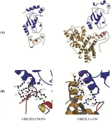
Figure 1.3 Conservation of E2 structures between species 13 Figure 1.4 Comparison between the UBE2D2:CNOT4 docking model and
the UBE2L3:c-Cbl crystal structure
14
Figure 1.5 The family of human E2 ubiquitin-conjugating enzymes 16 Figure 1.6 Illustration of the hierarchical structure and specificity within the
ubiquitin system
19
Figure 1.7 Structures of the catalytic domains of the five subclasses of DUBs with ubiquitin
23
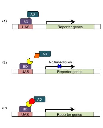
Figure 1.8 The Y2H system 28
Figure 1.9 Illustration of the configuration of an ITC reaction cell 30 Figure 1.10 Schematic operation of basic NMR spectrometer 32
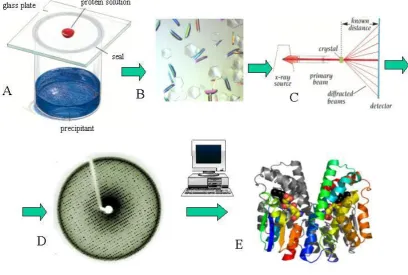
Figure 1.11 Steps of protein X-ray crystallography 34
Figure 3.1 Known E2:DUB interaction 65
Figure 3.2 Y2H vectors used in this study 68
Figure 3.3 The Gateway® system 73
Figure 3.4 Pooling and deconvolution strategy 79
Figure 3.5 Schematic representation of Y2H protocol 80
Figure 3.6 E2 bait-pooled DUB prey interactions 83
Figure 3.7 Pooled DUB preys deconvoluted in singe mating 84
Figure 3.8 Pooled E2 prey-DUB bait interactions 85
Figure 3.9 Pooled E2 preys deconvoluted in singe mating 86 Figure 3.10 Known interactions between E2 ubiquitin conjugating enzymes
and DUBs in humans
91
Figure 3.11 Probability of disorder for OTUB1 94
Figure 3.12 Superposition of OTUB1 and OTUB2 96
Figure 3.13 ΔNOTUB1:E2 interactions 98
Figure 4.1 Maps of pETM-11 vector 101
Figure 4.2 Generation of pETM-11 clones 103
Figure 4.3 Coomassie blue SDS-PAGE gels of uninduced and IPTG-induced samples from Ni2+ column elution
105
xii
before and after TEV protease cleavage
Figure 4.5 Typical column peaks for (i) OTUB1, (ii) UBE2D2, (iii) UBE2E1 and (iv) UBE2E2
108
Figure 4.6 1D-NMR spectrum 110
Figure 4.7 Typical gel filtration column peaks 113
Figure 4.8 ITC titration of UBE2E1 with OTUB1 115
Figure 4.9 UBE2E1 may form dimer in high concentration 115
Figure 4.10 NMR titration 15N-OTUB1:UBE2E1 119
Figure 4.11 NMR spectrum of ΔNOTUB1 120
Figure 4.12 Typical gel filtration column peak 122
Figure 4.13 ITC titration of UBE2E1 and UBE2D2 with ΔNOTUB1 124
Figure 4.14 2D NMR titration of ΔNOTUB1:UBE2D2 127
Figure 4.15 2D NMR titration ΔNOTUB1:UBE2E1 128
Figure 4.16 Superposition of free 15N-ΔNOTUB1, 15N-ΔNOTUB1:UBE2D2 and 15
N-ΔNOTUB1:UBE2E1 2D NMR spectrum
130
Figure 4.17 Superposition of 15N-UBE2D2:ΔNOTUB1 and known UBE2D2 profile
133
Figure 4.18 Predicted UBE2D2 binding interface with ΔNOTUB1 134
Figure 4.19 Crystal growth from initial trials 138
Figure 4.20 Coomassie blue 1D SDS-PAGE of newly purified ΔNOTUB1 141 Figure 4.21 2D NMR spectrum of double species ΔNOTUB1 142
Figure 5.1 Mutant UBE2D2 interactions in Y2H 153
Figure 5.2 Binding model of UBE2D2, OTUB1 and E3 RINGs 155 Figure 5.3 E2 shell-like model proposed to be important for selectivity of key
enzymes (E1, E3, Ub/UBL) in directing Ub/UBL-conjugation pathways
156
Figure 5.4 Map of pET151/D-TOPO® vector 158
Figure 5.5 Localisation of OTUB1 and ΔNOTUB1 singly transfected into HeLa cells
163
Figure 5.6 Localisation of UBE2N, UBE2E1 and UBE2D2 in HeLa cells 163 Figure 5.7 Co-localisation of UBE2D2, UBE2E1 and UBE2N with OTUB1
in HeLa cells
165
Figure 5.8 Co-localisation of UBE2D2 and UBE2N with ΔNOTUB1 in HeLa cells
165
Figure 6.1 Schematic and ribbon representation of Ub~UBE2D2-OTUB1-Ub complex
xiii
Figure 6.2 Stereo view of the OTUB1:UBE2D2 binding interface 172
xiv
List of Tables
Page Table 1.1 Known and putative UBLs in Saccharomyces cerevisiae 4
Table 1.2 Functions of various ubiquitin chains 9
Table 1.3 The comparison between ITC and NMR 31
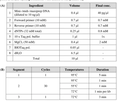
Table 2.1 Reaction mixture and cycling parameters used for typical KOD Hot Start PCR reactions
38
Table 2.2 List of Y2H medium and their uses 39-40
Table 2.3 Recipes for basic yeast media 41
Table 2.4 SD-X amino acids supplement mixes 41
Table 2.5 Dropout (DO) amino acid mixture 42
Table 2.6 Reaction mixture and cycling parameters for a typical YC-PCR 44 Table 2.7 Typical QuikChange® mutagenesis reaction setup 49 Table 2.8 Description of bacterial plasmid and E. coli strains 50 Table 2.9 Reaction mixture and cycling parameters for BC-PCR 51 Table 2.10 Reaction mixture and cycling parameters for mini-, midi- or
maxiprep-PCR
54
Table 2.11 Buffer recipes 57
Table 2.12 Recipe for 2M9 minimal media 59
Table 2.13 Recipe for SDS-PAGE resolving and stacking gel 59
Table 3.1 The ubiquitin conjugating enzyme (E2s) 70
Table 3.2 Y2H deubiquitinating enzymes (DUBs) clone collection 76-77
Table 3.3 E2:DUB interaction summary 87-89
Table 3.4 OTUB1:E2 interaction summary 99
Table 4.1 NcoI and HindIII restriction site sequences incorporated in forward and reverse primers
102
Table 4.2 Q/SP selections for each protein 107
Table 4.3 Comparison of ΔNOTUB1 and CNOT4 binding sites on UBE2D2 135 Table 5.1 Forward and reverse UBE2D2 mutagenesis primers 146
Table 5.2 TOPO® forward and reverse primers 159
Table 6.1 Specific residues that mediate interactions in Ub~ UBE2D2-OTUB1-Ub complex
168-169
Table 6.2 Comparison of OTUB1 (DUB) and CNOT4 (E3) binding sites on UBE2D2 (E2)
xv
List of Abbreviations
1D One-dimensional
2D Two-dimensional
3AT 3-amino-1,2,4-triazole
3D Three-dimensional
AD GAL4 activating domain
Ade Adenine
ATP Adenosine triphosphate
APS Ammonium persulphate
BC-PCR Bacterial colony-PCR
BD GAL4 DNA binding domain
BioGRID Biological General Repository for Interaction Datasets BMRB Biological Magnetic Resonance Bank
bp base pair
C-terminal Carboxy-terminal / COOH-terminal CaCl2 Calcium chloride
cDNA Complementary DNA
CO2 Carbon dioxide
D2O Deuterium oxide
Da Dalton
DAPI 4’,6-diamidino-2-phenylindole dH2O Distilled water
DMEM Dulbecco’s Modified Eagle’s Medium DNA Deoxyribonucleic acid
dNTPs Deoxynucleotide triphosphates
DO Dropout
DTT Dithiothreitol
DUB Deubiquitinating enzyme
EMBL The European Molecular Biology Laboratory FCCS Fluorescence cross correlation spectroscopy FeCl2 Iron (II) chloride / ferrous chloride
FL Full-length
GF Gel filtration
GFP Green fluorescent protein
xvi
HECT Homologous to E6-AP carboxy terminus
HEPES 4-(2-hydroxyethyl)-1-piperazineethanesulfonic acid
His Histidine
HSQC Heteronuclear Single Quantum Coherence HPRD Human Protein Reference Database
IEC Ion exchange chromatography
IPTG Isopropyl-β-D-thio-galactopyranoside ITC Isothermal titration calorimetry
Ka Binding affinity
Kd Dissociation constant
kcal Kilocalories
KH2PO4 Monopotassium phosphate
LacZ Gene encoding β-galactosidase
Lys Lysine
MgCl2 Magnesium chloride
MHz Megahertz
MINT Molecular Interactions Database MnSO4 Manganese (II) sulphate
MWCO Molecular weight cut-off N-terminal Amino terminal / NH2-terminal Na2HPO4 Sodium phosphate dibasic
NaN3 Sodium azide
NaOH Sodium hydroxide
NEAA Non-essential amino acids NH4Cl Ammonium chloride
Ni2+ Nickel
NMR Nuclear magnetic resonance
NCBI National Centre for Biotechnological information
OD Optical density
ORF Open reading frame
PAGE Polyacrylamide gel electrophoresis PCR Polymerase chain reaction
PDB Protein Data Bank
PEG Polyethylene glycol
pI Isoelectric point
xvii
ppm parts per million
rf radiofrequency
RING Really Interesting New Gene
RNA Ribonucleic acid
RONN Regional Order Neural Network
rpm revolutions per minute
RT Room temperature
SD Synthetic defined
SDS Sodium dodecyl sulphate
SOB Super optimal broth
SOC SOB with catabolite repression (added glucose) TCEP Tris(2-carboxyethyl)phosphine
TEMED N,N,N’,N’-tetramethylethylenediamine
TEV Tobacco etch virus
Tm Melting temperature
Tris Tris(hydroxymethyl)aminomethane
tRNA Transfer RNA
UAS Upstream activating sequence
Ub / Ubq Ubiquitin
UBL Ubiquitin-like
UPS Ubiquitin-Proteasome System
UV Ultraviolet
WT Wild type
X-Gal 5-bromo-4-chloro-3-indolyl-beta-D-galactopyranoside
Y2H Yeast two-hybrid
YC-PCR Yeast colony-PCR
ΔG Change in Gibbs energy
ΔH Change in enthalpy
xviii
Acknowledgements
First and foremost, I would like to thank the Ministry of Higher Education Malaysia (MOHE) and Universiti Sains Malaysia (USM) for providing the scholarship and study leave. Without their support, this PhD would not have been possible.
I owe my deepest gratitude to my supervisor Professor Chris M. Sanderson for the continuous encouragement, excellence supervision and extraordinary patience throughout the completion of this work. I am also hugely indebted to Professor Lu-Yun Lian for the access to the equipments and facilities in School of Biosciences and NMR centre. To Dr. Igor Barsukov, Dr. Paul Elliott and Dr. Martyna Pastok, a sincere thanks for the advice and technical assistance that they provided.
To current and former members of the Sanderson lab: Julie, Jonathan, Russell, Helen, Rob, Kelly, Amy, Emily and Dave, all have contributed to a happy tune in my daily lab life, therefore a very huge thanks to all of you. To Yvonne, Jia Lih and the rest of the 5th floor Nuffield Wing occupants, thank you for all of your help along the way. Further thanks go to the rest of the staff and fellow students in the department, always friendly and helpful since day one.
I would like to acknowledge everyone outside of the lab especially my ex-housemates in Liverpool, always get me smile and laugh along this tearful journey. Izyan, Shafiqah, Hada, Ad, Eeka, Maryam, Najwa and Intan; these girls are indeed true friends and the most irreplaceable pieces of my life. Also to my loyal old friends in Malaysia thank you for always being there. Especially to Linda, you are the only one that never leave.
Page | 1
1.1 Overview of the human ubiquitin system
Many vital body functions such as temperature regulation and maintenance of blood pressure
serve to maintain the state of homeostasis in our body. The same principle applies at the
level of a single cell where control of protein homeostasis is essential for regulating cellular
physiology or responding to adverse signals. One of the most important modes of
post-translational regulation is protein ubiquitination, a highly dynamic process that governs
nearly every function in human cells (Hershko and Ciechanover, 1998). Over the past two
decades, ubiquitination has been increasingly acknowledged as more than simply a signal for
protein degradation, but also a main regulator of a diverse array of different biological
processes (Weissman, 2001). It is a reversible post-translational modification of protein,
almost as common as phosphorylation, which involves the addition of the ubiquitous small
protein aptly named ubiquitin to the lysine residue of a target substrate (Hershko and
Ciechanover, 1998).
In general, ubiquitin attachment occurs through a sequential enzymatic cascade involving an
E1 activating enzyme, an E2 conjugating enzyme and an E3
ubiquitin-protein ligase, resulting in the modification of a specific ubiquitin-protein substrate (Pickart, 2001). In
the classical view of ubiquitination, a polyubiquitin chain is synthesised by serial addition of
ubiquitin moieties to the Lys48 residue of the previously conjugated ubiquitin (Hershko and
Ciechanover, 1998). This particular polyubiquitinated substrate is recognised as a target for
degradation by a multi-subunit ATP-dependent protease complex known as the 26S
proteasome (Tashiro et al., 1997; Pickart, 2001). The reversibility of ubiquitination is
provided by deubiquitinating enzymes (DUBs) which either prevent ubiquitin attachment to
Chapter One | INTRODUCTION
Page | 2
the target substrate or remove ubiquitin from specific target proteins (Komander et al., 2009;
Reyes-Turcu et al., 2009).
1.2 Ubiquitin and ubiquitin like molecules
Ubiquitin is a 76 amino acid globular protein with a molecular mass around 8.5 kDa and is
highly conserved throughout eukaryotes (Weissman, 2001). Since its discovery over three
decades ago, it has become abundantly clear that the ubiquitin system is an essential feature
of all aspects of eukaryotic biology. Due to its pervasive action, ubiquitin does not seem to
be produced in excess but the free pool of ubiquitin monomer is maintained at an adequate
level to ubiquitinate the large number of its potential substrates in human cells (Kimura and
Tanaka, 2010). In yeast as well as in higher eukaryotes, ubiquitin is initially expressed in the
form of a precursor either as polyubiquitin, a linear fusion protein consisting of four or more
ubiquitin copies in a head-to-tail configuration, or as fusion proteins between ubiquitin and
large and small essential ribosomal polypeptides, L40 and S27, respectively (Finley et al.,
1989; Redman and Rechsteiner, 1989). These ubiquitin precursors are cleaved by DUBs to
release identical functional monomeric ubiquitin units.
In the ubiquitination process, ubiquitin covalently attaches to the lysine residues of target
proteins via its carboxy-terminal glycine residue to form an isopeptide linkage in an
ATP-dependent fashion. Multiple ubiquitin can be covalently added to a substrate successively by
E1, E2 and E3 enzymes, producing a substrate conjugated with polyubiquitin (Pickart, 2001;
Dye and Schulman, 2007). The key feature of ubiquitin is the fact that it contains seven
lysines, each of which can potentially mediate attachment to other ubiquitin molecules,
allowing the formation of a range of structurally distinct polyubiquitin chains (Figure 1.1)
Chapter One | INTRODUCTION
Page | 3
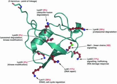
N- MQIFVKTLTGKTITLEVEPSDTIENVKAKIQDKEGIPPDQQRLIFAGKQLEDG RTLSDYNIQKESTLHLVLRLRGG -C
[image:21.595.115.526.182.476.2]
Figure 1.1 Ribbon diagram representing the tertiary structure of ubiquitin and the functional implications of specific linkages: The structure of ubiquitin reveals that all seven lysine residues (red, with blue nitrogen atoms) and methionine (with a green sulphur atom) reside on different surfaces of the molecule. The percentage numbers refer to the relative abundance of the particular linkage in S. cerevisiae (Komander, 2009). Above is the amino acids sequence of ubiquitin with the seven lysine residues highlighted in red.
In addition to ubiquitin itself, multiple polypeptides that are distinct from but related to
ubiquitin are also enzymatically conjugated to target substrates via processes very similar to
ubiquitination (Gill, 2004). These ubiquitin-like proteins (UBLs) participate in many
biological processes including gene transcription, signal transduction, autophagy and
Chapter One | INTRODUCTION
Page | 4
known to participate in chain formation (Tatham et al., 2001; Xirodimas et al., 2008; Matic
[image:22.595.151.480.203.529.2]et al., 2008). Members of the UBL family are listed in the table below:
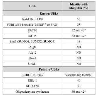
Table 1.1 Known and putative UBLs in Saccharomyces cerevisiae: Reviewed in Hochstrasser (2009). ND = Not detectable by standard BLAST search.
* = For each two ubiquitin-related domains.
UBL Identity with
ubiquitin (%) Known UBLs
Rub1 (NEDD8) 55
FUBI (also known as MNSF-β or FAU) 38
FAT10 32 and 40*
ISG15 32 and 37*
Smt3 (SUMO1, SUMO2, SUMO3) 18
Atg8 ND
Atg12 ND
Urm1 ND
UFM1 ND
Putative UBLs
BUBL1, BUBL2 Variable (up to 80%)
UBL-1 40
SF3A120 30
Chapter One | INTRODUCTION
Page | 5
1.3 Roles of ubiquitination
1.3.1 Intracellular protein degradation by ubiquitin-proteasome pathway: The classical
ubiquitin role
Protein degradation in eukaryotic cells is carried out either by lysosomal or proteasomal
degradation (Cooper, 2000). Lysosomes are membrane-enclosed organelles that contain a
large variety of hydrolytic enzymes which digest extracellular proteins taken up
by endocytosis. Most of the proteolysis of cytosolic proteins that occurs in lysosomes is
non-specific (Knopp et al., 1993). In contrast, the Ubiquitin-Proteasome System (UPS) uses
ubiquitin as a marker which can target cytosolic and nuclear proteins for selective
destruction by proteasomes (Pickart, 2001). It is the responsibility of the UPS to identify and
exterminate damaged and faulty proteins or those simply surplus for requirement to maintain
the right amount of proteins within cells. The UPS can malfunction in two ways: it can either
become overzealous and destroy useful protein inappropriately, or it can be restrained in
some way resulting in the build up of potentially harmful proteins, which can then reach
toxic levels. An imbalance in the UPS is thought to occur in common diseases especially
cancer (Scheffner et al., 1990; Loda et al., 1997; Joazeiro et al., 1999; Maxwell et al., 1999;
Waterman et al., 1999; Bignell et al., 2000). The UPS marks a target protein for destruction
by the addition of a specific form of ubiquitin modification, in essence, proteins tagged with
polyubiquitin chains of more than four ubiquitin residues (Thrower et al., 2000). There is a
lot of ubiquitin present in cells but it cannot attach itself to protein at random due to its
highly regulated and controlled process which has evolved ways of avoiding any unwanted
protein degradation.
The multi-stage ubiquitination process occurs by the sequential action of three different
enzymes (Pickart, 2001; Weissman, 2001; Markson et al., 2009). As can be seen in Figure
1.2, ubiquitination is initiated by the formation of high energy thioester intermediates,
E1-Cys~ubiquitin generated upon binding between the cysteine active site of E1
Chapter One | INTRODUCTION
Page | 6
process (Groettrup et al., 2008). The activated ubiquitin moiety is then transferred from the
E1 to the E2 ubiquitin-conjugation enzyme by transthiolation, again involving the carboxyl
terminus of ubiquitin to generate an E2-Cys~ubiquitin intermediate (Michelle et al., 2009).
The E2 protein acts as an escort for ubiquitin to its next destination, which is either an E3
HECT (homologous to E6-AP terminus) ligase or directly to a specific substrate protein via
E3 RING (Really Interesting New Gene). Unlike the situation with HECT type which really
involves in accepting and delivering the ubiquitin molecule to the substrate, E3-RINGs
primarily act as a platform on which the active E2-ubiquitin complex and target protein
substrate can meet and interact. The E3 proteins represent a pivotal part of ubiquitin cascade
processes as they define both substrate specificity and the recruitment of selective E2
enzymes. In each case, an E2 enzyme loaded with activated ubiquitin interacts with one of a
specific subset of E3 proteins in order to transfer ubiquitin to the target protein by the
formation of an isopeptide bond between ubiquitin's C-terminal glycine and the ε-amino
group of a lysine residue within the protein substrate or a selective lysine residue in
previously added ubiquitin, thereby forming polymeric chains of selective structure and
function (Deshaies and Joazeiro, 2009). The Lys48 polymeric chains target proteins to the
cell’s waste disposal unit, the proteasome (Hersko and Ciechanover, 1998). The proteasome
binds and removes the polyubiquitin chain and unfolds the protein. The protein is then
threaded through the proteasome chamber before being chopped up into component building
blocks, which are reused for the synthesis of new proteins while the ubiquitin is recycled
Chapter One | INTRODUCTION
[image:25.595.150.498.101.513.2]Page | 7
Chapter One | INTRODUCTION
Page | 8
1.3.2 The diversity of ubiquitin chain configurations
Protein ubiquitination is essential to the process of protein homeostasis. However, different
forms of protein ubiquitination confer different affects other than proteasome mediated
degradation (Weissman, 2001). Proteins can be modified through the conjugation of
monoubiquitin or polyubiquitin chains of variable length on any of the seven ubiquitin Lys
residues (Lys6, Lys11, Lys27, Lys29, Lys33, Lys48 or Lys63) or the amino-terminal Met
(Met1) of the ubiquitin monomer. Ubiquitin chains can thus be connected by at least eight
different homotypic linkages, as well as by a range of atypical chains such as heterologous,
forked or mixed chains (Ikeda and Dikic, 2008; Iwai and Tokunaga, 2009; Ye and Rape,
2009). The various conformations of ubiquitin chain create a range of molecular signals in
cells (Table 1.2).
Among these examples are non-canonical polyubiquitination through the Lys63 residues,
which are involved in DNA damage response (Nakada et al., 2010), stress response
(Arnason and Ellison, 1994), mitochondrial DNA inheritance (Fisk and Yaffe, 1999) and
ribosomal function (Spence et al., 2000). Another mode of conjugation is exemplified by
linking ubiquitin molecules via Lys29 of ubiquitin, which may also act as a signal for
degradation. However, Lys29-ubiquitinated Deltex, a regulator of Notch signalling was
found to be degraded by lysosomal rather than proteasomal degradation pathways
(Chastagner et al., 2006). Meanwhile, Lys11-linked chains have been implicated in ERAD
(endoplasmic-reticulum-associated degradation) where Lys11-linked ubiquitin chains were
co-purified with UBA/UBX family proteins (Alexandru et al., 2008) in which, the UBX
domains interact with the AAA (ATPase associated with various cellular activities) protein
Cdc48/p97, an important regulator of ERAD (Ye et al., 2001; Schuberth and Buchberger,
2008). On the other hand, monoubiquitination (or multi-monoubiquitination) of surface
receptors acts as a signal for internalisation mediated by ESCRT (endosomal sorting
complex required for transport) (Haglund et al., 2003; Raiborg and Stenmark, 2009). This
Chapter One | INTRODUCTION
Page | 9
proteins or associated transport modifiers serves as a signal for internalisation into the
[image:27.595.106.533.209.708.2]endocytotic pathway, ultimately resulting in lysosomal proteolysis (Hicke and Dunn, 2003).
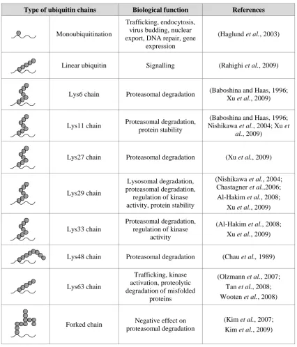
Table 1.2 Functions of various ubiquitin chains: The variety of biological functions of ubiquitination inside the human cells resulting from different types of ubiquitin chain formation.
Type of ubiquitin chains Biological function References
Monoubiquitination
Trafficking, endocytosis, virus budding, nuclear export, DNA repair, gene
expression
(Haglund et al., 2003)
Linear ubiquitin Signalling (Rahighi et al., 2009)
Lys6 chain Proteasomal degradation (Baboshina and Haas, 1996;
Xu et al., 2009)
Lys11 chain Proteasomal degradation,
protein stability
(Baboshina and Haas, 1996; Nishikawa et al., 2004; Xu et
al., 2009)
Lys27 chain Proteasomal degradation (Xu et al., 2009)
Lys29 chain
Lysosomal degradation, proteasomal degradation,
regulation of kinase activity, protein stability
(Nishikawa et al., 2004; Chastagner et al.,2006; Al-Hakim et al., 2008;
Xu et al., 2009)
Lys33 chain
Proteasomal degradation, regulation of kinase
activity
(Al-Hakim et al., 2008; Xu et al., 2009)
Lys48 chain Proteasomal degradation (Chau et al., 1989)
Lys63 chain
Trafficking, kinase activation, proteolytic degradation of misfolded
proteins
(Olzmann et al., 2007; Tan et al., 2008; Wooten et al., 2008)
Forked chain Negative effect on
proteasomal degradation
Chapter One | INTRODUCTION
Page | 10
1.4 Ubiquitin conjugation components 1.4.1 Human E1 ubiquitin-activating enzyme
The ubiquitin activating enzyme (UBE1) stands on the top of the ubiquitin hierarchy, being
responsible for activating ubiquitin and preparing it for one of a large number of distinct E2
ubiquitin-conjugating enzymes. Before the formation of the thioester bond between the
C-terminus of ubiquitin and E1, the ubiquitin was activated by adenylation via binding with
MgATP, leading to the formation of a ubiquitin adenylate intermediate that serves as the
donor of ubiquitin to a cysteine in the E1 active site (Haas et al., 1982; Groettrup et al.,
2008). Each charged E1 molecule carries two molecules of activated ubiquitin: one as a
thioester, and one as an adenylate. Only the thiol-linked ubiquitin is transferred to the E2 by
transthiolation. E1 is an efficient enzyme, justified by its maximum turnover number of
ATP-AMP exchange (1-2 s-1) involving all steps from ATP binding through thioester
formation (Haas et al., 1982) when compared to the catalytic rate (kcat) of substrate
ubiquitination which is 10- to 100-fold slower (Mastrandrea et al., 1999). This allows the
production of sufficient activated ubiquitin for all cellular ubiquitination reactions, even
though the concentration of the E1 protein is thought to be lower than the total concentration
of E2 proteins (Pickart, 2001).
The human genome encodes only two E1 genes: A1S9T (Zacksenhaus and Sheinin, 1990;
Zacksenhaus et al., 1990), which appeared to be an ubiquitin-activating enzyme and
designated later as UBE1, and UBA6 which was recently identified by homology searches
using one of the two ThiF-homology motifs that compose the adenylation domain (Pickett,
2007).
1.4.2 Human E2 ubiquitin-conjugating enzyme
The second component in the ubiquitin cascade are the E2 enzymes, which are the key
enzymes in a ubiquitin pathway. In contrast with E1 enzymes, E2 proteins will not forge
Chapter One | INTRODUCTION
Page | 11
the E2 conjugating enzyme through its ubiquitin fold domain to bring the catalytic cysteine
on the E2 into close enough proximity to the E1 active site, thereby allowing transfer of the
thioester bound from ubiquitin from the E1 to the E2 (Pickart, 2001). Once loaded with
ubiquitin, E2 proteins can donate the thioester linked ubiquitin to the active cysteine of a
HECT domain E3 ubiquitin ligase (E3) which then transfers ubiquitin to a protein substrate.
Alternatively, E2 proteins can transfer ubiquitin directly onto a lysine residue of a target
substrate protein. In this case, an E3 RING protein may assist the E2 protein in recognising
the appropriate protein target (Ptak et al., 2001).
The importance of E2 proteins lies in their role of determining the lysine preferences in
ubiquitination, hence dictates the topology of polyubiquitination (David et al., 2010). This
can be seen in BRCA/BARD, an E3 ligase which acts with UBE2K to generate
Lys48-conjugated chains, whereas with the UBE2N-UBE2V1 combination of E2 proteins, these
catalyse the formation of polyubiquitin chains conjugated through Lys63 (Christensen et al.,
2007). Also, the Pellino1 E3 protein acts with UBE2N-UBE2V1 to generate lysine
Lys63 chains, but when acting with UBE2R1, it actually catalyses the formation of
Lys48 chains. However, when this E3 functions with members of the UBE2D family
(Ubc4/5), it promotes the formation of Lys11 and Lys48 chains (Ordureau et al., 2008).
1.4.2.1 E2 structure
E2 proteins are distinguished by the presence of a UBC (ubiquitin conjugating) domain that
is approximately 35% conserved throughout eukaryotes. This domain consists of about 150
amino acids (Pickart, 2001), which is organised into four standard alpha helices (α1-4), a
short 310 helix, and four-stranded (S1-4) antiparallel β-sheets as demonstrated in Figure 1.3
(Pickart, 2001; Özkan et al., 2005). The β-sheets and α2 form a central region that is
bordered by the α1 region at one end and α3/α4 regions at the other. The cysteine residue of
the active site lies in a long loop that connects S4 to α2, and sits in a shallow groove formed
Chapter One | INTRODUCTION
Page | 12
(Pickart, 2001). A large proportion of the most highly conserved E2 residues are found
around the active site. Some of these residues interact with ubiquitin, and others are
presumed to interact with the E1 protein (Pickart, 2001). In contrast, the region of the E2
protein that faces the active site contains some of the most poorly conserved residues found
within the protein, perhaps to make the structural region easier to define. It is suggested that
a proportion of these residues may have diverged under low selective pressure due to a lack
of essential function. However, it is also possible that these differences may actually
facilitate specific partner interactions (Pickart, 2001). Support for the theory that the region
opposite the active site may be involved in specific E2 interactions is provided by the
analysis of the UbcH10 (UBE2C) protein, which shows that many residues in this region are
conserved among orthologues of UBE2C. However, these residues are not conserved among
different E2 enzymes within the same species, thus implying that these residues are specific
Chapter One | INTRODUCTION
Page | 13
[image:31.595.152.466.82.334.2]
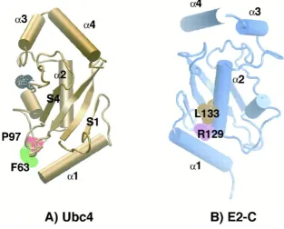
Figure 1.3 Conservation of E2 structures between species: (A) Yeast Ubc4 (UBE2D1 homologue). The active site cysteine (C86) is highlighted black, along with side chains corresponding to F63 (green) and P97 (red) of UBE2C. (B) Clam E2-C (UBE2C homologue). Relative to (A), the molecule was rotated ~180° along the y axis. The side chains of R129 (purple) and L133 (brown), corresponding to the first and fourth residues of a destruction box located in α2, are highlighted. Picture from Pickart (2001).
The structures of UbcH7 (UBE2L3) bound to RING E3 c-Cbl and of UbcH5B (UBE2D2)
bound to CNOT E3 RING have been solved (Zheng et al., 2000; Dominguez et al., 2004)
and would therefore provide a general explanation of E2 specificity. The CNOT4 RING
finger interacts specifically with UBE2D2 and not with UBE2L3 despite the fact that in both
complexes the same regions of the E2s are involved (helix α1, loops L1 and L2) in the
interaction with the RING (Winkler et al., 2004). This suggests that although the three
regions of UBE2D2 and UBE2L3 involved in the binding are similar, the binding properties
must be different. For example, several contacts between the H1 helix of UBE2D2:CNOT4
are not present in the UBE2L3:c-Cbl complex (Figure 1.4). Furthermore, UBE2L3:c-Cbl
involved mainly hydrophobic or uncharged residues while UBE2D2:CNOT4 governs the
Chapter One | INTRODUCTION
Page | 14
(A)
(B)
[image:32.595.130.502.108.525.2]UBE2D2:CNOT4 UBE2L3:c-Cbl
Chapter One | INTRODUCTION
Page | 15 1.4.2.2 E2 family diversity
The existence of at least 37 E2 enzymes suggest some degree of specificity in ubiquitination
events with regards to partners, substrates, and functions. Generally, the E2 family are
classified based on the existence of additional extensions to the UBC catalytic core (Figure
1.5). E2 class I consist of the catalytic domain only, while others with additional N- or C-
terminal extensions are classified as Class II and Class III, respectively. E2s that have
extension in both termini are categorised Class IV. These extensions are involved in
functional differences between E2s, which involve differences in subcellular localisation,
stabilisation of the interaction with E1 enzymes, or modulation of the activity of the
interacting E3s (van Wijk and Timmers, 2010).
While the majority of E2 enzymes contain predominantly the UBC domain alone along with
N- or C-terminal flanking regions, there are also a few examples of domains outside of the
UBC region, which are required for functions, both within and external to the ubiquitin
cascade. For example, it has been hypothesised that the ubiquitin binding UBA domain
C-terminal of the UBC domain in UBE2K may be involved in ubiquitin chain formation based
on a similar domain in the yeast Ubc1 protein (Merkley and Shaw, 2004). The massive
atypical E2 BIRC6 is annotated for an anti-apoptotic BIR domain distal to the C-terminal
UBC domain. At almost 5 000 amino acids in length it is likely however that BIRC6
contains many ordered domains. It is thought to be the sole chimeric E2:E3 ligase that
utilises the BIR domain to bind and mediate ubiquitination of SMAC and caspase-9(Bartke
Chapter One | INTRODUCTION
[image:34.595.121.531.137.526.2]Page | 16
Chapter One | INTRODUCTION
Page | 17
1.4.3 Human E3 ubiquitin-protein ligases
Human E3 ligases perform the final step in the ubiquitin pathway, having a vital role in the
recognition of specific protein substrates and mediating ubiquitin transfer from E2 enzymes
to specific protein substrates. Among all of the components of the ubiquitin cascade, they are
the most numerous and display the greatest diversity. Even though E3s are heterogeneous,
they can nevertheless be classified into two primary classes according to domain homology
and mechanism of action: HECT domain and RING finger-containing E3s (Glickman and
Ciechanover, 2002).
1.4.3.1 HECT domain E3s
HECT domain proteins harbour a 350aa sequence homologous to the E6-AP
carboxyl-terminal domain. This domain contains a conserved catalytic Cys residue that transfers the
activated ubiquitin from an E2 to an internal Cys residue within the E3 before conjugation of
ubiquitin to an NH2 group in the substrate (Glickman and Ciechanover, 2002). Indeed the
HECT domain has at least four biochemical activities: (1) it binds specific E2s; (2) it accepts
ubiquitin from the E2, forming a ubiquitin-thioester intermediate with its active-site cysteine;
(3) it transfers ubiquitin to the ɛ-amino groups of lysine side chains on the substrate by
catalysing the formation of an isopeptide bond; and (4) it transfers additional ubiquitin
molecules to the growing end of the multi-ubiquitin chain (Wang et al., 1999).
1.4.3.2 RING domain E3s
RING finger proteins were first thought to play a role in the dimerisation of proteins. It was
only in the late nineties that RING finger domains were identified as ubiquitin ligases
(Lorick et al., 1999). The RING finger proteins have been defined by a pattern of conserved
Cys and His residues that form a cross-brace structure allowing the binding of Zinc cations.
The conserved RING finger consensus is
Chapter One | INTRODUCTION
Page | 18
domains fall into three categories: RING-HC, RING-H2 and RING-CH depending on
whether a Cys or His occupies the fifth coordination site respectively. While RING fingers
are structurally diverse, all contain two interleaved Zinc-binding sites (Glickman and
Ciechanover, 2002). Unlike HECT E3s, RING E3s do not have recognisable catalytic active
sites that define classical enzymes. Instead, these E3s have large binding interfaces and act
Chapter One | INTRODUCTION
Page | 19
1.5 Hierarchical structure and specificity of the ubiquitin system
The structure of the ubiquitin system appears to be hierarchical. In human cells, only two
main E1 proteins carry out the activation of ubiquitin required for all modifications. These
enzymes are responsible for transferring ubiquitin to different E2 enzymes. Subsequently,
each E2 acts in conjunction with either one or more different E3 enzymes. Beside the two
main E1 activating enzymes and approximately 40 types of E2 conjugating enzymes, there
are about 600-800 E3 ligases in human cells, in other word there are nearly 50 000 different
potential combinations might operate in ubiquitin cascades, making the ubiquitin pathway
both versatile and highly complex (illustrated in Figure 1.6). The diversity of ubiquitin,
besides being contributed by more than 600 E3 ligases, is further expanded by the generation
of different forms of ubiquitin chains. Even though the type of ubiquitin chain formation is
depend on the E2 enzyme, it is still possible that the E3 ligase determines which substrate to
bind ubiquitin to (Nakada et al., 2010).
Chapter One | INTRODUCTION
Page | 20
1.6 The deubiquitinating enzyme
1.6.1 Classification of deubiquitinating enzyme
Deubiquitinating enzymes (DUBs) belong to the superfamily of proteases and they function
to remove covalently attached ubiquitin from proteins, either from the polyubiquitin chain or
from the substrate (Wilkinson, 1997). Among all ubiquitin machineries, the functions, targets
and regulation of DUBs are the most poorly understood due to their non-uniform structure
and function, though it is becoming increasingly apparent that DUBs regulate various
cellular processes (Ventii and Wilkinson, 2008). The human genome encodes approximately
95 DUBs, which fall into five major classes (Nijman et al., 2005) namely ubiquitin specific
proteases (USPs), ubiquitin C-terminal hydrolases (UCHs), ovarian tumour proteases
(OTUs), Josephins and the Jab1/MPN/MOV34 metalloenzymes (JAMMs, also known as
MPN+). The USP, UCH, OTU and Josephin families are cysteine proteases, whereas the
JAMM/MPN+ family members are zinc metalloproteases. Figure 1.7 show the structures of
the catalytic domains of all five subclasses of the DUBs. For the cysteine proteases, the
enzymatic activity relies on the thiol group of a cysteine in the active site. The adjacent
histidine, which is polarised by an aspartate residue helps in cysteine deprononation hence
these three make up the catalytic triad. During catalysis, the cysteine performs a nucleophilic
attack at the peptide bond between the target and the ubiquitin resulting in the release of the
target protein and formation of a covalent intermediate with the ubiquitin moiety. Upon
reaction of this intermediate with a water molecule, free enzyme and ubiquitin are released
(Nijman et al., 2005). On the other hand, metalloproteases generally use a Zn2+ bound
polarised water molecule to generate a non-covalent intermediate with the substrate. The
metal atom is stabilised by an aspartate and two histidine residues (Ambroggio et al., 2003).
The intermediate is further broken down by proton transfer from a water molecule causing
the release of the DUB (Nijman et al., 2005).
In spite of what appears to be a hierarchical system, an interaction between a DUB called
Chapter One | INTRODUCTION
Page | 21
conventional hierarchy and therefore much attention should be paid to elucidate the purposes
of such interaction.
1.6.1.1 USP domain
The largest and most diverse cysteine protease, the domain contains well-conserved Cys and
His boxes, which include all the catalytic triad residues as well as other residues in the active
site pocket (Amerik and Hochstrasser, 2004).
1.6.1.2 UCH domain
Generally small proteins and were originally identified by their ability to hydrolyse small
amides and esters at the C-terminus of ubiquitin (Amerik and Hochstrasser, 2004).
1.6.1.3 OTU domain
A novel family of cysteine proteases which display structural similarity in a presumed
catalytic core domain containing conserved Cys, His and Asp residues thought to comprise
the proteolytic/catalytic triad. OTU has been proven to have DUB activity by the ability of
its prominent members, Otubain 1 and 2, to cleave ubiquitin from either a ubiquitin–GFP
fusion protein or a tetraubiquitin fusion (Balakirev et al., 2003).
1.6.1.4 Josephin domain
Its representing member, Ataxin-3 has the typical properties of DUBs: the enzyme
disassembles ubiquitin–lysozyme conjugates, cleaves ubiquitin-7-amido-4-methylcoumarin
(ubiquitin-AMC), and binds to the DUB inhibitor ubiquitin aldehyde (Ubal) (Burnett et al.,
2003). The Josephin domain, which is found in over 30 predicted proteins, most of unknown
function, includes segments that show weak similarity to the His and Cys boxes of UBPs and
Chapter One | INTRODUCTION
Page | 22
like protease fold that characterises the other cysteine proteases (Amerik and Hochstrasser,
2004).
1.6.1.5 JAMM domain
The MPN+/JAMM motif DUB called AMSH (associated molecule with the SH3 domain of
STAM) was found to have deubiquitinating activity as well (McCullough et al., 2004). This
metalloprotease motif includes two absolutely conserved His residues and an Asp residue
Page | 23
Chapter One | INTRODUCTION
Page | 24
1.6.2 General function of DUB
Generally, DUBs activity can be divided into three major functional categories. Firstly, the
DUBs are responsible for processing linear ubiquitin precursor proteins into single ubiquitin
molecules. As explained earlier, ubiquitin is encoded as a polyubiquitin gene or ribosomal
fusion gene. Upon expression of ubiquitin protein, DUBs play a vital role in separating the
polyubiquitin and cleave the ribosomal protein to generate a free ubiquitin (Komander et al.,
2009a). Mutations in several DUBs have been shown to cause ubiquitin reduction and
therefore resulting in various serious defect in living cells. In yeast, deletion of DUB
encoding genes, including DOA4 and UBP6 were shown to reduce the amount of
monomeric ubiquitin (Swaminathan et al., 1999).
DUBs also functioned to remove ubiquitin chains from post-translationally modified
proteins, leading to the reversal of the ubiquitin signal hence rescuing the protein from either
proteasomal or lysosomal degradation. However, if a commitment to these degradative
machines has been made, DUBs act to recycle the ubiquitin released from the protesome (or
lysosome), thereby maintaining the free ubiquitin pool. Any ubiquitin that is released as an
oligomer could also be disassembled by DUBs (Komander et al., 2009a). Thirdly, DUBs can
also be used to edit the form of ubiquitin modification by trimming ubiquitin chains from the
distal end of the chain (Komander et al., 2009a).
The knowledge about cellular functions of DUB has increased significantly in recent years.
Due to the diversity in DUBs structure and functions, the prediction of DUBs main role in
cellular regulation has proved to be quite tricky. Nevertheless, essential progress that largely
focuses in elucidating the role of DUBs in the area of membrane trafficking, cell signalling
Chapter One | INTRODUCTION
Page | 25
1.7 Protein-protein interactions
Upon completion of Human Genome Project in 2003, approximately 23 000 genes of the
human genome were identified and mapped. However, this knowledge about the entire
human genome could not provide an understanding even of the basic principles in human
cellular systems. Entering the post-genomic era, the importance of protein-protein
interactions is becoming even more apparent in order to explain how the genetic information
in the form of DNA manage to generate functions. In order to tackle this question, we need
to understand how the gene products, particularly proteins, interact with each other to
perform many biological functions that build a living organism. Protein-protein interactions
(PPI) are fundamental to all biological processes. Determination of the PPI that take place
within an organism provides a framework for understanding the links between molecular and
cellular biology. Alteration of PPI are thought to be involved in the development in many
diseases for example neurodegenerative disorders, cancers and infectious diseases. Hence,
examination of when and how they are controlled is essential for understanding diverse
biological processes and elucidating the molecular basis of disease as well as identifying
potential targets for therapeutic interventions.
Various methodologies can be used to detect PPI. Each has its own strengths and weaknesses
with regard to the sensitivity and specificity of the method. Generally methods for
identifying interacting proteins can be divided into two types: (1) biology/biochemical
methods for example co-immunoprecipitation and genetic manipulation yeast two-hybrid
Chapter One | INTRODUCTION
Page | 26
1.7.1 Genetic in vivo methods
1.7.1.1 Yeast two-hybrid screen
Since its description in 1989 (Fields and Song, 1989), yeast two-hybrid (Y2H) analysis has
become widely used to detect binary protein-protein interaction. In this system, a protein of
interest (the bait) is fused to the DNA binding domain (BD) of a transcription factor (such as
GAL4) as can be seen in Figure 1.8. The bait’s potential interacting partner (the prey protein)
is fused to the transcription factor’s activation domain (AD). These fusions are carried out by
DNA cloning methods, allowing expression of the subsequent bait and prey fusion proteins
in the nucleus of the yeast host. The yeast strain used in this system carries a set of reporter
constructs which are under the control of an upstream sequence containing the binding sites
for the BD. If the bait-BD and prey-AD fusions interact, then a functional transcription
factor is reconstituted and expression of the reporter gene is activated (Fields and Song,
1989). The reporter genes produce a scorable phenotype such as growth on selective media
or colour change. For example, activation of the reporter genes ADE2 and HIS3, enables
growth on media lacking adenine and histidine respectively, and the activation of the lacZ
reporter gene produces a blue colour in an X-gal assay.
The first genome-wide Y2H interaction map was generated for bacteriophage T7, and
large-scale yeast two-hybrid screens have been conducted for several viruses, H. pylori, S.
cerevisae, P. falciparum, C. elegans, Drosophila and human (Flajolet et al., 2000; McCraith
et al., 2000; Uetz et al., 2000; Guo et al., 2001; Ito et al., 2001; Giot et al., 2003; Li et al.,
2004; Stanyon et al., 2004; Formstecher et al., 2005; LaCount et al., 2005; Rual et al., 2005;
Stelzl et al., 2005; Uetz et al., 2006). The data generated from these screens will be useful
for individual studies and for system wide studies (Parrish et al., 2006). For example the
human pathogen interaction maps have generated lead proteins that may interact during
pathogenesis and identified potential drug targets (Parrish et al., 2006). The potential for
proteome-wide interactome mapping in humans has been demonstrated by two major
Chapter One | INTRODUCTION
Page | 27
chosen on the basis of clone availability, and identified 2 754 protein interactions. Stelzl et
al. (2005) screened two-hybrid arrays generated from a human foetal brain cDNA library
and a collection of full-length ORFs, identifying 3 156 interactions. Combined, these two
datasets identified over 5 900 protein interactions, a large proportion of which are novel.
Our lab has used the Y2H system to perform both library screen and targeted matrix
experiments. A library screen allows the identification of novel interactive partners, whereas
a matrix approach enables the detection of an interaction between two specific proteins. A
library screen is applied as a tool for the discovery of binding partners for a specific bait, by
screening it against an AD-cDNA library (which contains potential cDNA prey clones fused
to the GAL4 AD). Direct yeast colony PCR of the diploid yeast colonies using prey specific
primers permits identification of the preys via sequencing. Meanwhile, the matrix method
involves pairs of proteins which are systematically tested for a binary two-hybrid interaction.
Here, arrays of yeast of one mating type containing bait clones are mated with arrays of yeast
of the opposing mating type containing individual prey clones. The advantage of the matrix
method is that there is no need to identify the interacting protein, as the clone is already
Chapter One | INTRODUCTION
Page | 28
(A)
(B)
[image:46.595.144.492.89.520.2](C)
Chapter One | INTRODUCTION
Page | 29
1.7.2 Biophysical and theoretical methods
1.7.2.1 Isothermal titration calorimetry (ITC)
ITC has been extensively used to study ligand binding and its application in
protein-protein binding are growing rapidly (Leavitt and Freire, 2001). It is the only technique that
determines directly the thermodynamic parameters of a given reaction which are the , and . Theoretically, the binding of two proteins cause a change in the thermodynamic potentials ( , and ) and these are measured directly by highly sensitive calorimetry (Velazquez-Campoy et al., 2004). ITC uses stepwise injections of one protein into a
calorimetric cell containing the second protein to measure the heat of the reaction for both
exothermic and endothermic processes (Figure 1.9). The underlying principles of ITC lies on
the chemical scheme based on a reversible association equilibrium:
where and are the interacting macromolecules. The strength of the interaction is
described by the association constant or the dissociation constant :
and
where and are the concentrations of the free reactants and is the concentration of the complex. These constants are related to the Gibbs energy of association
and dissociation and can be expressed in terms of the enthalpy, and entropy, , change in the process:
where is the gas constant (1.9872 cal/Kmol) and is the absolute temperature (kelvins).
Among the information available by doing ITC are simultaneous determination of the
Chapter One | INTRODUCTION
Page | 30
An ITC composed of two identical cells made of a highly efficient thermal conducting and
chemically inert material, surrounded by an adiabatic jacket (O’Brien et al., 2000). Sensitive
thermocouple circuits are used to detect temperature differences between the reference cell
filled with buffer and the sample cell filled with the first protein. Before the addition of
second protein, a constant power is applied to the reference cell. This directs a feedback
circuit, activating a heater located on the sample cell (MicroCal ITC manual). During the
experiment, ligand is titrated into the sample cell in known amount, causing heat to be either
taken up or evolved, depending on the nature of the reaction.
Chapter One | INTRODUCTION
Page | 31 1.7.2.2 Nuclear magnetic resonance (NMR)
The fundamental questions in protein-protein interaction are the location of binding
interface. A lot of biochemical or biophysical techniques are currently available to address
this question but among the most widely used is NMR chemical shift perturbation due to its
sensitivity to subtle changes in the chemical environment of proteins. NMR can provides
both specific and qualitative information for examples: (1) give specific information on
physical properties of individual functional groups such as ionisation states, pKa, and
hydrogen bonds; (2) provide site-specific information on backbone and side-chain dynamic
motions in solution; (3) be used to identify contacts between individual atoms of a protein
and its binding partners, as well as to study the kinetics of ligand binding (Gao et al., 2004).
Table 1.3 below shows the comparison between ITC and NMR.
Table 1.3 The comparison between ITC and NMR: Pros and cons of ITC and NMR. NMR, however, has an extra advantage since it can provides binding site information.
ITC NMR
Measure ΔH Δδ
Concentration P1 >1 μM >10 μM
Concentration P2 >10*[P] Any (>10*[P] opt)
Volume 0.3 ml 0.5 ml
Titration time ~30 min ~1 day
Binding parameter ΔH, Kd, TΔS Kd
Chemical shift arises from anisotropies in the electronic environment surrounding magnetic
nuclei. Some nuclei like 1H, 13C, 19F, 31P have nuclear spin that can create a magnetic
moment. By placing these nuclei in a magnetic field, they absorb electromagnetic radiation
at particular frequencies governed by their chemical environment and give an electronic
signal displayed as a peak (Figure 1.10). Their chemical environment is influenced by
Chapter One | INTRODUCTION
Page | 32
even the nuclei of the same element give rise to distinct spectral lines. The relative positions
of these spectral lines are therefore called chemical shifts. In other words, upon addition of
ligand or protein partner, shifting peaks, line width, and/or intensity changes indicate which
residues experience a change in their environment, and this in turn allow the identification of
the binding interface. In practice, this can be achieved by simply titrating the unlabelled
protein into an isotopically enriched (15N or 13C) protein sample and monitoring peak
perturbations in spectra of the labelled protein (Gao et al., 2004).